Chemical neurolysis in the management of muscle spasticity
Bạn đang xem bản rút gọn của tài liệu. Xem và tải ngay bản đầy đủ của tài liệu tại đây (143.41 KB, 15 trang )
8
Chemical neurolysis in the management
of muscle spasticity
A. Magid O. Bakheit
Introduction
Destruction of peripheral nerves with chemical
substances such as phenol and alcohol solutions
(chemical neurolysis)wasintroducedasanovelther-
apeutic modality in the 1930s, but it became a popu-
lar method of treatment of severe, intractable pain
associated with cancer in the mid-1950s (Maher,
1955; Brown,1958).A few yearslater peripheral nerve
blocks with local anaesthetics and neurolytic agents
were found to be effective in the management of
muscle spasticity and neurogenic bladder disorders
and more recently they have also been used to pre-
dict the outcome of certain surgical procedures such
as selective dorsal rhizotomy.
An important therapeutic use of peripheral nerve
and intrathecal blocks is in the treatment of severe or
intractablepain(e.g.pain associated with cancer and
with trigeminal and postherpetic neuralgia). Com-
plete symptomatic relief is achieved in more than
70% of patients with chronic pain due to neurogenic
causes or ischaemia (Hatangdi & Boas, 1975). Nerve
blocks have also been shown to be valuable in the
management of bladder dysfunction due to spinal
cord injury or disease. The selective chemical dener-
vation of S3 sacral segment in patients with a hyper-
active detrusor muscle increases bladder capacity
and reduces the uninhibited contractions. Conti-
nence is usually achieved in these patients without
sphincter disturbances or sexual dysfunction (Tor-
rens, 1974; Rockswold & Bradley, 1977). In addition,
chemical neurolysis has proved to be an effective
intervention in the management of severe upper
and lower limb muscle spasticity. In most patients
it relieves the muscle spasticity without significantly
affecting the strength of the voluntary muscle con-
traction (Brown, 1958; Khalili & Betts, 1967). This
confers chemical neurolysis a major advantage over
treatment with oral antispasticity drugs.
Chemical neurolysis can be achieved with periph-
eral nerve blocks, motor point (intramuscular) injec-
tions and the intrathecal administration of alcohol or
phenol. These procedures are generally safe, effec-
tive and relativelyeasytoperform. They arepreferred
to oral antispasticity drugs which often cause sys-
temic adverse effects and are nonselective in their
action, thus affecting spastic and nonspastic mus-
cles. The latter adverse effect may lead to functional
loss. In a study by Katrak and colleagues (1992) of
patients recovering from stroke, dantrolene reduced
muscle strength in the unaffected extremities with-
out significantly reducing muscle tone or improv-
ing function in the spastic limbs. Another disad-
vantage of systemic antispasticity drugs is that their
effectiveness diminishes with prolonged use due to
pharmacological tolerance. Tolerance to these drugs
usually develops after a few weeks or months of
treatment and progressive dosage increments are
often required to maintain the initial therapeutic
response.
Chemical neurolysis is only one of many methods
of treatment of muscle spasticity and the best clini-
cal outcomes are achieved when it is utilized as part
of an overall management strategy. Factors that pre-
cipitate or aggravate muscle spasticity, such as uri-
nary tract infections and faecal impaction, should
150
Chemical neurolysis in the management of muscle spasticity 151
be identified and treated. Empirical clinical experi-
ence also suggests that an intensive physiotherapy
programme enhances the beneficial effect of nerve
blocks and motor point injections. In some cases it
is more useful to combine chemical neurolysis with
serial splinting of the spastic limb, the application of
plaster casts or the use of an orthosis.
The effect of neurolytic agents is usually irre-
versible and their use should, therefore, only be
considered when a clear treatment goal has been
identified. There is a large variation in the way mus-
cle spasticity affects patients depending on the site
and chronicity of the upper motor neurone lesion,
its underlying cause, the degree of neural recovery
and the way the nervous system compensates for the
functional loss. Frequently spasticity is functionally
useful and an individualized approach to the man-
agement of this symptom is, therefore, essential.
Indications for treatment
Severe chronic muscle spasticity often causes con-
stant gnawing pain. In addition, it is frequently
associated with muscle spasms which occur spon-
taneously or when the patient attempts to move.
In severe cases the spasms may even be precipi-
tated by external stimuli, such as a sudden noise.
Spasmsof the hipflexors,extensors oradductorsmay
be accompanied by involuntary bladder emptying
and occasionally faecal incontinence. Other effects
of severe muscle spasticity include impaired motor
function and the development of deformities and
fixed contractures. Generally, treatment of spasticity
is indicated to alleviate distressing symptoms such
as pain or muscle spasms, to improve motor func-
tion, to facilitate activities of daily living (e.g. wash-
ing and dressing,urethral catheterization or perineal
hygiene) or to prevent or reduce the complications
often associated with muscle hypertonia (e.g. fixed
contractures or difficulties inmaintaining a comfort-
able position in bed or chair).
There is no research evidence at present to show
which patients are most likely to benefit from nerve
blocks and motor point injections. However, given
the fact that the beneficial effect of these procedures
usually lasts for several months and that good results
cannot be relied upon after two or three injections
(Bakheit et al., 1996a), it is likely that the technique
is most helpful for those whose spasticity may be
troublesome in the medium rather than the long
term. This would include patients recovering from
severehead injury or a recentrelapse ofmultiplescle-
rosis in whom spasticity is so severe that splinting
or the application of plaster casts is impracticable
because of the risk of soft tissue damage. Another
group of patients who are likely to benefit from
chemical neurolysis are those in whom spasticity is
preventing the acquisition of new motor skills, such
aschildrenwith cerebral palsy establishingincreased
independence in walking. A third group is subjects
who are likely to require future surgical treatmentfor
the complications of spasticity, such as the control
of pain, the relief of muscle spasms or the surgical
release of contractures, but in whom there are clini-
cal or technical advantages in delaying such surgery.
Indications for medial popliteal nerve blocks
Medial popliteal nerve blocks and motor point injec-
tions of the gastrosoleus muscle group are indicated
in cases of severe dynamic foot equinus (i.e. ankle
plantar flexion that is not due to a fixed contracture),
especially if resistant to serial casting or preventing
the effective use of an ankle-foot orthoses. In these
circumstances the foot equinus usually prevents the
correct placement of the patient’s foot in stance and
causes insufficient clearance of the foot from the
ground in the swing phase of the gait cycle, thus ren-
dering the patient’s gait unsafe. Another indication
for medial popliteal nerve blocks is when sustained
ankleclonusinterfereswith motor function orcauses
discomfort to the patient (e.g. if it prevents comfort-
able placement of the foot on the wheelchair foot-
plate). They are also useful, as a diagnostic test, in
the management of distal foot deformities in chil-
dren with cerebral palsy. For example, by reducing
the muscle imbalance in the lower limb a medial
popliteal nerve block provides valuable information
regarding the choice of the surgical procedure for
152 A. Magid O. Bakheit
the treatment of secondary foot deformities such as
hallux valgus or metatarsal subluxations (Carpenter,
1983).
Indications for obturator nerve blocks
The main indications for obturator nerve blocks in
ambulatory patients is ‘scissoring gait’. In nonam-
bulatory patients this treatment may be considered
when severe spasticity of the hip adductors prevents
easy urethral catheterization, washing and cleaning
the perineal area and seating or positioning in bed.
Occasionally, obturator nerve blocks are used to pre-
vent the development of, or to promote the healing
of, skin pressure sores on the medial aspect of the
knees.
Obturator nerve blocks have also been used in the
management of dislocation and subluxation of the
hip joint. This complication occurs in 25% of patients
with severespastic cerebral palsy and is often associ-
ated with severe pain. Treatment is usually effective
in pain relief probably due to reduced stretching of
the joint capsule and less friction of the femoral head
against the periosteum of the acetabulum (Trainer
et al., 1986).
Nerve blocks for upper limb muscle spasticity
In the upper limbs, chemical neurolysis seldom
improves motor function and is mainly indicated
to facilitate activities of daily living. For example,
the improved elbow extension following a successful
musculocutaneous nerve block often makes putting
on and removing upper body garments easier and
in some cases also increases the patient’s reach with
the paretic hand. Reduction of spasticity of the fin-
ger flexors is sometimes necessary to facilitate hand
hygiene and to prevent skin laceration in the palm
of a claw hand. Percutaneous phenol nerve blocks
are often successful in these cases but the proce-
dure involves a higher risk than when it is used for
lower limb spasticity. This is because the median and
ulnar nerves run in close proximity to the blood ves-
sels of the upper limb and an attempt to infiltrate
these nerves with the neurolytic agent may result in
vascular damage. Furthermore, both nerves contain
sensory fibres and the sensory loss following neurol-
ysis may cause loss or deterioration of hand function
and increase the risk of burns and injury. The use of
botulinum toxin is probably more appropriate than
alcohol or phenol for the management of upper limb
spasticity.
The diagnostic use of nerve blocks
Diagnostic nerve blocks with local anaesthetics are
sometimes necessary to assess the risk/benefit ratio
of chemical neurolysis. Although the effect of local
anaesthetics is not identical to that of phenol and
alcohol, their use often yields clinically valuable
information. Bupivacaine is best suited for this pur-
pose, as its effect lasts 7 to 8 hours when given in a
dose of 1 mg/kg body weight (0.5% Marcain contains
5.28 mg/ml of bupivacaine Hcl).
Diagnostic nerve blocks may beused to predictthe
effects of chemical neurolysis on motor function (e.g.
when severe spasticity of the wrist and finger flexors
is causing functional difficulties but the patient still
has some voluntary muscle strength in the affected
hand). They may also be used to assess the effects of
sensory loss on the patients’ functional ability when
injections of mixed sensory-motor nerves are being
considered. Diagnostic nerve blocks have also been
found valuable in predicting the functional outcome
of surgical procedures for spasticity, such as selec-
tive dorsal rhizotomy, and in the management offoot
dystonia (Bakheit et al., 1996b).
The pharmacological properties of
neurolytic agents
Phenol (a benzene derivative of carbolic acid) and
ethyl alcohol are the drugs most commonly used
for peripheral nerve and intrathecal blocks. Other
agents, such as cresol and chlorocresol, may also
be used. Although phenol and alcohol were initially
thought to reduce muscle tone by the selective inhi-
bition of gamma efferent pathways, their mode of
action was subsequently shown to be due to a local
Chemical neurolysis in the management of muscle spasticity 153
anaesthetic and neurolytic effect. The local anaes-
thetic effect is immediate and transient. As with
conventional local anaesthetics, nerve conduction
is initially blocked in the small fibres within the
nerve trunk (i.e. sympathetic and sensory fibres) and
then in the large motor axons. Braun et al. (1973)
attributed this selective effect of dilute solutions of
phenol or alcohol to the fact that fibres with a small
diameter have more relative surface contact area for
a given volume of nerve tissue than large alpha fibres.
Typically, recovery of nerve conduction occurs in the
reverse order. The neurolytic properties of alcohol
and phenol account for their more lasting clinical
effect.
Neurolytic agents in high concentration penetrate
the nerve tissue and coagulate protein. The applica-
tion of phenol or alcohol solutions causes nerve tis-
sue destruction, which is proportionaltotheconcen-
trationandvolumeof fluidinjected. Interestingly, the
myelin sheath is more susceptible than the axons
to this neurolytic injury. The pathological changes
resulting from chemical neurolysis occur in a pre-
dictable sequence. Histological changes consisting
of a marked inflammatory reaction in the nerve tis-
sue occur within hours of the application of the neu-
rolytic agent (Nathan et al., 1965). These are followed
in a few days by Wallerian degeneration that is maxi-
mal 2 weeks after the injection. In the event of severe
damage the nerve fibres are often replacedby fibrous
tissue. Finally, within a few weeks of the injection
evidence of partial nerve regeneration, mainly by
collateral sprouting, is usually evident; and by the
14thweek regeneration is almostcomplete (Burkell&
McPhee, 1970). The neurolytic effect is nonselective
and involves myelinated and nonmyelinated nerve
fibres. Very high concentrations of neurolytic agents;
for example, 15% phenol in saline or 10% phenol in
iophendylate (Myodil) may also cause localized vas-
culitis, tissue infarction and arachnoiditis (Baxter &
Schacherl, 1962).
Phenol is soluble in water, glycerine and other
organic solvents. Aqueous phenol is suitable for
peripheral nerve blocks and motor point injections,
whereas phenol in glycerine is preferred for intrathe-
cal block. Phenol in glycerine has a higher specific
gravity (i.e. heavier) than cerebrospinal fluid. This
allows the solution to be easily manipulated around
the desired nerve roots by the appropriate careful
positioning of the patient. Interestingly, chlorocre-
sol in glycerine (1: 50) is thought to be a better agent
than phenol for the management of pain in can-
cer patients. It was claimed to provide a more reli-
able symptomatic relief, presumably because it acts
partly by diffusion and spreads to a greater length
of the nerve root. Aqueous solutions of phenol have
been shown to have a more potent neurolytic effect
than phenol in glycerine.
Procedure of peripheral nerve blocks
Nerve blocks
Chemical neurolysis is most frequently used for
blocks of the medial popliteal, the obturator, the sci-
atic and the musculocutaneous nerve of the arm.
Nerve blocks are usually carried out percutaneously
as described below. However, occasionally ‘open’
blocks of the motor branches of mixed sensory-
motor nerves are performed. Following the surgical
exposureof the nerve, the motor division is identified
with an electrical stimulator and 2 to 5 ml of the neu-
rolytic agent are injected in a 2-cm segment of the
nerve beneath the neural sheath. The most effective
site of block depends on thecourse of thenerve in the
limb and where it divides to innervate the muscles
being considered for treatment.
An essential prerequisite for the success of periph-
eral percutaneous nerve blocks is the accurate place-
ment of the injection. This can be achieved easily
with an electrical stimulator utilizing a Teflon-coated
needle electrode as a probe. Alternatively, a standard
Venflon connected to the cathode of the stimulator
could be used. The electrode wire is wrapped around
the needle shaft at the top and the plastic sheath is
replaced to ensure that the needle is insulated except
at the tip. Although some clinicians use anatomi-
cal landmarks as the guide for needle placement,
this method is often inadequate and is associated
with up to 40% treatment failure (Ferrer-Brechner &
154 A. Magid O. Bakheit
Brechner, 1976). Nerve blocks require full coopera-
tion from the patient, and the frequent discomfort
that occurs afterwards means that children might
require a light general anaesthetic.
Medial popliteal nerve block
The medial popliteal (tibial) nerve is a continua-
tion of the sciatic nerve. It runs in the middle of
the popliteal fossa, where it gives off branches to the
two heads of the gastrocnemius from its proximal
portion approximately 1 cm above the head of the
fibula. Each of these divisions gives off three to five
terminal branches in the proximal fifth ofthe muscle.
The middle and distal branches enter deep into the
muscle and supply the main muscle mass and the
distal third, respectively. The branches to the soleus,
popliteus and tibialis posterior muscles arise more
distally. Further branches below the popliteal fossa
innervate the flexor digitorum longus and flexor hal-
lucis longus muscles. The terminal branches inner-
vate the toe flexors and the small muscles of the foot.
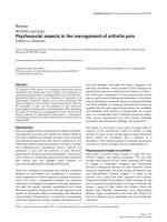
The medial popliteal nerve may be blocked at the
apex of the popliteal fossa or 2 to 3 cm lower, at
the level of the popliteal crease (Fig. 8.1). However,
injection placement in the latter site is thought to be
less effective than a moreproximal block (Felsenthal,
1974). This is presumably because the nerve fibres
are more dispersed distally. It is easier to do medial
popliteal nerve blocks with the patient lying prone.
Alternatively,the procedurecould beperformed with
the patient lying on his or her side and the limb
held in full extension by an assistant to prevent flex-
ion withdrawal. The location of the medial popliteal
nerve behind the knee can be easily identified at the
level of the tibial epicondyles with an electrical stim-
ulator, initially using surface electrodes delivering 5-
to 50-volt pulses of 0.1-msec duration. The skin is
then cleansed with iodine solution and infiltrated
with 1% lignocaine. The needle probe is then intro-
duced and manoeuvred in the tissue using stimulus
pulses of decreasing strength until a contraction of
the spastic muscles supplied by the nerve is obtained
in response to 0.5-mA electrical pulse with a stimu-
lus duration of 0.05 to 0.1 msec. Between 3 and 5 ml
of 4.5% phenol in water or 50% ethyl alcohol is then
injected over 3 to 4 minutes. Slowly the position of
the needle tip is readjusted in each plane to ensure
that the twitch had been fully suppressed. If a new
site is found during this manoeuvrea further 1to 2ml
of phenol should be injected. Ankle clonus is imme-
diately abolished or significantly attenuated with a
successful medial popliteal nerve block.
Obturator nerve blocks
The obturator nerve passes through the obturator
foramen into the thigh in the upper medial part of
the femoral triangle. (The femoral triangle is formed
by the lateral border of the adductor longus, the sar-
torius muscle and the inguinal ligament.) The nerve
emerges about 2 cm below the inguinal ligament and
just lateral to the origin of the tendon of the adductor
longus muscle (Fig. 8.2). It then immediately divides
into an anterior (superficial) and posterior (deep)
branches. Itis a predominantly motor nerve and sup-
plies the hip adductors. It also gives off branches to
the hip and knee joints and a cutaneous branch to
a small skin area on the medial aspect of the mid-
dle of the thigh. In one third of subjects there is an
accessory obturator nerve which emerges from the
pelvis above the superior pubic ramus and joins the
anterior branch of the main trunk approximately 4
to 5 cm below the inguinal ligament.
Localization of the obturator nerve is made with
the patient supine and both legs slightly abducted.
The tendon of the adductor longus muscle is usually
easily palpable in patients with hip adductor spas-
ticity. The femoral artery is approximately 2 cm lat-
eral to the obturator nerve and femoral pulsation is
another useful landmark. Stimulation of the nerve
may initially be carried out using a surface probe and
then a needle electrode as described in the above sec-
tion and is confirmed when a significant contraction
of the adductor muscles is seen. Following injection
of the anterior branch the needle is inserted 2 cm
deeper and perpendicular to the coronal plane to
block the posterior branch. A total of 4 to 5 ml of
phenol or alcohol equally divided between the two
sites is usually sufficient.
Chemical neurolysis in the management of muscle spasticity 155
Figure 8.1. Medial popliteal nerve block at the apex of the popliteal fossa (1) is more effective than a nerve
block at the level of the popliteal crease (2).