Management of spasticity in children
Bạn đang xem bản rút gọn của tài liệu. Xem và tải ngay bản đầy đủ của tài liệu tại đây (258.77 KB, 27 trang )
12
Management of spasticity in children
Rachael Hutchinson and H. Kerr Graham
Introduction
Spasticity can be defined as a velocity-dependent
resistance to passive movement of a joint and its
associated musculature (Lance, 1980; Rymer & Pow-
ers, 1989; Massagli, 1991). Although spasticity is usu-
ally present before contracture in children with cere-
bralpalsy, true muscle shortening or contracture also
appears at an early stage. The majority of children
will have a mixture of spasticity and contracture. Dis-
tinguishing spasticity from contracture is important
from a management point of view.
1. ‘Dynamic’ shortening is most commonly caused
by spasticity but may also be associated with
dystonia and mixed movement disorders. Typ-
ically, ‘dynamic’ contracture is recognized in
younger children with cerebral palsy or spas-
ticity of recent onset. Such children are likely
to exhibit hyperreflexia, clonus, co-contraction
and a velocity-dependent resistance to passive
joint motion. Children who exhibit ‘dynamic’
calf shortening may walk on their toes with an
equinus gait, but on the examination couch the
range of passive ankle dorsiflexion may be full or
almost full.
2. ‘Fixed’ shortening or ‘myostatic’ contracture
describes the typical stiffness found in mus-
cles of older children with cerebral palsy or
spasticity of longer duration. The stiffness
is much less velocity dependent and is still
present during couch examination and under
anaesthesia.
Causes of spasticity in children
With the eradication of poliomyelitis and the dra-
matic fall in the prevalence of spina bifida, the most
common motor disorder in children in developed
countries is cerebral palsy. The incidence of cere-
bral palsy in developed countries is static or even ris-
ing. The reductions in the prevalence of kernicterus
due to neonatal jaundice has been overshadowed
by improved survival of very low birth weight and
premature infants, many of whom suffer from spas-
tic diplegia and quadriplegia (Stanley & Alberman,
1984; Petterson et al., 1993a,b; Pellegrino & Dor-
mans, 1998; Marlow et al., 2005). Other common
causes of spasticity in children are acquired brain
injury and spinal cord injury. Table 12.1 shows the
cause of spasticity in a consecutive sample of 341
children seen in a variety of clinics at the Royal Chil-
dren’s Hospital in Melbourne in 1998.
Spasticity in children will continue to be a com-
mon and challenging problem for the foreseeable
future. While reduction in the incidence of cerebral
palsy would have the most impact in reducing the
overall incidence of spasticity in children, preven-
tion of traumatic brain injury and spinal cord injury
is probably more realistic (Glasgow & Graham, 1997).
The pathology of spasticity
Given that the most common cause of spasticity in
our clinics is cerebral palsy, subsequent discussion
214
Management of spasticity in children 215
Table 12.1. Aetiology of spasticity in 341 children
(cerebral palsy, orthopaedic and spasticity clinics)
Cerebral palsy 79%
Acquired brain injury 6%
Spina bifida 5%
Spinal cord injury 2%
Miscellaneous 8%
on pathology and management focuses mainly but
not exclusively on spasticity in the context of juve-
nile cerebral palsy. The effects of spasticity cannot be
separated from the overall effects of the upper motor
neurone (UMN) syndrome (Fig. 12.1). The child with
diplegia who walks on his toes because of calf spas-
ticity may also be unable to voluntarily control the
dorsiflexors of the ankle during gait. No matter how
effective management of the calf spasticity is, gait
may remain impaired because toe clearance cannot
be achieved during the swing phase of gait (Perry,
1985, 1992). Indeed there is virtually always an effec-
tive solution to calf spasticity/stiffness/shortening,
but inability to control the ankle dorsiflexors dur-
ing swing phase may mean life-long dependence on
an orthosis. Weakness and impaired selective motor
control have a much greater impact on gait and func-
tion than spasticity. They are also more difficult to
manage.
Fixed musculoskeletal pathology in cerebral palsy
is acquired during childhood. Children with cere-
bral palsy do not have contractures, dislocated hips
or scoliosis at birth. These common deformities are
acquired during childhood. Muscle growth in chil-
dren is a race between the pacemakers (i.e. the phy-
ses of the long bones) and the muscle tendon units,
in which the muscles are doomed to second place
(Graham & Selber, 2003). The prerequisites for nor-
mal muscle growth is frequent stretching of relaxed
muscle. In children with cerebral palsy, the muscles
do not readily relax because of spasticity, and they
are infrequently stretched because of reduced activ-
ity. Spasticity plus reduced activity leads to failure of
longitudinal muscle growth, contractures and fixed
deformities (Ziv et al., 1984; Cosgrove & Graham,
1994). The limb pathology can be considered in three
stages (Fig. 12.2):
In stage 1, typically the younger child with cerebral
palsy, the deformities are all dynamic or reversible.
This is the phase when spasticity management, gait
training and the use of orthoses may be most useful.
Orthopedic surgery is not indicated.
In stage 2, there are fixed contractures, which may
require surgical lengthening of muscles or tendons.
In stage 3, there are changes in bones and joints,
including torsionofthelongbonesand joint instabil-
ity. The most common torsional problems are medial
femoral torsion and lateral tibial torsion. Joint insta-
bility problems include hip subluxation and subtalar
collapse in the hindfoot (Graham & Selber, 2003).
Spasticity, dynamic and fixed contractures coexist
in varying proportions in most children. The tran-
sition from dynamic to fixed contracture occurs at
different rates in different topographical types of
cerebral palsy and at different rates in different limb
segments and even in different muscle groups in the
same limb segment. There appears to be a ‘biolog-
ical clock’ running at different speeds for different
muscles in children with cerebral palsy, governing
the timing of the transition from dynamic to fixed
contracture (Eames et al., 1999; Preiss et al., 2003).
In hemiplegia, there is an earlier transition from
dynamic to fixed contracture than in diplegia. The
dynamic component can be exploited by spasticity
management (Eames et al., 1999). In spastic hemi-
plegia, fixed contracture usually develops in the
lower limb earlier than in the upper limb. Spastic-
ity management may be appropriate in the upper
limb at an age when surgery is required for a fixed
equinus deformity. In the hemiplegic upper limb, the
first muscle to develop a fixed contracture is almost
invariably the pronatorteres (Preiss et al., 2003). This
may result more from the absence of active supina-
tion than increased spasticity in the pronator teres.
A useful strategy may be to combine a lengthening
or rerouting of the pronator teres, with spasticity
management of the wrist and finger flexors using
botulinum toxin A (BoNT-A). Recognition of these
types of patterns may greatly improve the outcome
of both spasticity and contracture management and
216 Rachael Hutchinson and H. Kerr Graham
Progressive musculo-skeletal pathology in CP
CNS pathology
PVL
Loss of
inhibition LMN
Positive features
of UMN syndrome
Neural
Musculoskeletal pathology
Muscle shortening
Bony torsion
Joint instability
Degenerative arthritis
Mechanical
• Weakness
• Fatiguability
• Poor balance
• Sensory deficits
• Spasticity
• Hyperreflexia
• Clonus
• Co-contraction
Negative features
of UMN syndrome
Loss of connections to LMN
(and other pathways)
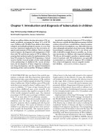
Figure 12.1. Progressive musculoskeletal pathology in cerebral palsy. (From Graham & Selber, 2003. Reproduced with
permission. Copyright
C
British Editorial Society of Bone and Joint Surgery.)
Management of spasticity in children 217
Figure 12.2. The stages of lower limb pathology in the child with cerebral palsy. (Modified after Rang, 1990.)
lead to the development of creative strategies to deal
with common clinical presentations (Preiss et al.,
2003)(Fig. 12.3).
Measuring spasticity in children: clinical
The Ashworth scale
There are few useful clinical measures of spastic-
ity and none validated for use in children. The Ash-
worth and modified Ashworth scales are blunt and
unresponsive tools in the assessment of the child
with cerebral palsy (Ashworth, 1964; Bohannon &
Smith, 1987). Their evaluations are subjective and
reliability between investigators may be a problem.
Most muscles in most children are grade 1+ to grade
3. Most useful clinical responses to spasticity man-
agement are within and not across a single Ashworth
grade. Of much greater utility is the measurement of
dynamic joint range, which can be used across most
major joints as a quantitative measure of spasticity
(Tardieu et al., 1954; Fosang et al., 2003).
The dynamic and static joint range of motion
The range of motion of joints in both the upper and
lower limbs is classically used as a proxy measure
of the length of muscles crossing that joint. In the
upper limb, the range of elbow extension is taken to
be a measure of the length of the elbow flexors, the
biceps and brachialis. Loss of elbow extension (fixed
flexion deformity) is taken to mean shortening of the
elbow flexors, although it should be noted that other
factors such as intrinsic joint contractures must be
excluded. In the lower limb, the range of dorsiflex-
ion at the ankle is considered to be a measure of the
calf muscle length. A further refinement is that the
range of ankle dorsiflexion with the knee flexed is
a measure of soleus length, and the range of ankle
dorsiflexion with the knee extended is a measure
of gastrocnemius length (Silfverskiold, 1924). This is
218 Rachael Hutchinson and H. Kerr Graham
Age 3
Age 7
Age 11
Age 19
Figure 12.3. The pathology in the lower limbs in children
with cerebral palsy is progressive as this sequence of hip
X-rays shows. At the age of 3 the hip X-ray is normal; at age
7 there is a very mild uncovering of the right hip. At age 11
the hip is subluxed and more than 50% is outside the
acetabulum. At age 19 there is painful degenerative
arthritis with few management options remaining.
the basis for the Silfverskiold test, and although it
may be only completely reliable under anaesthesia,
it is of great value as a simple test to differentiate
between gastrocnemius versus gastrocnemius and
soleus contracture. Typically, in hemiplegia there
is usually shortening of both the gastrocnemius
and soleus but in diplegia, isolated gastrocnemius
shortening is common. The criticism of the Sil-
fverskiold test (Silfverskiold, 1924) by Perry has in
our view led to an unwarranted devaluation of this
most useful clinical test (Perry et al., 1974, 1976,
1978).
Dynamic joint range of motion is measured by
provoking a stretch reflex if it is present. Typically
this first catch, or R1, comes in at a repeatable joint
angular position. This is usually 20 to 50 degrees
prior to R2, the static muscle length (Tardieu et al.,
1954). The variation is due to the proportion of
the deformity, which is dynamic, and not fixed. R2
approximates to the degree of ‘myostatic contrac-
ture’ or fixed shortening, which may require tendon
Example 1
A 3-year-old child with spastic diplegia has an equinus gait
affecting both lower limbs equally.
R1: −35 degrees (35 degrees of equinus)
R2: +5 degrees (5 degrees of dorsiflexion)
R2 − R1 = 40 degrees
Spasticity management is likely to be beneficial because
there are 40 degrees of dynamic shortening to be exploited
by spasticity management. Surgical lengthening of the heel
cord is contraindicated because the degree of fixed contrac-
ture is so small.
Example 2
A 10-year-old boy with hemiplegia walks with an equinus
gait on the affected side.
R1: − 30 degrees (30 degrees of equinus)
R2: −20 degrees (20 degrees of equinus)
R2 − R1: 10 degrees
Surgical lengthening of the Achilles tendon is indicated
because R2 minus R1 = 10 degrees. This is not enough
dynamic shortening for spasticity management and there
would be too much residual contracture.
Management of spasticity in children 219
lengthening and R1 the degree of spasticity or
dynamic shortening, which may respond to spas-
ticity management. These simple clinical tests of R1
and R2, static and dynamic muscle length can be per-
formed to assess the length of the adductors of the
hip, the hamstrings, quadriceps and the calf mus-
cles, some of the most important lower limb muscle
groups to be affected by spasticity.
The measurement of R2 and R1 are of great prac-
tical relevance in the management of spasticity
because they help to:
r
Differentiate between spasticity and contracture
r
Quantify the degree of spasticity present
r
Select which muscles might respond to spasticity
management
r
Serve to monitor the response to spasticity man-
agement
Measuring spasticity in children:
instrumented
Although we believe that dynamic joint range of
motion is a useful clinical tool in the measurement of
spasticity in children, there is a clear need for objec-
tive measurements with a greater degree of valid-
ity and repeatability. A number of techniques have
been described, and although most are useful within
research settings, none have become popular in clin-
ical practice.
Measurements of muscle stiffness address the
biomechanical rather than the neurophysiologi-
cal components of spasticity. These measurements
may also be obtained on the examination couch
or during walking. Static measurements include
measurements of muscle torque and resonant fre-
quency (Walsh, 1988; Corry et al., 1998; McLaugh-
lin et al., 1998). In a placebo-controlled clinical trial,
resonant frequency was found to be an objective
means to quantify muscle stiffness in the hemiplegic
upper limb. Reductions in resonant frequency were
recorded after injecting the forearm muscles with
BoNT-A (Corry et al., 1997).
Video recording of gait and aspects of the static
couch examination are very useful in clinical prac-
tice. Utility is further enhanced by split-screen, two-
dimensional recording with freeze-frame facilities
(Keenan et al., 2004). Careful editing and archiving
of patient records is also important.
Various scoringsystems or‘physician rating scales’
have been devised to increase the sensitivity and
objectivity of information gained from video record-
ings of children’s gait (Koman et al., 1993, 1994;
Corry, 1994). Although some have been tested for
repeatability, few have been tested for validity (Corry,
1994). Instrumented gait analysis, including kine-
matics and kinetics, provide the clinician with valu-
able information regarding the effects of spastic-
ity, contractures and other manifestations of the
UMN syndrome on gait (Gage et al., 1995). Typical
kinematic and kinetic patterns can be recognized
and interpreted in the light of the patient’s history
and clinical examination. Instrumented gait anal-
ysis is considered by many clinicians to be essen-
tial to plan such interventions as multilevel injec-
tions of BoNT-A and selective dorsal rhizotomy. The
dilemma is that only instrumented gait analysis gives
valid,repeatableand accurate measuresofthe effects
of spasticity and associated limb pathology on gait.
Instrumented gait analysis is limited in clinical utility
because of cost and availability. Furthermore, many
of the children who may need and benefit most from
spasticity management are too small and lacking in
co-operation for instrumented gait analysis using
current techniques.
Managing spasticity in children
In our preliminary open label study into the use of
BoNT-A in the lower limbs of children with cere-
bral palsy, the indications were summarized as ‘chil-
dren with dynamic deformities which were interfer-
ing with function, in the absence of fixed, myostatic
deformities’ (Cosgrove et al., 1994).
Although we believe that this statement remains
valid, we increasingly recognize the twin difficul-
ties in differentiating between dynamic and fixed
deformities and in measuring functional outcomes
in motor disabled children. Spasticity should not
be treated just because it is present. The natural
220 Rachael Hutchinson and H. Kerr Graham
history of spasticity in children is not sufficiently
well known nor are our present methods of manage-
ment sufficiently safe and effective to warrant such
an approach. Children with severe, ‘whole body’
involvement frequently use spasticity in functional
activities. A total extensor pattern may aid stand-
ing transfers. In this scenario, ‘successful’ spasticity
management, if measured by reduction in tone and
improved range of motion, might reduce rather than
enhance function. Hence the prime goal of spasticity
management must be improved function.
Understanding of motor development and meth-
ods of assessing function in children is also crucial.
A major characteristic of children who have cere-
bral palsy is the delayed acquisition of motor skills
(Rosenbaum et al., 2002). Given that spasticity man-
agement must often be undertaken against a back-
ground of growth and motor development, it is clear
that only controlled clinical trials can reliably sepa-
ratethe effects ofspasticity management on function
from gains made as part of normal motor develop-
ment. It is relatively straightforward to demonstrate
reduction in tone, improved joint range of motion
and improved muscle length after spasticity man-
agement, but evidence of functional gains is much
more demanding.
The Gross Motor Function Classification System
(GMFCS) is the most useful tool to stratify children
with cerebral palsy into five major groups (Palisano
et al., 1997). The Functional Mobility Scale is a use-
ful measure of functional mobility and is sensitive
to change after major interventions (Graham et al.,
2004). The Gross Motor Function Measure (GMFM)
is the most useful validated tool to measure func-
tional outcomes in children with cerebral palsy (Rus-
sell et al., 1989; Ketalaar et al., 1998; Wei et al., 2006).
The best candidates for spasticity management
are children who share the following features:
r
Mild to moderate spasticity
r
Good cognitive ability
r
No fixed contractures or deformities
r
Good selective motor control
r
Good general health
r
Stable supportive home environment
r
Access to appropriate physiotherapy
Management of spasticity
General
tnenamrePelbisreveR
Botulinum
toxin A
SDRITB
Oral
therapy
Focal
Figure 12.4. The four-way compass of spasticity
management with general versus focal (north and south)
and reversible versus permanent (west versus east).
r
Access to appropriate orthotics
Spasticity management may fail for a variety of
reasons including:
r
Spasticity, too severe and generalized
r
Poor cognitive ability
r
Fixed deformity
r
Poor selective motor control
r
Associated medical disease
r
Inadequate home support
r
No access to appropriate physiotherapy or
orthotics
Methods of spasticity management can be clas-
sified on a four-way compass (Fig. 12.4) according
to whether they are focal or general in effect and as
to whether the effects are permanent or temporary.
Within this four-way matrix (permanent-temporary,
focal-general) practical clinical guidelines may be
derived. The child with acquired spasticity sec-
ondary to acquired brain injury may have a relatively
short period of severe spasticity in a hemiplegic dis-
tribution. This could be managed by a program,
which may include intramuscular BoNT-A to large
muscle groups on the affected side including the
elbow flexors, the forearm muscles and the gas-
trosoleus. In this scenario the focal and temporary
nature of BoNT-A may be advantageous. Selective
dorsal rhizotomy (SDR) would be contraindicated
because it is permanent and bilateral.
Management of spasticity in children 221
A child with spastic diplegia who demonstrates
lower limb spasticity may respond favorably to
SDR; the permanence and generalized effects on
the lower limbs may be advantageous. Multiple,
repeated injections of BoNT-A would be less effec-
tive and risk systemic side effects.
The spasticity team and the spasticity clinic
Successful spasticity management in children
depends as much on teamwork as it does on tech-
niques and technology. Given that options in spastic-
ity management in children include administration
of drugs by oral and intrathecal routes, neurosurgi-
cal procedures and orthopaedic surgery, it should be
self-evident that spasticity management is a multi-
disciplinary exercise. In many centres, the concept
of a spasticity team and a spasticity clinic are well
developed. At the Royal Children’s Hospital in Mel-
bourne, the members of the team are drawn from the
following backgrounds:
r
Physical Medicine and Rehabilitation
r
Child Development and Rehabilitation
r
Physiotherapy
r
Occupational Therapy
r
Clinical Nurse Coordinators
r
Orthotics
r
Neurosurgery
r
Orthopaedic Surgery
r
Motion Analysis Laboratory
Many children are managed successfully by individ-
ual clinicians. However, there are a sufficient num-
ber of very difficult management problems to justify
a monthly spasticity clinic where the management
of a small number of problem children is discussed
in detail. Often investigations such as gait analysis
or examination under anaesthesia are requested to
aid decision making. We find the multidisciplinary
discussions stimulating and the communication
between specialties invaluable and management is
frequently altered with benefit to our patients. The
most frequent management issue is the interplay
between spasticity management and orthopaedic
surgery for deformity correction. Are the deformities
dynamic or fixed? To resolve this common dilemma,
an examination under the full relaxation of a general
anaesthetic may be invaluable.
Oral medications: generalized temporary
Oral medications for the management of spasticity
in children are in the temporary/generalized cat-
egory of the treatment compass. The agents most
frequently used are diazepam (Valium), baclofen
(Lioresal) and dantrolene sodium (Dantrium). In
general oral medications have a rather narrow ther-
apeutic window between efficacy and side effects.
Individual responses vary greatly, and a careful clin-
ical trial is necessary for many children to deter-
mine the individual response/side-effect profile.The
advantages and disadvantages of oral agents have
recently been discussed (Ried et al., 1998); see also
Chapter 7).
Diazepam
Most clinicians are familiar with the role of diazepam
as an anxiolytic agent. However evidence from ani-
mal work suggests that it possesses both muscle
relaxant and spinal reflex blocking properties. The
spinal actions of diazepam are the result of poten-
tiation of the presynaptic inhibitory effects of GABA
at GABA
A
receptors on spinal afferent presynaptic
terminals. Central effects in the brainstem reticular
formation result in sedation (Costa & Guidoffi, 1979;
Young & Delwaide, 1981a; Davidoff, 1989; Blackman
et al., 1992). Diazepam is rapidly and almost com-
pletely absorbed following oral or rectal adminis-
tration. Intravenous administration is occasionally
used to gain rapid control of muscle spasms in a
child who is excessively anxious and in pain after
orthopaedic procedures, but there is a risk of res-
piratory depression, and this route is not recom-
mended for routine use. Intramuscularinjections are
painful, rarely required and erratic in their absorp-
tion profile. Rectal administration is ideal when chil-
dren are fasting, nauseated or unable to take medi-
cation orally. The half-life in children is shorter than
in adults but still long at 18 hours. There tends to
be a cumulative effect of diazepam and it may take
222 Rachael Hutchinson and H. Kerr Graham
some time to reach the appropriate levels in body
tissues and optimal clinical effect. The drug’s vol-
ume of distribution is large, reflecting its extensive
tissue penetration within the body. It is metabolized
by the liver to pharmacologically active metabo-
lites, including nordiazepam and oxazepam (Green-
blatt et al., 1980). The most common side effects are
excessive sedation, respiratory depression, fatigue
and ataxia. Paradoxical effects may occur, including
hallucinations and increased spasticity. These must
be recognized and not managed by increasing the
dose.
Many children with cerebral palsy and other forms
of spasticity demonstrate increased spasticity when
theyare anxious and especiallywhen they arein pain.
Anxiety and pain seem to interact in a vicious cycle
to increase muscle tone after painful interventions
such as orthopaedic surgery (Baillieu et al., 1997).
Thecentral tranquilizingeffects and peripheral tone-
reducing effects of diazepam are extremely useful in
this situation. However, this means equally that there
is a very small threshold between effective reduction
in spasticity and sedation, invalidating diazepam for
chronic spasticity management in the vast major-
ity of children. We use diazepam almost routinely in
children with cerebral palsy who are facing painful
invasive procedures, including orthopedic surgery,
SDR, etc. Addiction and withdrawal symptoms are
reported in patients who use diazepam in the long
term (Young & Delwaide, 1981b). We have noted a
‘rebound’ phenomenon in children who have high
doses of diazepam postoperatively if it is stopped
suddenly. We routinely recommend that children
be ‘weaned’ slowly from diazepam use after short-
term/high-dose use.
Dantrolene
Dantrolene is valuable in the treatment and preven-
tion of malignant hyperthermia (Arens & McKinnon,
1971; Waterman et al., 1980). The main effect on
skeletal muscle appears to be direct muscle relax-
ation rather than a central or a spinal level of action.
Dantrolene inhibits the release of calcium from
the sarcoplasmic reticulum of muscle cells (Van-
Winkle, 1976; Desmedt & Hainaut, 1979; Molnar &
Kathirithamby, 1979). All muscles, both spastic and
normal, tend to be affected, ranging from relax-
ation through to weakness. Dantrolene is rapidly and
extensively absorbed, but there is a lack of pharma-
cokinetic data in children and especially in children
who have spasticity (Lietman et al., 1974; Young &
Delwaide, 1981a; Lerman et al., 1989). The utility of
dantrolene has been limited by the potential for hep-
atotoxicity (Utili et al., 1977; Wilkinson et al., 1979;
Chan, 1990). Fatal dantrolene-induced hepatitis has
been reported in adults but not in children. In chil-
dren, transaminase levels may rise, leading to a with-
drawal of therapy. Liver function should be assessed
prior to starting dantrolene therapy and at frequent
intervals thereafter (Ried et al., 1998).
A number of studies have been reviewed by Black-
man and colleagues, who note that the numbers of
patients within the published files are small and the
outcome measures not particularly objective (Black-
man et al., 1992). However, most studies do report
that in comparison with placebo, dantrolene has a
positive effect in reducing muscle tone but not nec-
essarily in improving function.
Tizanidine
Tizanidine is a benzothiodozol derivative of cloni-
dine and acts centrally as an alpha-2-adrenergic
agent. It may reduce spasticity by decreasing the
release of excitatory neurotransmitters from affer-
ent terminals and interneurones (Albright & Neville,
2000). Experience in children is limited and use is
limited by sedation.
Baclofen
Baclofen was introduced in the mid-1970s and
appears to act as a GABA agonist on the GABA
B
receptors (Rice, 1987). Baclofen inhibits transmit-
ter release by competitive inhibition of excitatory
neurotransmitters at the spinal level. There may be
actions in the spinal cord or more centrally which
are not yet fully described or understood (Pedersen
et al., 1974; Calta & Santomauro, 1976; Milla & Jack-
son, 1977; McKinlay et al., 1980; Young & Delwaide,
1981a; Dolphin & Scott, 1986; Fromm & Terrence,
Management of spasticity in children 223
1987). Pharmacokinetic data in respect of baclofen
children are lacking. Although baclofen is rapidly
absorbed after oral administration, levels in the cere-
brospinal fluid (CSF) are low because of its low lipid
solubility and 30% binding to plasma proteins. This
limits its transport across the blood–brain barrier
(Knutson et al., 1974; Gilman et al., 1990). It can
be administered orally or intrathecally but not par-
enterally. The response to baclofen in children varies
widely (Milla & Jackson, 1977). In general the thresh-
old between effective reduction in spasticity or mus-
cle tone and side effects such as dizziness, weakness
and fatigue is rather small. However, individual chil-
dren can respond well, and a careful trial of various
dose levels is worthwhile, although the majority will
have their medication discontinued because of side
effects. Hallucinations and seizures may occur with
abrupt withdrawal of baclofen; therefore, as with
diazepam, children who have become habituated to
larger doses should be weaned off the drug slowly. A
double-blind crossover trial of oral baclofen admin-
istration in children documented a decrease in spas-
ticity with little change in functional abilities, such
as ambulation and the performance of activities of
daily living (ADLs)(Milla & Jackson, 1977; Molnar &
Kathirithamby, 1979).
Much interest has been raised by the intrathe-
cal administration of baclofen (Knutson et al., 1974;
Penn & Kroin, 1985). Using this technique, the low
lipid solubility and binding to plasma proteins is
avoided by administration of the drug directly to the
target tissues. As will be seen in a later section, this
introduces a new ‘risk–benefit’ profile with specific
advantages and disadvantages.
Casting and orthoses: temporary/focal
The use of casting and orthoses can be classified as
focal/temporary. Casting, orthoses, neurolytic injec-
tions and physiotherapy are often used in vari-
ous combinations to manage spasticity in younger
children with cerebral palsy (see also Chapter 6).
The efficacy and duration of casting are related to
the proportions of dynamic and fixed contracture
before treatment and the responsiveness to the con-
nective tissue to stretching forces. Many clinicians
combine casting with intramuscular injections of
botulinum toxin. It is still unclear as to whether the
combined effect of injection and casting may be
better than either intervention on its own (Boyd &
Graham, 1997; Corry et al., 1997; Booth et al., 2004;
Kay et al., 2004); however, the evidence remains
anecdotal.
Spasticityof the gastrosoleus,resulting in dynamic
equinus, is usually treated by serial below-knee cast-
ing for periods of 1 to 4 weeks. Given the very
widespread utilization of the technique by phys-
iotherapists, there have been few outcome studies
(Corry et al., 1998; Brouwer et al., 2000). In a ran-
domized clinical trial, Corry and colleagues com-
paredserial casting with injection of botulinum toxin
in the management of dynamic equinus in chil-
dren with cerebral palsy. They concluded that both
interventions were effective but that the effects of
botulinum toxin lasted longer (Corry et al., 1998).
Flett et al. (1999) reported the inconvenience of cast-
ing and child and family preference for botulinum
toxin over serial casting.
Orthoses such as the ankle-foot orthosis (AFO) are
widely used in the management of younger children
who have calf spasticity. The effects of AFOs are dif-
ficult to study in younger children, but there are def-
inite biomechanical benefits, confirmed by motion
analysis (Rose et al., 1991; Ounpuu et al., 1993).
Intramuscular injections: chemoneurolysis:
temporary/focal
Intramuscular injections are focal in nature. The
duration depends on the agent, the concentration
used and the site of injection. ‘Chemoneurolysis’
refers to a nerve block resulting in impaired neu-
romuscular conduction by the destruction of neural
tissue, either temporarily or permanently (see Chap-
ter 8). Injection can be performed at many levels in
the peripheral nervous system from nerve root to
motor end plate (Glenn, 1990). The more proximal
the injection site, the more general and prolonged
the effect. Sciatic nerve block results in a variable
degree of weakness of all of the muscles supplied by
the sciatic nerve in the distal thigh and leg. Injec-
tion of the gastrocnemius muscle affects small local