Clinical use of orthodontic mini implants for intrusion and retraction a systematic review
Bạn đang xem bản rút gọn của tài liệu. Xem và tải ngay bản đầy đủ của tài liệu tại đây (370.4 KB, 14 trang )
Downloaded from search.informit.org/doi/10.3316/informit.180982068457349. RMIT University, on 02/10/2021 11:47 PM Australia time- Melbourne. © Australasian Orthodontic Journal, 2020.
Clinical use of orthodontic mini-implants for
intrusion and retraction: a systematic review
Sanjam Oswal,* Sanket S. Agarkar,† Sandeep Jethe,+ Sujata Yerawadekar,* Pradeep
Kawale,* Sonali Deshmukh* and Jayesh S. Rahalkar*
Department of Orthodontics and Dentofacial Orthopedics, Dr. D.Y. Patil Vidyapeeth University,* private practice† and
Department of Orthodontics and Dentofacial Orthopedics, Dr. D. Y. Patil Dental School,+ Pune, India
Background: Bimaxillary dental protrusion is common in many ethnic groups and is generally treated by the extraction of all first
premolars. However, temporary anchorage devices (TADs) are currently gaining popularity and most studies have focused on
anchorage loss, treatment duration, mini-implant success and failure rates, pain, discomfort and root resorption. Few studies have
focused on the clinical effectiveness of implants for the intrusion and retraction of anterior teeth.
Objectives: To assess the clinical use of orthodontic mini-implants for the intrusion and retraction of anterior teeth.
Methods: A systematic review of articles selected from PUBMED and Google Scholar was carried out to determine the clinical
use of orthodontic mini-implants for anterior tooth intrusion and retraction. Additional studies were hand searched to identify and
include clinical trials, prospective and retrospective studies, while excluding finite element method (FEM) studies and case reports.
A total of 598 articles were identified, of which 37 papers met the inclusion criteria and, following the elimination of duplicates,
20 articles were selected.
Results: Orthodontic mini-implants are more efficient for intrusion and retraction when compared to conventional intraoral and
extra-oral anchorage devices. A greater amount of intrusion and retraction is achieved when mini-implants are placed between
the first and second premolars without using any specific intrusive mechanics.
Conclusion: The present review highlights the clinical effectiveness of orthodontic mini-implants for anterior tooth intrusion and
retraction and the results suggest that orthodontic mini-implants are more effective than other conventional methods of anchorage
reinforcement.
(Aust Orthod J 2020; 36: 87-100)
Received for publication: March 2019
Accepted: August 2019
Sanjam Oswal: ; Sanket S. Agarkar: ; Sandeep Jethe: ;
Sujata Yerawadekar: ; Pradeep Kawale: ;
Sonali Deshmukh: ; Jayesh S. Rahalkar:
Introduction
Background
Bimaxillary dental protrusion is common in many
ethnic groups and is characterised by dentoalveolar
flaring of the maxillary and mandibular anterior teeth
with resultant protrusion of the lips and convexity
of the face. The present trend to treat bimaxillary
protrusion is by extraction of the four first premolars,
followed by anterior tooth retraction to obtain
the desired dental and soft-tissue profile changes.1
However, the extraction of premolars often raises the
query of anchorage demands.
© Australian Society of Orthodontists Inc. 2020
Orthodontic anchorage has always been an integral
aspect of treatment planning and execution. To address
the problem of anchorage loss, many appliances and
techniques have been devised, including the Nance
holding arch, transpalatal bars, extra-oral traction,
multiple teeth serving as one anchorage segment,
anchorage preparation, and the employment of light
forces.2 Recently, titanium-alloy mini-implants have
been suggested as a source of skeletal anchorage.3
There have been numerous studies conducted in
which mini-implants have been compared with
other anchorage devices. Sandler et al. showed that
there was no difference between the effectiveness of
Australasian Orthodontic Journal Volume 36 No. 1 May 2020
87
Downloaded from search.informit.org/doi/10.3316/informit.180982068457349. RMIT University, on 02/10/2021 11:47 PM Australia time- Melbourne. © Australasian Orthodontic Journal, 2020.
OSWAL, AGARKAR, JETHE, YERAWADEKAR, KAWALE, DESHMUKH AND RAHALKAR
TADs, a Nance button palatal arch, and headgear for
reinforcing anchorage during orthodontic anterior
retraction.4
Benson et al. showed that headgear and midpalatal
implants were equally effective in providing
anchorage;5 whereas Upadhyay et al. have shown that
TADs were more effective than other methods of
anchorage supplementation.1
Creekmore and Eklund were the first to report the use
of TADs, in a clinical report published in 1983.6 With
the recent emergence of mini-implant applications,
studies have been performed to investigate their
efficacy as an anchorage source for en-masse retraction
of anterior teeth.
Most of the studies have focused on anchorage loss,
treatment duration, mini-implant success and failure
rates, pain, discomfort and root resorption. Few
studies have focused on the clinical effectiveness of
implants for anterior tooth intrusion and retraction.
Although the anchorage control of posterior teeth
is superior with mini-implants, the nature of the
displacement of maxillary incisors with both methods
of space closure will be of interest for clinicians. The
type and direction of the resulting tooth movement
depends on the interaction between the line of force
and centre of resistance (Cr) of any specific tooth or
group of teeth.7 The line of force application, amount
of force, force decay and constancy, archwire-bracket
play and archwire deflection (regulated primarily
by the archwire properties) are critical factors for
controlling incisor retraction with mini-implant
supported anchorage.8
Therefore, the present study aimed to summarise the
clinical effectiveness of mini-implant use for incisor
intrusion and retraction.
Material and methods
Selection criteria
Inclusion criteria:
1. Articles published between January 2000 and
January 2018.
2. Articles stating the use of orthodontic miniimplants for anterior tooth intrusion and
retraction.
3.
88
RCT, clinical trials, prospective and retrospective
studies.
Australasian Orthodontic Journal Volume 36 No. 1 May 2020
Exclusion criteria:
1.
FEM studies.
2.
Case reports and animal studies.
PICO:
Participants: orthodontic patients
Intervention: mini-implants
Comparison: intraoral and extra-oral anchorage
reinforcement
Outcomes: intrusion and retraction
Information sources:
Two Internet sources of evidence were used by the
first author (S.O.) in the search for appropriate papers
satisfying the study purpose: The National Library of
Medicine (MEDLINE PubMed) and Google Scholar;
and a manual search was conduct using DPU college
library resources. All cross reference lists of the selected
studies were screened for additional papers that could
meet the eligibility criteria of the study. The databases
were searched until January 2018 using the keywords
provided in Table I and search strategy given in
Table II.
Study selection:
Various electronic databases were searched by the
first author (S.O.) using different strategies and the
key words and possible combinations. The number
of articles identified through the database search was
598. Duplicate articles were removed. After thorough
reading of titles and abstracts, the number of relevant
articles reduced to 27. Of these, 20 met the inclusion
criteria and were selected and confirmed by the other
authors (S.A. and J.R.).
Table I. Keywords.
Primary Keywords
Secondary Keywords
Orthodontic
Mini-implant
Micro-implant, mini screw,
temporary anchorage device,
TADs, skeletal anchorage
Intrusion
Incisor intrusion, incisor
displacement
Retraction
Anterior teeth retraction, en masse
retraction
CLINICAL USE OF ORTHODONTIC MINI-IMPLANTS FOR INTRUSION AND RETRACTION
Downloaded from search.informit.org/doi/10.3316/informit.180982068457349. RMIT University, on 02/10/2021 11:47 PM Australia time- Melbourne. © Australasian Orthodontic Journal, 2020.
Table II. Search strategy.
Sr. No.
Search strategy
Number of
articles found
Number of
articles selected
Reason for exclusion
SS1
Orthodontic AND mini implant AND
intrusion AND retraction
15
4
FEM study/case report/not
relevant to this study
SS2
Orthodontic AND micro implant AND
intrusion AND retraction
2
0
FEM study/case report/not
relevant to this study/duplicate
SS3
Orthodontic AND mini screw AND intrusion
AND retraction
10
0
FEM study/case report/not
relevant to this study/duplicate
SS4
Orthodontic AND temporary anchorage
device AND intrusion AND retraction
5
1
FEM study/case report/not
relevant to this study/duplicate
SS5
Orthodontic AND TADs AND intrusion AND
retraction
3
0
FEM study/case report/not
relevant to this study/duplicate
SS6
Orthodontic AND skeletal anchorage AND
intrusion AND retraction
25
2
FEM study/case report/not
relevant to this study/duplicate
SS7
Orthodontic AND mini implant OR micro
implant OR mini screw OR temporary
anchorage device OR TADs OR skeletal
anchorage AND intrusion AND retraction
46
2
FEM study/case report/not
relevant to this study/duplicate
SS8
Orthodontic AND mini implant AND
intrusion OR incisor intrusion OR incisor
displacement AND retraction
101
1
FEM study/case report/not
relevant to this study/duplicate
SS9
Orthodontic AND mini implant AND
intrusion AND retraction OR anterior teeth
retraction OR en masse retraction
384
10
FEM study/case report/not
relevant to this study/duplicate
Data collection process:
•
an endpoint appropriate to the aim of the study
The data collection process was performed by the first
author (S.O.). A Microsoft Excel Spreadsheet was
populated with the study data, which was re-evaluated
by the other authors (S.A. and J.R.).
•
sample size adequacy
•
distribution of sample size within the groups
•
adequate statistical analysis
•
main outcome to be measured is clearly described
in the introduction/methods section
•
intervention and sites of interest clearly described
•
and main findings of the study.
Data items:
The data items included were study ID, author’s name,
year of publication, location, study design, sample
size, population, implant specification, intervention,
comparison, outcome, results and conclusion.
Results
Risk of bias/quality assessment in
individual studies
The quality of the selected articles was analysed using
a self-modified MINORs checklist.9,10 A total of 10
criteria were analysed to grade the risk of the studies:
•
a clearly stated aim
•
an inclusion criteria of consecutive patients
•
data collection
The items were scored 0 (not reported), 1 (reported
but inadequate) or 2 (reported and adequate). If the
total score of each study was <15, it was considered a
low quality study, 15–17 was considered a moderate
quality study, and 18–20 was considered a high quality
study (Tables III, IV, V).
As this was a systematic review, the heterogeneity of
the selected studies was not assessed.
Study selection
The data search was carried out based on the title
relevance to the systematic review. A total of 598
titles were screened across various medical and
Australasian Orthodontic Journal Volume 36 No. 1 May 2020
89
OSWAL, AGARKAR, JETHE, YERAWADEKAR, KAWALE, DESHMUKH AND RAHALKAR
Downloaded from search.informit.org/doi/10.3316/informit.180982068457349. RMIT University, on 02/10/2021 11:47 PM Australia time- Melbourne. © Australasian Orthodontic Journal, 2020.
Table III. Quality of studies when mini-implants are compared with extra oral anchorage devices.
Sr. No.
Methodological items
Deguchi
et al.
Yao CC
et al.
Lai EH
et al.
Chen M
et al.
A.Y. Lee and
Y. H. Kim
Park HM
et al.
1.
Clearly stated aim
2
2
2
2
2
2
2.
Inclusion criteria of consecutive
patients
1
1
1
1
2
2
3.
Data collection
2
2
2
2
2
2
4.
Endpoint appropriate to the aim
of study
2
2
2
2
2
2
5.
Is the sample size adequate
1
1
1
1
1
1
6.
Distribution of sample size in
different groups
1
1
1
1
2
2
7.
Adequate statistical analysis
2
2
2
2
2
2
8.
Are the main outcome to be
measures are clearly described in
introduction/ methods section
2
2
2
2
2
2
9.
Are the intervention and sites of
interest clearly described
2
2
2
2
2
2
10.
Are the main finding of study
clearly described.
2
2
2
2
2
2
17
17
17
17
19
19
TOTAL
Interpretation: <15 = low quality studies, 15–17 = moderate quality study, 18–20 = high quality study.
The items are scored 0 (not reported), 1 (reported but inadequate) or 2 (reported and adequate).
Table IV. Quality of studies when mini-implants are compared with intra oral anchorage devices.
Sr. No.
Methodological items
Upadhyay
et al.
Liu YH
et al.
Liou and
Chang
Basha AG
et al.
1.
Clearly stated aim
2
2
2
2
2.
Inclusion criteria of consecutive patients
1
2
1
2
3.
Data collection
2
2
1
1
4.
Endpoint appropriate to the aim of study
2
2
2
2
5.
Is the sample size adequate
1
1
1
1
6.
Distribution of sample size in different groups
2
2
1
2
7.
Adequate statistical analysis
2
2
2
2
8.
Are the main outcome to be measures are
clearly described in introduction/ methods
section
2
2
2
2
9.
Are the intervention and sites of interest
clearly described
2
2
2
2
10.
Are the main finding of study clearly
described.
2
2
2
2
18
19
16
18
TOTAL
Interpretation: <15 = low quality studies, 15–17 = moderate quality study, 18–20 = high quality study.
The items are scored 0 (not reported), 1 (reported but inadequate) or 2 (reported and adequate).
90
Australasian Orthodontic Journal Volume 36 No. 1 May 2020
CLINICAL USE OF ORTHODONTIC MINI-IMPLANTS FOR INTRUSION AND RETRACTION
Downloaded from search.informit.org/doi/10.3316/informit.180982068457349. RMIT University, on 02/10/2021 11:47 PM Australia time- Melbourne. © Australasian Orthodontic Journal, 2020.
Table V. Quality of studies when mini-implants are used for intrusion and retraction.
Sr. No.
Methodological items
Upadhyay
et al.
Kim SH
et al.
Liu H
et al.
Lee KJ
et al.
Upadhyay
et al.
Victor D.
et al.
Jee JH et
al.
Monga
N. et al.
1.
Clearly stated aim
2
2
2
2
2
2
2
2
2.
Inclusion criteria of
consecutive patients
2
1
1
2
2
1
1
2
3.
Data collection
2
1
2
1
2
2
2
2
4.
Endpoint appropriate to
the aim of study
2
2
2
2
2
1
2
2
5.
Is the sample size
adequate
1
1
1
1
1
1
2
1
6.
Distribution of sample size
in different groups
1
1
2
1
1
2
1
2
7.
Adequate statistical
analysis
2
2
2
2
2
2
2
2
8.
Are the main outcome
to be measured clearly
described in introduction/
methods section
2
2
1
2
2
2
2
2
9.
Are the intervention and
sites of interest clearly
described
2
2
2
2
1
2
2
2
10.
Are the main findings of
study clearly described.
2
2
1
2
2
2
2
2
18
16
16
17
17
17
18
19
TOTAL
Interpretation: <15 = low quality studies, 15–17 = moderate quality study, 18–20 = high quality study.
The items are scored 0 (not reported), 1 (reported but inadequate) or 2 (reported and adequate).
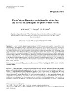
dental journals, of which 93 titles were short-listed.
On duplicate removal and a thorough review of the
abstracts, 27 full-text articles were obtained. A final
total of 20 articles met the selection criteria and were
selected for qualitative synthesis for the systematic
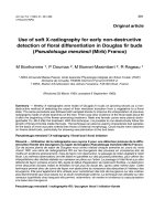
review. The outline of the selection process is
illustrated in Figure 1.
Table VI shows the effectiveness of mini-implants
when compared with extra-oral anchorage reinforcement such as J-hook headgear and conventional
headgears. It was evident that mini-implants provide
better vertical and sagittal control but do not
significantly decrease treatment time.
Table VII shows the effectiveness of miniimplants when compared with intraoral anchorage
reinforcement devices such as a Nance holding arch,
a transpalatal arch, or banding of the second molars.
Discussion
The present systematic review identified articles
in which the effectiveness of mini-implants was
compared with intraoral and extra-oral anchorage
reinforcement for anterior tooth intrusion and
retraction. Also, additional studies stated the
effectiveness of mini-implants for intrusion and
retraction without comparison against traditional
methods of anchorage reinforcement. Therefore, the
effectiveness of mini-implants may be evaluated under
the following headings:
a.
Effectiveness of mini-implants when compared
with extra-oral anchorage reinforcement.
b. Effectiveness of mini-implants when compared
with intraoral anchorage reinforcement.
c.
Effectiveness of mini-implants alone.
Table VIII shows the results when mini-implants are
used for intrusion and retraction without a comparison
with conventional anchorage reinforcement devices.
Australasian Orthodontic Journal Volume 36 No. 1 May 2020
91
OSWAL, AGARKAR, JETHE, YERAWADEKAR, KAWALE, DESHMUKH AND RAHALKAR
Iden0fica0on
Eligibility
Screening
Recordsiden%fiedthrough
databasesearching
(N = 591)
Addi%onalrecordsiden%fied
throughothersources
(N = 7)
Totalrecords
(N = 598)
Titlesscreened
(N = 598)
Recordsexcluded
a=erreviewof%tles
(N = 505)
Titlesscreenedfor
duplicateremoval
(N = 93)
Excluded-duplicates
(N = 56)
Abstractsscreened
(N = 37)
Recordsexcluded
a=erreviewof
abstracts
(N = 10)
Fulltextsscreenedon
basisof%tleand
abstract
(N = 27)
Included
Downloaded from search.informit.org/doi/10.3316/informit.180982068457349. RMIT University, on 02/10/2021 11:47 PM Australia time- Melbourne. © Australasian Orthodontic Journal, 2020.
Figure 1. PRISMA 2009 Flow Diagram.
Studiesexcludeda=erreview
offulltext(N = 7)
• Languageotherthan
English=0
• Studiesnotmee%ng
theinclusioncriteria=7
Studiesincludedin
qualita%vesynthesis
(N = 20)
Figure 1. PRISMA 2009 Flow Diagram.
Effectiveness of mini-implants when
compared with extra-oral anchorage
reinforcement
The present systematic review identified six articles
that compared the effectiveness of mini-implants with
extra-oral anchorage reinforcements such as J-hook
headgear and/or headgear anchorage.
92
Australasian Orthodontic Journal Volume 36 No. 1 May 2020
When comparing the intrusion effects between
implant anchorage and J-hook headgear on the
maxillary incisors, Deguchi et al.11 found that the
incisors intruded by 3.6 ± 1.7 mm and the molars
extruded by 0.1 ± 2.0 mm in the implant group. In the
J-HG group, the incisors intruded by 1.1 ± 1.6 mm
and the molars extruded by 1.3 ± 2.9 mm. There was
CLINICAL USE OF ORTHODONTIC MINI-IMPLANTS FOR INTRUSION AND RETRACTION
Downloaded from search.informit.org/doi/10.3316/informit.180982068457349. RMIT University, on 02/10/2021 11:47 PM Australia time- Melbourne. © Australasian Orthodontic Journal, 2020.
Table VI. Mini-implants compared with extra oral anchorage devices.
Author and
year
Study
design
Sample Comparison
size
Intervention
Outcome
T. Deguchi
et al.
2008
CS
18
MI and J-hook
headgear
MI: 8
J-HG: 10
MI group – more incisor intrusion
J-HG group – more molar extrusion
J-HG group – more root resorption
Yao CC
et al.
2008
RS
47
MI and HG
HG: 22
MI: 25
MI group – greater anterior tooth retraction
MI group – less maxillary molar mesialisation
MI group – more intrusion of the maxillary first molar
HG group – more extrusion of maxillary first molar
Lai EH et
al.
2008
RS
40
MI, miniplates
and HG
HG: 16
Miniplate group – significant intrusion of the maxillary
MI: 15
posterior teeth
Miniplates: 9 MI group – greater retraction of the maxillary anterior teeth,
MI group – less anchorage loss of the maxillary posterior teeth
MI group – maxillary molar intrusion
Chen M
et al.
2015
CS
31
MI and
HG with SL
brackets
MI: 15
HG: 16
MI group – treatment time almost similar
MI group – better control in both the antero-posterior and
vertical directions
MI group – more retraction of the maxillary incisors
MI group – less anchorage loss of the maxillary first molar
Ah-Young
Lee and
Young Ho
Kim 2011
CS
40
MI and HG
HG: 20
MI: 20
MI group –
maximum anchorage of molars,
greater retraction of incisors,
greater intrusion of incisor and molar
Park HM
et al.
2012
CS
24
MI and HG
HG: 12
MI: 12
MI group –
more backward movement of MXCI, MXLI, and MXC
more intrusion of MXCI and MXC
less forward movement of MXP2, MXM1, and MXM2
less contraction of MXP2 and MXM1
CS: clinical study, RS: retrospective study, MI: mini implant, HG: headgear, J-HG: J-hook headgear, SL: self ligating, MXCI: maxillary central incisor, MXLI:
maxillary lateral incisor, MXC: maxillary canine, MXP2: maxillary second premolar, MXM1: maxillary first molar, MXM2: maxillary second molar
more incisor intrusion in the implant group and more
molar extrusion in the J-HG group. To investigate
the effectiveness of bony anchorage during maxillary
dento-alveolar retraction in adults with Class II and
Class I malocclusions compared with traditional extraoral anchorage such as headgear, Yao et al.12 found
that the skeletal anchorage group had greater anterior
tooth retraction and less maxillary molar mesialisation
than the headgear group. Translational movement
of the incisors was more common than tipping
movement, and intrusion of the maxillary dentition
was greater in patients receiving miniplates compared
with those receiving screw-type bony anchorage.12 In
addition, in patients with a high mandibular plane
angle, those receiving skeletal anchorage had genuine
intrusion of the maxillary first molar whereas those
receiving headgear anchorage had extrusion of the
maxillary first molars.
When comparing the orthodontic outcomes of
maxillary dento-alveolar protrusion treated with
headgear, miniscrews, or miniplates for maximum
anchorage, Lai et al.13 found significant intrusion of
the maxillary posterior teeth in the miniplate group
but not in the miniscrew and headgear groups.
Greater retraction of the maxillary anterior teeth, less
anchorage loss of the maxillary posterior teeth, and the
possibility of maxillary molar intrusion all facilitated
correction of the Class II malocclusion, especially for
patients with a hyperdivergent face.
In a determination of the differences between the outcomes of treatment using micro-implant anchorage
compared with headgear anchorage in adult patients
with bimaxillary protrusion treated with self-ligating
brackets, Chen et al.14 reported that micro-implant
anchorage did not shorten the orthodontic treatment
period and that micro-implant anchorage achieved
better control in the antero-posterior and vertical directions during treatment when compared with headgear anchorage. Also, it was concluded that microimplant anchorage might result in more retraction of
Australasian Orthodontic Journal Volume 36 No. 1 May 2020
93
OSWAL, AGARKAR, JETHE, YERAWADEKAR, KAWALE, DESHMUKH AND RAHALKAR
Downloaded from search.informit.org/doi/10.3316/informit.180982068457349. RMIT University, on 02/10/2021 11:47 PM Australia time- Melbourne. © Australasian Orthodontic Journal, 2020.
Table VII. Mini-implants compared with intra oral anchorage devices.
Author
and year
Study
design
Sample
size
Comparison
Intervention Outcome
Upadhyay
M et al.
2008
CS
30
MI and CAR
MI : 15
CAR: 15
In MI group –
Distal movement of the MXM
Intrusive effect on the MXM
Intrusion of the MXC1
MXC1 retracted by controlled tipping and partly by translation
In CAR group –
Mesial movement of MXM
Extrusive effect on the MXM
MXC1 showed controlled tipping
Upadhyay
M et al.
2008
RCT
40
MI and CAR
MI : 20
CAR: 20
In MI group –
MXM distalised and intruded
MXC1 retracted and intruded
In CAR group –
MXM mesialised and extruded
MXC1 retracted and intruded
Liu YH et
al.
2009
CS
34
MI and TPA
MI : 17
TPA : 17
In MI group –
More retraction of MXC1
MXC1 and MXM were intruded
MXM distalised
In TPA group –
MXC1 and MXM were extruded
MXM mesialised
Liou and
Chang
2010
RS
50
MI and CAR
MI : 20
CAR: 30
In MI group –
Retraction at U1E (mm): 8.2 ± 2.4
Intrusion at U1E (mm): 0.4 ± 2.0
Retraction at U1A (mm): 3.0 ± 2.7
Intrusion at U1A (mm): 2.7 ± 1.8
In CAR group –
Retraction at U1E (mm): 6.5 ± 2.1
Intrusion at U1E (mm): 0.0 ± 1.6
Retraction at U1A (mm): 1.3 ± 1.6
Intrusion at U1A (mm): 2.5 ± 1.4
Basha AG
et al.
2010
CS
14
MI and CAR
CAR: 7
MI : 7
Anchor loss was statistically significant in CAR group (1.73 mm)
Retraction Time –
In CAR group: 0.92 mm per month (0.917)
In MI group: 0.85 mm per month (0.923)
S. Al-Sibaie
and M. Y.
Hajeer
2014
RCT
56
MI and TPA
MI : 28
TPA : 28
Mean treatment duration:
In MI group – 12.90 months
TPA group – 16.97 months
In MI group –
U1E: retracted (-5.92 mm) and intruded (-1.53 mm)
U1A: retracted (-4.56 mm) and intruded (-1.16 mm)
MXM: distalised (0.89 mm)
In TPA group –
U1E: retracted (-4.79 mm) and extruded(0.92 mm)
MXM: mesialised (1.50 mm) and extrusion seen
CS: clinical study, RS: retrospective study, RCT: randomised controlled trials, MI: mini implant, CAR: conventional anchorage reinforcement, TPA:
transpalatal arch, MXCI: maxillary central incisor, MXM: maxillary molars, U1E: maxillary central incisor edge, U1A: maxillary central incisor apex
94
Australasian Orthodontic Journal Volume 36 No. 1 May 2020
CLINICAL USE OF ORTHODONTIC MINI-IMPLANTS FOR INTRUSION AND RETRACTION
Downloaded from search.informit.org/doi/10.3316/informit.180982068457349. RMIT University, on 02/10/2021 11:47 PM Australia time- Melbourne. © Australasian Orthodontic Journal, 2020.
Table VIII. Effectiveness of mini-implants alone.
Author
and year
Study
design
Sample
size
Study type
Intervention
Outcome
Upadhyay PS
M et al.
2009
23
Cephalometric
study
MI
MXCI retracted and intruded
MXM distalised and intruded
Kim SH et
al.
2009
RS
17
Cephalometric
study
MI
MXM showed mesial movement, extrusion and mesial
tipping
MXCI retracted and slight amount of extrusion seen
Liu H et
al.
2011
CS
60
3D CT scan
MI
Retraction of MXU1E 5.94 ± 0.90 mm
Retraction of MXU1A 1.40 ± 0.23 mm
Intrusion of MXCI : 1.84 ± 0.26
Mesial drifting of MI seen
Lee KJ et
al.
2011
CS
36
Cephalometric
study
MI between
MXP2 and
MXM1
MI between
MXP2 and
MXP1
MI between MXP2 and MXP1 –
Greater intrusion (1.59 mm) of U1E
Upadhyay PS
M et al.
2012
32
Cephalometric
study
FFA group:
18
MI group: 14
In FFA group –
Extrusion and mesial movement of the lower molar
Lower incisor proclination
In MI group –
Distalisation and intrusion of the upper molar and incisor
Victor D
et al.
2014
CS
20
Cephalometric
study
MI group: 10
Control
group: 10
In MI group –
Distal tipping of molars,
Intrusion of incisor tip and apex
Intrusion of molar
In control group –
Mesial tipping molars,
Extrusion of incisor tip and apex
Extrusion of molars
Jee JH et
al.
2014
CS
31
Cephalometric
study using C
implants
Conventional
C-wire group
: 15
Preformed
C-wire group
: 16
Monga N RS
et al.
2016
18
Cephalometric
study
MI
In Preformed C-wires group –
Maximum retraction of the maxillary anterior teeth
Maintenance of posterior occlusions without mesialisation
of the molars.
Lesser treatment time
Easy and simultaneous levelling and space closure
MXM position
Sagittal – mesial movement
Vertical – extrusion
Angular – distal tipping
MXCI position
Sagittal – distal movement
Vertical – intrusion
Angular – distal tipping
PS: prospective study, RS: retrospective study, CS: clinical study, MI: mini implant, FFA: fixed functional appliance, MXCI: maxillary central incisor, MXP1:
maxillary first premolar, MXP2: maxillary second premolar, MXM: maxillary molars, MXU1E: maxillary central incisor edge, MXU1A: maxillary central
incisor apex, 3D CT: 3 dimensional computed tomography
Australasian Orthodontic Journal Volume 36 No. 1 May 2020
95
Downloaded from search.informit.org/doi/10.3316/informit.180982068457349. RMIT University, on 02/10/2021 11:47 PM Australia time- Melbourne. © Australasian Orthodontic Journal, 2020.
OSWAL, AGARKAR, JETHE, YERAWADEKAR, KAWALE, DESHMUKH AND RAHALKAR
the maxillary incisors and less anchorage loss of the
maxillary first molars when compared with the use of
headgear anchorage.
In a comparison of the anchorage loss in the upper first
molar and retraction of the upper central incisor in
cases with a Class I malocclusion between orthodontic
mini-implants (OMIs) and conventional anchorage
reinforcements (CARs), Lee and Kim15 determined
that the upper incisor edge retracted by 9.5 mm in a
mini-implant group and 7.1 mm in a control group.
The upper central incisors intruded by 0.9 mm and
the upper molars intruded by 1.0 mm in the miniimplant group, whereas the upper central incisors
extruded by 0.7 mm and the upper molars extruded
by 0.9 mm in the conventional group. Park et al.16
compared the effects of conventional and orthodontic
mini-implant anchorage (OMI) on tooth movement
and arch-dimension in the maxillary dentition in
Class II division 1 patients. It was found that, in the
OMI group, there was greater distal movement of the
maxillary incisors and canines. A greater amount of
maxillary central incisor and canine intrusion was
observed with less forward movement of the posterior
teeth compared with the conventional group.
The findings of the articles concluded that the use
of mini-implants provides better vertical and sagittal
control when compared with extra-oral anchorage
reinforcements like J hook headgear and conventional
headgear. Although mini-implants do not shorten
treatment duration significantly, they provide greater
anterior retraction and less molar mesialisation but
produce molar intrusion, whereas extra-oral anchorage
using headgear may result in molar extrusion and
molar mesialisation.
Effectiveness of mini-implants when
compared with intraoral anchorage
reinforcement
The present systematic review identified six articles
in which the effectiveness of mini-implants was
compared with intraoral anchorage reinforcement
such as transpalatal arches (TPA), Nance holding
arch, or banding of the second molars. When
comparing the changes in position of the molars
and incisors between the implant and conventional
method of anchorage reinforcement group, Upadhyay
et al.17 found that there was a net distal and intrusive
movement of the molar and the maxillary incisor
96
Australasian Orthodontic Journal Volume 36 No. 1 May 2020
intruded in the implant group. The maxillary central
incisors were retracted primarily by controlled tipping
and partly by translation in the implant group. In the
conventional anchorage group, there was net mesial
and extrusive movement of the molars and incisor
retraction showed significant amounts of controlled
tipping, but some uncontrolled tipping was also
noted.
In a RCT study, Upadhyay et al.1 compared the
dentoskeletal and soft-tissue treatment effects
during en-masse retraction of anterior teeth using
mini-implants as anchor units with conventional
methods of anchorage such as transpalatal arches and
banding of the second molars, in bimaxillary dental
protrusion patients undergoing the extraction of all
four first premolars. It was found that, in the implant
group, the maxillary and mandibular molars were
distalised by 0.78 ± 1.35 mm and 0.89 ± 1.23 mm
and were intruded by 0.22 ± 0.65 mm and 0.75 ±
0.84 mm respectively. In addition, the maxillary and
mandibular incisors were retracted and intruded. In
the non-implant group, the maxillary and mandibular
molars mesialised by 3.22 ± 1.06 mm and 2.67 ± 2.11
mm and were extruded by 0.67 ± 1.19 mm and 1.22
± 1.59 mm, respectively.1
In a comparison of the differences in cephalometric
parameters after active orthodontic treatment
using mini-screw implants or transpalatal arches as
anchorage in adult patients with bimaxillary dental
protrusion needing extraction of four premolars,
Liu et al.18 reported that the maxillary incisors were
retracted by 7.03 ± 1.99 mm and intruded by 1.91
± 2.33 mm, while the maxillary molars distalised by
1.42 ± 2.55 mm and intruded by 0.06 ± 1.40 mm
in the mini-screw implant group. In a TPA group,
the maxillary incisors retracted by 4.76 ± 1.67 mm
and extruded by 1.17 ± 1.99 mm while the molars
mesialised by 1.91 ± 1.75 mm and extruded by 1.47
± 1.15 mm. These results show that the maxillary
incisors and molars intruded in the implant group
and extruded in the TPA group.
In a retrospective study, when investigating apical root
resorption of maxillary incisors in patients requiring
en-masse maxillary anterior retraction and intrusion
using miniscrews and the factors disposing a patient
to apical root resorption, Liou and Chang19 found
retraction and intrusion at the incisor tip of 8.2 ± 2.4
mm and 0.4 ± 2.0 mm respectively in the mini-implant
group. Furthermore, at the incisor root apex, there
Downloaded from search.informit.org/doi/10.3316/informit.180982068457349. RMIT University, on 02/10/2021 11:47 PM Australia time- Melbourne. © Australasian Orthodontic Journal, 2020.
CLINICAL USE OF ORTHODONTIC MINI-IMPLANTS FOR INTRUSION AND RETRACTION
was retraction and intrusion of 3.0 ± 2.7 mm and 2.7
± 1.8 mm respectively. These values were greater when
compared with the conventional anchorage group.
When measuring and comparing the difference
between the rate of en-masse retraction with miniimplants and molar anchorage, Basha et al.20 found
that anchorage loss was statistically significant in a
non-implant group (1.73mm) when compared with
an implant group. Al-Sibaie and Hajeer21 conducted
a RCT to compare the skeletal, dental, and soft
tissue treatment outcomes between sliding en-masse
retraction of the upper anterior teeth employing miniimplants and a two-step sliding retraction approach
employing conventional anchorage in patients
presenting with a Class II division 1 malocclusion.
In the mini-implant group, the upper incisor edges
retracted (−5.92 mm) and intruded (−1.53 mm),
while the upper incisor apices retracted (−4.56 mm)
and intruded (−1.16 mm) and the upper molars were
distalised (0.89 mm). In the TPA group, the upper
incisor edges retracted (−4.79mm) and extruded (0.92
mm) and the upper molars were mesialised (1.50 mm)
and extrusion was seen.21
It was clear that the use of mini-implants provided
better anchorage control in the vertical and sagittal
planes and produced molar distalisation along with
the intrusion of the molars and incisors. Whereas
conventional anchorage reinforcements such as TPAs,
Nance holding arches, or the banding of second
molars resulted in greater molar mesialisation and the
extrusion of molars and incisors. Also, the incisors
retracted mainly by controlled tipping and partially
by translation when mini-implants were used.
Effectiveness of mini-implants alone
The present systematic review identified eight articles
in which the effectiveness of mini-implants was
evaluated for their ability to produce intrusion along
with retraction. In one study, the effectiveness of
mini-implants was evaluated according to the implant
placement site.
A study conducted to examine the skeletal, dental, and
soft tissue treatment effects of retraction of maxillary
anterior teeth using mini-implant anchorage in
non-growing Class II division 1 female patients by
Upadhyay et al.22 found that during anterior tooth
retraction, the maxillary central incisors were retracted
and intruded while the upper molars were distalised
and intruded (0.45 ± 0.79 mm and 0.64 ± 0.78 mm
respectively). In addition, the lower molars were
mesialised and extruded (0.64 ± 1.1 mm and 0.52 ±
0.75 mm, respectively). To achieve independent enmasse retraction of the anterior teeth while avoiding
the use of orthodontic appliances in the posterior
segments during the retraction period, Kim et al.23
retrospectively found that the maxillary molars showed
mesial movement, extrusion and mesial tipping, while
the mandibular molars showed slight extrusion. The
upper incisors were retracted with a minor amount of
extrusion and the lower incisor intruded slightly.
In a study to quantitatively evaluate the position of
miniscrews and molars subjected to an orthodontic
force (150 g) and using 3D CT registration evaluations,
Liu et al.24 found that the maxillary incisors retracted
at their edge and apex by 5.94 ± 0.90 mm and 1.40
± 0.23 mm respectively, and intruded by 1.84 ± 0.26
mm. It was also found that the miniscrews drifted
mesially at the head and apex by 0.23 ± 0.08 mm and
0.23 ± 0.07 mm respectively. Lee et al.25 evaluated the
anteroposterior and vertical displacement patterns of
the maxillary teeth in sliding mechanics determined
by the position of interradicular miniscrews after
the extraction of premolars. Implants were placed
between the maxillary second premolar and the first
molar (group A) and between the first and second
premolars (group B). In group A, the vertical position
of the incisal edge did not change significantly during
the retraction period. While in group B, a significantly
greater amount of intrusion (1.59 mm) was found
when compared with group A. Simultaneous
intrusion and retraction can be effectively obtained by
using miniscrews between the premolars in extraction
patients, without the need for additional intrusive
mechanics.
When comparing the treatment effects of maxillary
anterior tooth retraction with mini-implant anchorage
in young adults presenting with a Class II division 1
malocclusion involving the extraction of the maxillary
first premolars with comparative patients treated by a
fixed functional appliance, Upadhyay et al.26 reported
that in the mini-implant group the upper molar and
upper incisors intruded by 0.64 ± 0.78 mm and
1.32 ± 1.08 mm and distalised by 0.45 ± 0.79 mm
and 5.18 ± 2.74 mm, respectively. The lower molars
extruded and mesialised by 0.82 ± 0.75 mm and 0.64
± 1.1 mm and the lower incisors distalised by 1.77 ±
2.16 mm, respectively.
Australasian Orthodontic Journal Volume 36 No. 1 May 2020
97
Downloaded from search.informit.org/doi/10.3316/informit.180982068457349. RMIT University, on 02/10/2021 11:47 PM Australia time- Melbourne. © Australasian Orthodontic Journal, 2020.
OSWAL, AGARKAR, JETHE, YERAWADEKAR, KAWALE, DESHMUKH AND RAHALKAR
implants proved to be more efficient for producing
intrusion and retraction.
Victor et al.27 compared the torque of the incisors,
the tip of the molars and vertical control during
orthodontic treatment with and without mini screw
implants. The results indicated that there was mild
distal tipping of the molars, intrusion of the incisor
tip and apex and very mild intrusion of the molar in
the implant group. In the control group, there was a
mesial tipping of the molars, extrusion of the incisor
tip and apex and a mild extrusion of the molars.27
When evaluating the therapeutic effects of a preformed
assembly of nickel-titanium (NiTi) and stainless steel
(SS) archwires (preformed C-wire) combined with
temporary skeletal anchorage devices (TSADs) as the
sole source of anchorage and to compare these effects
with those of a SS version, of C-wire (conventional
C-wire) for en-masse retraction, Jee et al.28 found
that the maxillary anterior teeth were fully retracted
to close the extraction spaces. Uprighting of the
maxillary anterior teeth by controlled tipping was
observed. In addition, mesialisation and mesial tipping
of the maxillary and mandibular molars was noted in
the conventional C-wire group compared with the
preformed C-wire group. There was linguoversion
of the mandibular anterior teeth in both groups
and extrusion of the mandibular teeth was observed
in both groups, except in the anterior region in the
preformed C-wire group. In relation to the soft-tissues,
the upper and lower lips moved posteriorly.28 During
quantitative and qualitative assessment of anchorage
loss during en-masse retraction with indirectly loaded
miniscrews in patients with bimaxillary protrusion,
Monga et al.29 determined that the ratio of incisor
retraction to molar protraction was 4.2 in the maxilla
and 4.7 in the mandible. The first molars showed a
mean extrusion of 0.20 mm in the maxilla and 0.57
mm in the mandible while the mean angular change of
the first molars was -2.43° in the maxilla and -0.03° in
the mandible with a mean anchorage loss in reference
to the pterygoid vertical of 1.3 mm in the maxilla and
1.1 mm in the mandible. There was mesial movement
with extrusion and distal tipping of the molars and
distal movement with intrusion and distal tipping of
incisors.
En-masse space closure with miniscrew sliding
mechanics involved orthodontic movements of the
maxillary dentition simulated by the finite element
method. The relationship between force direction and
the movement patterns was clarified. When a power
arm was lengthened, rotation of the entire dentition
decreased. The posterior teeth were effective in
preventing rotation of the anterior teeth. In cases of a
highly-positioned miniscrew, bodily tooth movement
was almost achieved. The vertical component of the
force produced intrusion or extrusion of the entire
dentition.30
The use of mini-implants may therefore provide less
molar mesialisation along with intrusion of the molars
and incisors. Changing implant position by placement
between the premolars resulted in simultaneous
intrusion and retraction of the anterior teeth without
the use of intrusive mechanics. Therefore, mini-
The present review highlighted the clinical effectiveness of orthodontic mini-implants for anterior intrusion and retraction. The results of the review suggest
that:
98
Australasian Orthodontic Journal Volume 36 No. 1 May 2020
When using conventional mechanics, force application
is usually parallel to the occlusal plane and, hence,
the orthodontist is only required to analyse force
in that plane. However, because mini-implants are
usually placed apical to the occlusal plane into bone
between the roots of teeth, the force applied is always
at an angle (notably, the preferred location for miniimplant placement is between the roots of the second
premolars and first molars) close to the mucogingival
junction. However, care should be taken to ensure
that they are not inserted too far apically into movable
mucosa, as this can lead to failure due to persistent
inflammation around the insertion site.8
Limitations
The present review had limitations. Articles in
languages other than English were not included.
Moreover, the number of clinical trials investigating the
clinical use of orthodontic mini-implants for intrusion
and retraction was limited. After application of the
PRISMA guidelines, many articles were excluded and
a total of 20 were ultimately selected for the review.
This may be insufficient to come to a meaningful
conclusion. Therefore, further investigations of the
clinical effectiveness of orthodontic mini-implants
should be conducted.
Conclusions
1. Orthodontic mini-implants are more effective
Downloaded from search.informit.org/doi/10.3316/informit.180982068457349. RMIT University, on 02/10/2021 11:47 PM Australia time- Melbourne. © Australasian Orthodontic Journal, 2020.
CLINICAL USE OF ORTHODONTIC MINI-IMPLANTS FOR INTRUSION AND RETRACTION
than other conventional methods of anchorage
reinforcement for anterior tooth intrusion and
retraction.
2. Simultaneous intrusion and retraction can be
effectively obtained by using miniscrews placed
between the premolars.
Conflict of interest
None
Acknowledgment
A special thanks to all the faculty members and
my colleagues at Dr. D. Y. Patil Dental College
and Hospital, Pune for their constant support and
guidance throughout this study.
Corresponding author
Dr. Sanjam Oswal
Department of Orthodontics and Dentofacial
Orthopedics
Dr. D.Y. Patil Vidyapeeth University
Pimpri
Pune, Maharashtra
411018
India
Email:
References
1. Upadhyay M, Yadav S, Nagaraj K, Patil S. Treatment effects of miniimplants for en-masse retraction of anterior teeth in bialveolar dental
protrusion patients: a randomized controlled trial. Am J Orthod
Dentofacial Orthop 2008;134:18-29.e1.
2. Renfroe EW. The factor of stabilization in anchorage. Am J Orthod
Dentofacial Orthop 1956;42:86-97.
3. Costa A, Raffainl M, Melsen B. Miniscrews as orthodontic
anchorage: a preliminary report. Int J Adult Orthodon Orthognath
Surg 1998;13:201-9
4. Sandler J, Murray A, Thiruvenkatachari B, Gutierrez R, Speight P,
O’Brien K. Effectiveness of 3 methods of anchorage reinforcement
for maximum anchorage in adolescents: A 3-arm multicenter
randomized clinical trial. Am J Orthod Dentofacial Orthop
2014;146:10-20.
5. Benson PE, Tinsley D, O’Dwyer JJ, Majumdar A, Doyle P, Sandler
PJ. Midpalatal implants vs headgear for orthodontic anchorage—a
randomized clinical trial: cephalometric results. Am J Orthod
Dentofacial Orthop 2007;132:606-15.
6. Creekmore TD, Eklund MK. The possibility of skeletal anchorage. J
Clin Orthod 1983;17:266-9.
7. Jayaratne Y, Uribe F, Janakiraman N. Maxillary incisors changes
during space closure with conventional and skeletal anchorage
methods: a systematic review. J Istanb Univ Fac Dent 2017;51(3
Suppl 1):S90-S101.
8. Upadhyay M, Yadav S, Nanda R. Biomechanics of incisor retraction
with mini-implant anchorage. J Orthod 2014;41 Suppl 1:S15-23.
9. Slim K, Nini E, Forestier D, Kwiatkowski F, Panis Y, Chipponi
J. Methodological index for non-randomized studies (minors):
development and validation of a new instrument. ANZ J Surg
2003;73:712-6.
10. Downs SH, Black N. The feasibility of creating a checklist for the
assessment of the methodological quality both of randomised and
non-randomised studies of health care interventions. J Epidemiol
Community Health 1998;52:377-84.
11.Deguchi T, Murakami T, Kuroda S, Yabuuchi T, Kamioka H,
Takano-Yamamoto T. Comparison of the intrusion effects on the
maxillary incisors between implant anchorage and J-hook headgear.
Am J Orthod Dentofacial Orthop 2008;133:654-60.
12.Yao CC, Lai EH, Chang JZ, Chen I, Chen YJ. Comparison of
treatment outcomes between skeletal anchorage and extraoral
anchorage in adults with maxillary dentoalveolar protrusion. Am J
Orthod Dentofacial Orthop 2008;134:615-24.
13. Lai EH, Yao CC, Chang JZ, Chen I, Chen YJ. Three-dimensional
dental model analysis of treatment outcomes for protrusive maxillary
dentition: comparison of headgear, miniscrew, and miniplate skeletal
anchorage. Am J Orthod Dentofacial Orthop 2008;134:636-45.
14. Chen M, Li ZM, Liu X, Cai B, Wang DW, Feng ZC. Differences of
treatment outcomes between self-ligating brackets with microimplant
and headgear anchorages in adults with bimaxillary protrusion. Am J
Orthod Dentofacial Orthop 2015;147:465-71.
15. Lee AY, Kim YH. Comparison of Movement of the Upper Dentition
According to Anchorage Method: Orthodontic Mini-Implant versus
Conventional Anchorage Reinforcement in Class I Malocclusion.
ISRN Dent 2011;2011:321206.
16.Park HM, Kim BH, Yang IH, Baek SH. Preliminary threedimensional analysis of tooth movement and arch dimension change
of the maxillary dentition in Class II division 1 malocclusion treated
with first premolar extraction: conventional anchorage vs. miniimplant anchorage. Korean J Orthod 2012;42:280-90.
17. Upadhyay M, Yadav S, Patil S. Mini-implant anchorage for en-masse
retraction of maxillary anterior teeth: a clinical cephalometric study.
Am J Orthod Dentofacial Orthop 2008;134:803-10.
18.Liu YH, Ding WH, Liu J, Li Q. Comparison of the differences
in cephalometric parameters after active orthodontic treatment
applying mini-screw implants or transpalatal arches in adult patients
with bialveolar dental protrusion. J Oral Rehabil 2009;36:687-95.
19. Liou EJ, Chang PM. Apical root resorption in orthodontic patients
with en-masse maxillary anterior retraction and intrusion with
miniscrews. Am J Orthod Dentofacial Orthop 2010;137:207-12.
20.Basha AG, Shantaraj R, Mogegowda SB. Comparative study
between conventional en-masse retraction (sliding mechanics) and
en-masse retraction using orthodontic micro implant. Implant Dent
2010;19:128-36.
21. Al-Sibaie S, Hajeer MY. Assessment of changes following en-masse
retraction with mini-implants anchorage compared to two-step
retraction with conventional anchorage in patients with class II
division 1 malocclusion: a randomized controlled trial. Eur J Orthod
2014;36:275-83
22. Upadhyay M, Yadav S, Nagaraj K, Nanda R. Dentoskeletal and soft
tissue effects of mini-implants in Class II division 1 patients. Angle
Orthod 2009;79:240-7.
23. Kim SH, Hwang YS, Ferreira A, Chung KR. Analysis of temporary
skeletal anchorage devices used for en-masse retraction: a preliminary
study. Am J Orthod Dentofacial Orthop 2009;136:268-76.
24.Liu H, Lv T, Wang NN, Zhao F, Wang KT, Liu DX. Drift
characteristics of miniscrews and molars for anchorage under
orthodontic force: 3-dimensional computed tomography registration
evaluation. Am J Orthod Dentofacial Orthop 2011;139:e83-9.
Australasian Orthodontic Journal Volume 36 No. 1 May 2020
99
Downloaded from search.informit.org/doi/10.3316/informit.180982068457349. RMIT University, on 02/10/2021 11:47 PM Australia time- Melbourne. © Australasian Orthodontic Journal, 2020.
OSWAL, AGARKAR, JETHE, YERAWADEKAR, KAWALE, DESHMUKH AND RAHALKAR
25. Lee KJ, Park YC, Hwang CJ, Kim YJ, Choi TH, Yoo HM et al.
Displacement pattern of the maxillary arch depending on miniscrew
position in sliding mechanics. Am J Orthod Dentofacial Orthop
2011;140:224-32.
26.Upadhyay M, Yadav S, Nagaraj K, Uribe F, Nanda R. Miniimplants vs fixed functional appliances for treatment of young adult
Class II female patients: a prospective clinical trial. Angle Orthod
2012;82:294-303.
27.Victor D, Prabhakar R, Karthikeyan MK, Saravanan R, Vanathi
P, Vikram NR et al. Effectiveness of mini-implants in threedimensional control during retraction - a clinical study. J Clin Diagn
Res 2014;8:227-32.
100
Australasian Orthodontic Journal Volume 36 No. 1 May 2020
28. Jee JH, Ahn HW, Seo KW, Kim SH, Kook YA, Chung KR et al. Enmasse retraction with a preformed nickel-titanium and stainless steel
archwire assembly and temporary skeletal anchorage devices without
posterior bonding. Korean J Orthod 2014;44:236-45.
29. Monga N, Kharbanda OP, Samrit V. Quantitative and qualitative
assessment of anchorage loss during en-masse retraction with
indirectly loaded miniscrews in patients with bimaxillary protrusion.
Am J Orthod Dentofacial Orthop 2016;150:274-82.
30. Kojima Y, Kawamura J, Fukui H. Finite element analysis of the effect
of force directions on tooth movement in extraction space closure
with miniscrew sliding mechanics. Am J Orthod Dentofacial Orthop
2012;142:501-8.