Báo cáo y học: "Clinical Symptoms Associated with Asystolic or Bradycardic Responses on Implantable Loop Recorder Monitoring in Patients with Recurrent Syncope"
Bạn đang xem bản rút gọn của tài liệu. Xem và tải ngay bản đầy đủ của tài liệu tại đây (511.57 KB, 2 trang )
Int. J. Med. Sci. 2009, 6
114
I
I
n
n
t
t
e
e
r
r
n
n
a
a
t
t
i
i
o
o
n
n
a
a
l
l
J
J
o
o
u
u
r
r
n
n
a
a
l
l
o
o
f
f
M
M
e
e
d
d
i
i
c
c
a
a
l
l
S
S
c
c
i
i
e
e
n
n
c
c
e
e
s
s
2009; 6(3):114-115
© Ivyspring International Publisher. All rights reserved
Short Communication
Ocular Manifestations of West Nile Virus Infection
Salim Ben Yahia, Moncef Khairallah
Department of Ophthalmology, Fattouma Bourguiba University Hospital, Monastir (Tunisia)
Published: 2009.05.26
West Nile Virus (WNV), first isolated in 1937 in
the West Nile district of Uganda, is a single-stranded
RNA flavivirus. It is a member of the Japanese en-
cephalitis serogroup. WNV infection is a zoonotic
disease transmitted by a mosquito vector (type
Culex), with wild birds serving as its reservoir. The
disease is endemic in Europe, Australia, Asia, Africa,
and North and Central America since its appearance
in New York in 1999.
1
Most human infections are subclinical (80%) or
manifest as febrile illness (20%). Severe neurologic
disease (meningoencephalitis), frequently associated
with advanced age and diabetes, was initially re-
ported to occur in less than 1% of patients. However,
over time, WNV infection has increased in severity.
The diagnosis is confirmed by detection of IgM anti-
body in serum or cerebrospinal fluid.
1
A typical multifocal chorioretinitis, frequently
asymptomatic, is the most common ocular manifesta-
tion of WNV infection (80%).
2
Active chorioretinal
lesions appear as circular, deep, creamy lesions on
ophthalmoscopy, with early hyopofluorescence and
late staining on fluorescein angiography (FA). Inac-
tive chorioretinal lesions typically are partially atro-
phic and partially pigmented with a “targetlike ap-
pearance”: central hypofluorescence and peripheral
hyperfluorescence on FA (Figure 1). Chorioretinal
lesions vary in number and size, involving the mid-
periphery, with or without involvement of the poste-
rior pole. Linear clustering of chorioretinal lesions,
following the course of retinal nerve fibers, is a
prominent feature (> 80%). Indocyanine green an-
giography shows more lesions, in the form of hy-
pofluorescent spots, than those appreciated clinically
or by FA.
Other ocular manifestations of WNV infection
include anterior uveitis, retinal vasculitis, optic neuri-
tis, subconjunctival hemorrhage, sixth nerve palsy,
nystagmus, and congenital chorioretinal scarring.
2,3
Ocular disease usually has a self-limited course,
and visual acuity returns to baseline in most patients.
However, persistent visual loss may occur due to fo-
veal chorioretinal scar, choroidal neovascularization,
vitreous hemorrhage, tractional retinal detachment,
severe ischemic maculopathy, optic atrophy, and ret-
rogeniculate damage.
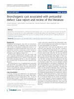
Figure 1. Midphase fluorescein angiogram of a 64-year-old
diabetic woman with a 20-day history of fever and headache
shows chorioretinal lesions with central hypofluorescence
and peripheral hyperfluorescence. Note the presence of
mild non-proliferative diabetic retinopathy.
There is no proven treatment for WNV infection.
In cases of severe disease, therapy is supportive, with
hospitalization, intravenous fluids, respiratory sup-
port, and prevention of secondary infection.
Prevention is the mainstay of WNV infection
control, with public health measures to reduce the
number of mosquitos (draining standing water, lar-
Int. J. Med. Sci. 2009, 6
115
vicides…) and personal protection against mosquito
bites (repellants, window screens, protective cloth-
ing,…). Vaccination, a long term solution, is still in the
research phase.
In conclusion, chorioretinal involvement, fre-
quently asymptomatic and self-limited, is present in
almost 80% of patients with WNV infection associated
with neurologic disease. The unique pattern of mul-
tifocal chorioretinitis can help establish an early di-
agnosis of the disease while serologic testing is
pending. Therefore, an ocular examination, including
ophthalmoscopy and FA in selected cases, should be
part of the routine evaluation of patients with clini-
cally suspected WNV infection.
References
1. Hayes EB, Gubler DJ. West Nile virus: epidemiology and clini-
cal features of an emerging epidemic in the United States. Annu
Rev Med 2006;57:181-94.
2. Khairallah M, Ben Yahia S, Ladjimi A, et al. Chorioretinal in-
volvement in patients with West Nile virus infection. Oph-
thalmology 2004;111:2065-70.
3. Garg S, Jampol LM. Systemic and intraocular manifestations of
West Nile virus infection. Surv Ophthalmol 2005;50:3-13.