Silychristin and isosilychristin from the fruits
Bạn đang xem bản rút gọn của tài liệu. Xem và tải ngay bản đầy đủ của tài liệu tại đây (102.27 KB, 5 trang )
106
Journal of Chemistry, Vol. 45 (1), P. 106 - 109, 2007
SILYCHRISTIN AND ISOSILYCHRISTIN FROM THE FRUITS OF
SILYBUM MARIANUM (L.) GAERTN.
Received 31 May 2006
TRINH THI DIEP
1
, PHAN VAN KIEM
2
, NGUYEN THUONG DONG
1
, NGUYEN HUU TUNG
2
,
BUI THI BANG
1
, CHAU VAN MINH
2
, ALESSANDRA BRACA
3
1
National Institute of Medicinal Materials, Ministry of Health
2
Institute of Natural Products Chemistry, VAST
3
Department of Bioorganic Chemistry and Biopharmaceutics University of Pisa, Italia
SUMMARY
By various chromatography methods, two flavonolignan silychristin (1) and isosilychristin (2)
were isolated from the fruits of Silybum marianum (L.) Gaertn. cultivated in the North of
Vietnam. Their structures were elucidated by analyses of the NMR (1D-, 2D-NMR) and ESI
spectra. This is the first report of both 1 and 2 from S. marianum cultivated in Vietnam.
I - INTRODUCTION
In previous papers, we have reported the
isolation and structural elucidation of
flavonolignan, flavonoid, and lignan
compositions of the fruits of Silybum marianum
(L.) Gaertn. cultivated in the North of Vietnam
and used in folk medicine [1 - 4]. Here, we
report the isolation and structural determination
of two flavonolignan silychristin (1) and
isosilychristin (2) isolated from the fruits of
Silybum marianum (L.) Gaertn. cultivated in the
North of Vietnam. The structures were
elucidated by analyses of the NMR and ESI
spectra, and by the reported data. This is the
first report of both 1 and 2 from S. marianum
cultivated in Vietnam.
II - EXPERIMENTAL
1. General experimental procedures
The Electronspray Ionization (ESI) mass
spectrum was obtained using a AGILENT 1100
LC-MSD Trap spectrometer. The NMR spectra
were recorded on a Bruker AM500 FT-NMR
spectrometer using TMS as the internal
standard. Column chromatography (CC) was
performed on silica gel (Kieselgel 60, 70 - 230
mesh and 230 - 400 mesh, Merck) or YMC RP-
18 resins (30 - 50 µm, FuJisilisa Chemical Ltd.).
Thin layer chromatography (TLC) was
performed on DC-Alufolien 60 F254 (Merck
1.05715) or RP18 F254s (Merck) plates.
2. Plant material
The fruits of Silybum marianum (L.) Gaertn.
were collected in Sapa, Lao Cai Province in
June, 2004 and were identified by Dr Nguyen
Tap, Department of Botany and Medicinal Plant
Resources, National Institute of Medicinal
Materials.
3. Extraction and isolation
Air-dried and powdered fruits of Silybum
marianum (L.) Gaertn (200 g) were defatted with
petroleum and then extracted with methanol.
After removal of the solvent under vacuum, the
residue (5.8 g) was obtained, which was then
repeated chromatographed on silica gel columns
to get 1 (30 mg) as colorless amorphous powder
and 2 (50 mg) as yellow crystals.
107
III - RESULTS AND DISCUSSION
Compound 1 was obtained as colourless
amorphous powder from methanolic extract of
the fruits of S. marianum (L.), mp. 174 - 176
o
C.
The
1
H-NMR spectrum of 1 exhibited two
doublets at
H
5.85 and 5.90 ppm (J = 1.5 Hz),
two doublets at
H
6.82 and 6.87 ppm, three
other aromatic protons at
H
6.79 (d, J = 8.0
Hz), 6.81 (dd, J = 8.0, 1.0 Hz) and 6.99 (d, J =
1.0 Hz). Two doublet of doublets of two protons
at trans configuration were at 5.00 (dd, J =
11.5, 2.5 Hz) and 4.49 (dd, J = 11.5, 5.5 Hz)
[5], a doublet at 5.46 (J = 7.0 Hz), a multiplet
at 3.49. The methylene group bearing oxygen
atom was at 3.64 (dd, J = 10.0, 10.5 Hz) and
3.71 (dd, J = 10.5, 5.5 Hz), and methoxyl group
was at 3.75 as a singlet signal. All the
1
H-NMR
data suggested the flavonolignan type of 1,
which was similar to the isolated compounds
from the same sources [1, 2].
Table 1: The NMR data of 1
C
C
a
C
b
,d
H
c
,d
(J in Hz)
2 85.2 83.20 5.00 (dd, 11.5, 2.5)
3 73.8 71.66 4.49 (dd, 11.5, 5.5)
4 198.2 197.53 -
5 165.2 163.25 -
6 97.4 96.01 5.90 (d, 1.5)
7 168.7 166.97 -
8 96.3 94.96 5.85 (d, 1.5)
9 164.4 162.49 -
1’ 130.0 128.99 -
2’ 116.6 115.26 6.87 (d, 1.5)
3’ 147.5 147.02 -
4’ 142.1 140.66 -
5’ 131.5 129.90 -
6’ 116.9 115.58 6.82 (d, 1.5)
89.1 86.96 5.46 (d, 7.0)
55.5 53.29 3.49 (m)
64.8 62.94 3.64 (dd, 10, 10.5); 3.71 (dd, 5.5, 10.5)
1” 134.8 132.38 -
2” 110.6 110.43 6.99 (d, 1.0)
3” 149.0 147.5 -
4” 147.5 146.33 -
5” 116.2 115.26 6.79 (d, 8.0)
6” 119.7 118.64 6.81 (dd, 1.0, 8.0)
OCH
3
56.4 55.64 3.75 (3H, s)
5-OH 11.89 (br s)
a
C
of silychristin [5],
b
125 MHz,
c
500 MHz,
d
Measured in DMSO-d
6
.
108
O
OH
O
O
HO
OH
2
3
4
5
6
7
8
9
10
1'
2'
3'
4'
5'
6'
OH
C
H
2
O
H
OH
OCH
3
1
"
2"
3"
4"
5
"
6"
1
O
OH
O
HO
OH
2
3
4
5
6
7
8
9
1
0
1'
2'
3
'
4
'
5'
6
'
1
"
2
"
3"
4"
5"
6"
O
H
3
CO OH
HOH
2
C
OH
2
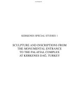
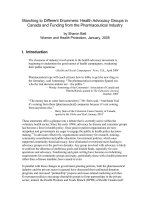
Fig.1. The structures of 1 & 2
The
13
C-NMR spectrum of 1 showed the
signals of 25 carbons including 12 quaternary,
11 methine, 1 oximethylene and 1 methoxyl
carbons, suggesting a flavonolignan compound.
The carbonyl group was at 197.53, the
methylene bearing oxygen atom was at 62.94
and methoxyl group was at 55.64. All NMR
assignments of 1 were made from analysis of
HSQC, HMBC spectra and by comparison with
those of isolated compound (dehydrosily-
christin) [3] as well as with those of silychristin
reported in the literature [5]. In the HMBC
spectrum, H-2 ( 5.00) correlated with C-3 (
71.66)/C-4 ( 197.53)/C-1’ (128.99)/C-2’
115.26), and H-3 ( 4.49) had only correlations
with C-2 ( 83.2) and C-1’ (128.99) confirming
the position of two oximethine carbons at C-2
and C-3. In addition, the H-C long-range
correlations were observed between H- (
5.46) and carbons C- ( 53.29)/C-1” (
132.38)/C-6” ( 118.64)/C-2” (110.43)/C-
(62.94), as well as the H-C long-range
correlations were observed between H- (
3.49) and carbons C1” ( 132.38)/C- (62.94)
also confirming the positions of C-, C- and
C- of two oximethine and oximethylene
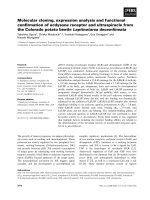
groups. The selected H-C correlations in the
HMBC spectrum of 1 were shown in Fig. 2.
Furthermore, the negative ESI mass spectrum of
1 exhibited an ion peak at m/z 481 [M-H]
+
with
strongest intensity, corresponding to the
molecular formula of C
25
H
22
O
10
. From the above
data, compound 1 was determined to be
silychristin, which was first isolated from
Silybum marianum (L.) Gaertn. cultivated in the
North of Vietnam. This compound also was
reported to be inhibitor of horseradish
peroxidases and lipoxygenase and
antihepatotoxic agent [5].
Compound 2 was obtained as yellow
crystals from methanolic extract of the fruits of
S. marianum (L.), mp. 155 - 157
o
C and had
similar NMR spectra as compound 1, suggesting
the same flavonolignan type. 25 carbons
corresponding to 25 carbon signals were
observed in the
13
C-NMR spectrum of 2
(including 12 quaternary, 11 methine, 1
oximethylene and 1 methoxy carbons as 1).
Detailed comparison from NMR spectrum of 2
with those of 1, especially at C-2, C-3, C- and
C- showed that 2 must be a derivative of 1, as
isosilychristin, which was also isolated from
Silybum marianum (L.) Gaertn [6]. The ESI
mass spectrum of 2 was taken confirming the
molecular formula of C
25
H
22
O
10
of 2 from the
appearance of an ion peak at m/z 483 [M+H]
+
in
the positive and an ion peak at m/z 481 [M-H]
+
in the negative spectra. All NMR assignments
of 2 were made carefully from HSQC and
HMBC and from the comparison with those of 1
as shown in table 2. The H-C long-range
correlations in the HMBC were shown in Fig.2
further confirming the structure of 2 as
isosilychristin, which also was first isolated
from Silybum marianum (L.) Gaertn. cultivated
the North of Vietnam
109
Table 2: The NMR data of 2
C
C
a
,b
H
a
,c
(J in Hz)
2 79.87 5.17 (d, 12.0)
3 71.48 4.66 (dd, 12.0, 6.0)
4 197.99 -
5 163.33 -
6 96.10 5.92 (d, 1.5)
7 166.83 -
8 95.03 5.87 (d, 1.5)
9 162.55 -
10 100.49 -
1’ 128.85 -
2’ 119.42 6.95 (d, 8.0)
3’ 116.08 6.74 (d, 8.0)
4’ 145.92 -
C
C
a,b
H
a,c
(J in Hz)
5’ 141.56 -
6’ 124.44 -
86.36 5.57 (d, 2.0)
51.97 3.68 (m)
63.49 3.46 (dd, 10, 10.5)
3.72 (dd, 5.5, 10.5)
1” 132.83 -
2” 110.28 6.86 (d, 1.5)
3” 147.44 -
4” 146.27 -
5” 115.15 6.71 (d, 8.5)
6” 118.48 6.76 (dd, 8.5, 1.5)
OCH
3
55.59 3.71 (3H, s)
5-OH 11.90 (br s)
a
Measured in DMSO-d
6
,
b
125 MHz,
c
500 MHz.
O
OH
O
O
HO
OH
2
4
5
6
7
8
9
10
1'
2'
3'
4'
5'
OH
CH
2
OH
OH
OCH
3
1"
2"
3"
4
"
5"
6"
O
OH
O
HO
OH
3
4
5
6
7
8
9
10
2'
3'
4'
5'
1"
2"
3"
4"
5"
6"
O
H
3
CO OH
HOH
2
C
OH
Figure 2: H-C long-range correlations in the HMBC of 1 and 2
Acknowledgements: The authors wish to thank
Mr Dang Vu Luong, the NMR Lab., Institute of
Chemistry, Vietnamese Academy of Science and
Technology for recording the NMR spectrum,
and Dr. Nguyen Tap, Department of Botany and
Medicinal Plant Resources, National Institute
of Medicinal Materials for the plant
identification. This study was supported by a
grant from the Vietnam- Italia international
cooperation project.
REFERENCES
1. Vo Van Chi. Vietnamese Medical Plant
Dictionary, Ha Noi Medicine Pub. (1997).
2. T. T. Diep, N. T. Dong, B. T. Bang, C. V.
Minh, P. V. Kiem. Journal of Medicinal
Materials, Vol. 10(6), 175 - 178 (2005).
3. T. T. Diep, N. T. Dong, B. T. Bang, C. V.
Minh, P. V. Kiem. Journal of Medicinal
Materials, Vol. 11(1), 9 - 13 (2006).
4. T. T. Diep, N. T. Dong, B. T. Bang, C. V.
Minh, P. V. Kiem. Vietnamese Journal of
Chemistry, 2006 (accepted).
5. H. Tanaka, M. Hiroo, K. Ichino, K Ito.
Chem. Pharm. Bull., Vol. 37(6), pp. 1441 -
1445 (1989).
6. M. Kalonga, Z. Naturforsch., B, Vol. 36,
262 - 266 (1981).
110
7.