Tai lieu thuc hanh danh cho lop Thu Y tien tien
Bạn đang xem bản rút gọn của tài liệu. Xem và tải ngay bản đầy đủ của tài liệu tại đây (977.15 KB, 20 trang )
<span class='text_page_counter'>(1)</span><div class='page_container' data-page=1>
1
Group ID:………..Semester:………Year:………
Name:………..Student ID:………
Class:………
Name:………..Student ID:………
Class:………
<b>TABLE OF CONTENTS </b>
<b>Practice 1. DNA extraction……….1 </b>
Task 1: Learning to use a pipette………1
Task 2: Extraction of DNA from swine blood………..6
<b>Practice 2. Polymerase Chain Reaction (PCR)………9 </b>
Task 1: Dilute your genomic DNA sample………..10
Task 2: Swine PCR………10
<b>Practice 3. Electrophoresis………..13 </b>
Task 1: Agarose electrophoresis………..13
Task 2: Analyse result from swine PCR………15
</div>
<span class='text_page_counter'>(2)</span><div class='page_container' data-page=2>
2
<b>Practice 1:DNA EXTRACTION </b>
<b>Task 1: </b>
<b>Learning to use a pipette</b>
Pipettes are instruments used to accurately measure and transfer small volumes of
liquids. Misusewill lead to inaccurate measurements and damage to the pipette. In this
task, you will learn to use a piston air displacement pipette with disposable tips.
<b>A. Parts of a micropipette </b>
a. A curved piece for catching hold pipette
b. Close the pipette with your fingers
c. Digital volume indicator
d. Tip ejector button
Before coming to class
You will need to bring a laboratory coat to this class, wear glove when conducting
experiment and tie back long hair
<b>e </b>
</div>
<span class='text_page_counter'>(3)</span><div class='page_container' data-page=3>
3
e. Plunger buttonand Volume adjustment dial
f. Attachment point for a disposable tip
<b>B. Sizes of micropipettes </b>
Different pipettes will measure a different range of volumes; this is written to the top of
the button on the top of the pipette or on the body of pipette. Do NOT wind the pipette
outside this range, as in this will result in a loss of calibration.
The dial on the pipette indicates of volume of liquid that will be transferred. The dial can
have decimal places indicated by number in red. The small strokes between each
bottom number are used to dial values between the printed numbers.
Here are the pipettes that we will use:
Pipette Dial numbers Comments
P10 00.0to 10.0
(0 µl to 10.0 µl)
Decimal numbers in red. Do Not wind below
zero or above 10.0
P20 02.0 to 20.0
(2 µl to 20.0 µl)
Decimal numbers in red. Do NOT wind below
2 or above 20.0
P40 05.0 to 40.0
(5 µl to 40.0 µl)
Decimal numbers in red. Do NOT wind below
5 or above 40.0
P100 010 to 100
(10 µl to 100 µl)
No decimal numbers. Do NOT wind below 10
or above 100.0
P200 020 to 200
(20 µl to 200 µl)
No decimal numbers. Do NOT wind below 20
or above 200.0
P1000
<i><b>010 to 100 </b></i>
(0.1 mL to 1.00 mL =
100µl to 100 µl)
</div>
<span class='text_page_counter'>(4)</span><div class='page_container' data-page=4>
4
Different tips fit different pipettes and measure different volumes:
<b>C. Adjusting Volume on micropipettes and inserting the Tip </b>
The tips are racked in plastic boxes with covers. When you receive a box, it will be
sterile. Please be careful when touching box or tips not to contaminate them. The box
should be closed when not in use to prevent airborn contamination.
Inserting the Tip:
1. Select the correct size tips.
2. Open the box without touching the tips with your hands.
3. Insert the micropipette shaft into the tip and press down firmly. This will attach the
tip to the shaft.
4. Remove the micropipettor with the tip attached.
5. Close the box without touching the tips with your hands.
Clear tips 0-10 µl
Yellow tips 10-200 µl
Blue tips 200-1000 µl
The plunger button (thumb knob) allows
you to adjust the volume that is measured.
It can be dialed to the left or right to
increase or decrease the volume.
</div>
<span class='text_page_counter'>(5)</span><div class='page_container' data-page=5>
5
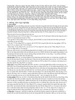
<b>Figure.</b>A-C, Three positions of the button of the pipette. D-H, Liquid handling with
pipettes. D, Adjusting the volume. E, Fastening an appropriate tip. F, Drawing of the
liquid. G, Transferring the liquid into an Eppendorf tube. H, Dispensing the liquid
<i>1.Which pipette will you use to measure the following volumes? What will the dial read </i>
<i>when set to measure the required volume? Which color tip will you use? </i>
Amount to
Measure Which Pipette? (name) Dial Reading
Which colour
tip?
2.3 µl
154.6 µl
783 µl
36.2 µl
19.0 µl
<b>D. Practice: </b>
Within your groups of 4 students, work in pairs to use pipette. You will have 3 Petri
dishes each containing a piece of filter paper.
</div>
<span class='text_page_counter'>(6)</span><div class='page_container' data-page=6>
6
Petrie dish 1 : 5 µlPetrie dish 2: 90 µlPetrie dish 3: 250 µl
b) Each pair should measure the given volume of coloured liquid into one side of the
Petri dish.
<b>How to do? </b>
1. Select the correct pipette and the dial required volume. If the dial doesn’t move,
push the locking bar down
2. Open the container containing the liquid to be aspirated and the container where
the liquid will be dispensed
3. Select the correct tip size
4. Hold the tip box steady with one hand. Holding the pipette in your dominant
hand and keeping the pipette vertically, place the pipette into the tip and twist
about 180 degrees to add the tip. Raise out of the box. Be careful to keep the tip
clean is not to touch anything with the tip.
5. Press and HOLD the top button (piston) down to the FIRST stop.
6. Keeping the pipette vertical, lower the tip just below the surface of the liquid to
be
7. Slowly raise the top button until you can let go of the button.
8. Raise the pipette tip out of the liquid.
9. Place the tip into the container where the liquid is to be dispensed. Commonly,
the tip is place against the side of the container.
10.Gently press the top button (piston) down to the FIRST stop, then continue
pressing to the SECOND stop (which blows out the small amount of liquid left in
the tip) and HOLD down the button while you lift the pipette out f the container.
11.Slowly release the button. Use the tip eject button (coloured) to expel the tip into
the waste container.
</div>
<span class='text_page_counter'>(7)</span><div class='page_container' data-page=7>
7
<b>Task 2</b>
<b>: </b><b>Extraction of DNA from swine blood </b>
<b>A.</b> <b>Purification of swine blood sample </b>
Eliminating all DNA – free components (plasma, serum, hemoglobin, platelet in mammal
blood). This step critically contributes to the purity of DNA after the purification process
is complete.
<i>Note: 2 students per blood sample </i>
<i>Clean up after class (including all used test tubes) </i>
<b>Tools</b>: Centrifuge, 15ml test tubes for containing samples, micropipette, pipette filler
bulb, jar for containing eliminated liquid
Chemical: NaCl 0.2%
<b>Procedure: </b>
1. Draw 5ml of NaCl 0.2% into test tube containg 1ml swine blood, mix thoroughly
and centrifuge at 3000rpm for 3 minutes
2. Remove the supernatant.
3. Repeat (1) and (2) 4 times
<b>B.</b> <b>Extract of DNA from swine blood </b>
The isolation of genomic DNA is the first step of many genetic protocols used in genetic
disease testing, paternity testing, forensics and in research. The methods we use today
are among the methods currently used in resaerch and testing laboratories. You will
gain experience in extracting DNA from swine blood.
<i>Note: 2 students per blood sample </i>
<i>Clean up after class (including all used test tubes) </i>
<b>Tools</b>: Centrifuge, micropipette, tips, jar for containing eliminated liquid
</div>
<span class='text_page_counter'>(8)</span><div class='page_container' data-page=8>
8
<b>Procedure: </b>
1. Add 3ml Lysis buffer and 30 µl Proteinase K to the sample-containing test tube,
mix thoroughly and incubate at 55oC over night (at least 8 hours)
<i>Q2.1 What is the role of the Proteinase K enzyme? </i>
2. Add 3ml Phenol, mix and centrifuge at 3000rpm for 5 minutes. Ensure that the
centrifuge is balanced before starting.
<i>Q2.2 Why are you centrifuging your sample? </i>
<i>Q2.3 Precipitation should appear in this step. Why can it be seen now? </i>
<i>Q2.4 Where is the DNA now? </i>
3. Transfer the supernantant into anew tube and add 3ml Phenol/CIAA, mix and
centrifuge at 3000rpm for 5 minutes. Ensure that the centrifuge is balanced
before starting.
</div>
<span class='text_page_counter'>(9)</span><div class='page_container' data-page=9>
9
4. Transfer the supernatant into a new tube and add 3ml CIAA
(Chloroform/Isoamyl Alcohol), mix and centrifuge at 3000rpm for 5 minutes.
<i> Q2.6 Where is the DNA now? </i>
5. Transfer the supernatant into a test tube containing 5ml ice-cold Ethanol and
invert gently to mix.
<i>Q2.7 White strand should appear. What is this and why can it be seen now? </i>
6. Centrifuge at 6000rpm for 5 minute You should see a pellet near the base of the
tube. Remove the supernatant, ensuring the pellet is not discarded.
7. Add 3ml Ethanol 70%, invert 50 times to wash the pellet and centrifuge at
3000rpm for 5 minutes
8. Remove all ethanol solution and let the sample dry
9. Add 200 µl TE 1X buffer to dissolve the pellet. Gentle flicking of the tube will
encourage the pellet to dissolve.
10.. Incubate at 370C over night.
</div>
<span class='text_page_counter'>(10)</span><div class='page_container' data-page=10>
10
<b>Practice 2: POLYMERASE CHAIN REACTION (PCR) </b>
The purpose of this practical is to give you some experience in a commonly used
approach for many making many copies of a specific region of the genome through in
vitro DNA replication using the <b>polymerase chain reaction</b> or<b> PCR</b>. PCR is the starting
point for many analytical techniques, including
microsatellite genotyping to identify individuals
the sequencing of genes to analyze variation associated with genetic diseases
the diagnosis of certain infectious diseases
molecular sexing where species do not show external, or early, indicators of sex
(e.g. birds) or where mature animals cannot be examined (e.g. embryos or
faeces/hair samples in conservation genetics)
In this way, PCR forms an integral part of most genetic tests for disease and disease
causing agents.
PCR is able to amplify even extremely small amount of DNA for analysis. This means that
it can be used to amplify DNA extracted from forensic samples like hair, traces of blood
or even single sperm. However, this also mean that very small amount of contaminating
DNA will also be amplified. You should show extreme caution in ensuring that only DNA
from your target sample will be in your PCR reaction!
<b>Within your group of 4 students, works in pair to set up the swine PCR. Each pair will </b>
<b>set up a PCR for the 4 DNA samples (1 from your own group and 3 from other group). </b>
<b>You will be allocated a group number. Record your group number here: _________ </b>
<i>So the group of 4 students will label in group 3 will have tubes labeled: 3-1,3-2 </i>
Before coming to class
You will need to bring a laboratory coat to this class, wear glove when conducting
experiment and tie back long hair
</div>
<span class='text_page_counter'>(11)</span><div class='page_container' data-page=11>
11
<b>Task 1:Dilute your Genomic DNA sample</b>
1. For each sample, you will need to dilute the DNA. Do this for your own swine DNA
sample.
a. record the concentration of your DNA sample in column C. This value will be
provided to you by your tutor
b. Calculate how much DNA and water you need to add to prepare 200 µl of DNA
diluted to 10 ng/µl using the following table
i. You can use the formula <i>initial concentration X initial volume = final </i>
<i>concentration X final volume or column D = (A x B)/C </i>
ii. How much water will you need to add to bring the total volume to 200 µl? i.e.
column E = B – D
A B C D E
Sample
ID
Final
concentration
Final volume Current
concentration
(“initial
concentration”)
Amount of DNA
to add
(“initial
concentration”)
Amount of
water to
add
10 ng/µl 200 µl
2. Use a new 1.5 mL eppendorf tube and add the amount of water from column E and
the amount of DNA from column D
<b>Task 2</b>
<b>: </b><b>Swine PCR </b>
1. To set up the PCR, you will need to make a Master Mix for your group, with one
reaction for each DNA (one from your own sample and the other three from other
group), one reaction for a negative (no DNA template) control for PCR .
</div>
<span class='text_page_counter'>(12)</span><div class='page_container' data-page=12>
12
2. You will need a 200µl eppendorf tube per group (2 students) to make up the master
mix. Label this tube with your group number.
3. Calculate how much of each reagent you will need to add to your master mix using
the table shown below
a. Column A shows you initial concentrattion. Column B shows you
concentration for running PCR. Column C shows shows you the amount
needed for 1 reaction. And column D shows the amount needed for 4
reaction plus 1 (negative)
b. To complete column C, calculate by these equations :
- “Rule of three”
- C = B x V∑/A
- H2O = V∑ - known reagents
Reagents A B C D
DNA 10ng/ µl 50ng
Sterile pure H2O
PCR buffer 10X 1X
dNTPs 1.0mM 150µM
Forward primer 10pmol/µl 50pmol
Reverse primer 10pmol/µl 50pmol
MgCl2 25mM 2,25mM
Enzyme (Taq DNA
polymerase)*
5UI/ µl 1UI
Total (V∑) 20 µl
4. Add each reagent to your master mix tube. Mix by gently flicking or vortexing.
Keep on ice.
</div>
<span class='text_page_counter'>(13)</span><div class='page_container' data-page=13>
13
<i>Record the details of the samples here: </i>
<i>1. </i>
<i>2. </i>
<i>3. </i>
<i>4. </i>
<i>5. </i>
6. Pipette out 15µl of the master mix into each of the small reaction tube.
7. Add 5µl of DNA of the relevent DNA into each tube following the list in step 2
above. Do not add anything to the N tube.
8. Give tubes to the tutor. Samples will be run on together on a PCR machine using
the programme:
INITIAL DENATURATION 940C, 10 min
Then
DENATURATION 940C, 30 sec
ANNEALING 550C, 45 sec Repeat this for 35 cycles
EXTENSION 720C, 45 sec
Then
FINAL EXTENSION 720C, 5 min
HOLD 40C
</div>
<span class='text_page_counter'>(14)</span><div class='page_container' data-page=14>
14
<b>Practice 3: ELECTROPHORESIS </b>
<b>Task 1: Agarose electrophoresis</b>
<b>A.</b> <b>Agarose 1% preparation (carried out by lecturer) </b>
1. Weigh 1g agarose and 100ml TAE 1X solution
2. Microwave to dissolve the agarose (achieve boiling point 3 times)
3. Wait until the agarose cools down (500C) and add 6 µl ETBR. Gently shake the
agarose over ice clockwise, then counterclockwise to avoid bubbles and help
ETBR dissolve completely in gel. ETBR intercalates into double-strand of DNA and
flouresces when placed under UV light.
4. Pour the gel into the mold and let it fix
<i>Q3.1 How many grams of agarose are needed to prepare 100ml agarose solution of </i>
<i>1.5%? </i>
<i>Q3.2 Why should bubbles be avoided when the gel is prepared? </i>
<b>B.</b> <b>Sample loading (carried out by students) </b>
1. When the groups located to a gel are ready, add the gel to electrophoresis tank.
Remember the DNA will migrate from black to red (negative to positive) so sit
your gel in the correct way.
2. Observe the tutor loads 5µl ladder 100 (ABM) into the wellof an agarose gel.
Before coming to class
</div>
<span class='text_page_counter'>(15)</span><div class='page_container' data-page=15>
15
3. Each student carefully pipette 5µl of PCR product into an agarose gel. Note that
each sample should be loaded and electrophoresis started without delay.
<i>Write here the your sample and order samples on gel </i>
4. Ensure the power pack is switched off, then place the lid in place. Connect the
electrodes from the electrophoresis tank to the power pack (red to positive, black
to negative).
5. Set the voltage to 100 volts and press Start. Run the eletrophoresis for 30
minutes.
6. When the electrophoresis run is complete, the tutor will turn the power pack off
and remove the gel, place your gel under UV light.
7. Observe and determinate your result.
<i>Q3.3 What result did you obtain from your PCR? </i>
<i>Q3.4. The size of PCR products:………bp </i>
<i>Q3.5 Your sample is (positive/negative)……… for </i>
<i>(PRRS/PED) ………. </i>
<i>(Note: to answer Q3.5, answer the questions in Task 2 first) </i>
<i>Q3.6 If one or more bands are seen in the ‘negative’ lane, what does it mean? </i>
</div>
<span class='text_page_counter'>(16)</span><div class='page_container' data-page=16>
16
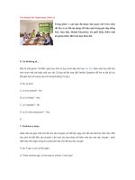
<i>Q3.8 Fill in the sizes corresponding to bands of ladder </i>
<b>Figure</b>. Electrophoresis with ladder 100
<b>Task 2: Analyse results from swine blood </b>
Each pair will obtain a result from your PCR which is expected to diagnosis of certain
infectious disease: PRRS or PED
<i>Q3.9 PRRS is the shorthand abbreviation of………. </i>
<i>………. </i>
<i>Also known as………..… </i>
<i>that occurs in………..……….……….., </i>
<i>caused by the ………..virus </i>
<i>Q3.10 PED is the shorthand abbreviation of:...……… </i>
<i>………….……… </i>
<i>caused by………..………virus. </i>
</div>
<span class='text_page_counter'>(17)</span><div class='page_container' data-page=17>
17
<i>F is………. </i>
<i>R is……….. </i>
<i>Q3.11 Find the primer sequences in the sequences shown below. Underline the </i>
<i>primer sequences in both the sequences. </i>
<i>(Hint: you need to write the reverse complement sequence of the reverse primer) </i>
<b>LOCUS AY612613 </b>
14281 GCAAAGTTGA GGTCGAAGGT CATCTGATCG ACCTCAAAAG AGTTGTGCTT GATGGTTCCG
14341 TGGCAACCCC TATAACCAGA GTTTCAGCGG AACAATGGGG TCGTCCTTAG ATGACTTCTG
14401 TCATGATAGC ACGGCTCCAC AAAAGGTGCT TTTGGCGTTT TCTATTACCT ACACGCCAGT
14461 GATGATATAT GCCCTAAAGG TGAGTCGCGG CCGACTGCTA GGGCTTCTGC ACCTTTTGAT
14521 CTTCCTGAAT TGTGCTTTCA CCTTCGGGTA CATGACTTTC GCGCACTTTC AGAGTACAAA
14581 TAAGGTCGCG CTCACTATGG GAGCAGTAGT TGCACTCCTT TGGGGGGTGT ACTCAGCCAT
14641 AGAAACCTGG AAATTCATCA CCTCCAGATG CCGTTTGTGC TTGCTAGGCC GCAAGTACAT
14701 TCTGGCCCCT GCCCACCACG TTGAAAGTGC CGCAGGCTTT CATCCGATTG CGGCAAATGA
14761 TAACCACGCA TTTGTCGTCC GGCGTCCCGG CTCCACTACG GTCAACGGCA CATTGGTGCC
14821 CGGGTTAAAA AGCCTCGTGT TGGGTGGCAG AAAAGCTGTT AAACAGGGAG TGGTAAACCT
14881 TGTCAAATAT GCCAAATAAC AACGGCAAGC AGCAGAAGAG AAAGAAGGGG GATGGCCAGC
14941 CAGTCAATCA GCTGTGCCAG ATGCTGGGTA AGATCATCGC TCAGCAAAAC CAGTCCAGAG
<b>Primer </b> <b>Sequences of the primers (5’ – 3’) </b> <b>Infection </b>
<b>detected </b>
<b>Amplified </b>
<b>target </b>
<b>gene </b>
<b>Size of </b>
<b>PCR </b>
<b>product </b>
ORF6-F GTGGCAACCCCTATAACCAGAG
PRRSv <i>ORF6 </i> 780 bp
ORF6-R ACGACAGACACAATTGCCGCTCAC
E pro-F CGCAGTTTACACACCTATAGGG
<i>PEDv </i> <i>E&M </i> 412 bp
</div>
<span class='text_page_counter'>(18)</span><div class='page_container' data-page=18>
18
15001 GCAAGGGACC GGGAAAGAAA AATAAGAAGA AAAACCCGGA GAAGCCCCAT TTTCCTCTAG
15061 CGACTGAAGA TGATGTCAGA CATCACTTTA CCCCTAGTGA GCGGCAATTG TGTCTGTCGT
15121 CAATCCAGAC CGCCTTTAAT
//
F:……….
R:………..
Size of PCR product:……….
<b>LOCUS JQ282909</b>
25561 GCTTCACTTG TCACCGGTTG TGTAATAGCG CAGTTTACAC ACCTATAGGG CGTTTGTATA
25621 GAGTTTATAA GTCTTACATG CAAATAGACC CCCTCCCTAG TACTGTTATT GACGTATAAA
25681 CGAAATATGT CTAACGGTTC TATTCCCGTT GATGAGGTGA TTCAACACCT TAGAAACTGG
25741 AATTTCACAT GGAATATCAT ACTGACGATA CTACTTGTAG TGCTTCAGTA TGGCCATTAC
25801 AAGTACTCTG CGTTCTTGTA TGGTGTCAAG ATGGCTATTC TATGGATACT TTGGCCTCTT
25861 GTGTTAGCAC TGTCACTTTT TGATGCATGG GCTAGCTTTC AGGTCAATTG GGTCTTTTTT
25921 GCTTTCAGCA TCCTTATGGC TTGCATCACT CTTATGCTGT GGATAATGTA CTTTGTCAAT
25981 AGCATTCGGT TGTGGCGCAG GACACATTCT TGGTGGTCTT
//
F:……….
R:………..
</div>
<span class='text_page_counter'>(19)</span><div class='page_container' data-page=19>
19
<b>General Practice </b>
Based on the paper, answer and complete the information (by Vietnamese):
1. The title of paper, name of authors?
2. Purposes and objects?
</div>
<span class='text_page_counter'>(20)</span><div class='page_container' data-page=20>
20
4. Complete the tables below:
<b> Reagent </b> <b>Amount needed </b>
<b>for 1 reaction </b>
<b>Running </b>
<b>programme </b> <b>Temperature </b> <b>Time </b>
DNA Initial
denaturation
Sterile pure H2O Denaturation
PCR buffer Annealing
dNTPs Extension
Forward primer Final extension
Reverse primer Number of cycle
MgCl2
Enzyme (Taq
DNA
polymerase)*
Total (V∑)
5. How long (approximately) will the PCR take?
</div>
<!--links-->