ĐẶC điểm MIỄN DỊCH của TRẺ EMhandout
Bạn đang xem bản rút gọn của tài liệu. Xem và tải ngay bản đầy đủ của tài liệu tại đây (1.04 MB, 27 trang )
ĐẶC ĐIỂM MIỄN DỊCH CỦA TRẺ EM
GV: Trần Thị Hồng Vân
1. SINH LÝ HỆ THỐNG MIỄN DỊCH
Chức năng chính của hệ thống miễn dịch là nhận biết “tự thân” và loại bỏ những
thực thể “không tự thân” ( các vi sinh vật, tế bào u, tế bào cấy ghép)
Hệ thống miễn dịch là môt mạng lưới phức tạp gồm:
Miễn dịch khơng đặc hiệu( bẩm sinh)
Miễn dịch đặc hiệu( thích ứng): thu được trong q trình sống
1.1. Miễn dịch khơng đặc hiệu (bẩm sinh)
Tạo ra phản ứng tương tự nhau với tất cả các kháng nguyên. Gồm:
1.1.1.Hệ thống thực bào (phagocytic system): là các tế bào có nhiệm vụ
nuốt và tiêu hóa các vi sinh vật, như: Neutrophils, monocytes trong máu,
Macrophages trong tổ chức, macrophages phế nang ở phổi, tế bào Kuffer ở gan, tế
bào hoạt dịch ở khớp…
1.1.2. Các protein bổ thể:
Bổ thể là hệ thống cần thiết trong đáp ứng miễn dịch không đặc hiệu. Định
nghĩa ban đầu của nó là thành phần bổ sung khơng đặc hiệu, khơng bền vững với
nhiệt, tác dụng cùng với các kháng thể đặc hiệu để dung giải vi sinh vật. Các yếu
tố bổ thể tương tác với nhau theo 3 con đường: cổ điển, lectin và đường thay thế
để tạo ra hoạt tính đầy đủ của nó.
TABLE 132-1 -- Constituents of the Complement System
SERUM COMPONENTS
Classical pathway
C1q
C1r
C1s
C4
C2
C3
Alternative Pathway
Factor B
Factor D
1
Lectin Pathway
Mannose-binding lectin (MBL), L- and H-ficolin
MBL-associated proteases 1,2,3
Membrane Attack Complex
C5
C6
C7
C8
C9
Control Protein, Enhancing
Properdin
Control Proteins, Downregulating
C1 inhibitor (C1 INH)
C4-binding protein (C4-bp)
Factor H
Factor I
S protein (vitronectin)
Clusterin
Anaphylatoxin inactivator
Membrane Regulatory Proteins
CR1
Membrane cofactor protein (MCP;CD46)
Decay-accelerating factor (DAF)
CD59 (membrane inhibitor of reactive lysis; protectin)
MEMBRANE RECEPTORS
CR1
CR2 (CD21)
CR3 (CD11b/CD18)
CR4 (CD11c/CD18)
C3a receptor
C5a receptor
2
C1q receptor
Other downregulating serum factors and membrane receptors have been described but
have not been fully defined. CR, complement receptor.
Figure 132-1 Sequence of activation of the components of the classical and lectin (MBL) pathways of complement and interaction
with the alternative pathway. Activation of C3 is the functionally essential target. The multiple sites at which inhibitory regulator
proteins (not shown) act are indicated by asterisks, emphasizing the delicate balance between action and control in this system that
is essential for host defense yet capable of mediating profound damage to host tissues. Ab, antibody (IgG or IgM class only); Ag,
antigen (bacterium, virus, tumor cell, or erythrocyte); B,D,P, factors B and D, I, and properdin; C-CRP, C carbohydrate–C-reactive
protein; C4-bp, C4-binding protein; MASPs, MBL-associated serine proteases; MBL, mannose-binding lectin.
1.1.3. Các chất phản ứng cấp và hàng rào bảo vệ (da và niêm mạc)
1.1.4. Cytokines: là các polypeptids không phải Immunoglobulin (non-Ig)
do các tế bào monocytes và lymphoctes sản xuất ra khi đáp ứng tương tác với các
kháng nguyên đặc hiệu, không đặc hiệu hoặc các tác nhân kích thích hịa tan
khơng đặc hiệu.
3
1.2. Hệ thống miễn dịch đặc hiệu: là miễn dịch thu nhận, thích ứng và ghi nhớ:
gồm:
- Thành phần tế bào: các lymphocytes
- Thành phần dịch thể: các Immunoglobulin (Ig)
1.1.1. Lymphocytes: gồm 3 nhóm
• Tế bào lympho T (T cell) : nguồn gốc từ tuyến ức, gồm có:
- Tế bào Th (T-helper: Th0, Th1 & Th2): tế bào T hỗ trợ
- Tế bào Ts (T suppressor) : tế bào T ức chế
- Tế bào Tc (T cytotoxic) : Tế bào T độc
•
Tế bào lympho B (B cell): nguồn gốc từ tủy xương, tiết các Ig đặc hiệu với
kháng nguyên
• Tế bào lympho non-T, non-B: TB diệt tự nhiên (natural killer-NK)
1.1.2. Các immunoglobulin (Ig):
Do các tế bào Lympho B tiết ra. Có 5 loại Ig.
1.1.2.1. IgM:
- Xuất hiện sớm nhất trong đáp ứng miễn dịch
- Là yếu tố kết dính và opsonin hóa có hiệu lực, gắn với bổ thể
- Là kháng thể chủ yếu chống lại các polysaccharides, vi khuẩn Gr(-), các ngưng
kết tố hồng cầu.
1.1.2.2. IgG (IgG1, IgG2, IgG3, IgG4):
- Là loại Ig có nhiều nhất, được tìm thấy trong dịch gian bào và các tổ chức
- Qua được nhau thai; IgG 1,2,3 kết hợp được với bổ thể
- Là kháng thể chủ yếu đối với các kháng độc tố, virus, vi khuẩn, là Ig chủ yếu
trong -globulin
1.1.2.3. IgA (IgA1, IgA2):
Là Ig chủ yếu trong các chất tiết của thanh niêm dịch.
1.1.2.4. IgD: chức năng chưa được xác định.
1.1.2.5. IgE: được tìm thấy trong các chất tiết của thanh niêm dịch
IgE tăng trong các bệnh nhiễm ký sinh trùng; phản ứng dị ứng; gắn với các
dưỡng bào ( mast cell); BC ưa bazơ.
1.3. Các cơ quan của hệ thống miễn dịch:
bao gồm: Amiđan và VA, hạch lympho và mạch bạch huyết, tuyến ức, lách, mảng
Peyer’s ở ruột, ruột thừa, tủy xương
4
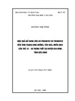
ảnh 1: các cơ quan của hệ thống miễn dịch
Đáp ứng với
kháng nguyên
Phơi nhiễm
Loại đáp ứng
Hệ thống MD không đặc
hiệu (bẩm sinh)
Hệ thống MD đặc hiệu
Không đặc hiệu
Đặc hiệu
Đáp ứng tức thì,
tối đa
Đáp ứng chậm
Dịch thể
Dịch thể và qua trung gian
TB
Tế bào MD
BC trung tính
Macrophages
Lymphocytes
(T, B cell, NK)
Ký ức MD
Khơng
Có
5
ảnh 2: Mạng lưới miễn dịch
2. SỰ PHÁT TRIỂN HỆ THỐNG MIỄN DỊCH Ở TRẺ EM
Các yếu tố bảo vệ cơ thể của mỗi cá thể phát triển với tốc độ khác nhau ở trong
bào thai. Vào lúc sinh ra, chức năng của hầu hết các cơ chế MD tương ứng với
tuổi thai và kém hơn người lớn, kể cả trẻ sinh đủ tháng, do vậy, trẻ sơ sinh và nhũ
nhi ( đặc biệt từ 3-12 tháng) có tình trạng thiếu hụt miễn dịch đáng kể, nhất thời, ở
tất cả các cơ chế của hệ thống miễn dịch, làm cho trẻ có nguy cơ cao mắc các bệnh
nhiễm trùng nặng. Trẻ sinh non, chấn thương khi đẻ, mắc bệnh từ trong bào thai,
stress, dùng một số thuốc có nguy cơ thiếu hụt miễn dịch cao hơn.
2.1. Hệ thống miễn dịch bẩm sinh:
2.1.1. Hệ thống thực bào :
Các tế bào thực bào xuất hiện đầu tiên vào giai đoạn túi noãn của bào thai. Các
bạch cầu hạt và BC đơn nhân có vào tháng thứ 2 và 4 của thai kỳ. Chức năng của
các tế bào trên tăng dần theo tuổi thai nhưng vẫn cịn kém lúc ra đời, do đó trẻ bị
giảm đáp ứng miễn dịch và dễ mắc các bệnh nhiễm trùng. Khi bị nhiễm trùng, trẻ
sơ sinh thường khơng có những dấu hiệu lâm sàng chỉ điểm như ở trẻ lớn (sốt, hội
chứng nhiễm trùng...)
Siêu cấu trúc của bạch cầu đa nhân trung tính ở trẻ sơ sinh thì bình thường
nhưng có sự biến dạng màng tế bào và giảm tính bám dính làm ảnh hướng đến các
chức năng của tế bào như tính hóa hướng động và thực bào.
Các monocyte trong máu là tiền thân của các đại thực bào ở tổ chức. Các đại thực
bào này có khả năng thực bào từ khi cịn trong bào thai và hoạt tính diệt vi khuẩn
6
của nó lúc đẻ bình thường hoặc hơi thấp. Các đại thực bào phế nang xâm nhập vào
phế nang lúc đẻ hoặc sắp đẻ giúp làm sạch các mảnh vụn của dịch ối và vi sinh
vật. Khả năng thực bào của các đại thực bào tổ chức còn hạn chế. Sự giảm năng
lực của hệ thống võng nội mô ở trẻ sơ sinh một phần là do sự giảm hoạt động
opsonin huyết thanh.
Tính hóa hướng động ở trẻ em đạt tới mức ở người lớn khi trẻ được vài năm
tuổi. Tính thực bào và diệt vi sinh vật đạt mức bình thường ở trẻ sơ sinh khỏe
mạnh sau 12 giờ tuổi, giảm ở trẻ đẻ thấp cân và trẻ đủ tháng có stress.
2.1.2. Opsonin:
Opsonin là các thành phần trong huyết thanh làm nhiệm vụ xâm nhập vào vi sinh
vật hay các kháng nguyên để giúp cho hoạt động thực bào của các phagocytes có
hiệu quả hơn.
Các yếu tố opsonin huyết thanh bao gồm IgG, IgM (bền vững với nhiệt) và bổ
thể ( khơng bền với nhiệt). IgM opsonin hóa các VK Gr(-) hiệu quả hơn IgG
IgG qua được nhau thai, IgM và bổ thể không qua được nhau thai.
Mức sản xuất IgM ở trẻ mới đẻ còn thấp, ngoại trừ trẻ bị nhiễm trùng trong bào
thai. Sự tổng hợp các bổ thể bắt đầu khi thai được 5,5 tuần. Nồng độ bổ thể vào
lúc sinh chỉ đạt 50-75% ở người lớn.
Hoạt động opsonin huyết thanh khác nhau theo tuổi thai, bị giảm ở trẻ đẻ non yếu
với tất cả các vi sinh vật được thử, ở trẻ đủ tháng khỏe mạnh chỉ bị giảm với vài
loại vi sinh vật, chủ yếu là vi khuẩn Gr(-)
2.1.3. Sự phát triển hệ thống bổ thể ( complement):
ảnh 3: Sự phát triển của hệ thốnng bổ thể ở trẻ em
7
2.1.4. Cytokine:
Chức năng của da khi sinh
Da là một cơ quan đa chức năng phức tạp, là nơi tiếp xúc của cơ thể với môi
trường. Chức năngcủa da bao gồm:s
- Ngăn cản sự mất nươc
- Điều hịa nhiệt
- Kiểm sốt nhiểm khuẩn
- Giám sát miễn dịch
- Sinh axít mantle: axít mantle là một lớp film mỏng mịn phủ trên mặt da có tác
dụng như một hàng rào ngăn cản vi sinh vật và các chất ô nhiễm khác xâm nhập
vào da. Các vi sinh vật và các chất ô nhiễm này có tính chất kiềm sẽ bị trung hịa
bởi axít mantle.
- Chống oxi hóa (antioxidant)
- Tổng hợp vitamin D3, đồng thời bảo vệ da khỏi tác dụng của tia cực tím
- Hàng rào bảo vệ các hóa chất.
- Xúc giác.
Trẻ sơ sinh non tháng có tỷ lệ tử vong cao, có liên quan đến sự chưa hồn thiện
của hàng rào biểu mô, làm cho vi khuẩn dễ xâm nhập vào cơ thể, cũng như dễ bị
hạ thân nhiệt.
Chức năng kháng khuẩn của niêm dịch:
8
Thành phần
Chức năng
Mucin
Hàng rào cơ học chống VK xâm nhập
Các kháng thể
Trung hịa độc tố, ngăn cản sư kêt dính vào niêm mạc
của VK hoặc độc tố VK.
Lactoferin
Tác dụng kìm khuẩn (tạo chelate với Fe3+)hoặc diệt
khuẩn (Lactoferricidin)
Lysozym
Tiêu vi khuẩn bằng cách phân tách peptido-glycan của
thành vi khuẩn
2.2. Hệ thống miễn dịch đặc hiệu:
2.2.1. Miễn dịch tế bào ( Tế bào T)
- Các tế bào mầm của tuyến ức được sinh ra từ biểu mô của túi hầu thứ 3 và
thứ 4 vào khoảng tuần thứ 6 của thai kỳ, đến tuần thứ 12 là có thể tham gia
vào đáp ứng miễn dịch.
- Tuyến ức hoạt động trong suốt thai kỳ và thời gian đầu sau sinh. Nó phát
triển nhanh trong bào thai và có thể nhìn thấy trên phim X-quang ở trẻ sơ
sinh bình thường, và sau đó hồn thiện dần qua nhiều năm.
- Tuyến ức được coi là trung gian giúp cơ thể chấp nhận các kháng nguyên
“tự thân” và cần thiết cho các tổ chức lympho ngoại biên phát triển và
trưởng thành. Các thành phần biểu mô ở tuyến ức tạo ra các chất dịch giúp
tế bào T biệt hóa và trưởng thành.
- Vào lúc sinh, các đáp ứng test tăng nhạy cảm da bị giảm rõ rệt, sự loại bỏ
da ghép cũng bị suy yếu. Các chức năng này tăng lên trong vài tháng đầu
của cuộc sống.
- Số lượng tế bào T, T hỗ trợ, T ức chế, và T sinh sản đáp ứng với tế bào
mitogens và allogeneic thì bình thường hoặc tăng.
- Một vài lymphokines (IL-1, IL-2, yếu tố hoại tử u (TNF), interferon (INFα), không bao gồm IL-4, INF-, được lymphocytes tạo ra với số lượng gần
bình thường ở trẻ sơ sinh.
- Các hoạt tính độc tế bào ( NK, Phụ thuộc Ab, tế bào T diệt độc tế bào) có
thể thấp hơn ở người lớn. Hoạt tính của T ức chế có thể tăng hơn ở người
lớn, có thể là do bất thường của điều hòa miễn dịch và sự giảm sản xuất Ab
gây thiếu hụt miễn dịch qua tế bào T. Các yếu tố như mẹ bị nhiễm virus,
9
tăng Bilirubine máu, uống thuốc (corticosteroids, chống chuyển hóa…)
trong giai đoạn cuối của thai kỳ có thể làm giảm chức năng tế bào T ở trẻ sơ
sinh.
2.2.2. Miễn dịch dịch thể ( tế bào B)
- Các tế bào B được tìm thấy ở tủy xương, máu, gan, lách trong bào thai vào
lúc 12 tuần của thai kỳ.
- Một lượng rất nhỏ IgM và IgG được tổng hợp vào lúc 20 tuần và IgA vào
lúc 30 tuần, chủ yếu là IgM.
- Nồng độ cao của IgM trong huyết thanh dây rốn (>20mg/dL) chứng tỏ có
sự tiếp xúc với kháng nguyên, thường là do nhiễm trùng bẩm sinh.
- Hầu hết các IgG là do chuyển từ mẹ sang con qua nhau thai. Vào lúc sinh,
IgG của trẻ bằng hoặc cao hơn ở người lớn (110% mức của mẹ). Trẻ sinh
non có mức IgG thấp tùy theo tuổi thai.
- IgG truyền qua nhau thai này bị giảm dần (dị hóa) với thời gian bán hủy là
25 ngày, gây hiện tượng “ giảm gammaglobulin máu” vào lúc 2-6 tháng,
sau 6 tháng thì tốc độ tổng hợp IgG sẽ tăng vượt quá tốc độ phân hủy IgG
từ mẹ.
- Trẻ sinh non có sự giảm mạnh gammaglobulin máu trong suốt 6 tháng đầu.
Đến 1 tuổi, mức IgG bằng khoảng 70% người lớn.
- IgG của mẹ truyền cho con sẽ hết trong máu trẻ vào lúc 9 tháng
- IgA, IgM, IgD, IgE không qua được nhau thai. Nồng độ các Ig này tăng lên
rất chậm và đạt 30% ở người lớn vào lúc 1 tuổi, IgM đạt 75% mức người
lớn vào lúc 1 tuổi.
IgM đạt mức xấp xỉ người lớn vào lúc 1 tuổi ,
IgG:
8 tuổi ,
IgA:
11 tuổi
10
ảnh 4: Nồng độ các Immunoglobulin trong huyết thanh bào thai và trẻ em
•
Miễn dịch thụ động từ mẹ sang:
- kháng thể IgG từ nhau thai sang và các yếu tố miễn dịch trong sữa mẹ giúp bù
đắp lại hệ thống miễn dịch chưa trưởng thành của trẻ sơ sinh và cho trẻ miễn dịch
với nhiều loại VK, VR nguy hiểm. Tuy nhiên các IgG thụ động này ngăn cản đáp
ứng của trẻ với tiêm chủng như sởi, rubella.
- Sữa mẹ có rất nhiều yếu tố chống vi khuẩn ( IgG, IgA tiết, BC, bổ thể, lysozyme,
lactoferrin) nằm trên bề mặt đường tiêu hóa, hơ hấp, giúp chống lại sự xâm nhập
của các tác nhân gây bệnh.
KẾT LUẬN
• Hệ thống miễn dịch của trẻ em, nhất là trẻ nhỏ, trẻ sơ sinh cịn non kém do
đó trẻ em rất đễ bị nhiễm trùng nặng
• Các yếu tố miễn dịch từ mẹ truyền cho con qua nhau thai và sữa mẹ là rất
quan trọng giúp trẻ chống lại bệnh nhiễm trùng.
TÀI LIỆU THAM KHẢO
1. BÀI GIẢNG NHI KHOA
2. Kliegman: Nelson Textbook of Pediatrics, 18th edition 2007. part III: immunology.
3. THE MERCK MANUAL- 16th edition
4. Sarrah S. Long, MD. Principle and Practice of Pediatric Infectious DiseasesThird edition- p 86-94
11
TABLE 121-1 -- Characteristic Clinical Patterns in Some Primary
Immunodeficiencies
FEATURES
DIAGNOSIS
IN NEWBORNS AND YOUNG INFANTS (0 TO 6 MONTHS)
Hypocalcemia, heart disease, unusual facies
DiGeorge anomaly
Delayed umbilical cord detachment,
leukocytosis, recurrent infections
Leukocyte adhesion defect
Diarrhea, pneumonia, thrush, failure to thrive
Severe combined immunodeficiency
Maculopapular rash, alopecia,
lymphadenopathy
Severe combined immunodeficiency with
graft-versus-host disease
Bloody stools, draining ears, eczema
Wiskott-Aldrich syndrome
Mouth ulcers, neutropenia, recurrent infections XL-Hyper IgM syndrome
IN INFANCY AND YOUNG CHILDREN (6 MONTHS TO 5 YEARS)
Severe progressive infectious mononucleosis
X-linked lymphoproliferative syndrome
Recurrent cutaneous and/or systemic
Hyper-IgE syndrome
staphylococcal abscesses, coarse facial features
Persistent thrush, nail dystrophy,
endocrinopathies
Chronic mucocutaneous candidiasis
Short stature, fine hair, severe varicella
Cartilage hair hypoplasia with shortlimbed dwarfism
Oculocutaneous albinism, recurrent infection
Chédiak-Higashi syndrome
Lymphadenopathy, dermatitis, pneumonia,
osteomyelitis
Chronic granulomatous disease
IN OLDER CHILDREN (OLDER THAN 5 YEARS) AND ADULTS
Progressive dermatomyositis with chronic
enterovirus encephalitis
X-linked agammaglobulinemia
Sinopulmonary infections, neurologic
deterioration, telangiectasia
Ataxia-telangiectasia
Recurrent neisserial meningitis
C6, C7, or C8 deficiency
Sinopulmonary infections, malabsorption,
splenomegaly, autoimmunity
Common variable immunodeficiency
Candidiasis with raw egg ingestion
Biotin-dependent cocarboxylase
deficiency
From Stiehm ER, Ochs HD, Winkelstein JA: Immunologic Disorders in Infants and
Children, 5th ed. Philadelphia, Elsevier/Saunders, 2004.
12
TABLE 121-2 -- Common Clinical Features of Immunodeficiency
Usually present Recurrent upper respiratory infections
Severe bacterial infections
Persistent infections with incomplete or no response to therapy
Paucity of lymph nodes and tonsils
Often present
Persistent sinusitis or mastoiditis (Streptococcus pneumoniae,
Haemophilus,
Moraxella catarrhalis, Staphylococcus aureus, Pseudomonas spp.)
Recurrent bronchitis or pneumonia
Failure to thrive or growth retardation for infants or children; weight loss
for adults
Intermittent fever
Infection with unusual organisms
Skin lesions: rash, seborrhea, pyoderma, necrotic abscesses, alopecia,
eczema, telangiectasia
Recalcitrant thrush
Diarrhea and malabsorption
Hearing loss due to chronic otitis
Chronic conjunctivitis
Arthralgia or arthritis
Bronchiectasis
Evidence of autoimmunity, especially autoimmune thrombocytopenia or
hemolytic anemia
Hematologic abnormalities: aplastic anemia, hemolytic anemia,
neutropenia, thrombocytopenia
History of prior surgery, biopsy
Occasionally
present
Lymphadenopathy
Hepatosplenomegaly
13
Severe viral disease (e.g., varicella, herpes simplex)
Chronic encephalitis
Recurrent meningitis
Deep infections: cellulitis, osteomyelitis, organ abscesses
Chronic gastrointestinal disease, infections, lymphoid hyperplasia,
sprue-like syndrome, atypical inflammatory bowel disease
Autoimmune disease such as autoimmune thrombocytopenia, hemolytic
anemia, rheumatologic disease, alopecia, thyroiditis, pernicious anemia
Pyoderma gangrenosum
Adverse reaction to vaccines
Delayed umbilical cord detachment
Chronic stomatitis or peritonitis
From Goldman L, Ausiello D: Cecil Textbook of Medicine, 22nd edition. Philadelphia,
WB Saunders, 2004; p. 1598.
TABLE 121-3 -- Characteristic Features of Primary Immunodeficiency
CHARACTERIS PREDOMINA PREDOMINAN GRANULOCY COMPLEME
TIC
NT T-CELL T B-CELL
TE DEFECT NT DEFECT
DEFECT
DEFECT
Age at the onset
of infection
Early onset,
Onset after
Early onset
usually 2–6 mo maternal
of age
antibodies
diminish, usually
after 5–7 mo of
age, later
childhood to
adulthood
Onset at any age
Specific
pathogens
involved
Bacteria:
mycobacteria
Bacteria:
Neisseria,
Escherichia coli
Bacteria:
streptococci,
staphylococci,
Haemophilus,
Campylobacter
Viruses: CMV, Viruses:
EBV,
enterovirus[*]
adenovirus,
14
Bacteria:
staphylococci,
Pseudomonas,
Serratia,
Klebsiella
PREDOMINA PREDOMINAN
CHARACTERIS NT T-CELL T B-CELL
GRANULOCY COMPLEME
TIC
DEFECT
DEFECT
TE DEFECT NT DEFECT
parainfluenza
3, varicella,
enterovirus
Fungi and
Fungi and
parasites:
parasites: giardia,
Candida;
cryptosporidia
opportunistic
infection, PCP
Affected organs
Special features
Failure to
thrive,
protracted
diarrhea,
extensive
mucocutaneous
candidiasis
Recurrent
sinopulmonary
infections, chronic
gastrointestinal
symptoms,
malabsorption,
arthritis,
enteroviral
meningoencephalit
is[*]
Graft-versus- Autoimmunity,
host disease
lymphoreticular
caused by
malignancy:
maternal
lymphoma,
engraftment or thymoma;
nonirradiated postvaccination
blood
paralytic polio
transfusion;
Postvaccination
, disseminated
BCG or
varicella;
hypocalcemic
tetany in
15
Fungi and
parasites:
Candida;
Nocardia,
Aspergillus
Skin
abscesses:
dermatitis,
impetigo,
cellulitis
Lymph
nodes:
suppurative
adenitis
Infections:
meningitis,
arthritis,
septicemia,
recurrent
sinopulmonary
infections
Oral cavity:
periodontiti
s, ulcers
internal
organs,
abscesses,
osteomyelit
is
Prolonged
attachment of
umbilical cord,
poor wound
healing
Rheumatoid
disorders: SLE,
vasculitis,
dermatomyositis
, scleroderma,
glomerulonephri
tis, angioedema
PREDOMINA PREDOMINAN
CHARACTERIS NT T-CELL T B-CELL
GRANULOCY COMPLEME
TIC
DEFECT
DEFECT
TE DEFECT NT DEFECT
infancy[†]
From Woroniecka M, Ballow M: Office evaluation of children with recurrent infection.
Pediatr Clin North Am 2000;47:1211–1224.
BCG, bacille Calmette-Guérin;CMV, cytomegalovirus; EBV, Epstein-Barr virus; PCP,
Pneumocystis carinii; SLE, systemic lupus erythematosus.
*
X-linked (Bruton's) agammaglobulinemia.
†
DiGeorge anomaly.
TABLE 121-4 -- Special Physical Features Associated with Immunodeficiency
Disorders
CLINICAL FEATURES
DISORDERS
DERMATOLOGIC
Eczema
T-cell defects, T- or B-cell immune deficiency,
Wiskott-Aldrich syndrome, IPEX
Sparse and/or hypopigmented hair Cartilage hair hypoplasia, Chediak-Higashi
syndrome, Griscelli's syndrome
Ocular telangiectasia
Ataxia-telangiectasia
Oculocutaneous albinism
Chediak-Higashi syndrome
Severe dermatitis
SCID with acute GVHD, Omenn syndrome
Recurrent abscesses, of lung
especially
Hyper-IgE syndrome
Recurrent organ abscesses, liver
and rectum especially
Chronic granulomatous disease
Recurrent skin infections, abscesses Leukocyte adhesion defect, hyper-IgE syndrome
Oral ulcers
Hyper-IgM syndrome, AID (cytokine deaminase)
Peridontitis, gingivitis, stomatitis
Neutrophil defects, hyper-IgM syndrome
Oral or nail candidiasis
T-cell immune defects; combined defects;
mucocutaneous candidiasis, hyper-IgE syndrome
Vitiligo
B-cell defects, mucocutaneous candidiasis
16
CLINICAL FEATURES
DISORDERS
Alopecia
B-cell defects, mucocutaneous candidiasis
Chronic conjunctivitis
B-cell defects
EXTREMITIES
Clubbing of the nails
Chronic lung disease due to antibody defects
Arthritis
Antibody defects, Wiskott-Aldrich syndrome, hyperIgM
ENDOCRINOLOGIC
Hypoparathyroidism
DiGeorge's syndrome, mucocutaneous candidiasis
Endocrinopathies (autoimmune)
Mucocutaneous candidiasis
Growth hormone deficiency
X-linked agammaglobulinemia
Gonadal dysgenesis
Mucocutaneous candidiasis
HEMATOLOGIC
Hemolytic anemia
B- and T-cell immune defects, ALPS
Thrombocytopenia, small platelets Wiskott-Aldrich syndrome
Neutropenia
Hyper-IgM syndrome, Wiskott-Aldrich variant
Immune thrombocytopenia
B-cell immune defects, ALPS
SKELETAL
Short-limb dwarfism
Short-limb dwarfism with T- and/or B-cell immune
defects
Bony dysplasia
ADA deficiency, SCID
From Goldman L, Ausiello D: Cecil Textbook of Medicine, 22nd edition. Philadelphia,
WB Saunders, 2004, p. 1599.
ADA, adenosine deaminase deficiency; AID, activation-induced cytidine deaminase;
ALPS, autoimmune lymphoproliferative syndrome; GVHD, graft-versus-host disease; Ig,
immunoglobulin; IPEX, X-linked immune dysfunction enteropathy polyendocrinopathy;
SCID, severe combined immunodeficiency.
TABLE 121-5 -- Initial Immunologic Testing of the Child with Recurrent Infections
17
COMPLETE BLOOD COUNT, MANUAL DIFFERENTIAL, AND
ERYTHROCYTE SEDIMENTATION RATE
Absolute lymphocyte count (normal result [see Chapter 716 ] rules against T-cell
defect)
Absolute neutrophil count (normal result [see Chapter 716 ] rules against
congenital or acquired neutropenia and [usually] both forms of leukocyte adhesion
deficiency, in which elevated counts are present even between infections)
Platelet count (normal result excludes Wiskott-Aldrich syndrome)
Howell-Jolly bodies (absence rules against asplenia)
Erythrocyte sedimentation rate (normal result indicates chronic bacterial or fungal
infection unlikely)
SCREENING TESTS FOR B-CELL DEFECTS
IgA measurement; if abnormal, IgG and IgM measurement
Isohemagglutinins
Antibody titers to tetanus, diphtheria, Haemophilus influenzae, and pneumococcus
SCREENING TESTS FOR T-CELL DEFECTS
Absolute lymphocyte count (normal result indicates T-cell defect unlikely) Candida
albicans intradermal skin test: 0.1 mL of a 1 : 1,000 dilution for patients ≥6 yr, 0.1 mL of
a 1 : 100 dilution for patients <6 yr
SCREENING TESTS FOR PHAGOCYTIC CELL DEFECTS
Absolute neutrophil count
Respiratory burst assay
SCREENING TEST FOR COMPLEMENT DEFICIENCY
CH50
18
Figure 121-1 A diagnostic testing algorithm for primary immunodeficiency diseases. DTH, delayed type hypersensitivity. (From
Lindegren ML, Kobrynski L, Rasmussen SA: Applying public health strategies to primary immunodeficiency diseases: A potential
approach to genetic disorders.MMWR Recomm Rep 2004;53(RR-1):1–29.)
19
Figure 121-2 Absolute lymphocyte counts in normal individual during maturation. (Data graphed from Altman PL: Blood and
Other Body Fluids. Prepared under the auspices of the Committee on Biological Handbooks. Washington, DC, Federation of
American Societies for Experimental Biology, 1961.)
TABLE 121-6 -- Laboratory Tests in Immunodeficiency
SCREENING TESTS
ADVANCED TESTS RESEARCH/SPECIAL TESTS
B-CELL DEFICIENCY
IgG, IgM, IgA levels
B-cell enumeration
20
Advanced B-cell phenotyping
SCREENING TESTS
ADVANCED TESTS RESEARCH/SPECIAL TESTS
(CD19 or CD20)
Isoagglutinin titers Ab
response to vaccine antigens
(e.g., tetanus, diphtheria,
rubeola, Haemophilus
influenzae)
IgG subclass levels IgD Biopsies (e.g., lymph nodes) Ab
and IgE levels
responses to special antigens
(e.g., φX, KLH)
Natural Ab titers (e.g., Mutation analysis
anti–streptolysin O,
Escherichia coli)
Ab responses to new
vaccines (e.g., typhoid,
pneumococcal
vaccines)
Lateral pharyngeal xray study for adenoidal
tissue
T-CELL DEFICIENCY
Lymphocyte count and
morphology
T-cell subset
enumeration (CD3,
CD4, CD8)
Advance flow cytometry
Chest x-ray examination for
thymic size[*]
Proliferative responses Cytokine and cytokine receptor
to mitogens, antigens, analysis
allogeneic cells
Delayed skin tests (e.g.,
Candida, tetanus toxoid)
HLA typing
Cytotoxic assays (e.g., NK, CTL).
Chromosome analysis
Enzyme assays (e.g., ADA, PNP)
Thymic imaging
T-cell receptor analysis
T-cell activation studies
Apoptosis studies
Biopsies
Mutation analysis
PHAGOCYTIC DEFICIENCY
WBC count, morphology
Adhesion molecule
assays (e.g.,
21
Enzyme assays (e.g., MPO,
G6PD, NADPH oxidase)
SCREENING TESTS
ADVANCED TESTS RESEARCH/SPECIAL TESTS
CD11b/CD18, selectin
ligand)
Respiratory burst assay
Bactericidal assays
Mutation analysis
IgE level
COMPLEMENT DEFICIENCY
CH50 activity
Opsonic assays
C3 level
Component assays
C4 level
Activation assays (e.g.,
C3a, C4a, C4d, C5a)
Alternative pathway activity
From Stiehm ER, Ochs HD, Winkelstein JA: Immunologic Disorders in Infants and
Children, 5th ed. Philadelphia, Elsevier/Saunders, 2004.
Ab, Antibody;ADA, adenosine deaminase; ADCC, antibody-dependent cellular
cytotoxicity; C, complement; CH, hemolytic complement; CTL, cytotoxic T lymphocyte;
DR, class II histocompatibility antigen; G6PD, glucose-6-phosphate dehydrogenase;
HLA, human leukocyte antigen; IFN, interferon; Ig, immunoglobulin; KLH, keyhole
limpet hemocyanin; MIF, migration inhibition factor; MPO, myeloperoxidase; NADPH,
nicotinamide adenine dinucleotide phosphate; NBT, nitroblue tetrazolium; NK, natural
killer; PNP, purine nucleoside phosphorylase; WBC, white blood cell;öX, phage antigen.
*
In infants only.
TABLE 121-7 -- 2003 Modified IUIS Classification of Primary and Secondary
Immunodeficiencies
GROUPS AND DISEASES
INHERITANCE
A. PREDOMINANTLY ANTIBODY DEFICIENCIES
XL agammaglobulinemia
XL
AR agammaglobulinemia
AR
Hyper-IgM syndromes
XL
a. XL
b. AID defect
c. CD40 defect
AR
d. UNG defect
22
GROUPS AND DISEASES
INHERITANCE
e. Other AR defects
AR
Ig heavy-chain gene deletions
AR
κ chain deficiency mutations
AR
Selective IgG class deficiencies
?
Selective IgA deficiency
Variable
Antibody deficiency with normal or elevated Igs
?
Common variable immunodeficiency
Variable
Transient hypogammaglobulinemia of infancy
?
B. COMBINED IMMUNODEFICIENCIES
T-B+ SCID
a. X-linked (γc deficiency)
XL
b. Autosomal recessive (Jak3 deficiency)
AR
c. IL-7 Rd deficiency
T-B- SCID
a. RAG-½ deficiency
AR
b. ADA deficiency
AR
c. Reticular dysgenesis
AR
d. Artemis defect
AR
T+B- SCID
a. Omenn syndrome
AR
b. IL-2Rα deficiency
AR
Purine nucleoside phosphorylase deficiency
AR
MHC class II deficiency
AR
MHC class I deficiency caused by TAP-2 defect
AR
CD3γ or CD3ε deficiency
AR
CD8 deficiency (ZAP-70 defect)
AR
C. OTHER CELLULAR IMMUNODEFICIENCIES
Wiskott-Aldrich syndrome
XL
23
GROUPS AND DISEASES
INHERITANCE
Ataxia-telangiectasia
AR
DiGeorge anomaly
?
Primary CD4 deficiency
Signal transduction deficiency
D. DEFECTS OF PHAGOCYTIC FUNCTION
Chronic granulomatous disease
a. XL
XL
b. AR
AR
1. p22 phox deficiency
2. p47 phox deficiency
3. p67 phox deficiency
Leukocyte adhesion defect 1
AR
Leukocyte adhesion defect 2
AR
Neutrophil G6PD deficiency
XL
Myeloperoxidase deficiency
AR
Secondary granule deficiency
AR
Schwachman syndrome
AR
Severe congenital neutropenia (Kostmann)
AR
Cyclic neutropenia (elastase defect)
AR
Leukocyte mycobacterial defects
AR
IFN-γR1 or R2 deficiency
AR
IFN-γR1 deficiency
AD
IL-12Rβ1 deficiency
AR
IL-12p40 deficiency
AR
STAT1 deficiency
AD
E. IMMUNODEFICIENCIES ASSOCIATED WITH LYMPHOPROLIFERATIVE
DISORDERS
Fas Deficiency
AD
Fas ligand deficiency
24
GROUPS AND DISEASES
INHERITANCE
FLICE or caspase 8 deficiency
Unknown (caspase 3 deficiency)
F. COMPLEMENT DEFICIENCIES
C1q deficiency
AR
C1r deficiency
AR
C4 deficiency
AR
C2 deficiency
AR
C3 deficiency
AR
C5 deficiency
AR
C6 deficiency
AR
C7 deficiency
AR
C8α deficiency
AR
C8β deficiency
AR
C9 deficiency
XL
C1 inhibitor
AD
Factor I deficiency
AR
Factor H deficiency
AR
Factor D deficiency
AR
Properdin deficiency
XL
G. IMMUNODEFICIENCY ASSOCIATED WITH OR SECONDARY TO OTHER
DISEASES
Chromosomal Instability or Defective Repair
Bloom syndrome
Fanconi anemia
ICF syndrome
Nijmegen breakage syndrome
Seckel syndrome
Xeroderma pigmentosum
Chromosomal Defects
25