Tài liệu ASM Metals HandBook P19 pptx
Bạn đang xem bản rút gọn của tài liệu. Xem và tải ngay bản đầy đủ của tài liệu tại đây (1.91 MB, 40 trang )
ASM
INTERNATIONAL ®
The Materials
Information Company
Publication Information and Contributors
Fractography, was published in 1987 as Volume 12 of the ASM Handbook. The Volume was prepared under
the direction of the ASM Handbook Committee.
Authors and Reviewers
• LAMET UFRGS
• D.L. Bagnoli Mobil Research & Development Corporation
• Kingshuk Banerji Georgia Institute of Technology
• Bruce Boardman Deere & Company
• R.D. Bucheit Battelle Columbus Laboratories
• H. Burghard Southwest Research Institute
• Theodore M. Clarke J.I. Case Company
• E. Philip Dahlberg Metallurgical Consultants, Inc.
• Barbara L. Gabriel Packer Engineering Associates, Inc.
• J. Gurland Brown University
• R.W. Hertzberg Lehigh University
• Jan Hinsch E. Leitz, Inc.
• Brian H. Kaye Laurentian University
• Victor Kerlins McDonnell Douglas Astronautics Company
• Campbell Laird University of Pennsylvania
• Robert McCoy Youngstown State University
• W.C. McCrone McCrone Research Institute
• C.R. Morin Packer Engineering Associates, Inc.
• Alex J. Morris Olin Corporation
• J.C. Murza The Timken Company
• D.E. Passoja Technical Consultant
• R.M. Pelloux Massachusetts Institute of Technology
• Austin Phillips Technical Consultant
• Robert O. Ritchie University of California at Berkeley
• Cyril Stanley Smith Technical Consultant
• Ervin E. Underwood Georgia Institute of Technology
• George F. Vander Voort Carpenter Technology Corporation
• George R. Yoder Naval Research Laboratory
• F.G. Yost Sandia National Laboratory
• Richard D. Zipp J.I. Case Company
Contributors of Fractographs
• R. Abrams Howmedica, Division of Pfizer Hospital Products Group, Inc.
• C. Alstetter University of Illinois
• C.-A. Baer California Polytechnic State University
• R.K. Bhargava Xtek Inc.
• H. Birnbaum University of Illinois
• R.W. Bohl University of Illinois
• W.L. Bradley Texas A&M University
• E.V. Bravenec Anderson & Associates, Inc.
• C.R. Brooks University of Tennessee
• N. Brown University of Pennsylvania
• C. Bryant De Havilland Aircraft Company of Canada Ltd.
• D.A. Canonico C-E Power Systems Combustion Engineering Inc.
• G.R. Caskey, Jr. Atomic Energy Division DuPont Company
• S.-H. Chen Norton Christensen
• A. Choudhury University of Tennessee
• L. Clements San Jose State University
• R.H. Dauskardt University of California
• D.R. Diercks Argonne National Laboratory
• S.L. Draper NASA Lewis Research Center
• D.J. Duquette Rensselaer Polytechnic Institute
• L.M. Eldoky University of Kansas
• Z. Flanders Packer Engineering Associates Inc.
• L. Fritzmeir Columbia University
• M. Garshasb Syracuse University
• D. Gaydosh NASA Lewis Research Center
• E.P. George University of Pennsylvania
• R. Goco California Polytechnic State University
• G.M. Goodrich Taussig Associates Inc.
• R.J. Gray Consultant
• J.E. Hanafee Lawrence Livermore National Laboratory
• S. Harding University of Texas
• C.E. Hartbower Consultant
• H.H. Honnegger California Polytechnic State University
• G. Hopple Lockheed Missiles & Space Company, Inc.
• T.E. Howson Columbia University
• D. Huang Fuxin Mining Institute People's Republic of China
• T.J. Hughel General Motors Research Laboratories
• N.S. Jacobson NASA Lewis Research Center
• W.L. Jensen Lockheed-Georgia Company
• A. Johnson University of Louisville
• J.R. Kattus Associated Metallurgical Consultants Inc.
• J.R. Keiser Oak Ridge National Laboratory
• C. Kim Naval Research Laboratory
• H.W. Leavenworth, Jr. U.S. Bureau of Mines
• P.R. Lee United Technologies
• I. Le May Metallurgical Consulting Services Ltd.
• R. Liu University of Illinois
• X. Lu University of Pennsylvania
• S.B. Luyckx University of the Witwatersrand South Africa
• J.H. Maker Associated Spring, Barnes Group Inc.
• K. Marden California Polytechnic State University
• H. Margolin Polytechnic Institute of New York
• D. Matejczyk Columbia University
• A.J. McEvily University of Connecticut
• C.J. McMahon, Jr. University of Pennsylvania
• E.A. Metzbower Naval Research Laboratory
• R.V. Miner NASA Lewis Research Center
• A.S. Moet Case Western Reserve University
• D.W. Moon Naval Research Laboratory
• M.J. Morgan University of Pennsylvania
• J.M. Morris U.S. Department of Transportation
• V.C. Nardonne Columbia University
• N. Narita University of Illinois
• F. Neub University of Toronto
• J.E. Nolan Westinghouse Hanford Company
• T. O'Donnell California Institute of Technology
• J. Okuno California Institute of Technology
• A.R. Olsen Oak Ridge National Laboratory
• D.W. Petrasek NASA Lewis Research Center
• D.P. Pope University of Pennsylvania
• B. Pourlaidian University of Kansas
• N. Pugh University of Illinois
• R.E. Ricker University of Notre Dame
• J.M. Rigsbee University of Illinois
• R.O. Ritchie University of California at Berkeley
• D. Roche California Polytechnic State University
• R. Ruiz California Institute of Technology
• J.A. Ruppen University of Connecticut
• E.A. Schwarzkopf Columbia University
• R.J. Schwinghamer NASA Marshall Space Flight Center
• H.R. Shetty Zimmer Inc.
• A. Shumka California Institute of Technology
• J.L. Smialek NASA Lewis Research Center
• H.J. Snyder Snyder Technical Laboratory
• S.W. Stafford University of Texas
• J. Stefani Columbia University
• J.E. Stulga Columbia University
• F.W. Tatar Factory Mutual Research Corporation
• J.K. Tien Columbia University
• P. Tung California Institute of Technology
• T.V. Vijayaraghavan Polytechnic Institute of New York
• R.C. Voigt University of Kansas
• R.W. Vook Syracuse University
• P.W. Walling Metcut Research Associates, Inc.
• D.C. Wei Kelsey-Hayes Company
• A.D. Wilson Lukens Steel Company
• F.J. Worzala University of Wisconsin
• D.J. Wulpi Consultant
• R.D. Zipp J.I. Case Company
Foreword
Volume 12 of the 9th Edition of Metals Handbook is the culmination of 43 years of commitment on the part of
ASM to the science of fracture studies. It was at the 26th Annual Convention of the Society in October of 1944
that the term "fractography" was first introduced by Carl A. Zapffe, the foremost advocate and practitioner of
early microfractography. Since then, the usefulness and importance of this tool have gained wide recognition.
This Handbook encompasses every significant element of the discipline of fractography. Such depth and scope
of coverage is achieved through a collection of definitive articles on all aspects of fractographic technique and
interpretation. In addition, an Atlas of Fractographs containing 1343 illustrations is included. The product of
several years of careful planning and preparation, the Atlas supplements the general articles and provides
Handbook readers with an extensive compilation of fractographs that are useful when trying to recognize and
interpret fracture phenomena of industrial alloys and engineered materials.
The successful completion of this project is a tribute to the collective talents and hard work of the authors,
reviewers, contributors of fractographs, and editorial staff. Special thanks are also due to the ASM Handbook
Committee, whose members are responsible for the overall planning of each volume in the Handbook series. To
all these men and women, we express our sincere gratitude.
Raymond F. Decker
President,
ASM International
Edward L. Langer
Managing Director,
ASM International
Preface
The subject of fractography was first addressed in a Metals Handbook volume in 1974. Volume 9 of the 8th
Edition, Fractography and Atlas of Fractographs, provided systematic and comprehensive treatment of what
was at that time a relatively new body of knowledge derived from examination and interpretation of features
observed on the fracture surfaces of metals. The 8th Edition volume also documented the resurgence of
engineering and scientific interest in fracture studies, which was due largely to the development and widespread
use of the transmission electron microscope and the scanning electron microscope during the 1960s and early
'70s.
During the past 10 to 15 years, the science of fractography has continued to mature. With improve methods for
specimen preparation, advances in photographic techniques and equipment, the continued refinement and
increasing utility of the scanning electron microscope, and the introduction of quantitative fractography, a
wealth of new information regarding the basic mechanisms of fracture and the response of materials to various
environments has been introduced. This new volume presents in-depth coverage of the latest developments in
fracture studies.
Like its 8th Edition predecessor, this Handbook is divided into two major sections. The first consists of nine
articles that present over 600 photographic illustrations of fracture surfaces and related microstructural features.
The introductory article provides an overview of the history of fractography and discusses the development and
application of the electron microscope for fracture evaluation. The next article, "Modes of Fracture," describes
the basic fracture modes as well as some of the mechanisms involved in the fracture process, discusses how the
environment affects material behavior and fracture appearance, and lists material defects where fracture can
initiate. Of particular interest in this article is the section "Effect of Environment on Fatigue Fracture," which
reviews the effects of gaseous environments, liquid environments, vacuum, temperature, and loading on
fracture morphology.
The following two articles contribute primarily to an understanding of proper techniques associated with
fracture analysis. Care, handling, and cleaning of fractures, procedures for sectioning a fracture and opening
secondary cracks, and the effect of nondestructive inspection on subsequent evaluation are reviewed in
"Preparation and Preservation of Fracture Specimens." "Photography of Fractured Parts and Fracture Surfaces"
provides extensive coverage of proper photographic techniques for examination of fracture surfaces by light
microscopy, with the emphasis on photomacrography.
The value of fractography as a diagnostic tool in failure analyses involving fractures can be appreciated when
reading "Visual Examination and Light Microscopy." Information on the application and limitations of the light
microscope for fracture studies is presented. A unique feature of this article is the numerous comparisons of
fractographs obtained by light microscopy with those obtained by scanning electron microscopy.
The next article describes the design and operation of the scanning electron microscope and reviews the
application of the instrument to fractography. The large depth of field, the wide range of magnifications
available, the simple nondestructive specimen preparation, and the three-dimensional appearance of SEM
fractographs all contribute to the role of the scanning electron microscope as the principal tool for fracture
studies.
Although the transmission electron microscope is used far less today for fracture work, it remains a valuable
tool for specific applications involving fractures. These applications are discussed in the article "Transmission
Electron Microscopy," along with the various techniques for replicating and shadowing a fracture surface. A
point-by-point comparison of TEM and SEM fractographs is also included.
Quantitative geometrical methods to characterize the nonplanar surfaces encountered in fractures are reviewed
in the articles "Quantitative Fractography" and "Fractal Analysis of Fracture Surfaces." Experimental
techniques (such as stereoscopic imaging and photogrammetric methods), analytical procedures, and
applications of quantitative fractography are examined.
An Atlas of Fractographs constitutes the second half of the Handbook. The 270-page Atlas, which incorporates
31 different alloy and engineered material categories, contains 1343 illustrations, of which 1088 are SEM,
TEM, or light microscope fractographs. The remainder are photographs, macrographs, micrographs, elemental
dot patterns produced by scanning Auger electron spectroscopy or energy-dispersive x-ray analysis, and line
drawings that serve primarily to augment the information in the fractographs. The introduction to the Atlas
describes its organization and presentation. The introduction also includes three tables that delineate the
distribution of the 1343 figures with respect to type of illustration, cause of fracture, and material category.
Fig. 1 Comparison of light microscope (top row) and scanning electron microscope (bottom row) fractographs
showing the intergranular fracture appearance of an experimental nickel-base precipitation-hardenable alloy
rising-load test specimen that was tested in pure water at 95 °C (200 °F). All shown at 50×. Courtesy of G.F.
Vander Voort and J.W. Bowman, Carpenter Technology Corporations. Additional comparisons of fractographs
obtained by light microscopy and scanning electron microscopy can be found in the article "Visual Examination
and Light Microscopy" in this Volume.
Officers and Trustees of ASM International
Officers
• Raymond F. Decker President and Trustee Universal Science Partners, Inc.
• William G. Wood Vice President and Trustee Materials Technology
• John W. Pridgeon Immediate Past President and Trustee John Pridgeon Consulting Company
• Frank J. Waldeck Treasurer Lindberg Corporation
• Trustees
• Stephen M. Copley University of Southern California
• Herbert S. Kalish Adamas Carbide Corporation
• William P. Koster Metcut Research Associates, Inc.
• Robert E. Luetje Kolene Corporation
• Gunvant N. Maniar Carpenter Technology Corporation
• Larry A. Morris Falconbridge Limited
• Richard K. Pitler Allegheny Ludlum Steel Corporation
• C. Sheldon Roberts Consultant Materials and Processes
• Klaus M. Zwilsky National Materials Advisory Board National Academy of Sciences
• Edward L. Langer Managing Director
Members of the ASM Handbook Committee (1986-1987)
• Dennis D. Huffman (Chairman 1986-;Member 1983-) The Timken Company
• Roger J. Austin (1984-) Materials Engineering Consultant
• Peter Beardmore (1986-) Ford Motor Company
• Deane I. Biehler (1984-) Caterpillar Tractor Company
• Robert D. Caligiuri (1986-) SRI International
• Richard S. Cremisio (1986-) Rescorp International Inc.
• Thomas A. Freitag (1985-) The Aerospace Corporation
• Charles David Himmelblau (1985-) Lockheed Missiles & Space Company, Inc.
• John D. Hubbard (1984-) HinderTec, Inc.
• L.E. Roy Meade (1986-) Lockheed-Georgia Company
• Merrill I. Minges (1986-) Air Force Wright Aeronautical Laboratories
• David V. Neff (1986-) Metaullics Systems
• David LeRoy Olson (1982-) Colorado School of Mines
• Paul E. Rempes (1986-) Champion Spark Plug Company
• Ronald J. Ries (1983-) The Timken Company
• E. Scala (1986-) Cortland Cable Company, Inc.
• David A. Thomas (1986-) Lehigh University
• Peter A. Tomblin (1985-) De Havilland Aircraft of Canada Ltd.
• Leonard A. Weston (1982-) Lehigh Testing Laboratories, Inc.
Previous Chairmen of the ASM Handbook Committee
• R.S. Archer (1940-1942) (Member, 1937-1942)
• L.B. Case (1931-1933) (Member, 1927-1933)
• T.D. Cooper (1984-1986) (Member, 1981-1986)
• E.O. Dixon (1952-1954) (Member, 1947-1955)
• R.L. Dowdell (1938-1939) (Member, 1935-1939)
• J.P. Gill (1937) (Member, 1934-1937)
• J.D. Graham (1966-1968) (Member, 1961-1970)
• J.F. Harper (1923-1926) (Member, 1923-1926)
• C.H. Herty, Jr. (1934-1936) (Member, 1930-1936)
• J.B. Johnson (1948-1951) (Member, 1944-1951)
• L.J. Korb (1983) (Member, 1978-1983)
• R.W.E. Leiter (1962-1963) (Member, 1955-1958, 1960-1964)
• G.V. Luerssen (1943-1947) (Member, 1942-1947)
• G.N. Maniar (1979-1980) (Member, 1974-1980)
• J.L. McCall (1982) (Member, 1977-1982)
• W.J. Merten (1927-1930) (Member, 1923-1933)
• N.E. Promisel (1955-1961) (Member, 1954-1963)
• G.J. Shubat (1973-1975) (Member, 1966-1975)
• W.A. Stadtler (1969-1972) (Member, 1962-1972)
• R. Ward (1976-1978) (Member, 1972-1978)
• M.G.H. Wells (1981) (Member, 1976-1981)
• D.J. Wright (1964-1965) (Member, 1959-1967)
Staff
This volume was published under the direction of Robert L. Stedfeld, Director of Reference Publications. ASM
International staff who contributed to the development of the Volume included Kathleen Mills, Manager of
Editorial Operations; Joseph R. Davis, Senior Technical Editor; James D. Destefani, Technical Editor; Deborah
A. Dieterich, Production Editor; Heather J. Frissell, Editorial Supervisor; George M. Crankovic, Assistant
Editor; Diane M. Jenkins, Word Processing Specialist; Donald F. Baxter Jr., Consulting Editor; Robert T.
Kiepura, Editorial Assistant; and Bonnie R. Sanders, Editorial Assistant.
Conversion to Electronic Files
ASM Handbook, Volume 12, Fractography was converted to electronic files in 1998. The conversion was based
on the Second Printing (1992). No substantive changes were made to the content of the Volume, but some
minor corrections and clarifications were made as needed.
ASM International staff who contributed to the conversion of the Volume included Sally Fahrenholz-Mann,
Bonnie Sanders, Marlene Seuffert, Scott Henry, Gayle Kalman, and Sue Hess. The electronic version was
prepared under the direction of William W. Scott, Jr., Technical Director, and Michael J. DeHaemer, Managing
Director.
Copyright Information (for Print Volume)
ASM International® The MaterialsInformation Society
Copyright © 1987 ASM International.
All rights reserved
First printing, March 1987
Second printing, May 1992
ASM Handbook is a collective effort involving thousands of technical specialists. It brings together in one book
a wealth of information from world-wide sources to help scientists, engineers, and technicians solve current and
long-range problems.
Great care is taken in the compilation and production of this volume, but it should be made clear that no
warranties, express or implied, are given in connection with the accuracy or completeness of this publication,
and no responsibility can be taken for any claims that may arise.
Nothing contained in the ASM Handbook shall be construed as a grant of any right of manufacture, sale, use, or
reproduction, in connection with any method, process, apparatus, product, composition, or system, whether or
not covered by letters patent, copyright, or trademark, and nothing contained in the ASM Handbook shall be
construed as a defense against any alleged infringement of letters patent, copyright, or trademark, or as a
defense against any liability for such infringement.
Comments, criticisms, and suggestions are invited, and should be forwarded to ASM International.
Library of Congress Cataloging in Publication Data
ASM International
Metals handbook.
Includes bibliographies and indexes.Contents: v. 1. Properties and selection--[etc.]--v. 9. Metallography and
Microstructures--[etc.]--v. 12. Fractography.
1. Metals--Handbooks, manuals, etc. I. ASM International. Handbook Committee.
TA459.M43 1978 669 78-14934
ISBN 0-87170-007-7 (v. 1)
SAN 204-7586
Printed in the United States of America
History of Fractography
Introduction
Fractography is the term coined by Carl A. Zapffe in 1944 following his discovery of a means for overcoming the
difficulty of bringing the lens of a microscope sufficiently near the jagged surface of a fracture to disclose its details
within individual grains (Ref 1). The purpose of fractography is to analyze the fracture features and to attempt to relate
the topography of the fracture surface to the causes and/or basic mechanisms of fracture (Ref 2).
Etymologically, the word fractography is similar in origin to the word metallography; fracto stems from the Latin fractus,
meaning fracture, and graphy derives from the Greek term grapho, meaning descriptive treatment. Alternate terms used to
describe the study of fracture surfaces include fractology, which was proposed in 1951 (Ref 3). further diversification
brought such terms as macrofractography and microfractography for distinguishing the visual and low magnification (≤
25×) from the microscopic, and optical fractography and electron fractography for distinguishing between studies
conducted using the light (optical) microscope and electron microscope.
This article will review the historical development of fractography, from the early studies of fracture appearance dating
back to the sixteenth century to the current state-of-the-art work in electron fractography and quantitative fractography.
Additional information can be obtained from the cited references and from subsequent articles in this Volume.
Acknowledgements
ASM wishes to express its appreciation to the following individuals for their assistance in compiling the historical data
used in this article: G.F. Vander Voort, Carpenter Technology Corporation; C.S. Smith, Massachusetts Institute of
Technology; R.O. Ritchie, University of California at Berkeley; C. Laird, University of Pennsylvania; J. Gurland, Brown
University; R.T. Kiepura, American Society for Metals.
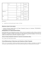
References
1. C.A. Zapffe and M. Clogg, Jr., Fractography--A New Tool for Metallurgical Research, Preprint 36,
American Society for Metals, 1944; later published in Trans. ASM, Vol 34, 1945, p 71-107
2. J.L. McCall, "Failure Analysis by Scanning Electron Microscopy," MCIC Report, Metals and Ceramics
Information Center, Dec 1972
3. C.A. Zapffe and C.O. Worden, Temperature and Stress Rate Affect Fractology of Ferrite Stainless, Iron Age,
Vol 167 (No. 26), 1951, p 65-69
History of Fractography
Fracture Studies Before the Twentieth Century
Valuable information has long been known to exist in the fracture surfaces of metals, and through the years various
approaches have been implemented to obtain and interpret this information (Ref 4). According to metallurgical historian
Cyril Stanley Smith, fracture surfaces have been analyzed to some degree since the beginning of the Bronze Age (Ref 5).
Early metalsmiths and artisans most likely observed specific fracture characteristics of metal tools and weapons and
related them to variables in smelting or melting procedures.
Sixteenth to Eighteenth Centuries. The first specific written description of the use of fracture appearance to gage
the quality of a metallurgical process was by Vannocio Biringuccio in De La Pirotechnia, published in 1540 (Ref 6). He
described the use of fracture appearance as a means of quality assurance for both ferrous and nonferrous (tin and copper-
tin bronzes) alloys.
Another early authority was Lazarus Ercker, who discussed fracture tests in a 1574 publication (Ref 7). The quality of
copper, for example, was determined by examining the fracture surface of an ingot that had been notched and then broken
by a transverse blow. Brass was similarly tested. A gray fracture surface was found to be associated with subsequent
cracking during working; this gray surface was the result of the use of a special variety of calamine, which caused lead
contamination of the ingot. Brittle fractures of silver were traced to lead and tin contamination.
In 1627, Louis Savot described in greater detail the use of the fracture test as a method of quality control of copper-tin-
bismuth cast bells (Ref 8). He recorded observations of grain size in fracture control samples as a guide for composition
adjustments to resist impact fracture when the bells were struck. In the same year, Mathurin Jousse described a method of
selecting high-quality grades of iron and steel, based on the appearance of fracture samples (Ref 9).
One of the most significant early contributions to the study of metal fractures was by de Réaumur (Ref 10), who
published a book in 1722 that contained engravings illustrating both the macroscopic and microscopic appearance of
fracture surfaces of iron and steel (although the microscope was invented circa 1600, at the time of de Réaumur it was
necessary to sketch what one saw and then transfer the sketch to metal, wood, or stone by engraving). In this classical
work, de Réaumur listed and illustrated seven classes of fracture appearance in iron and steel. These are described below
and shown in Fig. 1:
• Type I fracture: Large, irregularly arranged, mirrorlike facets, indicating inferior metal (Fig. 1a and b)
• Type II fracture: More regular distribution and smaller facets, indicating a slightly improved metal (Fig.
1c to e)
• Type III fracture: Interposed areas of fibrous metal between facets (Fig. 1f to h).
• Type IV fracture: Fibrous metal, with very few reflecting facets (Fig. 1j)
• Type V fracture: Framelike area surrounding an entirely fibrous center (Fig. 1k and m)
• Type VI fracture: An unusual type, with a few small facets in a fibrous background (Fig. 1n, p, and q)
• Type VII fracture: Characterized by a woody appearance (Fig. 1r)
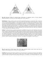
Fig. 1 Sketches from R.A.F. de Réaumur (Ref 10) depicting seven categories of fracture appearance in iron and
steel. (a) Type I fracture; large mirrorlike facets. (b) Same as (a), but as viewed with a hand lens. (c) Type II
fracture; smaller facets and more regular distribution. (d) Same as (c), but as viewed with a hand lens. (e)
Another type II fracture, but improved in regularity of distribution and reduced facet size compared with (c). (f)
type III fracture; advantageous occurrence of interposed areas of fibrous metal between facets. (g) Detail of
facets in (f). (h) Detail of fibrous metal in (f). (j) Type IV fracture; fibrous with very few reflecting facets. (k)
Type V fracture; framelike area surrounding an entirely fibrous center. (m) Same as (k), but as viewed with a
hand lens; a type VI fracture would look like this, except for finer grain size. (n) Type VI fracture; an unusual
type, with tiny facets in a fibrous background. (p) Detail of fibrous area in (n). (q) Detail of a small facets in
(n). (r) Type VII fracture; woody appearance.
A second plate from de Réaumur's book (Fig. 2) concerns the use of fracture surfaces in appraising the completeness of
conversion of iron to steel by the then current process of cementation (carburization). In his meticulous reproduction of
detail, he included phenomena still bothersome to metallurgists today, such as blistering, burning (overheating) brittle
fracture, and woody fracture. Descriptions of the fractures characteristics of the various stages of conversion are given
with Fig. 2. In summary, they are:
• Woody fractures characteristics of iron (Fig. 2a to c)
• Fractures characteristics of partly converted metal (Fig. 2d, f, and j)
• Fractures characteristics of steel (Fig. 2e and g)
Figure 2(h) shows a fracture that is typical of an iron that will convert easily to steel Fig. 2(j), a fracture typical of an iron
that will not convert to steel.
Fig. 2 Sketches from R.A.F. de Réaumur (Ref 10) defining fracture aspects that give evidence of the degree of
conversion of iron to steel by the cementation process. (a) Woody fracture, but without the distinctly clustered
appearance of the fracture in Fig. 1(r). (b) Woody fracture combined with minutely granular areas. (c) Fracture
exhibiting a combination of brittle facets, woody texture, and minutely granular areas. These fractures are
typical of iron. (d) Fracture in iron bar partly converted to steel, the outer minutely granular zone giving way to
an inner framework of brittle facets, which in turn surround the woody center. (e) Fracture in steel produced
from iron by cementation, showing a mass of small facets throughout the fracture, those in the center being
somewhat larger. (f) Fracture in iron bar converted to steel only from a-a to b-b and remaining as iron from a-a
to c-c because of overheating in the furnace. (g) Fracture in steel; the lusterless, rough facets resulted from
holding the specimen too long in the furnace. (h) Fracture in a type of iron that always produces very small
facets when converted to steel. (j) A type II fracture in iron (see Fig. 1), indicating that the iron will fail to
convert to steel in the center and will also produce an inferior fracture frame. (k) Fracture in forged steel
showing folding at left end, which could later cause cracking during heat treatment. In (d), (f), and (g), small
blisters are indicated by the letter g, large blisters by the letter G, and porosity by the letter O.
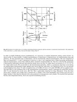
A third plate from de Réaumur (Fig. 3) displays his fracture studies at high magnifications and his fracture test. The
minute platelets shown in Fig. 3(f), which may have been pearlite or cementite in some form, were recorded a full century
and a half before the founding of metallography.
Fig. 3 Sketches from R.A.F. de Réaumur (Ref 10) showing notched-bar fracture test specimen, enlarged view
of grains, and details of fracture in cast iron. (a) A notched-bar fracture test, which allows two identical steel
bars to be broken with a single blow. (b) Grain ". . . prodigiously magnified [i.e., of the order of 50×] showing
the voids V and molecules M. . ." (probably crystallites) of which the grain is composed. (c) One of the
individual molecules of (b) showing the units of which it is composed. (d) Fracture in gray iron, strongly
resembling one in steel, except that the surface is brownish and the grains are coarser. (e) Detail of (d) at high
magnification showing the fracture to comprise". . . an infinity of branches. . ." Here de Réaumur's
magnification clearly exceeded 100×, although some of the enlargement may have been contributed to drawing
what he saw. (f) Detail of an individual branch (dendrite) showing structure of minute platelets (possibly or
cementite) placed one on another
Encouraged by his studies of the changes of fracture surfaces accompanying the conversion of iron into steel, de Réaumur
published in 1724 studies of cast metal fractures, including plates illustrating fractured antimony and lead ingots (Ref 11).
The results of these studies are reviewed by Smith in Ref 5.
Information on the nature of fracture of copper-zinc alloys was published in 1725 by Geoffroy (Ref 12). In his studies,
Geoffroy investigated the influence of the copper-to-zinc ratio on the appearance of the fracture surface and on grain size.
Somewhat later (1750), Gellert described the fracture characteristics of metals and semimetals (Ref 13), mentioning the
use of a fracture test for distinguishing among steel, wrought iron, and cast iron. The test was also used to appraise the
effects of carburization and heat treatment. Further, Gellert discussed causes of embrittlement of metals as disclosed by
inspection of fracture surfaces.
The German physicist and chemist Karl Franz Achard, in carrying out his studies on the properties of alloys, also realized
the importance of the appearance of fracture (Ref 14). Archard noted the appearance of the broken surfaces of nearly all
of the 896 alloys he tested (Ref 5). This number represented virtually every possible combination of all metals known at
that time.
Nineteenth Century. With the development of metallography as a metallurgical tool, interest in the further
development of fracture studies waned. An important exception to this was Mallet (Ref 15), who published a paper in
1856 that related fracture details in cannon barrels to the mode of solidification, referring to planes of weakness resulting
from sharp angles in the contours of the barrels. This may have been the first example of failure analysis and the first
recognition of the deleterious effects of stress concentration in design. At the same time, the U.S. Army Ordnance Corps
implemented fracture evaluation with mechanical testing for the study of ruptured cannon barrels (Ref 16).
In 1858, Tuner published a list categorizing fracture characteristics, citing such conditions as hot shortness, overheating,
and various types of tears (Ref 17). In 1862, Kirkaldy correlated the change in fracture appearance from fibrous to
crystalline with specimen configuration, heat treatment, and strain rate (Ref 18). He reported that crystalline fractures
were at 90° to the tensile axis, whereas fibrous fractures were irregular and at angles other than 90°.
The doctoral dissertation of E.F. Dürre in 1868 contains an excellent description of the many different textures and details
to be seen in the fracture of cast irons as well as a summary of the literature of the time (Ref 19). Dürre advocated the use
of low magnification to study the fracture of castings, but considered the high-magnification microscope impractical for
this purpose (Ref 5).
Two papers on steel, published by the Russian metallurgist D.K. Tschernoff, contributed significantly to fracture studies.
The first, published in 1868, discussed fracture grain size in relation to heat treatment and carbon content (Ref 20). In a
later paper, Tschernoff described the fracture of large-grain steel and, for the first time in history, accurately illustrated the
true shape of metal grain (Ref 21).
John Percy, a prolific author on metallurgical subjects, described by 1875 six general types of fracture patterns (Ref 22):
• Crystalline, with facets as in zinc, antimony, bismuth, and spiegeleisen
• Granular, with smaller facets, as in pig iron
• Fibrous, a general criterion of quality
• Silky, a finer variety of fibrous, such as in copper
• Columnar, typical of high-temperature fracture
• Vitreous, or glasslike
Adolf Martens (for whom martensite is named) undertook studies of metal structure by examining newly fractured
surfaces and polished-and-etched sections, both under the microscope. He published his first findings in Germany in 1878
(Ref 23, 24). His illustrations were hand engravings that reproduced meticulous pencil drawings in some figures and
photomicrographs in others.
A plate from a later article (1887) by Martens is shown in part in Fig. 4. This plate consists of fractographs produced by
photography and then printed by a photogravure process. The fractures shown in Fig. 4 illustrate features that Martens
termed Bruchlinien (fracture lines), which today would be called radial marks. The description of this fracture form by
Martens predates reports by other investigators who are usually credited for first treatment of this fracture feature.
Fig. 4 Steel fractures recorded by A. Martens (Ref 25, p 237, Plate X). Martens called attention to the radial
fracture marks in these fractographs, terming them Bruchlinien (fracture lines). (a) Ingot steel; tensile
strength, 765 MPa (111 ksi). (b) through (e) Tool steels from Böhler Bros. of Vienna and others: (b) extra
hard; (c) very hard, special; (d) moderately hard; (e) ductile. (f) Chisel steel with fracture lines. (a)through (e)
Actual size. (f) 6×
In the field of macrofractography, Martens observed the fracture surfaces obtained in tension, torsion, bending, and
fatigue. In describing the topography of these surfaces, he differentiated between coarse radial shear elements and fine
radial marks. He recognized that sharper radial marks occured in fine-grain material and that all radial marks diverge from
the fracture origin; that is, they point backward to the origin. More detailed information on the contributions of Martens to
metallurgy can be found in Ref 5.
Another important paper on fracture was written by Johann Augustus Brinell (the inventor of the Brinell hardness test) in
1885 (Ref 26). Brinell discussed the influence of heat treating and the resulting change in the state of carbon on the
appearance of steel fractures. Henry Marion Howe critically analyzed and subsequently praised Brinell's findings, stating
that the latter's work represented "the most important fracture studies ever made" (Ref 27).
Classic cup-and-cone tensile fracture was described by B. Kirsh before 1889 (Ref 28). He postulated a concept of crack
propagation in tensile specimens that is retained today. He theorized that the crack origin was at the tensile axis in the
necked region, that the origin grew concentrically in a transverse direction to produce the "bottom of the cup," and that
the sides of the cone were formed by a maximum shear stress at final separation.