Tài liệu Figure Drawing - Figure Anatomy pdf
Bạn đang xem bản rút gọn của tài liệu. Xem và tải ngay bản đầy đủ của tài liệu tại đây (734.08 KB, 18 trang )
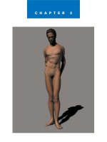
CHAPTER 3
Figure Anatomy
✎
33
U
nderstanding human anatomy will help you achieve greater
expressive ability in figure drawing. By understanding the many dif-
ferent aspects of the human form, you can better grasp how the fig-
ure works as a whole. For example, if you feel along the bone on the lower
part of your jaw, you will notice that there is a small indentation about
halfway between the chin and the back of the jaw. This indentation is to allow
a blood vessel to pass under the jaw. The indentation helps to protect the ves-
sel. The significance of this little indentation is that it affects the curvature of
the jaw. The jawbone is actually concave here, rather than convex. A slender
person who has little fat around the jaw will show this distinct feature of the
jaw more clearly than a heavy person will. Knowing this little aspect of the
figure can help the artist who wishes to express a thin person.
In a way, the study of anatomy increases your figure drawing arsenal. By
studying the underlying structure, you can develop a greater feel for the sur-
face, expanding creative possibilities. In essence, the human form is made up
of soft and hard tissue held together by tendons and ligaments. The bones
form the underlying structure of the body and in some cases act as protection
for delicate internal organs. Around and over the bones are muscles that are
used to drive movement. The whole system is controlled by an extensive
nervous system.
The human form is capable of extreme movement and flexibility. The muscles
that power body movement expand and contract, causing surface changes to
appear. Take a look at your arm. Hold it out in front of you with the palm
down. Now twist your arm so the palm is facing up. Notice how the muscle
beneath the skin move as the arm rotates. As the arm rotates, some muscles in
the forearm will expand and twist, while others will contract to cause the
34
Figure Drawing with Virtual Models
movement. Now bend your arm
up at the elbow. The muscles of
the upper arm will bunch to pull
the arm up. These muscles are
the biceps, so named because
there are two muscles.
Drawing from life, an artist is
often confronted with a number
of organic surfaces. It is helpful if
the artist understands not only
why the surface changes in
movement, but also what the
underlying structure is doing
during those changes. This will
help the artist to recognize the
subtle aspects of the figure that
might go unnoticed if the artist
didn’t have the proper instruc-
tion in anatomy.
The Skeleton
In Chapter 2 you created a simpli-
fied skeletal structure to use as a
base for drawing the figure. We
called it drawing from the inside
out. Now you will have the oppor-
tunity to better understand the
actual skeletal structure of the
human body. Figure 3.1 shows the
male skeleton. This skeleton comes
with Figure Artist’s bigger cousin,
Poser, and is available as additional
content for Figure Artist.
The human skeleton contains more
than 200 individual bones.
Although it might not be essential
for you to learn the names of all
200 bones, you should become
familiar with some of the major
bones listed here.
✎
Skull. The skull is the
bony framework upon
which the head is built.
It is composed of eight
cranial and 14 facial
bones. The cranial
bones are the dome-
shaped bones that
cover the top of your
head. They are very
near the surface. If you
press on the top of
your head, you can feel
the hard surface of the
bone just below the
skin. The shape of the
cranium pretty much
determines the shape
of the head. The facial
bones make up the
bones of the front of
the head and con-
tribute greatly to how a
person looks.
✎
Mandible. The
mandible, sometimes
referred to as the jaw-
bone, is actually one of
the facial bones of the
skull. It is the moveable
bone on the lower part
of the head. It is hinged
to the rest of the skull
up near the ears. It is
important to note
where the bone is
hinged because that
controls the bone
movement.
✎
Clavicle. The clavicle is
located on the upper
front of the chest near
the neck. It is a very
pronounced bone near
the surface, connecting
the arms to the chest.
There are two clavicle
bones—one on each
side of the body. The
clavicle’s flexible move-
ment allows for the
variety of movement in
the shoulder.
✎
Scapula. The scapula is
a plate-like triangular
bone located on the
right and left side of
the upper back. It is
sometimes called the
shoulder blade. It has
quite a range of move-
ment under the skin
and is more pro-
nounced in a slender
person. It will also pro-
trude more in a person
with poor posture.
35
Figure Anatomy
Figure 3.1 The skeleton forms the structure upon which the body is built.
36
Figure Drawing with Virtual Models
✎
Ribcage. The ribs are
actually a group of
bones that surround
the chest cavity and
serve as protection for
the delicate organs
housed in that area. All
together, the ribs form
a somewhat egg-
shaped structure that is
open at the bottom and
more closed at the top.
The ribcage also acts
as an anchor for many
of the muscles of the
upper back and chest.
✎
Sternum. The sternum
is located in the center
of the chest and con-
nects the ribs of the left
and right sides by way
of cartilage, which
gives the chest the flex-
ibility to expand and
shrink with breathing.
The sternum has a dis-
tinctive dagger shape
and is sometimes
referred to as the
breastbone.
✎
Spine. The spine is a
column of bones that
extends from the skull
to the pelvis. The spine
is a very flexible com-
bination of bones and
cartilage that encloses
and protects the spinal
cord. The spine is also
the structure that holds
the upper body erect.
There are 33 separate
irregularly shaped
bones called vertebrae
in the spinal column.
The top bone of the
spinal column is called
the Atlas, and the next
is called the Axis. The
shape of the Atlas
allows the head to nod
yes, and the shape of
the Axis allows the
head to shake no. The
vertebrae at the top of
the spinal column are
smaller than those near
the bottom. They con-
nect the ribcage in the
back and support most
of the major muscles of
the back. They can be
seen as a row of ridges
when a person bends
forward.
✎
Pelvis. The pelvis is
located in the lower
body and forms your
hips. There are actually
two pelvic bones—one
on either side of the
body. They are joined
together in the back by
the sacrum and in the
front by a muscle
called the pubic sym-
physis. The pelvis
serves to support the
body by anchoring the
spinal column, and it
also protects many of
the delicate organs of
the lower body. The
pelvis on a female is
wider, and the central
opening is larger than
on a male. This differ-
ence helps the female
to support a baby dur-
ing pregnancy. The
wider opening allows
for the baby to be born
because the baby must
pass through the
mother’s pelvis.
✎
Sacrum. The sacrum is
a V-shaped bone that is
actually several verte-
brae fused together as
a person reaches adult-
hood. This bone
attaches the spinal col-
umn to the pelvis
bones.
✎
Femur. The femur is
the large bone that
runs from the hip to the
knee. It would be the
largest bone in the
body except that there
are two of them, and
since they are usually
the same size, they
both share that honor.
The femurs are the
largest, longest, and
strongest bones in the
body. They support the
massive thigh muscles
37
Figure Anatomy
and are mostly sur-
rounded by those mus-
cles so that the bone is
not very close to the
surface, except near
the knee and around
the hip.
✎
Patella. The patella is
the small bone that fits
over the knee and is
sometimes called the
kneecap. It protects the
knee joint and strength-
ens the tendons of that
area. It is very near the
surface and is quite dis-
tinctive in the leg.
✎
Tibia. The tibia is the
bone that connects the
knee to the foot and is
the larger of the two
lower-leg bones. It is
sometimes referred to
as the shinbone. It is
near the surface on the
front of the leg, and its
curve is very evident
when you are viewing
the leg from the front.
✎
Fibula. The fibula is the
smaller of the two
lower-leg bones and is
primarily used for mus-
cle support in that area.
It is located on the out-
side of the body and is
mostly covered with
muscle, so it is less evi-
dent than the tibia.
✎
Foot bones. There are
26 bones in each foot
from the ankle to the
joints of the toes. The
largest bone is the cal-
caneus or heel bone.
The tarsal bones form
the ankle and are com-
posed of seven sepa-
rate bones. They are
near the surface and
protrude outward on
either side of the leg,
forming the knobs we
call ankles. The inside
knob is slightly higher
than the outside knob.
The bones of the foot
are closer to the sur-
face on the top of the
foot and toes than on
the bottom, which is
covered by thick
padding.
✎
Humerus. The humerus
is the largest and
strongest of the arm
bones and runs from
the shoulder to the
elbow. It connects to
the scapula at the
shoulder and the ulna
and radius at the
elbow. It is covered by
muscles and only nears
the surface at the
elbow, where it pro-
tects the ulnar nerve.
Sometimes when the
elbow is struck the
ulnar nerve causes a
tingling sensation,
leading the humerus to
sometimes be called
the funny bone.
✎
Ulna. The ulna is the
longer of the two
lower-arm bones and is
more firmly connected
to the humerus near
the back of the elbow.
The ulna is near the
surface, and if you run
your hand from the
back of the elbow to
your wrist, you can feel
the hard surface of this
bone.
✎
Radius. The radius is
also connected to the
humerus, but not as
firmly as the ulna. It is
also more responsible
for the movement of
the wrist.
✎
Hand bones. There are
27 bones in the hand
and fingers, running
from the wrist to the
tips of the fingers. The
underside of the hand
is covered with
padding and muscle
tissue, and the bones
are not as close to the
surface as they are on
the back of the hand,
where they can be very
distinctive.
38
Figure Drawing with Virtual Models
Try to learn how these bones look
and function even if you don’t
remember all their names. If you
can make sketches of each individ-
ual bone, such as the humerus in
Figure 3.2, you will gain a better
understanding of how the bones
affect the shape of the body.
If you look closely at the skeleton,
you will notice that there are no
straight bones. Every bone is
curved. In fact, every bone is made
up of many curves. If you draw the
bones of the body as straight, you
will end up with a stiff, robotic-
looking drawing. Your drawings
will look better if you understand
the direction of the curves of the
bones and emphasize those curves
in your figures.
Figure 3.3 shows the female skele-
ton. You can see from the skeleton
that there are many differences
between the male and female
skeletons even though they are
made up of the same number and
types of bones.
In the female, the pelvis is more
open and proportionally larger
than in the male skeleton. The
male ribcage is proportionally
larger, giving the male skeleton
wider shoulders and narrower
hips. They also give the female fig-
ure wider hips, a higher waistline,
and lower buttocks than the male.
In general, the female skeleton is
smaller and more delicate than the
male skeleton. The male skeleton is
build for power and lifting. If you
look at how the bones are built
and the muscles, you will see that
the male is more massive. The
bones are thicker, and the muscle
groups are bulkier. On the female
the bones are smaller and more
slender. The muscles are less bulky,
almost strap-like, except for the
area around the hip and thigh,
where the female tends to be more
bulky than the male does.
A good example of how the two
skeletons differ is in the elbow
joint. The male elbow is a fairly
direct joint, whereas the female
joint tends to have the lower arm
flare out. Take a look at your own
arm. Hold it out with the palm fac-
ing up. If you are male, the arm
should remain fairly direct. If you
are female you will notice that the
Figure 3.2 Sketch individual bones of the skeleton.