Identification of Pathogenic variants in IGHMBP2 gene and Application on Prenatal diagnosis of Rare Neuromuscular Disorders
Bạn đang xem bản rút gọn của tài liệu. Xem và tải ngay bản đầy đủ của tài liệu tại đây (834.77 KB, 82 trang )
CAO HA MY
IDENTIFICATION OF PATHOGENIC VARIAN TS
IN IGHMBP2 GENE AND APPLICATION ON
PRENATAL DLAGNOSIS OF
RARE NEUROMUSCULAR DISORDERS
Graduation Thesis
Major: General Practitioner
Major code: 52720101
Academic Supervisor
Associate Professor TRAN VAN KHANH. M.D, PhD.
ACKNOWLEDGEMENT
I first joined scientific research four years ago as an enthusiastic student
yearning for new knowledge. That full of difficulties but memorable experiences have
-■c -ÍM
V
changed me and become part of
myQỉ ugc
youth
I was lucks' enough to meet, learn, and
Hl
receive valuable guidance from many teachers and friends during niv journey. Their
support would be of great importance for me to complete my graduation thesis
confidently.
Firstly I would like to express my deepest gratitude to Associate Professor Tran
Van Khanh. MD PhD. Deputy Director of Center for Gene-Protein Research. Head of
Molecular Pathology Department of Hanoi Medical University, who provided me great
support and leadership which allowed me to conduct the research and finish this diesis
successfully I am thankful to her for presenting such excellent advice and guidance
despite having a tight schedule.
I heartily thank Professor Ta Thanh Van, M.D, Ph D. Chairman of University
Council of Hanoi Medical University and Director of die Center for Gen-Protein
Research. Professor The-Hung Bui MD, PhD Centre for Molecular Medicine. Clinical
Genetics Unit in Karolinska Universitat, and Assc. Professor Tran Huy Thinh. M.D., Ph
D Deput)' Head of Department of Biochemistry. Head of Department of Technology
and Scientific Research Management, who always created favorable conditions for me
to access scientific research and inspired me to be a good doctor and researcher with a
thorough understanding of medical ethics.
I am deeply
thankful
to
Dr always
Luong
Hoang
Longme
Department
Clinical
hospital
Allergy.
for
his patience,
Immunology
consistent
and Dermatology
motivation
National
and Eof
immense
insightful
knowledge.
comments
He
and
also
hard
questions
provided
during
with
my
scientific
research
projects.
-ÍM Qỉ ugc V
Hl
My sincere thank also goes to Ms. Le Thi Phuong and the Centre tor GeneProtein Research staff who carefully insttucted and supervised me to conduct several
molecular techniques.
The last words of thanks. 1 would like to send to my parents, who are the idols
in me and have always laid concrete encouragement to me in my life.
Hanoi. May 2021
Cao Ha My
r-u -ÍM CỊỈ ugc V
Hl
DECLARATION
LIST
OF ABBREVIATION
I hereby certify that this thesis incorporates original research, which has not been
previously submitted for a degree to any other institution, and the best of my
knowledge and belief, it does not contain any material previously published or written
by other persons except where reference has been made in the text
Hanoi. May 2021
CAO HA MY
AchR
AFP
Acetylcholine receptor
Alpha-fetoprotein
CNS
Central nervous system
CMT2S
CVS
Charcot-Marie-Tooth disease, type 2S
Chorionic villus sampling
DIA
Dimeric Inhibin-A
DNA
Deoxyribonucleic Acid
DSMA
hCG
Distal Spinal Muscular Atrophy
human Chorionic Gonadotropin
HMN
Hereditary Motor Neuropathy
HMSN
IGHMBP 2
Hereditary motor and sensory neuropathies
Immunoglobulin Mu DNA Binding Protein 2
IVF
in vứro Fertilization
NMD
Neuromuscular disorder
NGS
PNS
Next Generation Sequencing
Peripheral nervous system
Papp-A
Pregnancy-associated plasma protein A
PGD
Preimplantation Genetic Diagnosis
r-u -ÍM Qỉ ugc V Hl
PGS
Preimplantation
Screening
LIST Genetic
OF ABBREVIATION
PCR
Polymer Chain Reactions
RNA
Ribonucleic Acid
SMARD1
SMA
Spinal Muscular Atrophy with Respiratory distress 1
Spinal Muscular Atrophy
uE3
Unconjugated Estriol
r-u -ÍM Qỉ ugc V Hl
TABLE OF CONTENTS
INTRODUCTION
REFERE NCE
APPEND LX
-■c -ÍM Qỉ ugc V
Hl
LIST OF FIGURES
LIST OF T ABLES
Table 1.1. List of genes identified as distal hereditary motor neuropathies (dHMN)
Identification of Pathogenic Valiants in IGHMBP2 gene
and Application on Prenatal Diagnosis
of Rare Neuromuscular Disorders
ABSTRACT
Background spinal muscular atrophy with respiratory distress type 1 (SMARD
1) and Charcot - Marie - Tooth type 2S (CMT2S) are rare neuromuscular disorders
caused by biallelic pathogenic variants on IGHMBP2.
Objectives: 1) To identify pathogenic variants in IGHMBP2 gene and describe
four cases with IGHMBP2 mutation 2) To identify’ earners and application on prenatal
diagnosis
Subjects and methods: Four patients under 12 years of age with lower limbs
weakness (3 4 had respiratory disorders) and family members. Genetics analysis for
patients and family members was performed using Next Generation Sequencing and
Sanger Sequencing
Results We identified four IGHXỈBP2 mutations, in which C.1574T> c (p
Leu525Pro) is a novel mutation Both parents and sisters of four patients were identified
to be carriers. The mother of the third patient’s family was pregnant but had an abortion
after genetics testing of the fetus revealed compound heterozygous mutations on
IGHMBP2.
Conclusion: 1 patient was homozygous for IGH.\iBP2 mutation, and three out of
4 patients had compound heterozygous mutations. All parents were identified as
carriers, and we successfully applied the genetic testings on prenatal diagnosis for the
family of patient 03.
Key
words: IGHMBP2,
muscle
weakness neuromuscular
respiratory
distress.
Charcot-Marie-Tooth.
SMARD1 disorder
INTRODUCTION
Neuromuscular disorders (NMDs) include various conditions that affect
components of a motor unit, sensor.' and autonomic nen es, or their supportive
r-u -ÍM CỊỈ ugc V
Hl
structures, such as myopathies, disorders of the neuromuscular junction, and
neuropathies. The pattogenesis of NMD ranges from secondary to chronic medical
condition, trauma, nutritional deficiency to the hereditary cause. The presence of NMD
incurred health burden not only for the child’s disability and early death, lower quality
of life but also a financial burden for the family and society, and most importantly
emotional burden and stigma for the parents
Congenital NMD, or hereditary NMD is relatively rare. Therefore the chance of
encountering a case in daily clinical practice is meager Physicians would hardly
encounter such a case, and when they do. the condition is often misdiagnosed,
especially in Vietnam where many neuromuscular disorders were misdiagnosed as
either cerebral palsy or myasthenia gravis Definitive diagnosis is crucial for providing
the correct treatment and counseling direction for the patient In addition research at the
molecular level will unravel the mechanism of the disease, enabling scientists to
elaborate effective therapy for patients in the future.
/GẢBÍ5P2-related NMD is a congenital disease which was reported in two
clinical manifestations: SMARD1 and CMT2S with relatively different phenotype and
prognosis Differentiated and definitive diagnosis of these two diseases required both
extensive clinical examination and identification of genetic defects Deciphering the
exact pathogenic variants allows for thorough genetic counseling, prenatal diagnosis
and pre-implantation genetic diagnosis for high-risk families. Since there haven’t been
any curative treatments available for any NMD, detection of disease-causing variants
allowed physician to provide medical support (such as prenatal diagiosis) to family with
hope of having a healthy child
In order to better understand the natural history and mechanism of ĨGHMBP2related NMD and other rare diseases, it is necessary to create a domestic rare disease
network with a large cohort and longitudinal follow-up. Currently, some international
and regional rare disease network has already been established with the aim for helping
doctors and patients to raise awareness about the rare disease Therefore, our research on
IGHMBP2-related NMD will contribute initially to building the database of rare
diseases in Vietnam and a premise for further studies in the field of rare diseases,
r-u -ÍM CỊỈ ugc V
Hl
thereby merging our national rare disease data with an international database.
The study “Identification of Pathogenic variants in IGHMBP2 gene and
Application on Prenatal Diagnosis of Rare Neuromuscular Disorders" was conducted
with two primary objectives.
-
To identify IGH.\fBP2 pathogenic variants and to describe the clinical
manifestations of the patients
To detectdiagnosis
carriers among family members and apply on
prenatal
r-u -ÍM CỊỈ ugc V
Hl
1
3
C HAPTER 1. LITERATURE REVIEW
1.1.
Phenotypic spectrum of Neuromuscular Disorders
1.1.1.
The peripheral nervous systems
The primary function of the peripheral nervous system (PNS) is to transfer
information from the limbs to and from the central nervous system (CNS), which
consists of the brain and the spinal cord The nen es responsible for this include the
cranial nen es that link to the brainstem and 31 pairs of spinal nerves that branch out
between each vertebra of the spine, connecting to the spinal cord. Components of the
PNS are the sensory nen es, the motor nen es, and the autonomic nenous system. The
sensory nen es are the afferent nen es that convey information from the sensory organs
and limbs to the brainstem and the spinal cord, which typically have cell bodies located
in the dorsal root ganglia located close to the spinal cord Meanwhile, the motor nenes
arising from the ventral horn of the spinal cord transfer information from the brainstem
and the spinal cord to the neuromuscular junctions at the muscles The autonomic
nervous system is also considered part of the PNS as it works in conjunction with the
sensory and motor nen es However, the peripheral nenes include not only the nene fibers
but also several layers of connective tissue (endoneurium. perineurium, and epineurium)
and blood vessels [1] [2].
Individual nene fibers consist of long axons extending to the body's extremities
that may be myelinated or unmyelinated. Myelin in the peripheral nervous system
derives from Schwann cells, which adhere to nene cell membranes and create multiple
layers or wrappings of the membrane This myelin sheath results in an insulating lipidrich layer around the nene fibers, allowing for a higher conduction velocity depending
on the diameter of the sheath. Unmyelinated axons are solely enveloped by a single
layer of Schwann cell cytoplasm Thus they conduct very slowly by a continuous mode
of propagation of the electrical signal In the context of peripheral neuropadlies,
abnormalities can be found in both the axon and the myelin sheath, causing different
phenotypes [1].
The Motor unit is physiologic. Typically, striated muscle contraction is only
r-u -ÍM Qỉ ugc V Hl
1
4
possible through the firing of motor neurons that are activated either via descending
pathways or through reflex connections The muscle fibers in a motor unit respond in an
all-or-none fashion to excitation by the motor neuron, producing a quick twitch.
Conditions that damage some motor units (sparing others) usually result in overall
weakness of the muscle but high firing rates of individual motor units that are still intact
This is because of the decreased number of motor units that must be activated at
maximal frequencies to generate any muscle force. Therefore, weakness with a high
firing rate indicates a loss of motor neurons or motor axons [2]
1.1.2.
Classification
of
Neuromuscular
disorders
and
Congenital
Neuromuscular disorders
There has not been a unanimous classification of Neuromuscular disorders
However based on anatomical characteristics of the PNS, there are three basic types of
neuromuscular disorders, including damage to anterior horn cells (also called motor
neuron disease), damage to the peripheral nene fibers (myelin or axons), and damage to
the neuromuscular junction The etiology of this disease could either be congenital or
secondary' to prior medical conditions or acquired during a lifetime Thus, the term
congenital (or inherited) muscular disorders share some similarities with neuromuscular
disorders and include other types of diseases Currently, the congenital muscular disorder
can be divided into congenital neuromuscular disorder and congenital myopathy
Myopathy comprises two types: the disease that progressively destroys muscle fibers
such as Duchenne muscular dystrophy, Limb-Girdle muscular dystrophy and disease
leading to congenital functional defect of the muscle without distinct muscle wasting are
usually disease of the ion channel The following paragraphs will introduce the basic
classification of neuromuscular disorders
1.1.2.1. Motor neuron disease
Motor neuron disease results from damage to the anterior horn cells arising from
the spinal cord to the limbs (lower motor neurons) or the upper motor neurons of the
cerebral cortex that give rise to the descending tracts that control movements These
conditions can present weakness accompanied by lower motor sign s (atrophy,
fasciculations. and decreased reflexes) or upper motor neuron signs (spasticity, increased
r-u -ÍM CỊỈ
ugcVVHl
Qỉ ugc
Hl
1
5
reflexes, and upgoing toes). The most common condition affecting both the upper and
lower motor neurons is amyotrophic lateral sclerosis ALS. There are also variants of
motor neuron disease, which selectively involve the upper motor neurons (primary
lateral sclerosis), the lower motor neurons (progressive muscular atrophy), or the cranial
musculature (bulbar palsy). The cause for motor neuron disease can be genetic defects,
termed Hereditary Motor Neuropathy (HMN). For this thesis, a specific type of HMN
(called Spinal Muscular Atrophy with Respiratory distress) will be further discussed in
the following sections.
1.1.2.2.
Peripheral neuropathy
Peripheral neuropathy is a disease that affects the peripheral nerve fibers, which
includes mononeuropathy, mononeuropathy multiplex, or polyneuropathy- Therefore.
both abnormal motor and sensory features can be presented The symptoms can
determine the particular nerve or nerves that are affected. Symptoms may be "positive"
(including pain and dysesthesia), may be harmful (including loss of sensation, weakness,
or loss of reflexes), or may be irritative (such as fasciculationsor paresthesias).
Mononeuropathy
ưauma
of autoimmune
some
types.
and
radiculopathy
The
most
common
are
most
mononeuropathies
often
due
to
are
entrapment
as
carpal
tunnel
ofinterfering
nenes
syndrome
at
anatomically
(CTS),
ulnar
vulnerable
nene
enuapments
sites
such
al
the
Radiculopathy
elbow
and
radial
indicates
nene
damage
damage
to
fracture
nene
root(s)
of
the
humerus
typically
In
younger
occurs
individuals,
as
a
component
this
is
of
usually
several
due
spinal
to
diseases
intervertebral
degenerative
changes
disc
herniation.
in
the
discs,
This
bones
is
more
and
often
joints
due
in
to
older
rare
presentation
individuals
'Mononeuritis
of
certain
disorders
multiplex"
that
is
damage
aand
relatively
nerves
primarily
or
by
an
by
process
with
damaging
blood
flow
either
to
nerves
the
myelin
or
plexi
or
axon.
The
most
common
cause
is
diabetes
mellitus.
Polyneuropathy can affect either the axon, or myelin sheaths (demyelinating), or
both There are over 100 known acquired and inherited disorders that may cause
polyneuropathy Since the nen es to the lower limbs are the longest they are the most
dependent on a good supply of metabolic substrates and have the most significant
exposure to toxins or damaging the myelin Therefore, symptoms and signs are most
prominent in the feet Loss of sensation ("numbness”) is the most common finding but
paresthesias or dysesthesias (prickling, tingling, burning, etc.) are also common Causes
for polyneuropathy can be categorized as metabolic, nutritional (deficiency of vitamin
B). toxic (alcohol), infection (HIX’ syphilis), inflammatory, dysproteinemia and
hereditary There is an incredible number of peripheral neuropathies that may be fami
lial: some of the most common is Charcot Marie-Tooth disease with around 24 subtypes
[1], [3]. [4], For this thesis. Charcot- Marie-Tooth will be further discussed in the
following parts.
LI.2.3. End-plate (neuromuscular junction)
In most cases, each muscle fiber has only one end-plate, which plays as the plug
r-u -ÍM Qỉ ugc V Hl
1
6
for the nerve terminal branch Acetylcholine is the neurotransmitter produced from the
motor neuron stimulating specific muscles. Myasthenia gravis is the most common
disorder affecting neuromuscular transmission. Autoantibodies primarily mediate the
disease against die Acetylcholine receptor (AchR). The impairment of neuromuscular
transmission and muscular weakness is explained by several mechanisms including
functional blockade of AChR. increased degradation of AChR. and die complementmediated destruction of the postsynaptic folds [5], 1.1.3. Spinal .Muscular Atrophy with
Respiratory distress 1 (S.MARD1) LỈ.3.Ỉ. Overview of distal hereditary- motor
neuronopathy
The distal hereditary motor neuronopathies (dHMN) are a genetically
heterogeneous group of diseases characterized by distal lower-motor-neuron weakness
dHMN is also refened to as distal spinal muscular atrophy (dSMA). a reflection of the
commonly held but unproven belief that the pathology resides in the ventral horn of the
spinal cord of note. dHMN is often referred to as a 'neuronopathy' instead of a
'neuropath/ based on the hypothesis that the primary pathologic process resides in the
neuron cell body and not in the axons [6]
The dHMN usually presents as a classical peroneal muscular atrophy syndrome
without sensory symptoms [4] The overall clinical picture consists of progressive
weakness and wasting of the extensor muscles of the toes and feet Later on. weakness
and wasting also involve the distal upper limb muscles. Foot deformity is a common
feature. Often, unusual or additional features are present in ‘complicated' distal HMN.
including predominance in the hands, vocal cord paralysis, diaphragm paralysis, and
pyramidal tract signs In some families, several of these additional signs co-occur.
In
one
of
the
earliest
reports
on
distal
HMN,
a
family
was
described
and
onset
with
in
early
an
autosomal
adult
life
dominant
Also,
mode
kinship
of
inheritance
autosomal
reported
(7).
recessive
By
then,
inheritance
affected
individuals
isolated
with
cases
variable
were
onset
documented
ages,
The
ranging
diagnosis
from
infancy
of
dHMN
to
in
adult
a
patient
life,
with
were
a
distal
motor
whether
neuropathy
the
phenotype
phenotype
is
genetic
first
requires
Detection
considering
of
diseasecausing
novo
mutations,
variant
is
small
not
families,
always
straightforward
and
non-paternity
owing
A
de
detailed
informative.
history,
The
cardinal
as
is
often
feature
the
case,
is
usually
is
most
a
slowly
progressive
the
first
two
lengthdependent
decades
while
the
condition,
third
decade
often
is
starting
not
in
uncommon
progression
Poor
are
performance
valuable
clues,
in
school
whereas
and
insidious
a
short,
de
novo
history
acquired
in
etiology.
middle
age
Bulbar
should
involvement
prompt
search
other
than
for
an
the
recurrent
usually
confirms
laryngeal
distal
nerve,
wasting
is
rare
in
weakness
dHMN
The
with
examination
reduced
or
absent
amplitude
reflexes,
potentials
and
neurophysiology
associated
with
confirms
EMG
changes
reduced
suggesting
significant
chronic
proportion
distal
of
predominant
patients
classified
denervation.
as
dHMN
Ato
will
be
approach.
‘sporadic’
with
no
apparent
family
history'
using
this
Theoretically. dHMN is in contrast to Charcot-Marie-Tooth disease (CMT) and the
hereditary sensory neuropathies where sensory involvement forms a significant
component of the disease [4] Nevertheless, on clinical examination. dHMN can hardly
be distinguished from Charcot-Marie-Tooth (CMT) neuropathy (hereditary motor and
sensory neuropathies. HMSN) because unmistakable sensory- signs are often lacking in
these disorders Many forms of dHMN have minor sensory' abnormalities, and there is an
r-u -ÍM CỊỈ
ugcVVHl
Qỉ ugc
Hl
1
7
overlap between the axonal CMT (CMT2) and dHMN where the pathogenic variants in
the same gene (IGH.\!BP2) may cause both phenotypes (8] [9] Also, it is not uncommon
for patients with CMT2 and significant sensory involvement neurophysiologically to
have no sensory symptoms and minimal sensory signs. Therefore, nene conduction
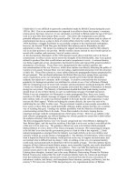
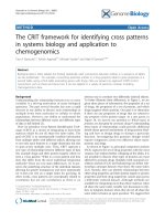
studies (NCS) are essential to support the diagnoses of CMT or distal HMN [10]. Figure
1.1 illustrated a Venn diagram of disease genes for CMT1. CMT2. and dHMN. The
genetic defect of dHMN and CMT. will be further elaborated in the following sections.
dHMN should also be differentiated with distal myopathy using electromyography
(EMG). Like dHMN. the distal myopathies are a genetically and phenotypically diverse
group of conditions Some such as Myoshi myopathy, may have neck flexion weakness
helpful in making the diagnosis, but others may present with isolated foot drop In such
scenarios. EMG is the most helpful test. In the upper limbs, the intrinsic hand muscles
are usually affected first in dHMN. whereas in the distal myopathies, it is often the
forearm flexors.
r-u -ÍM Qỉ ugc V Hl
1
8
Axonal CMT
Demyelinating CMT
Hereditary motor
neuropathy
u> wti. rexoM. mri.
UCS»T. OCỈHI. artiOHt.
UCOJ. WMtt UK MWH
n.*rrĩẠ
Figure 1.1. Venn diagram of disease genes for Charcot-Marie-Tooth disease
(subdivided into demyelinating and axonal CMT) and distal hereditary motor
neuropathy (dHMX). .AD = autosomal dominant; .AR = autosomal recessive, XL =
X-linked (reproduced from Sabine Rudnik-Schoneborn et aL, 2020) Ị1ỈỊ
1.1.3.2.
Classification of dHMX based on genetic etiology
Based
features,
onto
age
a
classification
at
onset,
mode
into
ofupdated
inheritance,
seven
subtypes
was
additional
proposed
into
[4],
[12].
the
existing
However,
classification
some
distal
and
represented
did
not
novel
fit
clinical
dHMN
subtypes'
and
genetic
genetic
entities.
etiology.HMN
Thefamilies
seems
genetic
to
beand
etiology
as
of
heterogeneous
was
reproduced
as
from
their
Rossor
clinical
et
al
manifestations
(2012)
and
RudnikTable
1.1.
Schonebom
related
et
this
al
(2020).
condition
which
(11],
[12]
currently
known
genes
r-u -ÍM CỊỈ
ugcVVHl
Qỉ ugc
Hl
19
Table J.1. List of genes identified as distal hereditary motor neuropathies
(dHM.X) based on the original classification by Harding and Thomas 1980
(Reproduced from Rosso et al., 2012) [12]
Disease group
dHMNtypel
Inheritance
Phenotype
AD
Gene
Juvenile onset with distal wasting and
HSPB1
HSPBS
weakness
GARS
DÌXCQHQ
dHMNtypell
AD
Aduh-onset with distal wasting and
HSPBÌ
HSPBS
weakness
BSCL2
HSPB3
Slowly progressive wasting and weakness
dHMN type in
AR
unknown
Slowly progressive wasting and
dHMXtypelV
AR
weakness with diaphragmatic paralysis
dHMNtypeV
AD
Upper-limb dominance
dHMN type VI
AR
dHMN type VII
AD
unimown
GARS
BSCL2
Spinal muscular atrophy with
respiratory distress type 1
IGHMBP2
DC7N1
X-linked dHMX
Adult-onset with vocal-cord paralysis
X-linked
Distal-onset wasting and weakness
dHMN and
pyTamidal features
.AD
DHNÍN and pyramidal signs
AD
Distal weakness at birth and arthrogryposis
TRPV4
ATP7A
SETX
BSCL2
Congenital distal
spinal muscular
TRPV4
atrophy
1.1.3.1.
Spinal Muscular Atrophy with Respiratory dìsưess ỉ (SMARD1)
SMARD 1 (OMIM 604320). also known as distal spinal muscular atrophy type 1
-■c -ÍM Qỉ ugc V
Hl
20
(DSMAl) or hereditary motor neuroncpathy type VI (dHMN VI), caused by the defect of
iht IGHMBP 2 gene Biallelic mutations in this gene result in degeneration of a-motor
neurons in the brain stem and the anterior horns of the spinal cord, causing spinal
muscular atrophy with respiratory distress type. In contrast to the classical s MN 1dependent spinal muscular atrophy, the first and predominant symptom of SMARD1 is
respirators’ distress due to diaphragmatic palsy, which typically arises as early as in the
first year of life Respiratory failure usually precedes weakness of the distal muscles,
which manifests as hand drops, fatty pads, finger contractures, and talipes. Muscular
atrophy and weakness become generalized within months and can lead to complete
tetraplegia.
Mellins et al. (1974) and Bertini et al (1989) delineated diaphragmatic spinal
muscular atrophy (SMA) as a variant of infantile SMA (SMA1; 253300) [13], [14], The
most prominent symptoms are severe respiratory distress resulting from diaphragmatic
paralysis with eventration shown on chest x-ray and predominant involvement of the
upper limbs and distal muscles. In contrast to classic SMA1 in diaphragmatic SMA the
upper spinal cord is more severely affected than the lower section. In a series of more
than 200 patients with early-onset SMA Rudnik- Schonebom et al (1996) found that
approximately 1% presented with diaphragmatic SMA and did not have a deletion of the
survival of motor neuron gene on chromosome 5q (11).
Gro-hmann
families
with
et and
al.
diaphragmatic
(1999)
reported
SMA
following
on
nine
patients
autosomal
from
3
recessive
SMARD
(spinal
inheritance.
muscular
They
atrophy
referred
with
respiratory
to(SIDS:
this
disorder
distress)
as
(15).
Italian
The
origin,
three
families
respectively.
were
of
In
Lebanese,
family
1.
German,
the
parents
andof
were
first
suspected
cousins
sudden
The
infant
first
affected
death
syndrome
son
died
al
10
272120).
weeks
One
daughter
difficulties
presented,
progressive
at the
age
respiratory
of
6 weeks,
distress.
with
feeding
-■c -ÍM Qỉ ugc V
Hl
21
Chest x-ray showed the eventration of the diaphragn She developed progressive
muscular atrophy with complete paralysis of the upper and lower limbs and mild
contractures of die knee and ankle joints Three other sibs died of respirator}- failure at
an earl}- age Autopsy specimens showed neurogenic atrophy of skeletal muscle without
signs of reinnervation. The diameter of the anterior of the spinal roots was reduced in the
upper spinal cord.
The remaining motor neurons showed chromatolysis In family 2. the first affected
child had severe muscular hypotonia and died at 9 weeks of cardiorespirator}- failure A
third child had been mechanically ventilated since die age of 3 months. Family 3 had
been reported in detail by Novelli et al. (1995). Grohmann et al (2003) reported the
clinical features of 29 infants with SMARD1 confirmed by a mutation in the IGHMBP2
gene [9], [16] Intrauterine growth retardation, prematurity, weak cry and foot
deformities were the earliest symptoms. Most patients' clinical manifestation appearcdat
the age of 1 to 6 months with severe respiratory distress due to diaphragmatic paralysis,
and progressive muscular weakness with predominantly distal lower limb muscle
involvement Sensory and autonomic nen es were also affected in some patients, as
demonstrated by decreased pain perception, excessive sweating, constipation, and
bladder incontinence
Grohmann et al (2001) demonstrated that SMARD type 1 results from mutations
in the gene encoding immunoglobulin mu-binding protein 2 ỰGHMBP2\ OMIM
600502). In 6 SMARD1 families. Grohmann et al. (2001) detected three recessive
missense mutations, two nonsense mutations, one frameshift deletion, and one splice
donor site mutation Mutations in mouse IGHMBP2 have been shown to be responsible
for spinal muscular atrophy in the ’neuromuscular degeneration' (nmd) mouse, whose
phenotype resembles the SMARD1 phenotype Among 29 infants with SMARD1,
Grohmann et al. (2003) identified 26 novel mutations in the IGHMBP2 gene, including
14 missense, six nonsense, four frameshifls. one inframe deletion, and one frameshift
insertion This gene is also associated with Charcot-Marie-Tooth type 2S (CTM2S),
which leads to weakness of distal limbs due to axonal damage. A detailed comparison of
the clinical manifestations of CMT2S and SMARD1 can be looked up in Table 1 3 [9]
Pitt et al (2003) reported 13 intanis with early-onset diaphragmatic palsy in
-c -ÍM Qỉ ugc V Hl
22
association with a progressive axonal neuropathy who showed similar characteristics.
The authors stated that none of the patients shared the exact characteristics of patients
with SMARD1. most notably the absence of pathologic changes in anterior horn cells of
1 patient examined Pitt et al. (2003) developed a set of diagnostic criteria to classify the
syndrome, including low birth weight (below the third percentile), onset within the first
three months of life, early onset of respirator}’ compromise with ventilator dependence,
and inability to wean slow’ motor nerve conduction velocities, and a general decrease in
the size of myelinated fibers on sural nene biopsy All eight patients tested were found to
have mutations in the IGHMBP2 gene, indicating that a broader spectrum of phenotypic
features may be associated with mutations in that gene (17]
To clinically delineate the various early- and late-onset forms of distal spinal
muscular atrophy from SMARD1. Guenther 2007 conducted a cluster analysis of
clinical symptoms showing that the probability of finding mutations in JGHMBP2 in
patients with respiratory' distress and suspected SMARD1 could be predicted by the
following items [18]:
(1) manifestation age of respiratory distress between 6 weeks and six months
(2) presence of diaphragmatic paralysis
(3) distal muscular weakness
(4) intrauterine growth retardation
1.1.4. Charcot-Marie-Tooth disease, type 2S
1.1.4.1.
Overview of Charcoi-Marie-Tooilt disease
Charcot-Marie-Tooth (CMT), also known as Hereditary Motor Sensory
Neuropathy (HMSN). functions as a term covering a group of clinically and genetically
heterogeneous inherited neuropathies [19], The prevalence of CMT in the general
population can vary but has an overall 1 in 6000 [20] Following the anatomical-based
classification of Neuromuscular disorders, CMT belonged to the group of peripheral
neuropathy. Theoretically, it should be differentiated from distal hereditary motor
neuronopathy (dHMN), a type of motor neuron disease with degeneration of the anterior
horn of the spinal cord However, clinically, there has not been a clear distinction
between these two groups, for example, patients with dHMN on the first examination
-c -ÍM
-■c
-ÍMQỉCỊỈ
ugcugc
V Hl
V
Hl
23
can develop sensory symptoms later in life and be classified as CMT. Some neurologists
even classify dHMN as the third type of CMT. also known as spinal CMT. besides the
two major types CMT1 and CMT2.
Due to the phenotypic variability the classification of CMT is not only based on
clinical presentation but also on neurophysiology’ or genetic testing. CMT can be
divided into demyelinating CMT (CMT1) with degeneration of the myelin sheath
around the axon and axonal CMT (CMT2) with the loss of myelinated axons slight
segmental degeneration. Generally, axonal loss occurs at the distal ends of fibers, a
process called “axonal dying back." Motor nerve conduction velocity (MNCV) is a vital
electrophysiology testing for the classification of CMT Uniformly slow MNCV Less
than 38 ms in the arms is characteristic for demyelinating CMT1, while MNCV above
this cut-oô' is typical of axonal CMT2. The intermediate form of CMT has intermediate
electrophysiological features, i.e., MNCV from 25 to 45 m s [21],
Besides the phenotypic variability seen in patients with CMT, a veryheterogeneous genotypic presence characterizes this disease. Pathogenic variants in
more than 80 genes have been found so far and more are being unraveled The genetic
background plays an essential role in classifying the disease and will be crucial to find
common pathways to explain the characteristic features seen in most patients. So far,
there have been more than 20 types of CMT based on genetic features and nen e
conduction velocity- [22].
1.1.4.2.
Demyelinating CMT (CMT1)
For the
demyelinating
form
of
CMT,
genes
are
often
associated
sunounding
with
thein
axon.
Schwann
Inheritance
cells
and
can
the
be
myelin
dominant,
sheath
recessive,
demyelinating
or
X-linked.
CMT.
PMP22
and
(Peripheral
the
autosomal
myelin
dominant
protein
form
22)
of
being
with
autosomal
the
most
common
dominant
mutated
inheritance
gene,
which
fashion
resulted
Patients
in
with
CMT1A
phenotype,
CMT1A
present
the
first
two
decades
with
a
classical
CMT
starting with foot deformities and difficulty walking There is mainly distal involvement
with wasting and weakness of the muscles and sensoạ- loss [Li. 2012. PMP22J.
Autosomal recessive CMTl. also known as CMT4, is relatively rare in the general
population although this varies depending on the community. The autosomal recessive
neuropathies tend to have an earlier onset and a more severe progression than the
autosomal dominant varieties. Except in consanguinity, they appear only in sibs or as
simplex cases [23].
1.1.4.3.
Axonal CMT(CMT2)
Axonal CMT also has both autosomal dominant (AD) and recessively inherited
-c -ÍM Qỉ ugc V Hl
24
fashion Several genes cause AD CMT2, but only a quarter of the patients receive a
molecular diagnosis. There is no primary gene responsible for most cases in that PMP22
is the primary gene explaining the AD CMT1 phenotype. From a genetic point of view,
point mutations in the A/F.V2 gene account for 20% of patients and are the most
common cause of CMT2 [24] Eleven additional genes have been identified to date :
causing this disease's dominant variant, most of them ubiquitously expressed (Table
1.2). The majority of these genes were not explicitly associated with die function of the
axon before mutations were discovered. By discovering these mutations, critical
pathways were revealed that are necessary for maintaining axonal integrity.
Axonal autosomal recessive neuropathies are very rare, and most cases found to
date have been restricted to specific geographical areas or families In a recent study,
mutations in the histidine triad nucleotide-binding protein 1 (HĨNH) gene have been
found in 11% of AR peripheral neuropathy patients with neuronivotonia In patients with
AR CMT2 and neuromyotonia, this percentage went up to 76%. Most of these families
were eastern European and presented with the same homozygous mutation, suggesting a
founder effect [25]. [26] In North Africa, multiple families were found with mutations in
Lamin AC (LMN.-L), causing CMT2B1 LMNA mutations can cause various phenotypes,
ranging from peripheral neuropathies and cardiac disorders to lipodystrophy and
premature aging disorders. More than ten different phenotypes have been shown to be
caused by mutations in this gene, and many of them show overlapping clinical features
[27]. In 2013. a patient was reported with a mutation in the tripartite motifcontaining 2
{TRIM2) gene, which also encodes for an E3-Ubiquitin ligase [28]. The loss of these
proteins in mouse models leads to neurodegeneration, indicating an essential role for
these proteins Figure 1.3. described the proposed pathomechanism of CMT and dHMN
(reproduced from Rudnik-Schoneborn et al., 2020) [11].
1.1.4.4.
Charcot-Marie-Tooth type 2S
CMT2S (OMIM 616155) is a recently discovered subtype of CMT2 in 2014 by
Ellen Cottenie et al. Truncating and missense pathogenic variants on IGHMBP2 gene is
found to be the genetic defect leading to this condition CMT2S is a relatively pure form
of autosomal recessive axonal neuropathy characterized by onset in the first decade of
-c -ÍM
-■c
-ÍMQỉCỊỈ
ugcugc
V Hl
V
Hl
25
slowly progressive distal muscle weakness and atrophy affecting the lower and upper
limbs. Patients have decreased reflexes and variable distal sensory impairment. This
gene has been found to be associated with Spinal Muscular Attophy with Respiratory
distress 1 or SMARD1 (also called distal Hereditary Motor Neuronopathy VI - dHMN
VI), a severe medical condition with very’ early-onset and respiratory compromise (as
discussed above) [8] A detailed comparison of the clinical manifestations of CMT2S and
SMARD1 can. be looked up in Table 1.3.
Cottenie
unrelated
et
families
al
(2014)
reported
axonal
15
neuropathy.
patients
from
The
families
11
were
European.
various
Serbian.
ethnic
Korean.
origins,
Pakistani.
including
The
English.
age
at
onset
was
in
half
the
of
first
patients
decade,
were
ranging
adults
from
at
the
1
to
time
10
of
years,
the
report
and
about
Most
patients
difficulties
presented
including
with
toe
delayed
walking,
motor
foot
development
drop,
and
and
steppage
gait
gait.
equinovarus.
Some
patients
Some
also
had
had
a used
foot
hand
deformity,
weakness
at
mainly
onset,
pes
and
almost
involvement.
all
eventually
Six
patients
developed
wheelchahs.
significant
and
hand
most
of
the
others
required
had
scoliosis
ankle-foot
and
orthoses
mild
proximal
for
walking
muscle
Some
weakness.
Physical
examination
showed absent reflexes and variable distal sensor)’ impairment Three patients had an
abnormal tongue shape, but otherwise, there was no bulbar or respirator)’ involvement
Electrophysiologic studies were reported to be consistent with a mild motor and sensor)’
axonal neuropathy with nene conduction velocities between 40 and 50 m s, althouiji
some patients had absent responses on testing Sural nene biopsy from 1 patient showed
a moderate reduction in density of large myelinated fibers whereas tiny myelinated
fibers were well presented: ultrastructural analysis showed occasional actively
degenerating axonal profiles [8].
Cortenie et al (2014) identified biallelic mutations in the IGHMBP2 gene as the
cause- of this CMT2 type. The pathogenic variants in the first family were found by
whole-exome sequencing; pathogenic variants in the remaining ten families were found
by targeted sequencing of a cohort of 85 families with recessive CNÍT2 Most of the
patients carried compound heterozygous mutations: many had a nonsense mutation in
the 5-prime region and a mutation in the last exon. Patient fibroblasts and
lymphoblastoid cells showed IGHMBP2 protein levels lower than controls but higher
than those observed in patients with SMARD1 (dHMN VI), suggesting that the milder
phenotype CMT2S may be related to residual protein levels [8].
Schottmann et al. (2015) reported five patients from three unrelated families with
CMT2S. There was variable severity of the disorder. One sib presented at age six
months with generalized hypotonia and had symptoms of respiratory insufficiency with
documented diaphragmatic paralysis in one family. He started using a wheelchau at age
six years. Additional features included bladder and gastrointestinal dysfunction with
achalasia. His sister presented with delayed motor development and distal muscle
weakness at age two. She did not have respiratory symptoms but lost free independent
-c -ÍM Qỉ ugc V Hl