Polyme phân hủy sinh học từ xylitol
Bạn đang xem bản rút gọn của tài liệu. Xem và tải ngay bản đầy đủ của tài liệu tại đây (240 KB, 6 trang )
DOI: 10.1002/adma.200702377
Biodegradable Xylitol-Based Polymers**
By Joost P. Bruggeman, Christopher J. Bettinger, Christiaan L.E. Nijst, Daniel S. Kohane,
and Robert Langer*
Synthetic biodegradable polymers have made a consider-
able impact in various fields of biomedical engineering, such as
drug delivery and tissue engineering. The design of synthetic
biodegradable polymers for bioengineering purposes is
challenging because of the application-specific constraints on
the physical properties, including mechanical compliance and
degradation rates, and the need for biocompatibility and low
cytotoxicity.
[1]
The monomer selection frequently limits the
range of required material properties. Our goal was to design a
class of synthetic biopolymers based on a monomer that
possesses a wide range of properties that are biologically
relevant. This monomer ideally should be: (1) multifunctional
to allow the formation of randomly crosslinked networks
and a wide range of crosslinking densities; (2) nontoxic;
(3) endogenous to the human metabolic system; (4) FDA
approved; and (5) preferably inexpensive. We chose xylitol as
it meets these criteria. We hypothesized that biodegradable
polyesters could be obtained through copolymerization
reactions with polycarboxylic acids; the hydration of such
biodegradable polymers could be controlled by tuning the
different compositions and stoichiometry of the reacting
monomer. Here, we describe xylitol-based polymers that
realize this design. Polycondensation of xylitol with water-
soluble citric acid yielded biodegradable, water-soluble
polymers. Acrylation of this polymer resulted in an elastomeric
photocrosslinkable hydrogel. Polycondensation of xylitol with
the water-insoluble sebacic acid monomer produced tough,
biodegradable elastomers with tunable mechanical and
degradation properties. These xylitol-based polymers exhib-
ited excellent in vitro and in vivo biocompatibility compared to
the well-characterized poly(L-lactic-co-glycolic acid) (PLGA),
and are promising biomaterials.
Sebacic acid (a metabolite in the oxidation of fatty acids)
and citric acid (a metabolite in the Krebs cycle) were chosen as
the reacting monomers for their proven biocompatibility;
[2,3]
they are also FDA-approved compounds. Polycondensation of
xylitol with sebacic acid produced water-insoluble waxy
prepolymers (termed PXS prepolymers). PXS prepolymers
with a monomer ratio of xylitol: sebacic acid of 1:1 and 1:2 were
synthesized and had a weight-average molecular weight (M
w
)
of 2443 g/mol (M
n
¼ 1268 g/mol, polydispersity index (PDI)
1.9) and 6202 g/mol (M
n
¼ 2255 g/mol, PDI 2.7), respectively.
The PXS prepolymers were melted into the desired form and
cured by polycondensation (120 8C, 40 m Torr for 4 days,
1 Torr ¼ 133.3 Pa) to yield low-modulus (PXS 1:1) and
high-modulus (PXS 1:2) elastomers. PXS prepolymers are
soluble in ethanol, dimethyl sulfoxide, tetrahydrofuran and
acetone, which allows processing into more complex geome-
tries. Polycondensation of xylitol with citric acid resulted in a
water-soluble prepolymer (designated PXC prepolymer), of
which the M
w
was 298 066 g/mol and the M
n
was 22 305 g/mol
(PDI 13.4), compared to linear poly(ethylene glycol) (PEG)
standards. To crosslink the water-soluble PXC prepolymer in
an aqueous environment, we functionalized the hydroxyl
groups of PXC with vinyl groups (designated PXCma) using
methacrylic anhydride, as previously described for photo-
crosslinkable hyaluronic acid.
[4,5]
During this reaction, the M
w
and M
n
of the polymer did not change appreciably. The
PXCma prepolymer was photopolymerized in a 10% (w/v)
aqueous solution using a photoinitiator. This is referred to as
the PXCma hydrogel. The synthetic route for these polymers is
summarized in Scheme 1.
Fourier-transform infrared (FT–IR) spectroscopy con-
firmed ester bond formation in all polymers (Fig. 1A), with
a stretch at 1740 cm
À1
, which corresponds to ester linkages. A
broad stretch was also observed at approximately 3448 cm
À1
,
which was attributed to hydrogen-bonded hydroxyl groups.
Compared to the FT-IR spectrum of PXC, the spectrum of
PXCma illustrated an additional stretch at 1630 cm
À1
, which
was associated with the vibration of the vinyl groups.
1
H-NMR
spectroscopy revealed a polymer composition of (1.10:1)
COMMUNICATION
[*] Prof. R. Langer, Dr. J. P. Bruggeman, C. L. E. Nijst
Department of Chemical Engineering
Massachusetts Institute of Technology
Cambridge, MA 02139 (USA)
E-mail:
Dr. J. P. Bruggeman
Department of Plastic and Reconstructive Surgery
Erasmus Medical Center, Erasmus University Rotterdam
3015 CE Rotterdam (The Netherlands)
Dr. C. J. Bettinger
Department of Materials Science and Engineering
Massachusetts Institute of Technology
Cambridge, MA 02139 (USA)
Dr. D. S. Kohane
Department of Anaesthesiology, Children’s Hospital
Harvard Medical School
Boston, MA 02114 (USA)
[**] J.P.B. acknowledges financial support from the J.F.S. Esser Stichting
and the Stichting Prof. Michae
¨
l-Van Vloten Fonds. CJB was funded
by a Charles Stark Draper Laboratory Fellowship. C.L.E.N.
acknowledges the financial support of Shell and KIVI. This work
was funded by NIH grant HL060435 and through a gift from Richard
and Gail Siegal.
Adv. Mater. 2008, 9999, 1–6 ß 2008 WILEY-VCH Verlag GmbH & Co. KGaA, Weinheim
1
COMMUNICATION
xylitol to sebacic acid for PXS 1:1, (1.08:2) xylitol to sebacic
acid for PXS 1:2, and (1.02:1) xylitol to citric acid for PXC. The
degree of substitution of xylitol monomers with a methacrylate
group was found to be 44% for the PXCma prepolymer
(average percentage of xylitol monomers modified with a
methacrylate group).
Ideally, the mechanical properties of an implantable
biodegradable device should match its implantation site to
minimize mechanical irritation to surrounding tissues and
should permit large deformations,
[2]
inherent to the dynamic in
vivo environment. All xylitol-based polymers revealed elastic
properties (Fig. 1B and C). The PXS 1:1 elastomer had an
average Young’s modulus of (0.82 Æ 0.15) MPa with an average
elongation at failure of (205.2 Æ 55.8%) and an ultimate tensile
stress of (0.61 Æ 0.19) MPa. Increasing the crosslink density by
doubling the feed ratio of the sebacic acid monomer resulted in
a stiffer elastomer. The PXS 1:2 elastomer had a Young’s
modulus of (5.33 Æ 0.40) MPa, an average elongation-at-failure
of (33.1 Æ 4.9%) and an ultimate tensile stress of (1.43 Æ 0.15)
MPa. The stress versus strain curves of PXS 1:1 and PXS 1:2
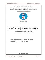
Scheme 1. Schematic representation of the general synthesis scheme of xylitol-based polymers. Xylitol (1), was polymerized with citric acid (2) or sebacic
acid (3) into poly(xylitol-co-citrate) (PXC) (4), and poly(xylitol-co-sebacate) (PXS) (5). Further polycondensation of PXS yielded elastomers. Photo-
crosslinkable hydrogels were obtained by acrylation of PXC in ddH2O using methacrylic anhydride (6) to yield PXC-methacrylate (PXCma) (7). PXCma was
polymerized into a hydrogel by free radical polymerization using a photoinitiator. A simplified representation of the polymers is shown. R can be H,
–OCH2(CH(OR))3CH2OR (xylitol), –CO(CH2)6COOR (sebacic acid), –CO(CH2)ROC(COOR)(CH2)COOR (citric acid), or –C(CH3)
–
–
CH2 (methacrylate
group).
2 www.advmat.de
ß 2008 WILEY-VCH Verlag GmbH & Co. KGaA, Weinheim Adv. Mater. 2008, 9999, 1–6
COMMUNICATION
were typical for low- and high-modulus elastomers (Fig. 1B).
[2]
DSC showed a glass-transition temperature of 7.3 and 22.9 8C
for PXS 1:1 and 1:2, respectively, indicating that these
elastomers are in a rubbery state at room and physiological
temperature. The mechanical properties of the PXS 1:1
elastomer were similar to those of a previously developed
elastomer, composed of glycerol and sebacic acid,
[2]
but PXS
1:1 showed a higher Young’s modulus for a comparable
elongation. Altering monomer-feed ratios of sebacic acid in
PXS elastomers resulted in a wide range of crosslink densities,
whilst maintaining elastomeric properties. The molecular
weight between crosslinks (M
c
) of the PXS polymers varied
by about one order of magnitude (from (10 517.4 Æ 102) g/mol
for PXS 1:1 to (1585.1 Æ 43) g/mol for PXS 1:2, Table 1) and
decreased as more crosslinking entities were introduced. Such
an appreciable difference cannot be obtained by changing the
condensation parameters of PXS 1:1. The increased crosslink
density in PXS 1:2 also resulted in significantly less equilibrium
hydration as determined by mass differential of PXS 1:2 in
ddH
2
O (24 h at 37 8C), when compared to PXS 1:1,
(4.1 Æ 0.3%) and (12.6 Æ 0.4%), respectively; PXS 1:2 also
showed a lower sol content (i.e. the fraction of free, unreacted
macromers within the elastomeric construct, Table 1). The
addition of more sebacic acid molecules to the polymer affects
the water-in-air contact angle (PXS 1:1 (26.58 Æ 3.68), PXS 1:2
(52.78 Æ 5.78), after 5 min), as more aliphatic monomers are
being introduced; this observation is in agreement with the
findings above.
The equilibrium hydration of PXCma hydrogels determined
by mass differential was (23.9 Æ 6.2%) after 24 h at 37 8C.
Volumetric-swelling analysis revealed that the polymer
volume fraction in the relaxed state (v
r
) was (6.9 Æ 0.1%)
and the polymer volume fraction in the swollen state (v
s
) was
(5.8 Æ 0.2%), whereby v
r
was measured immediately after
Table 1. Physical properties of xylitol-based polymers (PXS 1:1 and 1:2 are elastomers, PXCma is a photocured hydrogel). M
c
is the molecular weight
between crosslinks, which was calculated from Equation 1 for the PXS elastomers and from Equations 2 and 3 for the PXCma hydrogel (see Experimental
for details).
Polymer Young’s/compression
modulus [kPa]
Elongation/compression
at break [%]
Equilibrium
hydration by mass [%]
Sol content
[%]
Contact
angle [8]
Polymer
density [g/cm
3
]
Crosslink
density [mol/m
3
]
M
c
[gmol]
PXS 1:1 820 W 150 205.2 W 55.8 12.6 W 0.4 11.0 W 2.7 26.5 W 3.6 1.18 W 0.02 112.2 W 30.5 10 517.4 W 102.1
PXS 1:2 5 330 W 400 33.1 W 4.9 4.1 W 0.3 1.2 W 0.8 52.7 W 5.7 1.16 W 0.02 729.3 W 57.3 1 585.1 W 43.7
PCXma 5.8 W 1.2 79.9 W 5.6 23.9 W 6.2 31.7 W 10.6 n/a 1.51 W 0.05 136.4 W 27.9 11 072.1 W 115.6
0
5
10
15
20
25
30
35
40
100806040200
Strain (%)
Stress (kPa)
0.0
0.2
0.4
0.6
0.8
1.0
1.2
1.4
1.6
1.8
250200150100500
Elongation (%)
Stress (MPa)
720122017202220272032203720
Wavenumber (cm
-1
)
% Transmittance
PXS 1:1 PXS 1:2 PXC PXCma
0
20
40
60
80
100
120
302520151050
Time (weeks )
Mass remaining (%)
PXS 1:1 P XS 1:2 PXCma
B)
A)
C)
D)
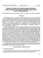
Figure 1. (A) FT–IR analysis of xylitol-based polymers. (B) Typical tensile stress versus strain curve of the PXS elastomers. (C) Typical compression stress
versus strain plot of the 10% (w/v) PXCma hydrogel with cyclic compression at 40%, 50%, and 75%, to failure (at $80%). (D) In vivo mass-loss over time.
Adv. Mater. 2008, 9999, 1–6 ß 2008 WILEY-VCH Verlag GmbH & Co. KGaA, Weinheim
www.advmat.de 3
COMMUNICATION
crosslinking, but before equilibrium swelling and v
s
was
determined at equilibrium swelling. Cyclic compression up to
75% strain of the PXCma hydrogel was possible without
permanent deformation and only limited hysteresis was
observed during cyclic conditioning, revealing the elastic
properties over a wide range of strain conditions. The PXCma
hydrogel failed at a compressive strain of (79.9 Æ 5.6%) and
showed a compressive modulus of (5.84 Æ 1.15) kPa (Fig. 1C).
The mechanical properties of the PXCma hydrogel discs were
similar to those of the previously reported photocured
hyaluronic acid hydrogels (50 kDa, 2–5% (w/v)),
[4]
although
the PXCma hydrogel showed a lower compression modulus for
a similar ultimate-compression stress. The
physical properties of the elastomers and the
hydrogel are summarized in Table 1.
Xylitol-based biopolymers degrade in
vivo. After subcutaneous implantation,
approximately 5% of the mass of the
hydrogel was found to remain after 10 days.
The degradation rate of PXS elastomers
varied according to the stoichiometric ratios.
PXS 1:1 had fully degraded after 7 weeks.
However, (76.7 Æ 3.7%) of the PXS 1:2
elastomer still remained after 28 weeks
(Fig. 1D). This demonstrates that the
in-vivo-degradation kinetics of xylitol-based
elastomers can be tuned in addition to the
crosslink density, surface energy, and equili-
brium hydration. Thus, this polymer platform
describes a range of physical properties that
allow a tuneable in vivo degradation rate.
The PXS 1:2 elastomers were optically
transparent during the first 15 weeks in
vivo and turned opaque upon degradation
(in week 28).
Compared to the prevalently used syn-
thetic polymer PLGA (65/35 LA/GA, high
M
w
), xylitol-based polymers show competi-
tive biocompatibility properties, both in vitro
and in vivo. Regardless of the eventual in vivo
application of these xylitol-based polymers, a
normal wound-healing process, which is
orchestrated by residential fibroblasts, is
mandatory upon implantation; we therefore
chose primary human foreskin fibroblasts
(HFFs) to test in vitro biocompatibility. All
xylitol-based elastomers and hydrogels were
transparent polymers, which facilitated char-
acterization of cell–biomaterial interactions.
HFFs readily attached to PXS elastomers and
proliferated into a confluent monolayer in 6
days. HFFs cultured on PXS elastomers
showed a similar cell morphology and pro-
liferation rate compared to HFFs grown on
PLGA (Fig. 2A and B). There was no cell
attachment on PXCma hydrogels. It is known
that cells in general do not attach to hydrogels, unless
attachment-promoting entities are incorporated.
[6]
We there-
fore examined the cytotoxicity of soluble PXCma prepolymers
in culture media. HFFs exposed for 4 or 24 h to PXCma
prepolymer fractions in the growth media (0.01–1% (w/v))
were not compromised in their mitochondrial metabolism, as
confirmed with a (1-(4,5- dimethylthiazol-2-yl)-3,5- diphenylte-
trazolium bromide) (MTT) assay, compared to HFFs with no
PXCma in the growth media (Fig. 2C). Clinical and histologic
assessments showed that none of the animals exhibited an
abnormal post-operative healing process after subcutaneous
implantation. The PXS 1:1 and 1:2 discs were encased in a
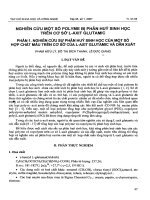
Figure 2. (A) Phase-contrast images (10x) of human primary fibroblasts after 5 days of in vitro
culture, seeded on PLGA (i), PXS 1:1 (ii) and PXS 1:2 (iii). Bars represent 250 mm. (B) Growth
rates of fibroblasts on PLGA, PXS 1:1 and PXS 1:2, expressed as cell differential. (C) MTT assay
of fibroblasts exposed to different PXCma prepolymer fractions in their growth medium.
(D) Representative images of H&E-stained sections of subcutaneous implantation sites of
(i) PLGA discs, (ii) PXS 1:1 discs, (iii) PXS 1:2 discs, (iv) 10% (w/v) PXCma hydrogel discs, 1 week
after implantation. (v) Shows the PXS 1:1 implantation site at week 5 ($73% had degraded) and
(vi) shows PXS 1:2 at week 12 (no degradation). The arrow (i) points to a vessel of the fibrous
capsule surrounding the PLGA implant, where some perivascular infiltration is observed.
P ¼ polymer, FC ¼ fibrous capsule, M ¼ muscle. Inserts are 5x overviews, full images are
magnified 25Â. Bars represent 100 mm.
4 www.advmat.de
ß 2008 WILEY-VCH Verlag GmbH & Co. KGaA, Weinheim Adv. Mater. 2008, 9999, 1–6
COMMUNICATION
translucent tissue capsule after one week, which did not
become more substantial throughout the rest of the study.
Histological sections confirmed that the polymer/tissue inter-
face was characterized by a mild fibrous-capsule formation
(Fig. 2Dii and iii). No abundant inflammation was seen in the
surrounding tissues and the sections showed a quiet polymer/
tissue interface, which was characteristic for the PXS
elastomers after the first week in vivo. Furthermore, no
perivascular infiltration was noted in the surrounding tissues of
the PXS discs. This quiescent tissue response was evident when
compared to the tissues in contact with the PLGA implants
(Fig. 2Di). A more substantial vascularized fibrous capsule
with minor perivascular infiltration (arrow) was seen surround-
ing the PLGA implants. A comparable thickness of fibrous-
capsule formation was noted for the 10% PXCma hydrogel at
day 10 (Fig. 2Div). No PXCma hydrogel was found at day 14
after repetitive sectioning of the explanted tissue. Long-term
histological sections of PXS 1:1 and 1:2 at week 5 and 12
demonstrated that even upon degradation the fibrous capsule
remained quiescent: at week 5 the PXS 1:1 elastomer had
degraded by approximately 73%, whereas the PXS 1:2 polymer
showed no degradation at all at week 12. Thus, xylitol-based
polymers exhibited excellent biocompatibility compared to
PLGA.
Our goal was to develop a polymer synthesis scheme that
required very simple adjustments in chemical composition to
achieve a wide range of material properties. We have described
a process for the synthesis of xylitol-based polymers. Xylitol is
well studied in terms of biocompatibility and pharmacokinetics
in humans.
[7,8]
It is a metabolic intermediate in the mammalian
carbohydrate metabolism with a daily endogenous production
of 5–15 g in adult humans.
[9]
The entry into metabolic pathways
is slow and independent of insulin, and does not cause rapid
fluctuations of blood glucose levels.
[10]
As a monomer, xylitol is
an important compound in the food industry, where it has an
established history as a sweetener with proven anticariogenic
activity.
[11]
Moreover, it has an antimicrobial effect on
upper-airway infections caused by Gram-positive strepto-
cocci.
[12–15]
Although xylitol has been studied in polymer
synthesis, others have typically utilized it as an initiator
[16]
or
altered xylitol to yield linear polymers by protecting three
of the five functional groups.
[17]
They were produced in
sub-kilogram quantities without the use of organic solvents or
cytotoxic additives. Xylitol-based polymers are endotoxin-free
and do not impose a potential immunological threat like
biological polymers extracted from tissues or produced by
bacterial fermentation, such as collagen and hyaluronic
acid.
[18,19]
In addition, the mechanical properties of xylitol-
based elastomers correspond to biologically relevant values
that fall close to or are equal to those of various tissues, such as
acellular peripheral nerves,
[20]
small diameter arteries,
[21]
cornea
[22]
and intervertebral discs.
[23]
In this report, we have
shown only three examples of possible polymers based on this
monomer. Potential combinations for the chemical composi-
tion of xylitol-based polymers are numerous and therefore it
provides a platform to tune mechanical properties, degradation
profiles and cell attachment.
Experimental
Synthesis and Characterization of the Polymers: All chemicals were
purchased from Sigma-Aldrich unless stated otherwise. Appropriate
molar amounts of the polyol and reacting acid monomer were melted in
a round-bottom flask at 150 8C under a blanket of inert gas and stirred
for 2 h. A vacuum ($50 mTorr) was applied to yield the prepolymers
PXS 1:1 (12 h), PXS 1:2 (6 h) and PXC (1 h). The PXC polymer was
dissolved in ddH
2
O and lyophilized. Methacrylated PXC prepolymer
(PXCma) was synthesized by the addition of methacrylic anhydride in
a $20-fold molar excess, as previously described for the methacrylation
of hyaluronic acid, [5] dialyzed in double-distilled water (ddH
2
O, M
w
cutoff: 1 kDa) and lyophilized. PXCma hydrogels were fabricated
by dissolving 10% (w/v) PXCma in a phosphate-buffered saline
(PBS) solution containing 0.05% (w/v) 2-methyl-1-(4-(hydroxyethoxy)
phenyl)-2-methyl-1-propanone (Irgacure 2959, I2959) as the photo-
initiator under exposure of $4 mW/cm
2
ultraviolet light (lamp model
100AP, Blak-Ray). All PXS 1:1 and 1:2 elastomers were produced by
further polycondensation (120 8C, 140 mTorr for 4 days). The
prepolymers were sized using gel permeation chromatography using
THF or filtered ddH
2
O as eluentia and Styragel columns (series of
HR-4, HR-3, HR-2, and HR-1, Waters, Milford, MA, USA). FT-IR
analysis was carried out on a Nicolet Magna-IR 550 spectrometer.
1
H-NMR spectroscopy was performed on a Varian Unity-300 NMR
spectrometer;
1
H-NMR spectra of the PXS prepolymers were
determined in C
2
D
6
O and spectra of the PXCma prepolymers were
obtained in D
2
O. The chemical composition of the prepolymers was
determined by calculating the signal integrals of xylitol and compared
to the signal integrals of sebacic acid or citric acid. The signal intensities
showed peaks of (–OC
H
2
(CH(OR))
3
CH
2
O–) at 3.5–5.5 ppm from
xylitol, (–C
H
2
–) at 2.3–3.3 ppm from citric acid, and peaks of
(–COC
H
2
CH
2
CH
2
–) at 1.3, 1.6 and 2.3 ppm from sebacic acid. The
final degree of substitution after acrylation of the PXC prepolymer was
calculated by the signal integral of the protons associated with
(–C(C
H
3
)
–
–
CH
2
) at 1.9, 5.7 and 6.1 ppm from the methacrylate groups.
Tensile tests were performed on hydrated (ddH
2
Oat378C > 24 h), dog
bone-shaped polymer strips and conducted on an Instron 5542
(according to the American Society for Testing and Materials (ASTM)
standard D412-98a). Compression analysis of the photocrosslinked
PXCma hydrogels was performed as described previously. [5]
Differential scanning calorimetry (DSC) was performed as reported
previously. [24] The mass density was measured using a pycno-
meter (Humboldt, MFG. CO).Thecrosslinkdensity(n)and
M
c
were calculated from the following equations for an ideal
elastomer: [25]
n ¼
E
0
3RT
¼
r
M
c
(1)
where E
0
is the Young’s modulus, R the universal gas constant, T
temperature and r is the mass density. According to Peppas et al., [26]
this rubber-elasticity theory can also be utilized to calculate the
effective M
c
for hydrogels that show elastic behavior and were
prepared in the presence of a solvent:
t ¼
rRT
M
c
1 À
2M
c
M
n
a À
1
a
2
ÀÁ
v
s
v
r
1
3
(2)
where t is the compression modulus of the hydrogel, v
s
(0.058 Æ 0.002)
is the polymer volume fraction in the swollen state, and v
r
(0.069 Æ 0.001) is the polymer volume fraction in the relaxed state.
Adv. Mater. 2008, 9999, 1–6 ß 2008 WILEY-VCH Verlag GmbH & Co. KGaA, Weinheim
www.advmat.de 5