Periodontal disease symptom treatment and prevention
Bạn đang xem bản rút gọn của tài liệu. Xem và tải ngay bản đầy đủ của tài liệu tại đây (14.48 MB, 393 trang )
DENTAL SCIENCE, MATERIALS AND TECHNOLOGY
PERIODONTAL DISEASE: SYMPTOMS,
TREATMENT AND PREVENTION
No part of this digital document may be reproduced, stored in a retrieval system or transmitted in any form or
by any means. The publisher has taken reasonable care in the preparation of this digital document, but makes no
expressed or implied warranty of any kind and assumes no responsibility for any errors or omissions. No
liability is assumed for incidental or consequential damages in connection with or arising out of information
contained herein. This digital document is sold with the clear understanding that the publisher is not engaged in
rendering legal, medical or any other professional services.
DENTAL SCIENCE, MATERIALS AND TECHNOLOGY
Additional books in this series can be found on Nova‘s website
under the Series tab.
Additional E-books in this series can be found on Nova‘s website
under the E-books tab.
DENTAL SCIENCE, MATERIALS AND TECHNOLOGY
PERIODONTAL DISEASE: SYMPTOMS,
TREATMENT AND PREVENTION
SHO L. YAMAMOTO
EDITOR
Nova Biomedical Books
New York
Copyright © 2011 by Nova Science Publishers, Inc.
All rights reserved. No part of this book may be reproduced, stored in a retrieval system or transmitted
in any form or by any means: electronic, electrostatic, magnetic, tape, mechanical photocopying,
recording or otherwise without the written permission of the Publisher.
For permission to use material from this book please contact us:
Telephone 631-231-7269; Fax 631-231-8175
Web Site:
NOTICE TO THE READER
The Publisher has taken reasonable care in the preparation of this book, but makes no expressed or
implied warranty of any kind and assumes no responsibility for any errors or omissions. No liability is
assumed for incidental or consequential damages in connection with or arising out of information
contained in this book. The Publisher shall not be liable for any special, consequential, or exemplary
damages resulting, in whole or in part, from the readers‘ use of, or reliance upon, this material. Any
parts of this book based on government reports are so indicated and copyright is claimed for those parts
to the extent applicable to compilations of such works.
Independent verification should be sought for any data, advice or recommendations contained in this
book. In addition, no responsibility is assumed by the publisher for any injury and/or damage to persons
or property arising from any methods, products, instructions, ideas or otherwise contained in this
publication.
This publication is designed to provide accurate and authoritative information with regard to the subject
matter covered herein. It is sold with the clear understanding that the Publisher is not engaged in
rendering legal or any other professional services. If legal or any other expert assistance is required, the
services of a competent person should be sought. FROM A DECLARATION OF PARTICIPANTS
JOINTLY ADOPTED BY A COMMITTEE OF THE AMERICAN BAR ASSOCIATION AND A
COMMITTEE OF PUBLISHERS.
Additional color graphics may be available in the e-book version of this book.
Library of Congress Cataloging-in-Publication Data
Periodontal disease : symptoms, treatment, and prevention / editor, Sho L.
Yamamoto.
p. ; cm.
Includes bibliographical references and index.
ISBN 978-1-61122-112-1 (eBook)
1. Periodontal disease. I. Yamamoto, Sho L.
[DNLM: 1. Periodontal Diseases. WU 240]
RK361.P453 2010
617.6'32 dc22
2010033087
New York
Contents
Preface vii
Chapter I Aesthetic Periodontal Therapy – Root Coverage 1
A. L. Dumitrescu, Liviu Zetu and Silvia Teslaru
Chapter II Periodontal Diseases in Children and Adolescents: Clinical
Features and Molecular Biological Analyses 31
Kazuhiko Nakano, Atsuo Amano and Takashi Ooshima
Chapter III Biomechanics of Rehabilitating the Perioprosthetic Patient 67
Petros Koidis and Manda Marianthi
Chapter IV Biomarkers of Periodontal Disease: Past, Present
and Future Challenges 93
Fionnuala T. Lundy
Chapter V Inflammatory Mediators and Oxidative Stress in Periodontal
Disease 107
Luigi F. Rodella, Paolo Brunamonti Binello,
Barbara Buffoli, Davide Merigo
and Mauro Labanca
Chapter VI Tobacco: A Risk Factor for Periodontal Disease 121
Nouf Al-Shibani, Nawaf Labban, Eman Allam,
and L. Jack Windsor
Chapter VII A Novel Cytodiagnostic Fluorescence Assay for the Diagnosis
of Periodontitis 137
Marco Giannelli, Lucia Formigli and Daniele Bani
Chapter VIII The Healthy Periodontium, the Diseased Periodontium 153
Leena Palomo and Nabil Bissada
Chapter IX Clinical Effects of 2% Chlorhexidine Gel on Patients
Undergoing Orthodontic Treatment 205
Abdolreza Jamilian, Mahmood Ghasemi Dariush Gholami
and Bita Kaveh
Contents vi
Chapter X Periodontal Disease and Systemic Diseases: Interrelationships
and Interactions 215
Giuseppe Pizzo, Rosario Guiglia and Giuseppina Campisi
Chapter XI Obesity Revised 247
Ayse Basak Cinar
Chapter XII Invasion of Host Cells by
Porphyromonas Gingivalis
in
Polymicrobial Infection 257
Atsushi Saito, Satoru Inagaki, Eitoyo Kokubu,
Ryuta Kimizuka and Kazuyuki Ishihara
Chapter XIII HMGB1: A Novel Inflammatory Mediator
in Chronic Periodontitis 273
Yoko Morimoto-Yamashita, Masayuki Tokuda, Kiyoshi Kikuchi,
Ikuro Maruyama, Mitsuo Torii, and Ko-ichi Kawahara
Chapter XIV Risk Factors for Chronic Periodontal Diseases 287
Daniela da Silva Feitosa, Mauro Pedrine Santamaria,
Márcio Zaffalon Casati, Enilson Antonio Sallum,
Francisco Humberto Nociti Júnior and Sérgio de Toledo
Chapter XV The Role of Antimicrobial Peptides in Periodontal Disease 321
Suttichai Krisanaprakornkit and Sakornrat Khongkhunthian
Index 353
Preface
Periodontal disease is a chronic bacterial infection characterized by persistent
inflammation, connective tissue breakdown and alveolar bone destruction. The chronic
inflammation associated with periodontal disease represents the host response to bacterial
plaque, mediated by the environment in which the response occurs. This book presents topical
research data in the study of periodontal disease, including aesthetic periodontal therapy and
root coverage techniques; clinical features of periodontal diseases in children and adolescents;
biomechanics and the perioprosthetic patient; maternal periodontitis and perinatal outcomes;
identifying patients with enhanced disease susceptibility in periodontal disease; and
inflammatory mediators and oxidative stress in periodontal disease.
Chapter I - Aesthetic considerations have influenced the management of dental maladies
in varying degrees for many years. For many years the goals of periodontal surgery have been
determined by functional aspects only. During recent years periodontal surgery has shifted its
focus from achieving more functional goals toward a combination of both good functional
and esthetic results. While accomplishing the best possible functional result, esthetics should
not only be maintained, but also enhanced. Sometimes the esthetic outcome is the only
important factor and function becomes secondary (e.g. treatment of recessions or the creation
of papillae). Predictability becomes the key word in this type of periodontal surgery. Patient
awareness and expectations have increased recently to the point that less than optimal
esthetics is no longer an acceptable outcome. Periodontal plastic surgery would accordingly
be defined as ―surgical procedures performed to prevent or correct anatomic, developmental,
traumatic or disease induced defects in the gingiva, alveolar mucosa or bone‖. The present
chapter is presenting and discussing the clinical outcomes of several root coverage
techniques: pedicle soft tissue grafts, rotational flaps, coronally advanced flap, semilunar flap,
free soft tissue graft, nonsubmerged grafts, submerged grafts etc.
Chapter II - The clinical features of periodontal diseases in children and adolescents
differ from those in adults. Periodontitis is extremely rare in children, except those
complicated with certain kinds of systemic diseases, whereas gingivitis is commonly
encountered. Childhood gingivitis can be reversed by professional mechanical tooth cleaning
in combination with tooth brushing instruction. On the other hand, gingivitis becomes
increasingly prevalent with age through the adolescent period, and early diagnosis and
appropriate interventions are necessary to prevent the onset of marginal periodontitis during
adolescence. Since most children with periodontitis possess a background of abnormal
immune responses, they have a lower likelihood of good prognosis,even though diligent
interventions are performed. Other types of periodontal diseases include gingival recession,
Sho L. Yamamoto viii
which is mainly caused by traumatic occlusion, and gingival overgrowth, which has a
hereditary background and is associated with specific medication such as
antiepilepticphenytoin. In addition, cases with a rapid loss of gingival attachment and alveolar
bone due to mechanical injury at the periodontal sulcus, termed ―acute periodontitis,‖ are also
encountered. Furthermore, an unintentional attachment loss, when materials such as small
plastic tubes being fitted to the teeth are inserted, is a unique type of periodontitis in young
children. It should be noted that periodontitis associated with anatomical anomalies, which
are derived from fragile periodontal attachment, is also encountered.
Considering the etiology of periodontitis, it is important to identify periodontitis-related
bacterial species, since the disease is generally known to be caused by specific bacteria.
However, most of those belong to the obligate anaerobic group, and it is difficult and time-
consuming to isolate them. On the other hand, recent developments in molecular biological
techniques have enabled rapid identification of species using bacterial DNA extracted from
various kinds of clinical specimens. Such approaches do not require isolation of viable
bacteria and even small amounts of DNA can be detected using PCR techniques. With such
modern techniques, the author have evaluated the distribution of periodontal bacterial species
in children, changes of species in the same subjects over a long interval, combinations of
species simultaneously detected, and mother-to-child transmission. In addition, the
distributions of bacterial species in children with Down‘s syndrome and other developmental
disabilities have been analyzed. The authors‘ results have provided valuable information
regarding bacterial profiles in clinical specimens, which should lead to further beneficial
methods for clinical use in the near future.
Chapter III - In advanced perioprosthetic cases where the periodontium‘s integrity is
severely compromised and the dental barrier‘s function is extremely disrupted, the
biomechanical response to the extrinsic mechanical stimuli of the system including the
prosthetic restoration supported by the biological tissues is quite altered. The differentiated
altered experience of the functional loading due to the lowered periodontium‘s threshold
along with the apical shift of the system fulcrum due to the periodontium‘s structure reduction
require a modified design of the restoration‘s metal framework as a critical factor in the
system‘s survival in order to secure the expected longevity of both the restorative and
biological structures, capturing the failure initiation of either progressive tissular or technical
collapse. So, the purpose of the present study was to: a. analyze the way by which the
periodontium reacts to the developing forces and how its integrity is related to the experience
of the stress field on the perioprosthetic patient; b. determine the parameters defining the
tooth prognosis in the perioprosthetic patient and how the restoration type is involved; c.
report the clinical significance of tooth splinting by cantilever cross arch fixed partial denture
applied on the perioprosthetic patient and the way it is related to the response of the reduced
periodontium and finally d. investigate the clinical significance of the specific design of the
metal framework in cantilever cross-arch fixed partial dentures via a theoretical finite element
model.
Chapter IV - Periodontal disease is a chronic bacterial infection characterised by
persistent inflammation, connective tissue breakdown and alveolar bone destruction. The
chronic inflammation associated with periodontal disease represents the host response to
bacterial plaque, mediated by the environment in which the response occurs. Periodontitis is
both site-specific and episodic in nature and thus biomarker development could prove
Preface ix
invaluable in identifying sites with active disease, predicting sites that may develop disease,
monitoring response to therapy or identifying patients with enhanced disease susceptibility.
In periodontal disease gingival crevicular fluid (GCF) flows from the gingival
microcirculation into the periodontal pockets and the volume increases in proportion to the
severity of the local inflammatory process. The study of GCF samples, from defined sites of
chronic periodontal inflammation, allows non-invasive access to an inflammatory exudate
that could be used for biomarker discovery. GCF contains proteins synthesised and secreted
in the inflamed gingival tissues and carried by the GCF to the gingival crevice/pocket. Here,
they are augmented by proteins released from bacteria and host cells, particularly
polymorphonuclear leukocytes (PMNs), present in the periodontal pocket. The constituents of
GCF are therefore derived from a number of sources including microbial plaque, host
inflammatory cells, serum and tissue breakdown products. Saliva has also been studied in the
search for biomarkers of periodontal disease. Saliva is a more complex fluid, comprising
glandular secretions, components of GCF, components of serum and also particles (including
bacteria) from a variety of oral and airway sources. Although saliva has the advantage of
being easily collected, its biochemical complexity may hinder detection of biomarkers
specific for periodontal disease. Furthermore the fact that saliva bathes the whole mouth
negates the use of salivary biomarkers for site-specific identification or monitoring of
periodontal disease.
Despite an impressive list of possibilities, biomarkers have yet to reach routine clinical
use as reasonable predictors of periodontal status. This chapter reviews the analysis of GCF
and saliva for monitoring periodontal health and disease. Potentially important biomarkers of
disease in both GCF and saliva are highlighted and their merits are described in further detail.
Putative biomarkers from both host and bacterial sources are considered and the use of
multiple biomarkers is discussed. Following the technological revolution in both genomic and
proteomic analysis over the last decade it is tempting to speculate that the next decade could
bring much waited progress in the field of biomarker identification and application in the field
of periodontal disease.
Chapter V - Periodontal disease represents today the main cause of teeth loss after the
third decade of life. About 60% of dental extractions are due to etiopathogenetic periodontal
factors. After 35 years, the frequency of marginal periodontal disease varies from 80% to
100% of world population, depending on statistical method used and the demographic areas
considered, showing a similar frequency in both sexes, slightly higher in female.
Two important and interrelated factors are involved in its physiopathological progression:
1) the activation of immune system and the release of inflammatory mediators, such as IL-1β,
IL-6 and TNF-α, which could overflow into the blood system and induce a systemic
inflammatory response; 2) the production of oxygen radicals and their related metabolites.
A recent focus of the dental research is the individuation of biomarkers, which can be
easily used as diagnostic tools. Among them, metalloproteinases (MMPs) and heat shock
proteins (HSPs) could provide potential biomarkers, which could be useful for evaluating
both the periodontitis development and the incidence of the related cardiovascular diseases.
Recent studies, in fact, have shown a direct correlation between periodontal and
cardiovascular diseases: in particular, both diseases have systemic and local causes, and the
constant bacterial contamination of oral cavity could be linked not only to periodontopathy
but also to the development of cardiovascular diseases.
Sho L. Yamamoto x
To date, the periodontal disease therapy available is based on the individuation and the
elimination of the causing factors. Nevertheless, new innovative surgical and pharmacological
therapies could be developed.
The aim of this work is to review the literature data focusing on the role of inflammatory
mediators and oxidative stress in periodontal disease and related factors.
Chapter VI - Periodontal disease results from complex interactions between infectious
agents and host factors. The disease expression can be modified by environmental, acquired,
and genetic risk factors. Tobacco usage, especially smoking, is considered a major modifiable
risk factor for periodontal disease. In addition to periodontal disease, tobacco usage is also a
risk factor for oral cancer and its recurrence, dental cariesand congenital defects in children
from mothers who smoke while pregnant. In periodontal disease, smokers have deeper
probing depths, more gingival recession, more alveolar loss and more furcation involvement
than non-smokers. They also show less favorable responses to various kinds of periodontal
treatments including non-surgical, surgical, regenerative procedures and dental implants. It is
clear from epidemiology studies that tobacco usage is correlated with periodontal disease.
This chapter reviews the evidence for the association between periodontal disease and
tobacco, and describes what is currently known about how tobacco and its components affect
the periodontal tissues that result in tissue damage.
Chapter VII - A topical issue in periodontology is to find objective diagnostic methods
which may be combined with the classical clinical inspection parameters to yield a reliable
grading of the severity and extent of periodontal disease. This study deals with a novel
cytodiagnostic fluorescence test, performed on exfoliation samples taken from
periodontal/oral tissues, useful to assess the severity of periodontal disease. Twenty-one
patients with different degrees of periodontitis were subjected to clinical and
histopathological grading and the results compared with those obtained from the
cytodiagnostic fluorescence assay. The author found that the amount of blood cells
(polymorphonuclear and mononuclear leukocytes, erythrocytes), the occurrence of
morphologically abnormal epithelial cells, and the number of spirochetes showed a
statistically significant correlation with the clinical and histopathological diagnostic
parameters, the latter being considered as the most reliable predictors of the severity of
periodontal disease. On these grounds, the author suggest that this cytodiagnostic method
may greatly help dental practitioners to achieve a chair-side, reliable and objective evaluation
of the degree and activity of periodontitis at first dental visit, and to perform a targeted
treatment and an accurate follow up of the patients during supportive periodontal therapy.
Chapter VIII -Differentiation of health from disease is central to understanding diagnosis
and treatment of periodontal diseases. It is logical to begin with an in-depth examination of
the structure and physiology of the healthy periodontium.
Chapter IX - Objectives: The purpose of this study was to compare the short-term clinical
effects of a single intrasulcular injection of 2% chlorhexidine gluconate gel (CG) and placebo
gel (PG) in orthodontic patients with fixed appliances and established gingivitis aged from 12
to 20 years.
Methods and Materials: 50 patients (31 females, 19 males) were divided into two groups
(CG and PG) of 50 subjects. This study was single blind randomized split mouth clinical trial.
As randomly assigned by coin toss, the first permanent molars on the right or left side of the
mouth received either CG or PG. Probing depth (PD) was measured with a Michigan 0 probe.
The gingival index (GI) of Löe and SILNESS and papilla bleeding index (PBI) of
Preface xi
MÜHLEMANN were recorded on the first permanent molars. These indices were measured
at baseline, and in treatment on second, fourth, eighth, and the twelfth weeks. T-test and chi-
square test were used to analyze the data.
Results: T-test showed that PD was reduced in experimental group in comparison with
the control group in the 4th week and following intervals (p<0.001). Chi-square showed that
PBI was improved in experimental group in comparison with the control group in the 2nd
week and following intervals (p<0.001). The same test showed that GI was improved in
experimental group in the 2nd week and following intervals (p<0.001).
Conclusion: The data indicate that the use of a single application of 2% CG was effective
in reducing gingivitis related to banded first permanent premolars in adolescents undergoing
orthodontic treatment in short time.
Chapter X - The focal infection theory, which for almost half a century justified
indiscriminate extraction of teeth to cure focal infections, since the end of the 1940s has
become progressively a discarded concept. In parallel with the declining importance assigned
to pulp and periapical infections in the pathogenesis of focal diseases, over the last decade
there has been increasing interest in the possible relationship between periodontal infection
and systemic diseases. Periodontal pathogens and their products, as well as inflammatory
mediators produced in gingival tissue, might enter the bloodstream through ulcerated pocket
epithelium, causing systemic effects (focal diseases).
On the basis of this mechanism, chronic periodontitis has been implicated as risk factor
for cardiovascular diseases associated to atherosclerosis, bacterial endocarditis, diabetes
mellitus, respiratory disease preterm delivery, rheumatoid arthritis, and more recently
osteoporosis, pancreatic cancer, metabolic syndrome, renal diseases and neurodegenerative
diseases such as Alzheimer‘s disease. Numerous hypotheses, including common
susceptibility, systemic inflammation, direct bacterial infection and cross-reactivity, or
molecular mimicry, between bacterial antigens and self-antigens, have been postulated to
explain these relationships. In this context, the association of periodontal disease with
systemic diseases has introduced the concept of ―periodontal medicine‖, which ultimately
guides the medical community in therapeutic approaches to improve not only the patient oral
health but also systemic health.
This chapter summarizes the pathophysiology of periodontal disease and presentsan
update on interrelationships and interactions between periodontal disease and systemic
diseases. Moreover, this chapter reviews the published literature that describes the effects of
periodontal treatment on cardiovascular diseases, adverse pregnancy outcomes, diabetes
mellitus, and respiratory disease.
Chapter XI - Obesity, diabetes and oral diseases (dental cariesand periodontal diseases),
largely preventable chronic diseases, are described as global pandemic due their distribution
and severe consequences. WHO has called for a global action for prevention and promotion
of these diseases as a vital investment in urgent need.
Diabetes and obesity, showing an increasing trend, lead to disabilities and negatively
impacts on the quality of life through life course along with oral diseases. WHO projects that
the prevalence of diabetes and deaths/year attrituble to diabetes complications will double
worldwide by 2030. Globally, more than 1 billion adults are overweight; almost 300 million
of them are clinically obese. Being obese/overweight raises steeply the likelihood of
developing DM2. Approximately 85% of people with diabetes are DM2, and of these 90% are
obese or overweight. Obesity increases the likelihood of periodontitis which is one of the
Sho L. Yamamoto xii
most common chronic diseases worldwide, described as pandemic, and closely related to
DM2. Promoting good oral health is significantly essential for prevention and reducing the
negative consequences of periodontal diseases, DM2 and obesity, and to maintain good
health, as proposed by European health goals by WHO.
Chapter XII - Periodontitis is one of the predominant polymicrobial infections of humans.
Since periodontitis results from complex interactions of multiple microorganisms, it is
important to investigate interactions between different periodontal bacteria and host cells.
Porphyromonas gingivalis, a gram-negative anaerobe, is a major colonizer of gingival tissues
and has been etiologically implicated in periodontal as well as cardiovascular diseases.
Cellular invasion by periodontal pathogens including P. gingivalis has been proposed as a
possible virulence factor, affording protection from the host immune responses and
contributing to tissue damage. In recent periodontal research, polymicrobial infection models
have been used to study host response profiles. However, data on the potential of host cell
invasion by periodontal pathogens in polymicrobial infection are scarce. The author
investigated the ability of periodontal pathogens to modulate invasion of human gingival
epithelial cells and aortic endothelial cells by P. gingivalis. Among the pathogens,
Fusobacterium nucleatum was shown to significantly enhance the P. gingivalis invasion. The
author describe the complex interaction between periodontopathogens and host cells, with a
particular focus on the co-infection by P. gingivalis and F. nucleatum.
Chapter XIII -Periodontitis is a major chronic inflammatory disease that destroys
periodontal tissue and eventually results in tooth loss. Although periodontitis is a local
disease, its chronic status triggers systemic inflammatory diseases including severe type 2
diabetes, heart disease, cancer and atherosclerosis. Therefore, the development of new
treatments for periodontitis contributes to the effective inhibition of systemic inflammatory
diseases.
High Mobility Group Box-1 (HMGB1), a primarily nuclear protein, is present in many
eukaryotic cells and is highly conserved between species. HMGB1 appears to have distinct
functions in cellular systems. It acts as an intracellular regulator of transcription and plays a
crucial role in the maintenance of DNA function. Extracellular HMGB1 released by various
cell types (i.e. macrophages/monocytes, endothelial cells and pituicytes), or necrotic cells,
stimulated by lipopolysaccharide (LPS) or tumor necrosis factor- -
proinflammatory cytokine through the multi-ligand receptor for advanced glycation end-
products (RAGE) and toll-like receptors (TLRs) 2 and 4. Extracellular HMGB1 plays a
critical role in the progression of chronic inflammatory diseases, such as septic shock,
rheumatoid arthritis, diabetes and atherosclerotic lesions. Recent studies show that HMGB1 is
continuously released from gingival epithelial cells modulated by TNF- and expressed in
epithelial tissues of patients with periodontitis. HMGB1 may be involved in the progression
of periodontitis as a novel inflammatory mediator. Therefore, understanding the mechanisms
underlying the functions of HMGB1 may lead to novel therapeutic approaches for chronic
periodontitis and help to prevent systemic inflammatory diseases.
This review summarizes the current knowledge on HMGB1, including its correlation
with disease and preventive medicine.
Chapter XIV - Chronic periodontal diseases include a group of inflammatory diseases
that affect periodontal supporting tissues of the teeth and encompass destructive and
nondestructive conditions. Periodontal diseases are multifactorial and the role of dental
biofilm in their initiation is primary. However, whether dental biofilm affects a particular
Preface xiii
subject, what form the disease takes and how it progresses, are all dependent of a wide variety
of factors. Therefore, the objective of this chapter is to outline the risk factors described for
the most prevalent chronic periodontal diseases (plaque induced gingivitis and chronic
periodontitis) and to explain some basic concepts related to the current understanding of the
role of these risk factors based on in vitro, animal and human studies. The review will focus
on the factors that may be associated with a direct increase in the likelihood of occurrence of
disease or an increase in its severity. The following factors will be discussed: 1) host
characteristics, such as age, gender and race; 2) social and behavioral factors (socioeconomic
status, cigarette smoking and emotional stress); 3) systemic factors, e.g. diabetes mellitus and
osteoporosis; 4) genetic factors; 5) tooth-level factors (root grooves, tooth position, caries,
occlusal discrepancies, iatrogenic restorations, root abnormalities and periodontal
parameters); and 6) the microbial composition of dental biofilm. Finally, this chapter will also
present literature-based evidence on predictive factors associated with patients and tooth
susceptibility for recurrence of periodontitis after the end of the active periodontal therapy
and will examine the use of some prognostic models which may be useful for clinicians in the
identification high-risk groups of patients.
Chapter XV - The oral cavity is a warm, moist environment, in which a number of
microorganisms colonize and live in harmony as a community, a so-called biofilm. In this
environment, antimicrobial peptides may play a critical role in maintaining normal oral health
and controlling innate and acquired immune systems in response to continuous microbial
challenges in periodontal disease. Two major families of antimicrobial peptides, found in the
oral cavity, are defensin and cathelicidin. Members of the defensin family are cysteine-rich
peptides, synthesized by plants, insects, and mammals. These peptides vary in length and in
the number of disulfide bonds, and have a beta-sheet structure. In the oral cavity, four alpha-
defensins are synthesized and stored in neutrophil granules, which are converted into active
peptides by proteolytic processing, while three human beta-defensins (hBDs), hBD-1, hBD-2,
and hBD-3, are predominantly produced by oral epithelial cells. The only member of the
cathelicidin family found in humans is LL-37, an alpha-helical peptide that contains 37 amino
acids and begins with two leucines at its NH3-terminus. LL-37 is derived from enzymatic
cleavage of a precursor peptide, namely, human cationic antimicrobial peptide-18. Clinically,
differential expression of antimicrobial peptides has been reported in specific types of
periodontal disease, and their presence has been shown in saliva and gingival crevicular fluid.
Current evidence suggests that alpha-defensins, beta-defensins, and LL-37 have distinct, but
overlapping, roles in antimicrobial and pro-inflammatory activities. Several studies have
shown antimicrobial activities of hBD-2, hBD-3, and LL-37 against several periodontal
pathogens, suggesting their potential role as antimicrobial agents for periodontal disease. The
clinical significance of antimicrobial peptides in periodontal disease has recently been
demonstrated in morbus Kostmann syndrome, a severe congenital neutropenia, in which
chronic periodontal infection in young patients, resulting from a deficiency of neutrophil-
derived antimicrobial peptides, causes early tooth loss. Although researchers initially focused
their attention on antimicrobial activities, it is now becoming evident that defensins and LL-
37 are multifunctional molecules that mediate various host immune responses, and may thus
represent essential molecules of innate immunity in periodontal disease. In this chapter, basic
knowledge and the clinical importance of antimicrobial peptides in periodontal disease will be
discussed in detail.
In: Periodontal Disease: Symptoms, Treatment and Prevention ISBN 978-1-61761-739-3
Editor: Sho L. Yamamoto, pp. 1-29 © 2011 Nova Science Publishers, Inc.
Chapter I
Aesthetic Periodontal Therapy –
Root Coverage
A. L. Dumitrescu
1
, Liviu Zetu
2
and Silvia Teslaru
2
1. Institute of Clinical Dentistry, Tromsø, Norway
2. U.M.F. "Gr.T. Popa", Iashi, Romania
Abstract
Aesthetic considerations have influenced the management of dental maladies in
varying degrees for many years. For many years the goals of periodontal surgery have
been determined by functional aspects only. During recent years periodontal surgery has
shifted its focus from achieving more functional goals toward a combination of both good
functional and esthetic results. While accomplishing the best possible functional result,
esthetics should not only be maintained, but also enhanced. Sometimes the esthetic
outcome is the only important factor and function becomes secondary (e.g. treatment of
recessions or the creation of papillae). Predictability becomes the key word in this type of
periodontal surgery. Patient awareness and expectations have increased recently to the
point that less than optimal esthetics is no longer an acceptable outcome. Periodontal
plastic surgery would accordingly be defined as ―surgical procedures performed to
prevent or correct anatomic, developmental, traumatic or disease induced defects in the
gingiva, alveolar mucosa or bone‖.The present chapter is presenting and discussing the
clinical outcomes of several root coverage techniques: pedicle soft tissue grafts, rotational
flaps, coronally advanced flap, semilunar flap, free soft tissue graft, nonsubmerged grafts,
submerged grafts etc.
The name and address of the author responsible for correspondence: Alexandrina L. Dumitrescu DDS, PhD, BA
Psychology, Associate Professor, Institute of Clinical Dentistry, Faculty of Medicine, University of Tromsø,
9037 Tromsø, Norway. Phone: +47 77 64 91 43, Mobile: +47 93 65 11 78, Fax: +47 77 64 91 01. E-mail:
A. L. Dumitrescu, Liviu Zetu and Silvia Teslaru 2
1. Introduction
Aesthetic considerations have influenced the management of dental maladies in varying
degrees for many years. For many years the goals of periodontal surgery have been
determined by functional aspects only. During recent years periodontal surgery has shifted its
focus from achieving more functional goals toward a combination of both good functional
and esthetic results. While accomplishing the best possible functional result, esthetics should
not only be maintained, but also enhanced. Sometimes the esthetic outcome is the only
important factor and function becomes secondary (e.g. treatment of recessions or the creation
of papillae). Predictability becomes the key word in this type of periodontal surgery (Hurzeler
and Weng, 1999).
Mucogingival surgery is a broaderterm that includes nonsurgical procedures such as
papilla reconstruction by means of orthodontic or restorative therapy (Takei et al.,
2006).Periodontal plastic surgery is defined as ―surgical procedures performed to prevent or
correct anatomic, developmental, traumatic or disease induced defects in the gingiva, alveolar
mucosa or bone‖. Among treatment procedures that may fall within this definition are various
soft and hard tissue procedures aiming at: gingival augmentation, root coverage, correction of
mucosal defects at implants, augmentation of edentulous ridges, removal of aberrant
frenulum, prevention of bridge collapse associated with tooth extraction, crown lengthening,
mucogingival tattoo, open interproximal space, gingival enlargement and exposure of teeth
that are not likely to erupt (Wennström and Pini Prato, 1997; McGuire, 1998).
The present chapter is presenting and discussing the clinical outcomes of several root
coverage techniques.
2. Gingival Recession
Gingival recession is characterized by the displacement of the gingival margin apically
from the cementoenamel junction, or CEJ, or from the former location of the CEJ in which
restorations have distorted the location or appearance of the CEJ. Gingival recession can be
localized or generalized and be associated with one or more surfaces. The resulting root
exposure is not esthetically pleasing and may lead to sensitivity and root caries(Kassab and
Cohen, 2003).
In USA, it was revealed that the prevalence of ≥1 mm recession in persons 30 years and
older was 58%, representing 61.3 million adults, and the extent of ≥1 mm recession averaged
22.3% teeth per person. The extent of ≥1 mm recession was 38.4% teeth per person among
persons with gingival recession. The prevalence and extent of recession increased steadily
with the age of the cohort, regardless of the threshold level used in defining recession. In the
youngest age cohort (30 to 39 years), the prevalence of recession was 37.8% and the extent
averaged 8.6% teeth. In contrast, the oldest cohort, aged 80 to 90 years, had a prevalence of
90.4% (more than twice as high), and the extent averaged 56.3% teeth (more than six times as
large). A comparisonby gender and race/ethnicity showed that the prevalence and extent of
recession were significantly higher in males than females (P< 0.001) after adjusting for age
and race/ethnicity, and in blacks than in whites (P< 0.002), after adjusting for age and gender
(Albandar and Kingman, 1999).
Aesthetic Periodontal Therapy – Root Coverage 3
Several factors were related to the etiology of gingival recession (Kassab and Cohen,
2003):
Aging
Anatomical factors that have been related to recession include fenestration and
dehiscence of the alveolar bone, abnormal tooth position in the arch, aberrant path of
eruption of the tooth, individual tooth shapeand presence/lack of attached gingiva
Physiological factors may include the orthodontic movement of teeth to positions
outside the labial or lingual alveolar plate, leading to dehiscence formation.
Various forms of trauma—such as vigorous toothbrushing, aberrant frenal
attachment, occlusal injury, operative procedures and tobacco chewing—have been
thought to play a role in the etiology of recession.
According to Miller (1985), recession defects can be classified into four groups taking
into consideration the anticipated root coverage that can be obtained:
Class I: Marginal tissue recession not extending to the mucogingival junction. No
loss of interdental bone or soft tissue.
Class II: Marginal tissue recession extends to or beyond the mucogingival junction.
No loss of interdental bone or soft tissue.
Class III: Marginal tissue recession extends to or beyond the mucogingival junction.
Loss of interdental bone. Interdental soft tissue is apical to the cemento-enamel
junction, but coronal to the apical extent of the marginal tissue recession.
Class IV: Marginal tissue recession extends beyond the mucogingival junction. Loss
of interdental bone and to a level corresponding to the apical extent of the marginal
tissue recession.
While complete root coverage can be achieved in Class I and II type recession defects,
only partial coverage may be expected in recessions of Class III and IV.
However, this classification has some limitations (Bouchard et al., 2001):
The position of the tooth and the alveolar ridge are not taken into account.
Recessions in teeth in a labial position may require orthodontic treatment prior to
surgical procedures.
The size of the defect in both vertical and horizontal dimensions must be considered.
As a rule of thumb, the literature classifies the defects as shallow (<3 mm), moderate
(3 to 5 mm) or deep (>5 mm). On average, clinical studies indicate a defect width of
4.5 mm. A 5-mm width should be viewed as wide. It is to be assumed that the larger
the recession area, the less root coverage should be expected.
The residual depth of the vestibule also seems to be of importance for the selection of
procedures.
A new two-figure Index of Recession (IR) was described by Smith (1997). The
horizontal component - the first digit - is expressed as a whole number value from the range
0-5 depending on what proportion of the CEJ is exposed, on either the facial or lingual
A. L. Dumitrescu, Liviu Zetu and Silvia Teslaru 4
aspects of the tooth, between the mesial and distal midpoints (MM-MD distance)
approximally. The criteria are as follows:
0; no clinical evidence of root exposure
1: as 0, but a subjective awareness of dentinal hypersensitivity in response to a 1
second air blast is reported and/or there is clinically detectable exposure of the CEJ
for up to 10% of the estimated MM-MD distance: a slit like defect
2; horizontal exposure of the CEJ >I0% but not exceeding 25% of the estimated
MM-MD distance
3: exposure of the CEJ >25% of the MM-MD distance but not exceeding 50%
4; exposure of the CEJ >50% of the MM-MD distance but not exceeding 75%
5: exposure of the CEJ >75'/o of the MM-MD distance up to 100%
Allocation of these codes does not imply that the extent of recession is equally dispersed
about the facial or lingual midpoints of the area of exposed roots.
The second digit of the IR gives the vertical extent of recession measured in whole mm
on a range 0-9. The precise criteria proposed are as follows:
0: no clinical evidence of root exposure
1: as 0, but a subjective awareness of dentinal hypersensitivity is reported and/or there is
clinically detectable exposure of the CEJ not extending >1 mm vertically to the gingival
margin
2-8: root exposure 2-8 mm extending vertically from the CEJ to the base of the soft tissue
defect
9: root exposure>8 mm from the CEJ to the base of the soft tissue defect.
An asterisk is afixed to the second digit whenever the vertical component of the soft
tissue defect encroaches into the muco-gingival junction or extends beyond it into alveolar
mucosa. The absence of an asterisk thus implies either absence of muco-gingival junction at
the indexed site or its non-involvement in the soft tissue defect. The prefixed F (or L) denotes
whether gingival recession is facial {or lingual) to the involved root.
3. Gingival Biotypes
The gingival morphology of the maxillary anterior region plays an important role in
determining the final esthetic outcome (Fu et al., 2010).
Clinical observations have led clinicians to identify two basic human periodontal forms
(Ochsenbein and Ross, 1973; Claffey and Shanley, 1986; Seibert and Lindhe, 1989). The
more prevalent, the thick flat type, occurs in over 85% of the patient population; the other, the
thin scalloped type, occurs in less than 15% of cases (Sanavi et al., 1998).
In the thick flat type there is this normal rise and fall of the gingival and bone, but there is
not a great disparity between the direct facial and that found interproximally. The gingiva is
thick or dense and is fibrotic in nature. Usually this type of periodontiurn has, quantitatively
and qualitatively, adequate amounts of attached masticatory mucosa. The teeth found in the
Aesthetic Periodontal Therapy – Root Coverage 5
thick flat periodontium are usually characterized by being more bulbous and square in form.
Contact areas are located more apically and usually are broad inciso gingivally and
faciolingually. The interproximal papillae filling the space between the teeth terminate at the
contact areas, hence, a flat periodontium. When irritated by tooth preparation, impression
procedures, extraction, or other clinical techniques, this periodontium usually reacts with
inflammation, followed by migration of the junctional epithelium apically, with resultant
periodontal pocket formation or redundant tissue (Sanavi et al., 1998). Predicable soft and
hard tissue contour after healing following surgery and minimal ridge resorption occurs after
extractions (Kao et al., 2008).
The thin scalloped type of periodontium, on the other hand, is distinguished by a
pronounced disparity between the height on the direct facial and that found interproximally.
The underlying bone is usually thin on the facial with dehiscences and fenestrations
commonly found. Usually there is less attached masticatory mucosa, from both quantitative
and qualitative perspectives. In the thin scalloped periodontium, the tooth form is usually
more subtle and somewhat triangular. Contact areas are located more incisally and are small
incisogingivally and faciolingually. The cervical convexity is less prominent. Since the
contact areas are located more incisally, the interproximal papilla is also positioned more
incisally, hence, the scalloped form. Excessive irritation of this type of periodontium usually
leads to recession both facially and interproximally (Sanavi et al., 1998). In this gingival
biotype after surgery it is difficult to predict where tissue will heal and stabilize and extensive
ridge resorption in the apical and lingual direction usually occurs after extractions (Kao et al.,
2008).
Many methods have been proposed to measure gingival tissue thickness:
direct measurements (Greenberg et al., 1976)
probe transparency (DeRouck et al., 2009; Kan et al., 2003). This evaluation was
based on the transparancy of the periodontal probe through the gingival margin while
probing the sulcus at the midfacial aspect of the examined tooth. If the outline of the
underlying periodontal probe could be seen through the gingival, it was categorized
as thin; if not, it was categorized as thick.
ultrasonic devices (Müller et al., 2000)
cone-beam computer tomography (CBCT) (Januário et al., 2008; Barriviera et al.,
2009; Fu et al., 2010).
The identification of the gingival biotype may be important in clinical practice since
differences in gingival and osseous architecture have been shown to exhibit a significant
impact on the outcomes of periodontal therapy (Claffey and Shanley., 1986; Anderegg et al.,
1995; Baldi et al., 1999), root coverage procedures (Huang et al., 2005; Hwang and Wang,
2006), orthodontic therapy (Wennström et al., 1990, 1996) and implants esthetics (Zigdon et
al., 2008; De Rouck et al., 2009; Evans and Chen, 2008; Romeo et al., 2008).
Hwang and Wang (2006) reviewed the current literature to verify the presence of any
association between gingival thickness and root coverage outcomes.Fifteen investigations
were included. All of these reported at least 0.7mm of flap thickness, although measurement
locations varied. Treatment modalities included coronally advanced flap, connective tissue
graft, and guided tissue regeneration with and without adjuncts. A significant moderate
A. L. Dumitrescu, Liviu Zetu and Silvia Teslaru 6
correlation occurred between weighted flap thickness and weighted mean root coverage and
weighted complete root coverage (r = 0.646 and 0.454, respectively; weighted mean of
gingival thickness accounted for 41.7% of variability in weighted mean root coverage results
and a lesser proportion (20.7%) in weighted complete root coverage (Hwang and Wang,
2006).
The paradigm shift proposedby Kao et al. (2008) was that by taking into consideration
the gingival tissue biotype during treatment planning, more appropriate strategies for
periodontal management may be developed, resulting in more predictable treatment
outcomes.
4. Root Coverage Procedures
Surgical procedures used in the treatment of recession defects may basically be classified
as (1) pedicle soft tissue graft procedures and (2) free soft tissue graft procedures
(Wennström et al., 2008).
4.1. Pedicle Soft Tissue Grafts
The pedicle graft procedures are, depending on the direct of transfer, grouped as (1)
rotational flap procedures (e.g. laterally sliding flap, double papilla flap, oblique rotated flap)
or (2) advanced flap procedures (e.g. coronally repositioned flap, semilunar coronally
positioned flap). Regenerative procedures are also included within the group of pedicle graft
procedures, i.e. rotational and advanced flap procedures involving the placement of a barrier
membrane between the graft and the root or the application of enamel matrix proteins
(Wennström et al., 2008).
4.1.1. Rotational Flaps
Grupe and Warren (1956) introduced the first technique for covering a localized gingival
recession. The laterally sliding flap consists of the removal of the collar of the gingiva around
the area of recession and elevation of a full-thickness flap on the adjacent tooth.This flap is
positioned laterally and sutured over the denuded root surface. The limitations of the
procedure are the amount of the attached tissue and the thickness of the labial bone at the
donor site. Leaving a thin labial plate exposed on the donor tooth risks recession at this site.
An laterally positioned pedicle graft cannot be performed unless there is significant gingival
lateral to the site of recession. A shallow vestibule also may jeopardize outcomes. Although
the use of the laterally positioned pedicle graft provides an ideal color match, it often is
inadequate for the treatment of multiple recessions (Kassab et al., 2010).
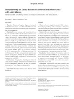
The technique is as follows (Figure 1.):
Recipient area.Initially, the recipient area for the laterally moved flap is prepared. A
reverse bevel incision is made all along the soft tissue margin of the defect. After removal of
the dissected pocket epithelium, the exposed root surface is thoroughly curetted. Two
superficial incisions are then delineating a 3 mm wide recipient area, at the one side of the
Aesthetic Periodontal Therapy – Root Coverage 7
defect as well as apical to the defect, where the epithelium together with the outer portion of
the connective tissue is removed by sharp dissection (Weneström et al., 2008).
The flap design is outlined by two vertical incisions that extended from the horizontal
incision to several millimeters apically to the mucogingival junction. A horizontal incision is
performed either at the gingival or 3 mm apically, following the marginal gingival contour,
thus joining the vertical incisions. A beveled linear horizontal incision is performed to
optimize the content of keratized tissue in the flap when the donor site is an edentulous site.
The flap is elevated as full thickness in the portion adjacent to the recession and as partial
thickness in the portion distal to the recession. Partial-thickness dissection is continued
apically and laterally to obtain passivity of flap movement and absence of muscle pull or
periosteal adhesion. The flap is rotated laterally to cover the recession defect completely and
extend for approximately 1 mm coronal to the cemento-enamel junction. Careful flap suturing
is performed to position and secure the soft tissues over the root surface by means of sling
and simple sutures (Santana et al., 2010).
Figure 1. Schematic drawing of rotational flap procedure.
Figure 2. Schematic drawing of double papilla flap technique.
A. L. Dumitrescu, Liviu Zetu and Silvia Teslaru 8
Following removal of the dressing and the sutures, usually after 10-14 days, the patient is
instructed to avoid mechanical tooth cleaning for further 2 weeks, but to use twice daily
rinsing with chlorhexidine solution as a means of infection control (Weneström et al., 2008).
Several modifications have been described to overcome the problem of dehiscence at the
donor site. Staffileno (1964) used a split-thickness pedicle flap so as not to denude the
adjacent site. This approach compromises vascularity and does not preclude bone resorption
at the donor site (Bahat et al., 1990). Other modifications of the procedure are the oblique
rotated flap (Pennel et al., 1965), the rotation flap (Patur, 1977), the double papilla flap
(Cohen and Ross, 1968) (Figure 2.) and the transpositioned flap (Bahat et al., 1990).
Zucchelli et al. (2010) revealead that present data do not seem to indicate the laterally
moved flap is an highly predictable and effective root coverage surgical procedure. From the
studies reviewed, the reported mean percentage of root coverage ranges between 34% and
82% (Smuckler,1976; Guinard and Caffesse, 1978; Espinel and Caffesse 1981; Waite, 1984;
Zade and Hirani, 1985; Oles et al., 1985) and only Oles et al. (1988) reported data relating the
―percentage of complete (up to the cemento-enamel junction) root coverage‖ and the range
was between 40% and 50% (Zucchelli et al., 2010).
4.1.2. Advanced Flaps Procedures
Since the lining mucosa is elastic, a mucosal flap raised beyond the mucogingival
junction can be stretched in coronal direction to cover exposed root surfaces. The coronally
advanced flap procedure has been described by several authors (Allen and Miller Jr, 1985;
Harris and Harris, 1994; Milano, 1998; Romanos et al., 1993; Wennström and Zucchelli,
1997; Bernimoulin et al., 1975).
The coronally advanced flap is the first choice surgical technique when there is adequate
keratinized tissue apical to the recession defect. Optimum root coverage results, good color
blending of the treated area with respect to adjacent soft tissues, and recuperation of the
original morphology of the soft tissues margin can be predictably accomplished using this
surgical approach. Furthermore, the coronally advanced flap is very effective in treating
multiple recession defects affecting adjacent teeth with obvious advantages for the patient in
terms of esthetics and morbidity. Some unfavorable local anatomic conditions may render the
coronally advanced flap contraindicated: 1) the absence of keratinized tissue apical to the
recession defect; 2) the presence of gingival (―Stillman‖) cleft extending in alveolar mucosa;
3) the marginal insertion of frenuli; 4) the presence of deep root structure loss; or 5) presence
of a very shallow vestibulum. In these situations the clinician should take the soft tissues
located laterally to the recession defect into consideration to evaluate the possibility to
perform a laterally moved flap (Zucchelli et al., 2010; Wennström and Zucchelli, 1996;
Zucchelli and De Sanctis, 2000).
The coronally positioned pedicle graft has many advantages over other surgical
procedures used to cover exposed roots. It does not require a separate surgical site to obtain a
graft. The tissue utilized will be a perfect color and contour match with the surrounding
tissue. Additionally, the procedure is simple to perform and does not require a lot of time
(Harris and Harris, 1994).
In aim to evaluate the predictability of the procedure several clinical studies have been
evaluated by Bouchard et al., 2001. The mean depth of the recession defects treated was 3.7
mm (3.3–4.1mm). The mean % of root coverage for advanced flaps was reported to be 77%
Aesthetic Periodontal Therapy – Root Coverage 9
(55–98), while the % of teeth with complete root coverage was 45% (9-84%) (Bouchard et
al., 2001).
More recently, Cairo et al. (2008) reviewed the clinical outcomes of the coronally
advanced flap on a total of 794 Miller Class I and II gingival recessions in 530 patients from
25 RCTs. This systematic review confirms that the coronally advanced flap procedure is a
safe and reliable approach in periodontal plastic surgery and is associated with consistent
recession reduction and frequently with complete root coverage. The results of meta-analyses
showed that only two combinations (coronally advanced flap + connective tissue graft and
coronally advanced flap + enamel matrix derivative) provided better results than coronally
advanced flap alone. Coronally advanced flap + connective tissue graft resulted in better
clinical outcomes for both complete root coverage (OR=2.49) and recession reduction (10.49
mm) compared with coronally advanced flap, and no other therapy provided better results
than coronally advanced flap + connective tissue graft. The combination of coronally
advanced flap + enamel matrix derivative was associated with a higher probability to obtain
complete root coverage (OR=3.89) and a higher amount of recession reduction (0.58 mm)
than coronally advanced flap alone. A possible benefit following root coverage procedures
may be the augmentation of keratinized tissue. This systematic review showed that coronally
advanced flap + connective tissue graft was associated with better clinical outcomes in terms
of keratinized tissue gain following therapy.
The technique for the coronally advanced flap procedure is:
The coronally advanced flap is initiated by two horizontal bevelled incisions (3mm in
length), mesial and distal to the recession defect located at a distance from the tip of the
anatomical papillae equal to the depth of the recession plus 1 mm.Two bevelled oblique,
slightly divergent, incisions starting at the end of the two horizontal incisions and extending
to the alveolar mucosa. The resulting trapezoidal-shaped flap is elevated with a split–full–
split approach in the coronal–apical direction. In order to permit the coronal advancement of
the flap, all muscle insertions present in the thickness of the flap are eliminated. This is done
keeping the blade parallel to the external mucosal surface. Coronal mobilization of the flap is
considered ―adequate‖ when the marginal portion of the flap was able to passively reach a
level coronal to the CEJ of the tooth with the recession defect. In fact, the flap should be
stable in its final coronal position even without the sutures. The root surface is mechanically
treated with the use of curettes. It must be considered that only the portion of the root
exposure with loss of clinical attachment (gingival recession1 probeable gingival
sulcus/pocket) is instrumented. Exposed root surfaces belonging to the area of anatomic bone
dehiscence were not instrumented not to damage connective tissue fibres still inserted in to
the root cementum. The facial soft tissue of the anatomic inter-dental papillae coronal to the
horizontal incisions is disepithelized to create connective tissue beds to which the surgical
papillae of the coronally advanced flap are sutured. By moving the flap coronally to reach the
tip of the disepitelized anatomical papillae, the vestibular soft tissue should be positioned 1
mm coronal to the cemento-enamel junction to account for soft tissue shrinkage. The suture
of the flap is started with two interrupted periosteal sutures performed at the most apical
extension of the vertical releasing incisions; then, it proceeded coronally with other
interrupted sutures, each of them directed, from the flap to the adjacent buccal soft tissue, in
the apical–coronal direction. This is done to facilitate the coronal displacement of the flap and
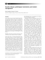
to reduce the tension on the last coronal sling suture (De Sanctis and Zucchelli, 2007) (Figure
3.).
A. L. Dumitrescu, Liviu Zetu and Silvia Teslaru 10
Figure 3. Coronally advaced flap procedure. a. A recession defect on the lower canine. b. Close suturing
of the pedicle graft to cover the exposed root surface. c. Healing outcome 3 months post-operatively. d.
Healing outcome 1 year post-operatively.
For the treatment of isolated gingival recession, Zucchelli et al. (2004) proposed the use
of a laterally moved and coronally advanced flap. Thereafter, the proposed surgical technique
combined the root coverage and esthetic advantages of the coronally advanced flap with the
increase in gingival thickness and in the amount of keratinized tissue associated with the use
of the laterally moved flap and resulted in a very high mean percentage of root coverage
(96%) and complete soft tissue root coverage (up to the CEJ) accomplished in 80% of treated
cases.
The main modification of the present surgical technique, with respect to those previously
proposed, was the elimination of all muscle insertions in the thickness of the flap to permit
the coronal advancement of the laterally moved flap. Furthermore, the coronal advancement
of the flap allowed the surgical papillae to cover the anatomic papillae which represented the
most coronal areas for anchoring the flap and a critical source for vascular exchanges. In
addition, coronal advancement of the flap beyond the cemento-enamel junction likely
compensates for the post-surgical soft tissue contraction, resulting in no exposure of the root
surface (Zucchelli et al., 2004).
The different thickness during flap elevation (greater in the central area than in the more
peripherical portions of the flap) represented another aspect of the proposed surgical
technique. In a thicker flap the amount of vascularized connective tissue increases and the
post-surgical soft tissue contraction decreases. Both these factors improve the possibility of
accomplishing and maintaining root coverage (Zucchelli et al., 2004).