Tài liệu Molecular Sensors for Cardiovascular Homeostasis P2 pptx
Bạn đang xem bản rút gọn của tài liệu. Xem và tải ngay bản đầy đủ của tài liệu tại đây (420.42 KB, 10 trang )
P1: OTE/SPH P2: OTE
SVNY334-Wang February 14, 2007 15:15
34 Christopher J. Benson and Edwin W. McCleskey
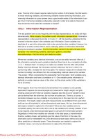
Figure 2.1. Sensory innervation of theheart. The myocardium is innervated by sympathetic
afferents that follow the sympathetic efferent nerves back to their cell bodies located in the
upper thoracic DRG, and cardiac vagal afferents that follow the vagal nerves to the nodose
ganglia. Afferent neurons from the pericardium follow the phrenic nerves to their cell bodies
in the upper cervical DRG (C
3
–C
5
). From the respective sensory ganglia, central projections
synapse in the spinal cord or brainstem. From Benson et al. (1999).
serve a nociceptive function, and the encoding mechanism for the signal, remain
controversial.
14,15
The first question pertains to the nature of the stimulus that is sensed by cardiac
afferents during myocardial ischemia. In the early 1900s a mechanical hypothe-
sis held sway: It was believed that distortion or distention of the ischemic cardiac
chambers activated mechanoreceptors on the heart (much like pain generated from
P1: OTE/SPH P2: OTE
SVNY334-Wang February 14, 2007 15:15
2. ASICs Function as Cardiac Lactic Acid Sensors During Myocardial Ischemia 35
other hollow visceral organs)
16
; however, experimental evidence has since proven
otherwise. While most cardiac afferents are responsive to mechanical stimuli, this
does not seem to correlate with pain responses in animals or humans.
17
Addition-
ally, current clinical practice in cardiology tells us that catheter-based valvular
balloon distention, myocardial puncture or biopsy, and radiofrequency-produced
burns within the myocardium are all painless procedures in conscious patients.
In the 1930s, Lewis put forth a chemical hypothesis that substances released
from ischemic muscle generate pain signals.
18
Since then, it has been generally
accepted that sensory activation during myocardial ischemia results from one
or more chemical stimuli. Various substances have been implicated, and have
been shown to activate cardiac afferent neurons: bradykinin,
19,20
adenosine,
21
serotonin,
22
histamine,
23
ATP,
24
prostaglandins,
25
reactive oxygen species,
11,26
and lactic acid.
27,28
Although incompletely understood, activation of cardiac affer-
ents in the setting of myocardial ischemia probably represents a complex interplay
between multiple mediators. Even less is understood regarding the molecular na-
ture of the chemical receptors that transduce these various stimuli into an electrical
signal. In this chapter we will focus on our efforts to identify chemical activators
of cardiac afferents and the underlying molecular nature of their receptors.
2.3. Acidic Metabolites are Likely Mediators of Sensation
During Cardiac Ischemia
The heart is an organ of high metabolic activity and is susceptible to rapid drops in
pH during ischemia. Under normal aerobic conditions, the heart readily consumes
lactic acid to generate ATP via the respiratory cycle. For example, maximally ex-
ercising skeletal muscle generates and releases lactic acid into the circulation. The
heart uses this as an energy source: the concentration of lactate within the coronary
arteries supplying the heart is generally higher than that in the venous drainage from
the heart. However, with insufficient blood supply and oxygen, cardiac myocytes
will attempt to maintain contractile function by switching to anaerobic glycolysis.
Consequently, lactic acid is generated and accumulates within the cells, which
along with the associated drop in pH, inhibits contractile function and contributes
to cell death.
29
Myocytes respond by pumping out lactic acid, primarily via a spe-
cific lactate transporter, which in turn acidifies the extracellular interstitial spaces
within the heart.
30
Additionally, ischemia also contributes to build-up of lactate
and other metabolites because low perfusion leads to reduced washout.
What are the concentrations of lactate and H
+
in the heart during ischemia?
In isolated ischemic hearts, myocardial intracellular pH drops from about 7.0 to
6.0.
31,32
The extracellular pH, which would be the signal available to trigger sen-
sory neurons, drops within 5 minutes from 7.4 to 7.0. It gets lower only when
there is complete loss of blood flow for prolonged times, conditions that cause
necrosis.
33,34
Occlusion of coronary blood flow in vivo generates a similar drop
in pH to the 7.0 range (Figure 2.2A).
28
It is the subtle change—the drop to near
P1: OTE/SPH P2: OTE
SVNY334-Wang February 14, 2007 15:15
36 Christopher J. Benson and Edwin W. McCleskey
C
D
B
A
Figure 2.2. Myocardial ischemia induces a drop in pH that contributes to cardiac afferent
activation. Epicardial pH is lowered during 5 minutes of ischemia (A); this is prevented
by infusion of isotonic neutral phosphate buffer into the pericardial sac (B). Frequency
histograms of action potentials recorded from a cardiac sympathetic afferent during control,
ischemia, and reperfusion before (C) and after (D) pericardial infusion of isotonic neutral
phosphate buffer. From Pan et al. (1999).
P1: OTE/SPH P2: OTE
SVNY334-Wang February 14, 2007 15:15
2. ASICs Function as Cardiac Lactic Acid Sensors During Myocardial Ischemia 37
neutral—that occurs at the time associated with pain.
35
If such a small pH change
can be the cause of the pain, there must be a very sensitive detector expressed in
cardiac muscle.
Can acidic metabolites associated with ischemia activate cardiac afferents?
Uchida and Murao
27
first showed that lactic acid applied to the surface of the
heart caused excitation of cardiac sympathetic afferent fibers, although relatively
high concentrations were required—correlating with a pH of 4.58. This pH value
is below that achieved during myocardial ischemia, and consequently it has been
argued that the H
+
concentrations associated with myocardial ischemia are not ad-
equate to activate cardiac afferents and produce pain.
7,36
However, it appears that
buffering within interstitial spaces keeps extracellular pH from ever approaching
the low value applied to the surface of the tissue. Pan et al.
28
measured the actual
pH achieved in the myocardium during acid application by placing a pH-sensitive
needle electrode into the myocardium within 1.0–1.5 mm of the surface. They
found that a lactic acid concentration of 50 μg/ml (pH 5.42) produced a robust
cardiac afferent activation, even though this only produced a drop in measured
myocardial pH to 7.0—a pH value readily achieved within minutes of myocardial
ischemia.
To evaluate the role of endogenously produced H
+
, Uchida and Murao
27
injected
sodium bicarbonate to buffer pH and reported a greater than 50% attenuation
of cardiac sympathetic afferent activation induced by coronary artery occlusion.
Similarly, Pan et al.
28
added a pH buffer into the pericardial sac surrounding the
heart to effectively prevent pH changes during ischemia, and they also found
afferent activation was inhibited by greater than 50% (Figure 2.2C,D). Thus, the
data indicate that acidosis associated with myocardial ischemia is sufficient to
excite cardiac afferents. In addition, while several chemicals probably contribute
to normal levels of cardiac afferent activation during ischemia, acidic metabolites
are a necessary component.
2.4. Isolated Cardiac Afferents Are Activated by Protons
To identify the molecular components that sense myocardial ischemia, we isolated
cardiac afferent neurons in culture. The cultivation of sensory neurons has proven
to be a useful model to study different sensory modalities; the cell bodies in vitro
seem to retain the molecular components necessary for sensory transduction at the
nerve terminals in vivo.
37
To distinguish cardiac from other sensory neurons, we
used a fluorescent tracer dye to label cardiac afferents in vivo so that they could
later be identified in primary dissociated culture (Figure 2.3A,B). Having isolated
labeled cardiac afferents, we first applied a variety of chemicals (implicated in
cardiac pain) to isolated rat cardiac and non-cardiac (unlabeled) sensory neurons,
and measured the resultant ionic currents by whole-cell patch-clamp.
38
The most important finding of this experiment was that acidic pH evoked
large inward currents in almost all cardiac sympathetic afferents (Figure 2.3C-E).
P1: OTE/SPH P2: OTE
SVNY334-Wang February 14, 2007 15:15
38 Christopher J. Benson and Edwin W. McCleskey
A
B
100
80
60
40
20
0
% responders
pH
ATP 5HT Cap Ach BK Aden
10
8
6
4
2
0
mean amplitude (nA)
*
*
pH
ATP 5HT
Cap Ach BK Aden
DRG heart
DRG unlabeled
Nodose heart
400 pA
2 nA
500 pA
2 sec
2 sec
5HT
2 sec
ATP Cap
10 sec
100 pA
C
D
E
pH 5
P1: OTE/SPH P2: OTE
SVNY334-Wang February 14, 2007 15:15
2. ASICs Function as Cardiac Lactic Acid Sensors During Myocardial Ischemia 39
←
Figure 2.3. Acidic pH activates large currents in isolated cardiac afferents. (A) Corre-
sponding phase (left) and fluorescence (right) micrographs of myocardium 3 weeks after
surgical injection of fluorescent tracer dye into the pericardial space. (B) Phase (left) and
fluorescence (right) micrographs of two cardiac sympathetic afferents in primary dissoci-
ated culture of DRG neurons. (C) Currents evoked by application of indicated agents to
cardiac sympathetic afferents. (D) The percentage of cardiac sympathetic (DRG heart),
cardiac vagal (nodose heart), and noncardiac (DRG unlabeled) neurons that responded to
various agents: [pH, 5.0; ATP, 30 μM; serotonin (5HT), 30 μM; capsaicin (Cap), 1 μM;
acetylcholine (ACh), 200 μM; bradykinin (BK), 500 nM; or adenosine (Aden), 200 μM].
(E) Mean amplitudes of the evoked currents of the responding neurons.
∗
P <.01 vs. pH-
evoked current in DRG heart. From Benson et al. (1999).
Consistent with this, all cardiac sympathetic afferent fibers fire action potentials in
response to epicardial application of lactic acid in whole animal models.
12,27
By
comparison, a much smaller percentage of noncardiac DRG neurons responded to
acid and their currents were significantly smaller. Moreover, the response to other
potential chemical mediators generated currents in a lower percentage of cells,
and the activated currents were far smaller than those evoked by acid. Thus, while
activation of cardiac afferents in the setting of myocardial ischemia most certainly
represents a complex interplay between multiple mediators, we have focused on
acid and the molecular nature of the pH sensor, as it seems to be expressed at very
high levels in cardiac-specific sensory neurons.
2.5. ASICs Are the Proton Sensors in Cardiac Afferents
H
+
-gated ion channels were first characterized by Krishtal and co-workers in
the early 1980s using electrical recordings of isolated sensory neurons.
39
They
describe a channel that opens in response to extracellular acidification, has the
unusual characteristic of preferentially passing Na
+
ions through its pore, and is
blocked by the diuretic amiloride. Further characterization demonstrated multiple
different types of H
+
-activated currents, and it became apparent that multiple
molecules were involved.
40,41
In the mid 1990s, two classes of ion channels were cloned that probably account
for the bulk of H
+
-activated currents described in native neurons. TRPV1 channels
are best known for their ability to detect noxious heat and capsaicin, the pungent
component of pepper.
42−44
However, they also integrate multiple signals, including
voltage, temperature, lipid metabolites, and extracellular acidity.
45−47
At 37
◦
, they
are reported to activate at about pH 6.0.
45
While this is much more acidic than
that associated with cardiac pain, it is possible that the complex swirl of altered
chemistry that accompanies tissue ischemia may increase the acid sensitivity of
these molecules.
At the same time, a second class of H
+
-gated ion channels was cloned in an
effort to identify related members of the DEG/ENaC family of ion channels. This
P1: OTE/SPH P2: OTE
SVNY334-Wang February 14, 2007 15:15
40 Christopher J. Benson and Edwin W. McCleskey
family includes the epithelial Na
+
channel, ENaC, which mediates Na
+
reab-
sorbtion in the kidneys, lungs, and colon,
48
and the degenerins in C. elegans,
which participate in mechanosensation.
49
All members in the family are selec-
tive for Na
+
, and are blocked by amiloride, properties shared by H
+
-gated ion
channels in sensory neurons. This analogy, along with the fact that several of the
newly cloned DEG/ENaC channels were expressed in sensory neurons, led the
Lazdunski group to describe the first acid-sensing ion channel (ASIC).
50
We now
know three genes within the DEG/ENaC family that encode H
+
-gated channels:
ASIC1,
50,51
ASIC2,
52
and ASIC3.
53
ASIC1 and ASIC2 both have alternative splice
forms involving the amino-termini. Although ASIC4 shows homology, it is not
gated by protons.
54,55
We suspect there are no additional ASIC genes; searches
of the recently completed mammalian genome sequences have not revealed novel
homologous sequences.
Like all DEG/ENaC proteins, ASICs have a large extracellular loop connecting
two transmembrane domains, with the amino and carboxyl termini inside the cell.
Expression of the ASICs individually in heterologous cells generates transient H
+
-
gated Na
+
currents (Figure 2.4A). Moreover, when coexpressed in combination,
they heteromulterize, producing currents with unique functional properties.
56−58
Expression of the ASICs is restricted to neurons, and mRNA corresponding to each
of the subunits is present in sensory neurons.
59−62
Furthermore, ASIC proteins have
been detected at nerve terminals,
61−63
where they are poised to transduce sensory
stimuli.
With this molecular background in mind, we set out to investigate the iden-
tity of the cardiac pH sensor. The biophysical and pharmacological properties
of the H
+
-evoked currents in cardiac afferents provided the answer. Application
of pH less than 7 activated a transient (rapidly activating and desensitizing) cur-
rent, which was followed by a sustained current only when the pH dropped fur-
ther, to pH 6 and below (Figure 2.4B). The EC
50
(pH 6.6) was less acidic than
previously reported by other investigators for acid-evoked currents in unselected
rat DRG neurons,
64
suggesting that cardiac afferents are particularly sensitive to
acidic changes. The transient current was Na
+
-selective, and the sustained cur-
rent was nonselective. Finally, the transient current was inhibited by the amiloride
(Figure 2.4C). These properties: the distinct kinetics, exquisite pH sensitivity,
Na
+
selectivity, and amiloride block, all indicate that H
+
-sensing channels in car-
diac afferents are ASICs. While our data suggests a minor role of TRPV1 in cardiac
sensation (capsaicin generated small amplitude currents in a smaller number of
cardiac afferents; Figure 2.3D and E), recent data supports TRPV1 expression in
rat cardiac afferents, and a role for TRPV channels in cardiac afferent activation
during ischemia.
65,66
To determine which of the three ASICs contribute to H
+
-gated channels in car-
diac afferents, we compared the biophysical properties of the native currents to
the properties generated by expression of ASIC1 (1a and 1b), ASIC2 (only 2a
is expressed in rat sensory neurons), and ASIC3 in heterologous cells.
67
Impor-
tantly, the pH sensitivity of ASIC3 most closely matches that of the cardiac afferent
channel (Figure 2.4D), and the threshold of activation (pH 7) is well within the
P1: OTE/SPH P2: OTE
SVNY334-Wang February 14, 2007 15:15
2. ASICs Function as Cardiac Lactic Acid Sensors During Myocardial Ischemia 41
ASIC 3 ASIC 1b ASIC 1a ASIC 2a
5 sec
pH 5.0 +/- 100
μ
M amiloride
2 sec
2 nA
*
1
0
I/ I
max
8 7 6 5
pH
Cardiac
ASIC 3
A
CD
pH 7.0 pH 6.5 pH 5.0 pH 4.0
5 nA
5 sec
B
Figure 2.4. ASIC3 reproduces the functional properties of the acid-evoked currents in
cardiac afferents. (A) Representative acid-evoked currents from COS cells expressing the
indicated ASIC subunits. The bars represent a solution change from pH 7.4 to 6, except for
ASIC2a, which is evoked by pH 5. (B) Currents evoked by applying various pH solutions
to a cardiac sympathetic afferent neuron. (C) Superimposed currents evoked by pH 5.0 and
by pH 5.0 plus 100 μM amiloride ([). (D) Average fractional current vs. pH for cardiac
afferents (filled circles) and COS-7 cells expressing ASIC3 (open circles). Adapted from
Benson et al. (1999), Sutherland et al. (2001), and Benson et al. (2001).
range attained during myocardial ischemia.
28,34
Other properties were also best
matched by ASIC3, suggesting it likely is the major constituent of the H
+
-gated
channel in rat cardiac afferent neurons. However, to match some properties re-
quired co-expression of multiple ASIC subunits.
56
For example, we found that
co-expression of ASIC3 and ASIC2 reproduced the cation nonselective sustained
currents occasionally observed in native neurons. Moreover, the characterization
of ASIC channel subunit composition in mice, taking advantage of mice lack-
ing specific ASIC genes, seems to indicate that a majority of ASIC channels in
sensory neurons are heteromultimers that consist of ASIC3 in combination with
other ASIC subunits.
68
P1: OTE/SPH P2: OTE
SVNY334-Wang February 14, 2007 15:15
42 Christopher J. Benson and Edwin W. McCleskey
2.6. ASICs Are Lactate Sensors
It has been observed in whole animal models that lactate is a more potent acti-
vator of visceral afferents than H
+
derived from other acid sources.
27
Panetal.
28
demonstrated that application of lactic acid to the surface of the heart to produce
a pH of 7.0 potently activated cardiac afferents. In contrast, application of acidic
phosphate buffer or inhalation of CO
2
caused no effect or only slightly increased
activity, respectively, despite producing equivalent drops in myocardial pH. Lac-
tic acid is also a more potent stimulator of intestinal and pulmonary afferents.
69,70
This seemingly paradox of lactic acid potency can now be explained by our further
understanding of how ASIC channels are activated.
Muscle ischemia causes extracellular lactate to rise to about 15 mM from a
resting level below 1 mM.
71,72
Applying 15 mM lactate concentration to iso-
lated cardiac afferents resulted in a ∼60% increase in current generated by pH
7 (Figure 2.5). This property was precisely reproduced by applying lactate to
heterologously expressed ASIC3. The mechanism involves a shift in the pH sen-
sitivity of the channel, making the channel an even better sensor of the subtle pH
changes that occur in the setting of cardiac ischemia. Lactate acts not through
a specific binding site, but rather it decreases the concentration of extracellular
divalent ions, which are known blockers of ASIC channels.
73
Decreasing extra-
cellular divalent ions can itself open ASIC channels and it potently increases their
sensitivity to protons.
74
This unique property of ASICs—to integrate both lactate
and H
+
—provides a molecular mechanism underlying the observed lactic acid
paradox (further supporting a role for ASICs as pH sensors in vivo), and makes
the channels ideal sensors of the metabolic changes associated with myocardial
ischemia.
pH 8.0
Control
pH 7.0
20 mV
Control
15 Lactate
15 Lactate
250 ms
500 ms
1 nA
A.
B.
Figure 2.5. Lactate potentiates ASICs. Voltage (A) and current (B) recordings from a
labeled cardiac sympathetic afferent neuron exposed to pH 7.0 in the presence or absence
of 15 mM lactate. The channels are ASICs because the current selectively passed Na
+
and
was blocked by 10 μM amiloride (data not shown). Adapted from Immke and McCleskey
(2001).
P1: OTE/SPH P2: OTE
SVNY334-Wang February 14, 2007 15:15
2. ASICs Function as Cardiac Lactic Acid Sensors During Myocardial Ischemia 43
2.7. ASICs May Integrate Multiple Mediators
During Ischemia
Multiple chemicals can activate cardiac afferents, and there is some evidence sug-
gesting an additive or synergistic effect. In a rat model of cardiac nociception,
Euchner-Wamser et al.
75
found that a mixture of chemical agents led to more
avoidance behavior and greater neuronal activation than bradykinin alone. More-
over, in the skin it has been proposed that a combination of chemical mediators
produces a more intense sensory activation than any individual mediator alone,
76
and that acid plays a dominant role in this setting.
77
Recent data suggests that ASICs, in addition to their role as lactate sensors,
might integrate multiple chemical signals. Pre-application of a mixture of chem-
ical mediators has been shown to increase H
+
-activated ASIC-like currents in
sensory neurons.
78
In part, this result is due to transcriptional up-regulation of
ASIC expression.
78−80
In addition, some chemicals can increase ASIC current
within minutes, suggesting a cellular signaling mechanism.
81
There are a couple
of potential signaling mechanisms that might, in part, explain an interaction be-
tween ASICs and other agents. First, ASIC2 can be phosphorylated and its function
potentiated by protein kinase C (PKC).
82
Recently, Deval et al.
81
demonstrated that ASIC3 + 2b heteromeric channels
(potentially an important ASIC channel in cardiac and other sensory neurons)
are positively regulated by a 2-minute pre-application of serotonin or bradykinin
via PKC pathway activation. The effect is similar to that produced by lactate: an
increase in the pH sensitivity of the channel. Both serotonin and bradykinin can
activate PKC via their respective G-protein-coupled receptors, leading to sensi-
tization of sensory neurons and inflammatory hyperalgesia.
83−85
Data suggests
ASIC currents are subsequently potentiated by PKC phosphorylation of purported
sites on the ASIC2b and –3 subunits.
81
Secondly, ASIC1 and ASIC3 can be phos-
phorylated by cAMP dependent protein kinase (PKA),
86
although the functional
significance is yet unknown. PKA signaling pathways are also important for sen-
sory neuron receptor function.
87,88
Multiple agents that have been implicated in
cardiac sensation, including adenosine, serotonin, histamine, and PGE
2
, can acti-
vate PKA.
89−92
and potentially regulate ASICs.
Evidence suggests multiple chemical mediators may be important to activate
cardiac afferents in the setting of ischemia; we hypothesis that lactic acid is a
major signal, and ASICs are a major sensor, and that other mediators could, in
part, produce effects by modulating ASIC channels.
2.8. Significance
We found that sensory neurons that innervate the heart express high levels of ASIC3
and we showed that it is particularly sensitive to lactic acid at concentrations that