Tài liệu Comparison of Gait of Young Women and Elderly Women pdf
Bạn đang xem bản rút gọn của tài liệu. Xem và tải ngay bản đầy đủ của tài liệu tại đây (1.46 MB, 8 trang )
1986; 66:1382-1387.PHYS THER.
Patricia A Hageman and Daniel J Blanke
Women
Comparison of Gait of Young Women and Elderly
found online at:
The online version of this article, along with updated information and services, can
Collections
Women's Health: Other
Geriatrics: Other
Gait Disorders
in the following collection(s):
This article, along with others on similar topics, appears
e-Letters
"Responses" in the online version of this article.
"Submit a response" in the right-hand menu under
or click onhere To submit an e-Letter on this article, click
E-mail alerts to receive free e-mail alerts hereSign up
by guest on December 24, 2012 from
Comparison
of
Gait of Young Women
and Elderly Women
PATRICIA A. HAGEMAN
and DANIEL J. BLANKE
The purpose of our study was to describe and compare free-speed gait patterns
of healthy young women with healthy elderly women. The evaluation was com-
pleted with high-speed cinematography using synchronized front and side views
of 26 healthy volunteers. One group was composed of 13 subjects 20 to 35 years
of age, and the other group was composed of 13 subjects 60 to 84 years of age.
Each subject participated in one test session consisting of three filmed trials of
free-speed ambulation down a 14-m walkway. The processed film was analyzed
for 10 gait characteristics. Differences in gait characteristics between the two
groups were examined using a correlated t test (p < .01). The elderly women
demonstrated significantly smaller values of step length, stride length, ankle
range of motion, pelvic obliquity, and velocity when compared with the younger
women. The results of our study suggest that the physical therapist should not
establish similar expectations for young women and elderly women during gait
rehabilitation.
Key
Words:
Gait,
Physical
therapy.
Understanding the effects of aging on movement and func-
tion is becoming increasingly important because of longer
average life spans and a growing elderly population. Changes
in stereotypic movements such as walking patterns have been
reported as early as 60 years of
age.
1,2
Because elderly people
frequently utilize physical therapy services to achieve the
maximal functional ability of motor activities such as gait,
the collection of
gait
analysis data of healthy elderly subjects
is essential to establish realistic rehabilitation expectations of
the elderly population.
Although advances in technology have made objective
methods of measuring gait more available, the physical ther-
apy literature contains only a few studies of gait comparisons
of healthy elderly women with healthy young women.
3-5
These studies report that elderly women demonstrate shorter
step and stride lengths, lower average velocities,
3,5
and
greater
variability in stride width
4
when compared with young
women. None of
these
studies, however, provides conclusive
evidence of the effects of aging on the gait patterns of the
elderly population because of small sample sizes and limita-
tions in the number of specific gait characteristics examined.
Additional comparisons of
the
gait
characteristics of healthy
young women and healthy elderly women are necessary be-
cause gait training is a major portion of geriatric physical
therapy rehabilitation and because women constitute a ma-
jority of the over-60-years age group. The purpose of this
study was to describe and compare the free-speed gait char-
acteristics of matched groups of healthy young women and
healthy elderly women.
METHOD
Subjects and Selection Procedure
Twenty-six female volunteers, thirteen 20 to 35 years of age
and
thirteen
60
years
of
age or
older,
were
accepted
as
subjects.
Each subject provided informed consent in accordance with
the procedures of the University of Nebraska Institutional
Review Board.
The health status of each subject
was
evaluated on the basis
of a medical review and an objective examination by a
registered physical therapist, and all subjects
were
found to be
free of disabling physical conditions or minor ailments that
could affect
or
influence locomotion. Specifically, the subjects
were without musculoskeletal or neurological involvement or
medication for these conditions. Because leg length is an
important determinant of
stride
length,
6
leg length was meas-
ured to ensure that each subject was without a leg-length
discrepancy (± 1.9 cm), as defined by Subotnick.
7
The per-
centage of body fat of each subject
was
determined using
skin-
fold measurements to ensure that no subjects who were ex-
tremely lean or obese would be included in the study. All
subjects were
within one
standard
deviation of the age-specific
average percentage of body fat listed by Jackson et al.
8
For
those subjects 20 to 35 years of
age,
the mean percentage of
body fat was 22.7 ± 6.8; for those subjects 60 years of age
and older, the mean percentage of body fat was 31.1 ± 7.5.
The elderly women meeting these criteria were tested first.
Young women meeting these criteria were recruited to match
the elderly women on the basis of right leg length. The
matching of right leg lengths was within the same range
suggested
by
Subotnick
for
leg-length
discrepancies
7
to
achieve
Mrs.
Hageman is Instructor, Division of Physical Therapy Education, Uni-
versity of Nebraska Medical Center, 42nd and Dewey Ave, Omaha, NE 68105
(USA).
Dr.
Blanke
is
Associate Professor, Department of Health, Physical Education,
and Recreation, University of Nebraska at Omaha, Omaha, NE 68182.
This article
was
submitted
June
4,1985;
was
with
the
authors for
revision
12
weeks;
and
was
accepted
January
29,
1986.
1382
PHYSICAL THERAPY
by guest on December 24, 2012 from
RESEARCH
a close pairing of subjects between the young and elderly
groups.
Instrumentation
High-speed cinematography
was
used to record the subjects'
gait. The motion-recording system included two high-speed
cameras positioned to orient their optical axes at 90 degrees
with one camera parallel to and the other camera perpendic-
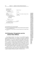
ular to a 14-m walkway (Figure). The front camera, a Photec
IV* fitted with a 50-mm Nikon
†
lens, was positioned 15.6 m
from the center of the walkway. The side camera, a LoCam*
fitted with a 25-mm Cosimcar* lens, was positioned
8
m from
the center of the walkway. The cameras were positioned
according to the procedure of Sutherland and Hagy.
9
Each
camera
was
set to run at
100
frames a second. A 1-m reference
scale was included in the field of view of both cameras. A
lighting device also was placed in the view of both cameras to
provide a common reference point so that frames could be
matched later to analyze any phase of the walking cycle.
The processed film was displayed on a Lafayette Data-
viewer
§
rear projection system. This system projects the film
image of the subject onto a viewing screen and allows the film
to be viewed frame by frame or advanced up to 24 frames a
second, depending on the viewer's needs for each variable
that is measured. The desired measurements were made di-
rectly from the projected image. A Numonics" digitizer was
used in conjunction with the projection system to assign
separate X,Y-coordinate values for any landmark from both
front- and side-view films. The coordinate values for the
landmarks were stored in a computer
#
and were used for
calculating the variables.
Procedure
Each subject was scheduled for one 45-minute testing ses-
sion. All testing was performed at the Gait Analysis Labora-
tory at the University of Nebraska at Omaha. Appropriate
shorts and sleeveless shirt were the required dress. A
1.9-cm
white dot with a 0.6-cm blue center was placed on the
following anatomical points in accordance with the proce-
dures of Sutherland and Hagy
9
and Sutherland et al
10
: right
and left anterior-superior iliac spines (ASISs), center of both
right and left patellae with the knee
flexed
to 25 degrees, right
and left malleoli, and the space between the second and third
metatarsals of both the right and left feet. Other markers
included a pelvic stick that consisted of a 15.5-cm dowel
directed perpendicularly from an Orthoplast
®
base. The base
was attached to a web belt with buckle closure. The belt was
placed on the subject so that the stick projected anteriorly
from a point midway between the ASISs. A tibial stick of
similar construction was placed around the maximal circum-
ference of the calf
so
that the stick projected anteriorly from
the tibial crest.
The subjects then walked barefoot along the 14-m walkway.
Each subject was advised to walk at the pace she normally
would choose when walking on a clear sidewalk. The first
Camera
Lighting
Figure. Walkway plan
of
Gait Analysis Laboratory.
4.75 m of the walkway allowed each subject to accelerate to
her chosen walking speed before reaching the filmed area.
The area from which measurements were taken was 3.25 m
long, allowing one to two gait cycles, depending on the size
of the subject and her walking speed. The last 6 m of the
walkway ensured that each subject did not decelerate until
she had left the filmed walkway area. Each subject performed
three trials.
Measurements
We used the procedure that was described by Sutherland
and Hagy
9
and validated in 1980 by Sutherland et al
10
to
obtain the measurements with the processed film. Reliability
of the measurements taken from our processed film was high
when test-retest results were compared during a pilot study.
The same observer (P.A.H.) made all of the test-retest meas-
urements from the film, recording a maximum deviation of
2.5 degrees for rotational measurements and a maximum
deviation of
2
cm for distance measurements.
* Photomic Systems, Inc, 265 H Sobrante Way, Sunnyvale, CA 94086.
†
Nikon, Inc, 623 Stewart Ave, Garden City, NY 11530.
‡ Redlake Corp, 1711 Dell Ave, Campbell, CA 95008.
§ Lafayette Instrument Co, PO Box 5729, Lafayette, IN 47903.
║Numonics Corp, 418 Pierce St, Lansdale, PA 19446.
#
Model 4052, Tektronix, Inc, PO Box 500, Beaverton, OR 97077.
Volume 66 / Number 9, September 1986
1383
by guest on December 24, 2012 from
Variables measured from the side view included: ankle
plantar-flexion and dorsiflexion range of motion, average
velocity of the center of gravity, step length, stride length, and
vertical excursion of the center of gravity. Step length was
measured as the distance in the line of travel between the
right heel-strike and the following left heel-strike, beginning
with the first full-body side view of the right heel-strike. A
scale factor was calculated using the 1-m reference scale in
the
cameras'
field
of view. The measured film step length was
multiplied by the scale factor to determine the actual step
length.
Stride length was measured as the distance in the line of
travel between successive points of foot-floor contact of the
right foot, beginning with the first total-body view involving
a right foot step. Actual stride length was calculated by mul-
tiplying the scale factor by the measured film stride length.
Average walking velocity was recorded as the total distance
traveled
by
the subject's center of gravity during one
gait
cycle
divided by the time elapsed during the movement, recorded
in centimeters per second. Center of gravity was calculated at
the initial point of the
right
foot-floor contact during the first
total-body view and the successive points of right foot-floor
contact. Center of gravity was determined according to the
segmentation method.
11
We calculated cycle time
by
counting
the number of frames for one gait cycle and dividing the
number by the film speed.
Vertical center-of-gravity excursion was calculated by com-
paring the center of
gravity
of digitized frames at mid-stance
and double-support phases of the gait cycle. Vertical center-
of-gravity excursion was determined
by
subtracting the lowest
vertical point from the highest vertical point.
A side view provided the method for determining the ankle
plantar-flexion-dorsiflexion
range
of motion. The total degree
of movement at the ankle formed by the line between the
knee and ankle center and the line along the bottom of the
foot was recorded in degrees. These measurements were ob-
tained directly from the viewing screen with a protractor.
The front-view camera provided the data for determining
stride width, lateral center-of-gravity excursion, pelvic ob-
liquity, pelvic rotation, and tibial rotation. Stride width was
measured as the horizontal distance between two consecutive
steps measured from a point between the second and third
metatarsals of each foot. Because the subject image became
larger as the subject approached the camera, the appropriate
scale factor was used for each point measured and confirmed
by measurements from the side view. The actual distance
between the metatarsal points of both feet was the stride
width.
Lateral center-of-gravity excursion was considered to be the
total lateral movement the body's center of gravity traveled
during one gait cycle. The center-of-gravity (X,Y) coordinates
for the mid-stance position of full weight bearing on the right
foot
and
on
the
left foot
were
calculated. Using
the
appropriate
scale factor for each center of gravity, the actual distance
between the two center-of-gravity points was calculated.
Pelvic obliquity was the arc of upward and downward
movement from the horizontal plane of the
right
ASIS during
one gait cycle. This measurement was obtained directly from
the viewing screen with a protractor. When the right ASIS
was at the highest vertical point, the angle of upward move-
ment was measured as the angle formed by the intersection
of two lines. The
first
line was the segment between the right
ASIS and the center point between the right and left ASISs
located at the base of the pelvic-stick attachment. The second
line was the segment between the base of the pelvic-stick
attachment to the horizontal plane. The angle of downward
movement was determined in a similar fashion and added to
the angle of upward movement for the total
arc
of movement.
Pelvic rotation was the degree of rotation that the pelvis
moved about
a
vertical
axis.
At 0
degrees
of rotation, the 15.5-
cm pelvic stick pointed directly
ahead.
At the point of greatest
observed rotation to the
right,
the distance that the tip of
the
pelvic stick had rotated to the
right
from the neutral position
was measured. This distance was converted into an actual
distance using the appropriate scale factor. Considering this
distance and the stick length to be two sides of a triangle, we
used
a
trigonometric function
to
calculate
the
angle
of rotation
to the right. The same method was used for recording the
greatest observed rotation to the left. The total rotation was
the sum of the degrees of
rotation
to the
right
arid left.
Tibial rotation was the degree the tibia rotated during foot-
floor contact of the
right
foot. The rotation of the tibial stick
about a vertical axis was calculated in the same manner as
the pelvic-stick rotation.
Data Analysis
Descriptive
statistics were
calculated
for
each
variable
meas-
ured in both groups. An independent t test was used to
compare the basic descriptive characteristics between the
groups. Because the groups were nonrandom and matched
for leg length, which may have affected the experimental
variables, a correlated t test was used to compare gait char-
acteristics between the groups. All comparisons were evalu-
ated at the .01 level of significance.
RESULTS
The basic descriptive characteristics of both groups of
women are presented in Table 1. The groups appeared to be
well matched for leg length because no statistical differences
were found between the two groups for either
right
or left leg-
length comparisons. Although no differences were found in
height or weight between the groups, the elderly women had
a higher percentage of body fat than the younger women.
Both groups, however, were within the normal range for
percentage of body fat based on their age ranges.
Gait characteristics measured from the
film
of the side-view
camera are reported in Table 2. A comparison of
the
means
of these variables reveals significant differences of all variables
except the vertical excursion of the center of gravity. The
younger female group demonstrated a longer step length and
stride length than the elderly women. Greater ankle move-
ment was observed in the walking patterns of the young
women than
in
those
of
the
elderly women.
The
young women
also ambulated at a significantly faster rate than the elderly
women.
Table 3 lists the comparison of variables measured from
the front-view camera. The only variable that revealed a
significant difference between the two groups of women was
pelvic obliquity because the young women demonstrated
substantially greater pelvic obliquity compared with the el-
derly women. We found no significant differences between
the groups for pelvic or tibial rotation or for lateral
center-of-
gravity excursion.
1384
PHYSICAL THERAPY
by guest on December 24, 2012 from
RESEARCH
TABLE 1
Basic Descriptive Characteristics of the Groups
Variable
Age
(yr)
Height (cm)
Mass (kg)
Body fat (%)
Leg length (cm)
Left
Right
66.85
161.00
61.43
25.27
86.98
86.69
Elderly Women
(n
= 13)
s
7.60
9.16
17.04
5.79
4.71
4.66
Range
(60.0-84.0)
(138.4-172.7)
(37.7-107.9)
(17.2-35.1)
(78.5-95.6)
(78.3-96.9)
23.92
165.10
60.43
20.37
86.81
86.65
Young Women
(n-13).
s
3.57
8.15
8.20
2.79
4.24
4.40
Range
(20.0-33.0)
(154.9-182.9)
(49.8-74.9)
(16.0-25.4)
(80.0-94.1)
(80.9-95.6)
t
-1.20
a
0.19
a
2.75
a,b
0.39
c
0.10
c
DISCUSSION
Our study resulted from a need to gain a better understand-
ing of the gait characteristics of young women and elderly
women. Whether physical therapists should expect elderly
women to have the same rehabilitation potential as young
women is not conclusive. Previous gait studies of healthy
women either did not consider the specific matching of
groups
3
or used a smaller sample size
5
than that used in our
study. Previous studies comparing the gait characteristics of
young and elderly women have emphasized gait intercycle
variability
4
and the influences of heel height on gait.
5
This
study of the linear, temporal, and rotational aspects of the
gait patterns of healthy young women and healthy elderly
women may be helpful to the physical therapist who uses gait
characteristics to evaluate a patient's progress.
We adhered strictly to the criteria established for subject
selection in this study. Matching the young group with the
elderly group using leg-length measurements was considered
crucial because of the influence of leg length on stride length.
6
The results of our study are in close agreement with the
findings of other gait studies involving adult women and those
involving adult men. The step- and stride-length measure-
ments of the elderly women in our study are similar to values
TABLE 2
Comparison of Gait Characteristics Measured from the Side-
View Camera
Variable
Step length
(cm)
Stride length
(cm)
Ankle range of
motion (°)
Velocity (cm/
sec)
Vertical center-
of-gravity ex-
cursion (cm)
Elderly Women
(n = 13)
66.34
134.92
24.62
131.94
2.87
s
6.77
14.71
4.61
23.85
1.34
Young Women
(n
= 13)
80.68
162.70
31.31
159.53
3.51
s
5.43
10.84
5.22
16.39
1.77
t
a
7.10
b
6.47
b
-3.93
b
-4.90
b
-1.33
published in a study by Murray et al
5
involving 30 women
aged 20 to 70 years and in a study by Chao et al
12
involving
37 women aged 32 to 85 years. Finley et al
3
reported shorter
step and stride lengths for the young women and elderly
women in their study than those of our study. The shorter
step and stride lengths of the subjects in the Finley et al study
may have represented the effects of cumbersome equipment
worn by their subjects during walking. The healthy elderly
women in our study demonstrated similar step- and stride-
length values to those reported by Sutherland and associates
10
for 15 healthy men aged 19 to 40 years.
The young women demonstrated significantly larger values
for step length than the elderly women. The values of the
young women in our study were similar to the values of 30
healthy men aged 20 to 65 years recorded during free-speed
gait by Murray et al.
13
The mean step and stride lengths of
the men were 78 cm and 156 cm, respectively. The young
women in our study demonstrated a greater walking velocity
than the women aged 20 to 36 years in a 1984 study by
Murray etal.
14
Our finding of larger means for step and stride lengths for
the young women is not surprising because the mean walking
velocity of the young group was significantly greater than the
TABLE 3
Comparison of Gait Characteristics Measured from the Front-
View Camera
Variable
Lateral
center-of-
gravity excur-
sion (cm)
Stride width (cm)
Pelvic obliquity (°)
Pelvic rotation (°)
Tibial rotation (°)
Elderly
Women
(n
= 13)
3.03
10.02
6.77
11.77
15.31
s
2.13
3.58
2.05
4.30
7.10
Young
Women
(n
= 13)
2.39
8.31
9.86
11.77
16.69
s
1.50
3.12
2.38
4.44
5.50
t
a
0.10
1.58
-3.65
b
0.00
-0.51
a
ctf=24.
*p<.01.
c
df=12.
a
(df=12.
b
p<.01.
a
df=12.
b
p<01.
Volume
66 /
Number
9,
September 1986
1385
by guest on December 24, 2012 from
mean walking velocity of the elderly group (p < .01). The
ambulation rate of the elderly women, however, was not
abnormally slow. Their mean walking speed of 131.9 ± 23.9
cm/sec was similar to values obtained by Murray et al
5
involving women aged 20 to 70 years whose mean free-speed
velocity was 130.0 ±15.0 cm/sec. The mean walking velocity
of the elderly women also was similar to values obtained in a
study by Sutherland et al involving men aged 19 to 40 years
whose mean walking velocity was 121.6 cm/sec.
10
A progressive increase in pelvic obliquity corresponding to
increased walking speeds has been reported.
614
Pelvic ob-
liquity was greater in the young women, as compared with
the elderly women. The pelvic obliquity values of adult men
reported in the literature ranged from five to eight degrees,
10
which is similar to the findings of our study.
Both groups of women maintained lateral and vertical
center-of-gravity excursions within a 5-cm range reported in
the literature.
6
Stride widths, however, were extremely varia-
ble among both groups. Gabell and Nayak reported similar
findings.
4
Young subjects (21-47 years of age) and elderly
subjects (66-84 years of age) in their study demonstrated
variability within the gait cycle, primarily in stride width.
The elderly women demonstrated substantially less move-
ment at the ankle during free-speed gait than the young
women. In a study by Murray et al that compared the gait
patterns of young men and elderly men, the elderly men (60-
65 years of age) also showed a marked reduction of ankle
movement during ankle plantar flexion at the end of ipsilat-
eral stance.
13
The reason for the decrease in ankle movement
is not clear, but it may have resulted from slower gait speeds,
as suggested previously.
5
Rotation about the thigh and tibia have been reported in
phase with pelvic rotation. The rotary displacement increases
progressively from the pelvis to the thigh to the tibia with
values of
8
degrees of rotation documented at the pelvis to 19
degrees of rotation measured at the tibia.
15
This progressive
increase in rotation from the pelvis to the tibia was demon-
strated by both young and elderly subjects in our study, and
no significant differences were found between groups. The
values obtained in our study were similar to reported ranges
for adult women during free-speed gait,
5
but were larger than
the values reported for the free-speed gait patterns of men.
610
Because pelvic rotation facilitates forward movement of the
hip joint of the swinging leg, increased pelvic rotation would
be expected during an increased stride length. A significant
increase in pelvic rotation was not demonstrated by the young
women, however, even though they demonstrated a signifi-
cantly larger stride length when compared with the elderly
women. This
finding
may be attributed to individual variation
in the interaction between stride length, walking velocity, and
pelvic rotation.
4
Despite significant differences among several gait charac-
teristics of both the young and elderly women, the elderly
women demonstrated values of step length, stride length,
walking velocity, pelvic rotation, and tibial rotation that were
similar to or exceeded those values of healthy young men and
women in other studies. These findings suggest that both the
young women and the elderly women from our study walk
faster today than their counterparts of
15
to 20 years ago.
The differences in the results of our study and those of
previous studies may have been caused by differences in
subject selection and measuring techniques. Subject cooper-
ation and ability to follow directions may have influenced the
results. Because
we
used a small subject
sample,
a true random
sampling of the age groups may not have been represented.
Some subjects may have had an undiagnosed or unrecognized
pathological condition that affected their gait, despite our
adherence to the guidelines established for subject selection.
Clinical Implications
The results of our study suggest that the clinician should
not expect the same gait training rehabilitation potential for
both young women and elderly women because differences
exist between the gait characteristics of healthy young women
and healthy elderly women. The degree to which a patholog-
ical condition may further affect the rehabilitation expecta-
tions of both groups during gait training is beyond the scope
of this study.
Based on the results of our study, the clinician may expect
an elderly woman to ambulate with a smaller step and stride
length, a slower walking speed, less pelvic obliquity, and less
ankle movement than a younger woman with a similar leg
length. No differences between the young and elderly women
would be expected in center-of-gravity excursion, pelvic and
tibial rotation, or stride width.
Physical therapists are involved in the gait training of
geriatric patients. Because of the frequency with which they
treat elderly women, clinicians must know the effects of age
on gait to understand the potential of gait training rehabili-
tation for this patient group.
Further study in this area is needed before definitive state-
ments can be made about the effects of
aging.
Care must be
taken when applying the results of this study to other popu-
lations. Further research could focus on the comparison of
additional gait characteristics such
as
hip motion, hip rotation,
angling of the feet, and upper extremity movement of young
women and elderly women.
CONCLUSIONS
For the sample of subjects we examined, the following
conclusions can be made:
1.
The young women and the elderly women did not dem-
onstrate significant differences in vertical center-of-gravity
excursion, lateral center-of-gravity excursion, stride width,
pelvic rotation, or tibial rotation.
2.
The young women demonstrated significantly larger values
than those of the elderly women in step length, stride
length, ankle range of motion, pelvic obliquity, and walk-
ing velocity.
3.
The values of the gait characteristics of both the young
women and the elderly women in this study were larger
than those of their counterparts of 15 years ago. Despite
these apparent changes, the effects of aging were observed
in these gait characteristics: step length, stride length, ankle
range of motion, pelvic obliquity, and walking velocity.
1386
PHYSICAL THERAPY
by guest on December 24, 2012 from
RESEARCH
REFERENCES
1.
Fisher M, Birren J: Age and strength. J Appl Physiol 31:490-497, 1947
2.
Berry G, Fisher R, Lang S: Detrimental incidents, including falls, in the
elderly institutional population. J Am Geriatr Soc 29:322-324, 1981
3.
Finley F, Cody F, Finizie R: Locomotive patterns in elderly women. Arch
Phys Med Rehabil 50:140-146, 1969
4.
Gabell A, Nayak V: The effect of age on variability in gait. J Gerontol
39:662-666, 1984
5. Murray M, Kory R, Sepic S: Walking patterns of normal women. Arch Phys
Med Rehabil 51:637-650, 1970
6. Inman V, Ralston H, Todd R: Human Walking. Baltimore, MD, Williams &
Wilkins, 1981
7. Subotnick S: The short leg syndrome. J Am Podiatr Med Assoc 66:720-
723,
1976
8. Jackson A, Pollock M, Ward A: Generalized equations for predicting body
density of women. Med Sci Sports Exerc 12:175-182, 1980
9. Sutherland D, Hagy J: Measurement of gait movements from motion
picture film. J Bone Joint Surg [Am] 54:787-797, 1972
10.
Sutherland D, Olsen R, Cooper L, et al: The development of mature gait.
J Bone Joint Surg [Am] 62:336-353, 1980
11.
Nutter J, Blanke D, Wang T: Microcomputers aid movement analysis.
Collegiate Microcomputer 3:1-11, 1985
12.
Chao E, Laughman R, Schneider E, et al: Normative data of knee joint
motion and ground reaction forces in adult level walking. J Biomech
16:219-232 1983
13.
Murray M, Drought A, Kory R: Walking patterns of normal men. J Bone
Joint Surg [Am] 46:335-360, 1964
14.
Murray M, Mollinger L, Gardiner G, et al: Kinematic and EMG patterns
during slow, free, and fast walking. J Orthop Res 2:272-280, 1984
15.
Levens A, Inman V, Blosser J: Transverse rotations of the segments of
the lower extremity in locomotion. J Bone Joint Surg [Am] 30:859, 1948
Volume 66
/
Number 9, September 1986
1387
by guest on December 24, 2012 from
1986; 66:1382-1387.PHYS THER.
Patricia A Hageman and Daniel J Blanke
Women
Comparison of Gait of Young Women and Elderly
Cited by
/>articles:
This article has been cited by 13 HighWire-hosted
Information
Subscription
/>Permissions and Reprints />Information for Authors /> by guest on December 24, 2012 from