Tài liệu The Pancreas: An Integrated Textbook of Basic Science, Medicine, and Surgery_2 docx
Bạn đang xem bản rút gọn của tài liệu. Xem và tải ngay bản đầy đủ của tài liệu tại đây (14.12 MB, 523 trang )
Introduction
Chronic pancreatitis is an inflammatory disorder of the pan-
creas that leads to changes in the structure of the gland, ulti-
mately resulting in impairment of its endocrine and exocrine
functions [1]. As opposed to acute pancreatitis, where injury
to the gland is transient, the morphologic and functional
changes associated with chronic pancreatitis are irreversible
[1–3]. Because of variations in presentation, the true preva-
lence of chronic pancreatitis has been difficult to study,
although most estimates range from 0.04 to 5% [1]. The vari-
ous causes of chronic pancreatitis are discussed in detail in
Chapters 39–43.
While the gold standard for the diagnosis of chronic pancre-
atitis is histologic, such an invasive approach is not feasible for
most patients. As such, the diagnosis of chronic pancreatitis is
typically made by other tests of pancreatic structure and func-
tion [4]. Endoscopic retrograde cholangiopancreatography
(ERCP), magnetic resonance cholangiopancreatography
(MRCP), and endoscopic ultrasound (EUS) are three imaging
modalities which, over the past decade, have rapidly changed
both the diagnostic and the therapeutic approach to chronic
pancreatitis. This chapter discusses the role of each of these
modalities in the diagnosis of chronic pancreatitis.
Diagnosis
In patients with advanced disease, the diagnosis of chronic
pancreatitis can be made by virtually any available test, obvi-
ating the need for invasive testing [4,5]. Although a history of
alcohol abuse and longstanding epigastric pain coupled with
the finding of pancreatic calcifications on plain abdominal
radiography is pathognomonic of chronic pancreatitis, this
occurs in only 30% of cases [1]. The presentation of chronic
pancreatitis can be highly variable, with differing pain pat-
terns and duration; up to 20% of patients may present with
so-called “painless pancreatitis.” Indeed, some patients may
be minimally symptomatic or “presymptomatic” despite
advanced degrees of pancreatic fibrosis [6]; these patients may
often have normal laboratory and imaging studies. In this
group of individuals with so-called “early” chronic pancreati-
tis, the diagnosis may be particularly challenging [7].
Endoscopic retrograde
cholangiopancreatography
Historically, ERCP has been thought to be the most specific
and sensitive imaging technique for the diagnosis of chronic
pancreatitis [8–10]. In most studies, the sensitivity and speci-
ficity of ERCP for the diagnosis of chronic pancreatitis have
ranged from 70 to 90% and 80 to 100%, respectively
[5,11–16] (Table 49.1). Ductal abnormalities detected using
ERCP can be classified from normal to severe depending on
the appearance of the main pancreatic duct, the number of
abnormal ductal side branches identified, and the presence or
absence of additional features such as evidence of ductal
obstruction, severe dilation, or irregularity. Together, these cri-
teria comprise the Cambridge classification of pancreato-
graphic findings in chronic pancreatitis [17] (Table 49.2).
Alternating strictures with ductal dilations, also known as
the “chain-of-lakes” appearance, are pathognomonic for
chronic pancreatitis. Other common findings include a dif-
fusely dilated pancreatic duct and the presence of visible side
branches (Fig. 49.1). The sensitivity and specificity of ERCP
for the diagnosis of chronic pancreatitis are greatest when obvi-
ous, advanced ductal abnormalities such as these are present.
The sensitivity and specificity decrease as the ductal changes
477
Endoscopic retrograde
cholangiopancreatography, magnetic
resonance cholangiopancreatography,
and endoscopic ultrasound in chronic
pancreatitis
Andrew S. Ross and Irving Waxman
49
9781405146647_4_049.qxd 1/30/08 11:46 AM Page 477
The Pancreas: An Integrated Textbook of Basic Science, Medicine, and Surgery, Second Edition
Edited by H. G. Beger, A. L. Warshaw, M. W. Büchler, R. A. Kozarek, M. M. Lerch, J. P. Neoptolemos,
K. Shiratori, D. C. Whitcomb, and B. M. Rau © 2008 Blackwell Publishing Limited, ISBN: 978-1-405-14664-7
become more subtle [11,18]. This is likely due to the fact that
subtle ductal abnormalities can be caused by diseases other
than chronic pancreatitis. In addition, less dramatic pancre-
atographic changes are open to a certain degree of subjectivity,
resulting in a high degree of interobserver variation in inter-
pretation [4,11].
Several additional factors may work to negatively impact
the accuracy of ERCP in the diagnosis of chronic pancreatitis.
The quality of the study may have a significant effect on diag-
nostic accuracy. A good-quality ERCP is defined as filling of
the pancreatic duct to the second generation of side branches
in the absence of a movement artifact [4,11,19]. Some have
suggested that up to one-third of all studies do not meet these
criteria [4]. In addition, many of the pancreatographic find-
ings associated with chronic pancreatitis are nonspecific.
Normal aging, pancreatic carcinoma, acute pancreatitis, and
pancreatic stent placement may produce changes similar to
those found in chronic pancreatitis [4,11,20,21]. As always,
all radiographic findings should be interpreted within the con-
text of the clinical history.
ERCP has traditionally been used to establish the diagnosis
of chronic pancreatitis in symptomatic patients with normal
abdominal radiographs and the absence of steatorrhea. In
most patients, abnormalities on ERCP correlate with func-
tional pancreatic impairment. Ductal abnormalities detected
using ERCP may or may not correlate with the degree of func-
tional pancreatic impairment. Bozkurt et al. [16] prospectively
compared ERCP findings and pancreatic function in 48
patients with an established diagnosis of chronic pancreatitis.
Pancreatic insufficiency was found in none of the patients
with a normal pancreatogram, whereas almost all of those
with markedly abnormal studies (Cambridge class III) were
found to have abnormal functional pancreatic testing.
However, some patients, especially those with early chronic
pancreatitis, have a normal pancreatogram [17]. In such cases,
where clinical suspicion remains high despite a normal ERCP,
additional diagnostic testing is warranted. Aside from the
diagnosis of chronic pancreatitis, ERCP has been used to pro-
vide a “road-map” of the pancreatic duct in patients with
severe disease or complications requiring surgery. The use of
therapeutic ERCP in chronic pancreatitis is discussed in
Chapter 53.
Despite its wide availability and high sensitivity for the diag-
nosis of chronic pancreatitis, ERCP is an invasive procedure
with a significant associated complication rate [22]. Short-term
complications including perforation, hemorrhage, infection,
cardiopulmonary problems, and pancreatitis have been
reported to occur in 5–10% of all patients undergoing the pro-
cedure, regardless of whether endoscopic sphincterotomy was
performed [22,23]. In addition, ERCP carries a 0.1–0.5% mor-
tality risk [11]. The risk of complications associated with
ERCP is closely related to operator skill and experience [22].
Given these risks, the use of other imaging modalities (such as
CHAPTER 49
478
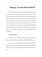
Figure 49.1 Pancreatogram revealing a diffusely dilated main
pancreatic duct with multiple visible side branches. These findings
are consistent with severe chronic pancreatitis.
Table 49.1 Sensitivity and specificity of endoscopic retrograde
cholangiopancreatography (ERCP) and endoscopic ultrasound (EUS)
for the diagnosis of chronic pancreatitis.
Imaging Sensitivity Specificity
technology (%) (%)
ERCP 70–90 80–100
EUS 79–87 72–91
Table 49.2 Cambridge classification of pancreatographic findings of chronic pancreatitis. (From ref. 10 with permission.)
Terminology Main pancreatic duct Duct side branches Additional features
Normal Normal None None
Equivocal Normal Ͻ3 None
Class I Normal Ն3 None
Class II Abnormal Ն3 None
Class III Abnormal Ն3 One or more of large cavity, filling
defects, severe dilation, or irregularity
9781405146647_4_049.qxd 1/30/08 11:46 AM Page 478
EUS and MRCP) to establish the diagnosis of chronic pancre-
atitis has greatly increased, thus relegating ERCP to a more
therapeutic role [24].
Endoscopic ultrasound
Although ERCP has high sensitivity for the diagnosis of
chronic pancreatitis, it is limited because it is only able to visu-
alize the pancreatic duct; pancreatic parenchymal changes
cannot be appreciated. As discussed previously, the diagnostic
sensitivity of ERCP for chronic pancreatitis is therefore great-
est when ductal changes consistent with severe advanced
chronic pancreatitis are present [7,11]. EUS was developed in
the 1980s as an imaging modality designed to perform high-
resolution imaging of the entire pancreas [25–27]. The use of
high-frequency transducers allows the user to detect subtle
parenchymal changes and minor ductal abnormalities in
patients with chronic pancreatitis [28,29]. In addition, the use
of EUS overcomes the major obstacles to pancreatic imaging
by transabdominal ultrasound, namely intestinal bowel gas
and fat [30].
EUS of the normal pancreas reveals a homogeneous echo-
texture that is more echogenic than the liver. The main pan-
creatic duct diameter is approximately 2.4 mm in the head,
1.8 mm in the body, and 1.2 mm in the tail. Side branches are
visible using EUS; however, they are narrow, with the greatest
diameter (0.8 mm) occurring in the head of the gland [30,31].
The diagnosis of chronic pancreatitis by EUS is based on the
presence of up to nine abnormalities of the pancreatic duct
and parenchyma [29] (Table 49.3). Ductal abnormalities
include increased wall echogenicity, irregular caliber or dila-
tion of the main pancreatic duct, dilation of side branches,
and the presence of calculi (Fig. 49.2). Parenchymal changes
include focal areas of reduced echogenicity, hyperechoic foci,
the presence of cysts, and lobular morphology (Fig. 49.3).
Studies vary with regard to the number of abnormalities
required to make the diagnosis of chronic pancreatitis by EUS,
ERCP, MRCP AND EUS IN CHRONIC PANCREATITIS
479
Table 49.3 Endoscopic ultrasound features of chronic pancreatitis. (From Ref. 32
with permission.)
Parenchymal
Focal areas of reduced echogenicity
Hyperechoic foci (Ͼ3 mm diameter)
Gland size, cysts
Accentuation of lobular pattern (hypoechoic areas surrounded by hyperechoic septae)
Ductal
Increased duct wall echogenicity
Irregular caliber of main pancreatic duct
Dilation of main pancreatic duct (Ͼ3 mm in head, Ͼ2mm in body, Ͼ1 mm in tail)
Dilation of side branches
Calculi
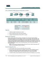
Figure 49.2 Endoscopic ultrasound (EUS) image revealing a dilated
pancreatic duct with increased echogenicity of the duct wall. These
ductal changes are commonly seen when EUS is performed in
patients with chronic pancreatitis.
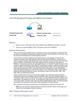
Figure 49.3 Endoscopic ultrasound (EUS) image revealing a
heterogeneous pancreas with hyperechoic foci. These findings are
consistent with the parenchymal changes often identified during EUS
performed in patients with chronic pancreatitis.
9781405146647_4_049.qxd 1/30/08 11:46 AM Page 479
although almost all require a minimum of three [7,29,32–36].
Minimal standard terminology for the description of
endosonographic changes consistent with chronic pancreatitis
has been developed [37] (Table 49.4).
The sensitivity and specificity of EUS for the diagnosis of
chronic pancreatitis (see Table 49.1) remains the subject of
much controversy. By definition, the sensitivity and specificity
of any diagnostic test are determined by comparison with the
“gold standard” test for the condition of interest. A reliable
gold standard for the diagnosis of chronic pancreatitis has not
been universally agreed [7]. The ideal gold standard for the
diagnosis of chronic pancreatitis would be pancreatic histol-
ogy, but this is clearly not feasible due to the high risk associ-
ated with pancreatic biopsy. In many studies, ERCP is chosen
as the diagnostic gold standard, although this technique is not
without its problems [7]. Chronic pancreatitis can exist in the
setting of a normal pancreatogram [4,17], a high degree of
interobserver variability exists in the interpretation of pancre-
atograms [4,38], and the ERCP changes of chronic pancreati-
tis are nonspecific [4,7,38,39].
Because of the lack of an approved gold standard diagnostic
test, EUS has been compared with several different modalities
in order to better understand its sensitivity and specificity for
the diagnosis of chronic pancreatitis. Multiple studies have
compared EUS with ERCP [7,28,29,32–36] for the diagnosis
of chronic pancreatitis. In three studies [29,32,35], both stan-
dard EUS criteria and the Cambridge classification for ERCP
were used and the results can therefore be compared with each
other directly [30]. If three endosonographic criteria are used
as a cutoff for the diagnosis of chronic pancreatitis, EUS and
ERCP agree in approximately 80% of cases [7,30]. Agreement
is highest in cases of severe advanced chronic pancreatitis.
However, in the majority of cases where the two tests disagree,
EUS demonstrated abnormalities when ERCP was normal.
The major question that has arisen is whether EUS is more
sensitive than ERCP or whether endosonographers are simply
overdiagnosing chronic pancreatitis [7,30]. The overall sensi-
tivity and specificity of EUS using ERCP as the gold standard
are 87% and 75%, respectively [29,32,35]. The sensitivity
and specificity of EUS compared with ERCP vary with respect
to the number of endosonographic criteria required to make
the diagnosis of chronic pancreatitis [30].
When pancreatic function testing is used as the comparison
gold standard for the diagnosis of chronic pancreatitis, EUS has
a sensitivity of 79% and specificity of 72% [29,32]. Agreement
was seen between the two tests in 75% of cases; however, simi-
lar to the case with ERCP, of the 25% of cases where there was
disagreement, 71% had abnormal EUS in the setting of normal
pancreatic function testing [30], again raising the issue as to
whether EUS is “overdiagnosing” chronic pancreatitis.
One small study compared pancreatic histopathology with
EUS for the diagnosis of chronic pancreatitis [40].
Histopathology was obtained by pancreatectomy or pancreatic
biopsy in 34 patients, all of whom had undergone prior EUS.
Using a threshold of three endosonographic criteria for the diag-
nosis of chronic pancreatitis, the sensitivity and specificity of
EUS were 87% and 64%, respectively. As the number of criteria
was increased, the sensitivity and specificity moved in opposite
directions. When six or more endosonographic criteria were
required to diagnose chronic pancreatitis, the sensitivity and
specificity were 43% and 91%, respectively. The results of this
study suggested that the use of four or more endosonographic
criteria (sensitivity 78%, specificity 73%) was ideal for the diag-
nosis of chronic pancreatitis [30]. While pancreatic biopsy to
obtain histopathology is highly invasive and associated with sig-
nificant operative risk, the use of EUS-guided fine-needle aspira-
tion (FNA) is less so. Although limited to cytology, the addition
of FNA has expanded the utility and diagnostic accuracy of EUS
for a variety of conditions. A single study [41] found that adding
FNA to EUS increased the negative predictive value of EUS to
CHAPTER 49
480
Table 49.4 Minimal standard terminology (MST) definitions for endoscopic ultrasound (EUS) findings in chronic
pancreatitis. (From ref. 37.)
EUS criteria for chronic pancreatitis MST definition
Hyperechoic foci Small distinct reflectors
Hyperechoic strand Small string-like hyperechoic structures
Lobular out gland margin No MST definition
Lobularity Containing lobules: rounded homogeneous areas separated by strands of another
echogenicity
Cyst Abnormal anechoic round or oval structure
Stone Hyperechoic lesion with acoustic shadowing within a duct or gallbladder
Calcification Hyperechoic lesion with acoustic shadow within a parenchymal organ or a mass
Ductal dilation No MST definition
Side-branch dilation No MST definition
Duct irregularity Coarse, uneven outline of the duct
Hyperechoic duct margins No MST definition
Atrophy No MST definition
Nonhomogeneous echo pattern No MST definition
9781405146647_4_049.qxd 1/30/08 11:46 AM Page 480
100% and the specificity to 64% when compared with ERCP as
the diagnostic gold standard. Although no standardized histo-
logic or cytologic criteria exist for the diagnosis of chronic pan-
creatitis by FNA, a scoring system was used which graded each
specimen with regard to the presence of an inflammatory cellular
infiltrate. The results of this study suggest that FNA is most help-
ful for excluding chronic pancreatitis when mild or patchy
parenchymal abnormalities with unclear significance are identi-
fied on EUS. FNA was generally well tolerated; mild acute pan-
creatitis occurred in 2 of 27 patients studied.
Given the ability to visualize both the pancreatic parenchyma
and duct, in addition to its excellent sensitivity and low associ-
ated procedural risk, the use of EUS for the diagnosis of chronic
pancreatitis has increased over the past decade. As such, the use
of minimal standard terminology to describe endosonographic
findings and the appropriate number of endosonographic
abnormalities required to make the diagnosis of chronic pancre-
atitis are of critical importance. The accuracy of any diagnostic
test is related to the reproducibility of its results [7]. When 11
experienced endosonographers who were blinded to the clinical
history independently evaluated previously taped examinations
for the presence of EUS criteria of chronic pancreatitis, diagnos-
tic agreement was reached at a rate comparable with other
endoscopic or radiographic tests [42]. Agreement was highest
for ductal dilatation and lobularity. As with any diagnostic test,
the clinical history is key to interpreting the results of EUS in the
diagnosis of chronic pancreatitis.
Magnetic resonance cholangio-
pancreatography
While ERCP has been associated with an incidence of acute
pancreatitis in up to 10% of individuals who undergo this
procedure [32], MRCP is able to provide high-quality imaging
of the pancreatic and biliary ducts in a noninvasive manner
[43]. Wallner et al. [44] first described MRCP in 1991. At that
time, the study was time-consuming with questionable image
quality. Over the past 15 years, however, the acquisition time
for single images has gone from 5 min to 2 s, allowing more
widespread use of this technology. In most centers, the imple-
mentation of high-quality MRCP into clinical practice has
replaced diagnostic ERCP [24].
Takehara et al. [45] first compared MRCP, specifically mag-
netic resonance pancreatography, with ERCP for the diagnosis
of chronic pancreatitis. High-quality images of the pancreatic
duct in the head, body, and tail of the gland were obtained in
70%, 64%, and 53%, respectively, of patients, all of whom
had been previously diagnosed with chronic pancreatitis based
on ERCP. Agreement between the two tests was observed in
83–92% of cases of ductal dilatation, 70–92% of cases of
ductal narrowing, and 92–100% of cases with ductal filling
defects. This study also found low interobserver variation for
most findings, although MRCP did tend to overestimate the
extent of pancreatic ductal stenosis [43,45]. Other studies
have yielded similar findings [46].
Secretin is a hormone secreted by the gastrointestinal tract
that leads to rapid secretion of a bicarbonate-rich fluid from
the exocrine pancreas [43,47]. As a result, the volume of fluid
in the pancreatic duct increases. The administration of intra-
venous secretin to improve imaging of the pancreatic duct was
first described in combination with transabdominal ultra-
sonography for the diagnosis of chronic pancreatitis [48,49].
Because of the tendency of MRCP to overestimate pancreatic
ductal stenosis, Takehara et al. [50] studied the use of secretin
stimulation during the acquisition of images in order to
improve signal intensity and imaging of the pancreatic duct in
patients suspected of having pancreatic disease. The investiga-
tors found that the use of secretin improved evaluation of the
main pancreatic duct and its side branches compared with
imaging not using secretin stimulation (Fig. 49.4). These
results have been replicated by other groups [47,51].
Since this initial study, several investigations have focussed
specifically on secretin-enhanced MRCP for the diagnosis of
chronic pancreatitis [52,53]. Manfredi et al. [52] studied this
modality in 31 patients with chronic pancreatitis. The use of
secretin increased the percentage of visible pancreatic duct
segments from 91 to 100% and side branches from 71 to
100%. Although the improved ductal visualization with
secretin was not statistically significant, the authors noted that
improved visualization of the ductal side branches may allow
earlier diagnosis of chronic pancreatitis, thus reducing the
ERCP, MRCP AND EUS IN CHRONIC PANCREATITIS
481
Figure 49.4 Secretin-stimulated magnetic resonance
cholangiopancreatography revealing a markedly dilated main
pancreatic duct with multiple visible side branches. These findings
are consistent with severe chronic pancreatitis.
9781405146647_4_049.qxd 1/30/08 11:46 AM Page 481
false-negative rate and improving the specificity of MRCP for
this diagnosis. Standardized criteria for the diagnosis of
chronic pancreatitis by MRCP have yet to be developed.
Aside from improving delineation of the pancreatic ductal
morphology, secretin-enhanced MRCP may have value in the
measurement of pancreatic exocrine function. Matos et al. [47]
performed MRCP in 10 volunteers and 13 patients with sus-
pected pancreatic disease. Pancreatograms were obtained prior
to and then at 30-s intervals following the administration of
secretin. The volume of filling within the duodenum was used
as a quantitative measure of pancreatic function. The results
were compared with ERCP and secretin stimulation testing.
The study found that the mean duodenal filling score was sig-
nificantly lower in patients with known reduced exocrine func-
tion compared with that in volunteers, thus providing the first
evidence that secretin-stimulated MRCP has the potential to
detect impaired pancreatic exocrine function. These results
have been confirmed by other investigators [52,54–56].
Direct comparisons of MRCP with EUS for the diagnosis of
chronic pancreatitis have yet to be performed. In comparison
with EUS and ERCP, MRCP is certainly the least invasive.
Secretin-stimulated MRCP has the additional advantage of
evaluating pancreatic function, an attribute not shared by EUS
or ERCP. Although not yet studied, this feature may enhance
the specificity of MRCP for the diagnosis of chronic pancreati-
tis. ERCP does not provide detailed images of the pancreatic
parenchyma; this is a potential disadvantage compared with
EUS, which has the ability to detect both ductal and parenchy-
mal abnormalities. Magnetic resonance imaging (MRI) of the
pancreas is possible at the same time as MRCP, although this
adds cost and time to the examination. The MRI findings asso-
ciated with chronic pancreatitis [57] are beyond the scope of
this chapter. Due to its minimally invasive nature and high cor-
relation with ERCP findings, MRCP is often ordered as the
first test for the diagnosis of chronic pancreatitis in cases where
advanced imaging modalities are required [52].
Diagnostic approach
The use of advanced imaging modalities such as ERCP, MRCP,
and EUS for the diagnosis of chronic pancreatitis is not
required in the majority of cases. Indeed, in many cases of
chronic alcoholic pancreatitis, the clinical history alone can be
sufficient to make the diagnosis [11]. However, in some cases,
especially early chronic pancreatitis, advanced imaging is
required. Of these three modalities, EUS likely has the greatest
ability to diagnose early disease. Although it is an invasive
diagnostic test, the complication rate associated with EUS is
less than that of ERCP and it has the ability to detect both mor-
phologic and ductal abnormalities. While MRCP is clearly the
least invasive, it is an expensive test with results that may be
center-dependent. In addition, the ability to visualize the pan-
creatic duct alone may decrease its diagnostic sensitivity for
early disease. Finally, it is the least studied of the three modali-
ties discussed in this chapter. While it is often considered the
gold standard, the high rate of procedure-related complications
associated with ERCP has limited its use in chronic pancreatitis
to the performance of therapeutic interventions.
References
1. Steer ML, Waxman I, Freedman S. Chronic pancreatitis. N Engl J
Med. 1995;332:1482–90.
2. Singer MV, Gyr K, Sarles H. Revised classification of pancreatitis.
Report of the Second International Symposium on the
Classification of Pancreatitis in Marseille, France, March 28–30,
1984. Gastroenterology 1985;89:683–5.
3. Sarner M, Cotton PB. Classification of pancreatitis. Gut
1984;25:756–9.
4. Forsmark CE, Toskes PP. What does an abnormal pancreatogram
mean? Gastrointest Endosc Clin North Am 1995;5:105–23.
5. Niederau C, Grendell JH. Diagnosis of chronic pancreatitis.
Gastroenterology 1985;88:1973–95.
6. Pitchumoni CS, Glasser M, Saran RM, Panchacharam P, Thelmo
W. Pancreatic fibrosis in chronic alcoholics and nonalcoholics
without clinical pancreatitis. Am J Gastroenterol 1984;79:382–8.
7. Forsmark CE. The diagnosis of chronic pancreatitis. Gastrointest
Endosc 2000;52:293–8.
8. Axon AT. Endoscopic retrograde cholangiopancreatography in
chronic pancreatitis. Cambridge classification. Radiol Clin North
Am 1989;27:39–50.
9. Novis BH, Narunsky L, Bank S. Endoscopic retrograde cholan-
giopancreatography in the evaluation of pancreatic disease. S Afr
Med J 1976;50:1501–5.
10. Bolan PJ, Fink AS. Endoscopic retrograde cholangiopancreatog-
raphy in chronic pancreatitis. World J Surg 2003;27:1183–91.
11. Forsmark CE. Chronic pancreatitis. In: Feldman M, Friedman L,
Sleisenger M, eds. Gastrointestinal and Liver Disease. Philadelphia:
WB Saunders, 2002: 953.
12. Girdwood AH, Hatfield AR, Bornman PC, Denyer ME, Kottler
RE, Marks IN. Structure and function in noncalcific pancreatitis.
Dig Dis Sci 1984;29:721–6.
13. Malfertheiner P, Buchler M, Stanescu A, Ditschuneit H. Exocrine
pancreatic function in correlation to ductal and parenchymal mor-
phology in chronic pancreatitis. Hepatogastroenterology 1986;
33:110–14.
14. Lankisch PG, Seidensticker F, Otto J et al. Secretin–pancreozymin
test (SPT) and endoscopic retrograde cholangiopancreatography
(ERCP): both are necessary for diagnosing or excluding chronic
pancreatitis. Pancreas 1996;12:149–52.
15. Braganza JM, Hunt LP, Warwick F. Relationship between pancre-
atic exocrine function and ductal morphology in chronic pancre-
atitis. Gastroenterology 1982;82:1341–7.
16. Bozkurt T, Braun U, Leferink S, Gilly G, Lux G. Comparison of
pancreatic morphology and exocrine functional impairment in
patients with chronic pancreatitis. Gut 1994;35:1132–6.
17. Axon AT, Classen M, Cotton PB, Cremer M, Freeny PC, Lees
WR. Pancreatography in chronic pancreatitis: international defi-
nitions. Gut 1984;25:1107–12.
18. Lehman GA. Role of ERCP and other endoscopic modalities in
chronic pancreatitis. Gastrointest Endosc 2002;56(6
suppl):S237–S240.
19. Johanson JF, Cooper G, Eisen GM et al. Quality assessment of
ERCP. Endoscopic retrograde cholangiopacreatography.
Gastrointest Endosc 2002;56:165–9.
CHAPTER 49
482
9781405146647_4_049.qxd 1/30/08 11:46 AM Page 482
20. Anand BS, Vij JC, Mac HS, Chowdhury V, Kumar A. Effect of
aging on the pancreatic ducts: a study based on endoscopic retro-
grade pancreatography. Gastrointest Endosc 1989;35:210–13.
21. Sherman S, Hawes RH, Savides TJ et al. Stent-induced pancreatic
ductal and parenchymal changes: correlation of endoscopic ultra-
sound with ERCP. Gastrointest Endosc 1996;44:276–82.
22. Freeman ML. Adverse outcomes of endoscopic retrograde
cholangiopancreatography. Rev Gastroenterol Disord 2002;
2:147–68.
23. Masci E, Toti G, Mariani A et al. Complications of diagnostic
and therapeutic ERCP: a prospective multicenter study. Am J
Gastroenterol 2001;96:417–23.
24. Anon. NIH state-of-the-science statement on endoscopic retro-
grade cholangiopancreatography (ERCP) for diagnosis and ther-
apy. NIH Consens State Sci Statements 2002;19:1–26.
25. Dimagno EP, Regan PT, Clain JE, James EM, Buxton JL. Human
endoscopic ultrasonography. Gastroenterology 1982;83:824–9.
26. DiMagno EP, Buxton JL, Regan PT et al. Ultrasonic endoscope.
Lancet 1980;i:629–31.
27. Hisanaga K, Hisanaga A, Nagata K, Ichie Y. High speed rotating
scanner for transgastric sonography. Am J Roentgenol 1980;
135:627–9.
28. Kahl S, Glasbrenner B, Leodolter A, Pross M, Schulz HU,
Malfertheiner P. EUS in the diagnosis of early chronic pancreati-
tis: a prospective follow-up study. Gastrointest Endosc 2002;
55:507–11.
29. Wiersema MJ, Hawes RH, Lehman GA, Kochman ML, Sherman
S, Kopecky KK. Prospective evaluation of endoscopic ultrasonog-
raphy and endoscopic retrograde cholangiopancreatography in
patients with chronic abdominal pain of suspected pancreatic ori-
gin. Endoscopy 1993;25:555–64.
30. Raimondo M, Wallace MB. Diagnosis of early chronic pancreati-
tis by endoscopic ultrasound. Are we there yet? JOP 2004;5:1–7.
31. Wiersema MJ, Wiersema LM. Endosonography of the pancreas:
normal variation versus changes of early chronic pancreatitis.
Gastrointest Endosc Clin North Am 1995;5:487–96.
32. Catalano MF, Lahoti S, Geenen JE, Hogan WJ. Prospective eval-
uation of endoscopic ultrasonography, endoscopic retrograde
pancreatography, and secretin test in the diagnosis of chronic
pancreatitis. Gastrointest Endosc 1998;48:11–17.
33. Nattermann C, Goldschmidt AJ, Dancygier H. Endosonography
in chronic pancreatitis: a comparison between endoscopic retro-
grade pancreatography and endoscopic ultrasonography.
Endoscopy 1993;25:565–70.
34. Buscail L, Escourrou J, Moreau J et al. Endoscopic ultrasonogra-
phy in chronic pancreatitis: a comparative prospective study with
conventional ultrasonography, computed tomography, and
ERCP. Pancreas 1995;10:251–7.
35. Sahai AV, Zimmerman M, Aabakken L et al. Prospective assess-
ment of the ability of endoscopic ultrasound to diagnose,
exclude, or establish the severity of chronic pancreatitis found by
endoscopic retrograde cholangiopancreatography. Gastrointest
Endosc 1998;48:18–25.
36. Hastier P, Buckley MJ, Francois E et al. A prospective study of
pancreatic disease in patients with alcoholic cirrhosis: compara-
tive diagnostic value of ERCP and EUS and long-term signifi-
cance of isolated parenchymal abnormalities. Gastrointest
Endosc 1999;49:705–9.
37. Aabakken L. Standardized terminology in endoscopic ultra-
sound. Eur J Ultrasound 1999;10:179–83.
38. Schmitz-Moormann P, Himmelmann GW, Brandes JW et al.
Comparative radiological and morphological study of human
pancreas. Pancreatitis like changes in postmortem ductograms
and their morphological pattern. Possible implication for ERCP.
Gut 1985;26:406–14.
39. Hayakawa K, Tanaka F, Torizuka T et al. Bronchial artery
embolization for hemoptysis: immediate and long-term results.
Cardiovasc Intervent Radiol 1992;15:154–8; discussion 8–9.
40. Zimmerman M, Mishra G, Lewin D et al. Comparison of EUS
findings with histopathology in chronic pancreatitis [Abstract].
Gastrointest Endosc 1997;45:AB185.
41. Hollerbach S, Klamann A, Topalidis T, Schmiegel WH.
Endoscopic ultrasonography (EUS) and fine-needle aspiration
(FNA) cytology for diagnosis of chronic pancreatitis. Endoscopy
2001;33:824–31.
42. Wallace MB, Hawes RH, Durkalski V et al. The reliability of EUS
for the diagnosis of chronic pancreatitis: interobserver agreement
among experienced endosonographers. Gastrointest Endosc
2001;53:294–9.
43. Merkle EM, Baillie J. Exocrine pancreatic function: evaluation
with MR imaging before and after secretin stimulation. Am J
Gastroenterol 2006;101:137–8.
44. Wallner BK, Schumacher KA, Weidenmaier W, Friedrich JM.
Dilated biliary tract: evaluation with MR cholangiography with a
T2-weighted contrast-enhanced fast sequence. Radiology
1991;181:805–8.
45. Takehara Y, Ichijo K, Tooyama N et al. Breath-hold MR cholan-
giopancreatography with a long-echo-train fast spin-echo
sequence and a surface coil in chronic pancreatitis. Radiology
1994;192:73–8.
46. Barish MA, Yucel EK, Soto JA, Chuttani R, Ferrucci JT. MR
cholangiopancreatography: efficacy of three-dimensional turbo
spin-echo technique. Am J Roentgenol 1995;165:295–300.
47. Matos C, Metens T, Deviere J et al. Pancreatic duct: morphologic
and functional evaluation with dynamic MR pancreatography
after secretin stimulation. Radiology 1997;203:435–41.
48. Bolondi L, Gaiani S, Gullo L, Labo G. Secretin administration
induces a dilatation of main pancreatic duct. Dig Dis Sci
1984;29:802–8.
49. Glaser J, Hogemann B, Krummenerl T et al. Sonographic imaging
of the pancreatic duct. New diagnostic possibilities using secretin
stimulation. Dig Dis Sci 1987;32:1075–81.
50. Takehara Y, Ichijo K, Tooyama N et al. [Enhanced delineation of
the pancreatic duct in MR cholangiopancreatography (MRCP)
with a combined use of secretin.] Nippon Igaku Hoshasen
Gakkai Zasshi 1995;55:255–6.
51. Nicaise N, Pellet O, Metens T et al. Magnetic resonance cholan-
giopancreatography: interest of IV secretin administration in the
evaluation of pancreatic ducts. Eur Radiol 1998;8:16–22.
52. Manfredi R, Costamagna G, Brizi MG et al. Severe chronic pan-
creatitis versus suspected pancreatic disease: dynamic MR
cholangiopancreatography after secretin stimulation. Radiology
2000;214:849–55.
53. Manfredi R, Costamagna G, Vecchioli A, Colagrande C, Spina S,
Marano P. [Dynamic pancreatography with magnetic resonance
after functional stimulus with secretin in chronic pancreatitis.]
Radiol Med (Torino) 1998;96:226–31.
54. Cappeliez O, Delhaye M, Deviere J et al. Chronic pancreatitis:
evaluation of pancreatic exocrine function with MR pancreatog-
raphy after secretin stimulation. Radiology 2000;215:358–64.
55. Heverhagen JT, Battmann A, Kirsch M et al. Magnetic resonance
hydrometry: non-invasive quantification of the exocrine pancre-
atic function. ROFO 2002;174:291–6.
56. Punwani S, Gillams AR, Lees WR. Non-invasive quantification of
pancreatic exocrine function using secretin-stimulated MRCP.
Eur Radiol 2003;13:273–6.
57. Miller FH, Keppke AL, Wadhwa A, Ly JN, Dalal K, Kamler VA.
MRI of pancreatitis and its complications: part 2, chronic pancre-
atitis. Am J Roentgenol 2004;183:1645–52.
ERCP, MRCP AND EUS IN CHRONIC PANCREATITIS
483
9781405146647_4_049.qxd 1/30/08 11:46 AM Page 483
484
Introduction
In the minority of patients (i.e., 5.8–20%), chronic pancreatitis
takes a primarily painless course [1–7]. Exocrine and endocrine
insufficiency are the dominating symptoms. For the majority of
patients, however, pain is the decisive symptom, causing much
discomfort in their daily lives. Some studies have correlated the
course of pain in chronic pancreatitis with the duration of the
disease, progressing exocrine and endocrine pancreatic insuffi-
ciency, and morphologic changes such as pancreatic calcifica-
tion and duct abnormalities. Furthermore, the course of pain
has been studied following alcohol abstinence and after surgery
in some groups.
Pain decrease and duration of chronic
pancreatitis
Whether progressive parenchymal destruction of the pancreas
leads to pain decrease has been repeatedly debated [8,9].
Ammann’s group has claimed that pain decreases with increas-
ing duration of the disease [3,10,11]. In one long-term study,
85% of 145 patients with chronic pancreatitis felt no more pain
after 4.5 years (median) from onset of the disease [3]. In another
series, in which the interval between the onset of alcohol-
induced chronic pancreatitis and pain relief was compared in
surgically and nonsurgically treated patient groups, the curves
were virtually parallel: pain relief was obtained in about 50%
within 6 years and in more than 80% within 10 years from the
onset of illness [12].
The reports from Zürich are at variance with the studies from
Japan and Germany. Miyake et al. [6] found that only 48.2% of
patients with chronic pancreatitis became free of pain within 5
years, but 66–73% became free of pain after more than 5 years.
This showed that every third or fourth patient still suffered
from relapsing pain attacks even after a long observation
period. Our group reported that the incidence of relapsing pain
attacks decreased during the observation period, but more than
half of the patients (53%) still suffered from relapsing pain
attacks after more than 10 years of observation [7].
At present, the course of pain in alcoholic and idiopathic
chronic pancreatitis remains unclarified. Layer et al. [13]
investigated a group of patients with idiopathic chronic
pancreatitis who had never consumed alcoholic beverages dur-
ing their lifetime. They found that patients with early-onset
pancreatitis (under 35 years of age) have a long course of severe
pain from the start of their illness, whereas patients with late-
onset pancreatitis (over 35 years) have a mild and often painless
course. Both forms differ from alcoholic pancreatitis in having
an equal gender distribution and a much slower rate of calcifi-
cation. In contrast, our group has found that the course of pain
is the same in alcohol- and nonalcohol-induced chronic pancre-
atitis [14]. Even when we divided the nonalcoholic group into
teetotallers and patients with little alcohol consumption, and
separately compared their course of pain with alcoholics, there
were no differences concerning pain relief among the three
groups [15]. Further studies are required.
Pain decrease and progressing exocrine
and endocrine pancreatic insufficiency
The Swiss group have repeatedly observed pain decrease when
exocrine and endocrine pancreatic function declines [8–11].
Similarly, Girdwood et al. [16] have reported from South
Africa that pain decreases when exocrine pancreatic function
deteriorates.
Conversely, groups from Denmark and Germany have
reported the opposite. Thorsgaard Pedersen et al. [17] from
Copenhagen found no correlation between pain and exocrine
pancreatic function. Our group in Göttingen [7] have used the
secretin–pancreozymin test and fecal fat analysis to evaluate
exocrine pancreatic insufficiency, whereas the Swiss group had
used only indirect pancreatic function tests, i.e., chymotrypsin
measurements, to evaluate exocrine pancreatic insufficiency
[3]. We used a clear-cut grading of the severity of exocrine pan-
creatic insufficiency: mild impairment was defined as reduced
enzyme output, moderate impairment as a decreased bicarbon-
ate concentration along with reduced enzyme output but nor-
mal fecal fat excretion, and severe impairment was equated
with an abnormal secretin–pancreozymin test plus steatorrhea.
At the end of the observation period, 141 (45%) of 311 patients
with painful chronic pancreatitis had severe exocrine pancreatic
insufficiency. The majority of them (81/144, 57%) still suffered
from pain attacks.
Additionally, we studied the course of pain in correlation with
endocrine pancreatic insufficiency. Endocrine pancreatic insuffi-
ciency was classified as absent, moderate (diabetes mellitus
Natural course of chronic pancreatitis
Paul Georg Lankisch
50
9781405146647_4_050.qxd 1/30/08 11:46 AM Page 484
The Pancreas: An Integrated Textbook of Basic Science, Medicine, and Surgery, Second Edition
Edited by H. G. Beger, A. L. Warshaw, M. W. Büchler, R. A. Kozarek, M. M. Lerch, J. P. Neoptolemos,
K. Shiratori, D. C. Whitcomb, and B. M. Rau © 2008 Blackwell Publishing Limited, ISBN: 978-1-405-14664-7
treated only by diet with or without oral medication), and severe
(requiring insulin). At the end of the observation period, 117
(38%) patients were classified as having severe endocrine pan-
creatic insufficiency. The majority of them (69/117, 59%) still
suffered from pain attacks [7,18].
Thus, according to our results, the progression of exocrine
and endocrine pancreatic insufficiency has limited, if any,
influence on the course of pain in chronic pancreatitis.
Pain decrease and development of
morphologic changes in the pancreas
(pancreatic calcifications and/or duct
abnormalities)
The Swiss group [3,10] showed an increased incidence of pan-
creatic calcifications, which in turn was associated with pain
decrease. However, in a later survey the same group reported
regression of pancreatic calcifications in a long-term study of
patients with chronic pancreatitis [19]. Thus, the prognostic
role of pancreatic calcifications in determining the course of
pain is unclear.
Furthermore, the Swiss results are at variance with two
other studies. Malfertheiner et al. [20] found that 89% of
patients had pain despite pancreatic calcifications observed on
computed tomography, of whom 39% had very intense pain.
In our study, freedom from pain was significantly higher in the
calcification group compared with the noncalcification group.
However, the majority of patients with pancreatic calcifica-
tions (56%) still had relapsing pain attacks [7].
The correlation between pain and pancreatic duct changes or
pressure in the duct system is also not clear. Ebbehøj et al.
[21,22] measured pancreatic tissue fluid pressure percuta-
neously or intraoperatively and found a significant correlation
with pain in patients with chronic pancreatitis but not with the
results of endoscopic retrograde cholangiopancreatography
(ERCP), i.e., regional pressure tended to be highest in the region
of the pancreas with the largest but not the smallest duct diam-
eter. Jensen et al. [23] found no correlation between pancreatic
duct changes and pain. Warshaw et al. [24] found that 2 of 10
patients, 1 year after lateral pancreaticojejunostomy, had no
pain relief despite a patent anastomosis detected by ERCP.
Two investigations have confirmed the nonparallelism between
pancreatic duct changes and pain relief. Malfertheiner et al. [20]
found severe pain in only 62% of patients who had advanced
pancreatic duct changes demonstrated by ERCP. We found no
significant correlation between pancreatic duct abnormalities
detected by ERCP and pain in 88 patients with chronic pancre-
atitis [7]. Severe pancreatic duct abnormalities, as defined by the
Cambridge classification [25], were present in 42 patients, but
only 16 (31%) of these became free of pain. Despite a normal
pancreatic duct in 14 patients, 10 (71%) suffered from persisting
pain [7].
Thus, morphologic changes such as pancreatic calcifications
or pancreatic duct abnormalities are not necessarily helpful in
determining the prognosis of chronic pancreatitis or predict-
ing the course of pain. Recently it has been shown that smok-
ing has an effect on the natural course of the disease since it
increases the risk of pancreatic calcification in late-onset but
not early-onset idiopathic chronic pancreatitis [26].
Pain decrease and alcohol abuse
Since alcoholism is the leading etiologic factor in chronic pan-
creatitis, several studies have investigated whether alcohol
abstinence influences pain or progression of the disease. Sarles
and Sahel [27] reported that 50% of their patients with
chronic pancreatitis experienced pain relief when alcohol
abuse was discontinued, whereas Trapnell [28] reported a fig-
ure of 75%.
Two other investigations have confirmed that abstinence can
be helpful. Miyake et al. [6] demonstrated pain relief in 60% of
their patients who discontinued or reduced alcohol intake,
whereas spontaneous pain relief was seen in only 26% of the
group who continued drinking. In another study, 66 (31%) of
214 patients with alcoholic chronic pancreatitis were motivated
to stop drinking [7]. Pain relief was obtained in only 52% of
these patients, whereas spontaneous relief in alcoholics was
seen in 37%. Thus, alcohol abstinence in every second patient
with chronic pancreatitis will probably lead to some improve-
ment of pain, but why exactly abstinence helps in some cases
but not others remains to be investigated.
Pain decrease and interventional
procedures
Interventional procedures for pain treatment in chronic pancre-
atitis include fragmentation of stones by extracorporeal shock-
wave lithotripsy (ESWL), endoscopic stone extraction, and
bridging of pancreatic strictures by stent applications. Reports of
the effect of these procedures on pain are controversial and con-
trolled studies are lacking. A large Japanese study of 555 patients
who underwent ESWL for pancreatic stones reported a success
rate of 92.4% (fragmentation of stones) and a complete stone
clearance rate after ESWL alone or in combination with inter-
ventional endoscopy of 72.6%. Symptom relief was achieved in
91.1% of the patients. Complications developed in 6.3% of the
patients, including acute pancreatitis in 5.4%. A total of 504
patients were followed up for a mean of 44.3 months, during
which 122 (22%) suffered stone recurrence (mean time to recur-
rence, 25.1 months); 22 (4.1%) required surgery [29]. In another
series from Japan, a total of 117 patients with pancreatic stones
underwent ESWL and endoscopic treatment. Immediate pain
relief was achieved in 97% and complete removal of stones in
56%. During long-term follow-up over 3 years, 70% of the
patients continued to be asymptomatic [30]. These results are at
variance with a smaller German study in 80 patients with chronic
pancreatitis, in whom ESWL was always followed by a further
NATURAL COURSE OF CHRONIC PANCREATITIS
485
9781405146647_4_050.qxd 1/30/08 11:46 AM Page 485
CHAPTER 50
486
endoscopic procedure. Treatment success was defined as com-
plete clearance of the main pancreatic duct or partial clearance
that allowed implantation of a pancreatic stent. Successful treat-
ment was more frequent in patients with solitary stones. The
mean duration of follow-up was 40 (range 24–92) months. Pain
relief and necessity for further analgesia was independent of
ESWL results [31] (Table 50.1). Thus, in this study pancreatic
drainage by ESWL and endoscopy had almost no effect on pain
in chronic pancreatitis in the long term [32].
The effect of pancreatic stents on pain in chronic pancreatitis
is even more controversial. Patients undergoing pancreatic duct
stent placement for disrupted ducts, isolated strictures, pancreas
divisum, and hypertensive pancreatic sphincters showed subse-
quent ductal changes consistent with chronic pancreatitis in
36%, even though 72% of these patients had a normal initial
pancreatogram [33]. Furthermore, patients with preoperative
endoscopic pancreatic stenting had frequent postoperative com-
plications, mostly septic, and a prolonged hospital stay [34].
A surgical review of the pitfalls and liminations of stenting in
chronic pancreatitis reported that the indications for surgery
in patients with a pancreatic stent were severe abdominal pain
in 100%, relapsing pain attacks in 77%, and necrotizing pan-
creatitis in 14%. Before being selected for surgery, 4.5 ERCPs
and 3.7 stent exchanges were performed per patient. Thus, from
the surgical point of view, endoscopic pancreatic duct stenting
in chronic pancreatitis seems not to be indicated because of a
low success rate and a substantial risk of complications [35].
The latter results are in sharp contrast to a long-term out-
come study of pancreatic stenting in severe chronic pancreati-
tis in 100 patients from Belgium. The majority (70%) of
patients who responded to pancreatic stenting remained pain-
free after definitive stent removal. However, a significantly
higher restenting rate was observed in patients with chronic
pancreatitis and pancreas divisum [36]. Obviously, the results
are also different in special subgroups. Endoscopic stenting of
biliary strictures in chronic pancreatitis provided an excellent
short-term but only moderate long-term result in another
study from Germany. Patients without calcifications of the
pancreatic head benefit from biliary stenting. However,
patients with calcifications had a 17-fold increased risk of fail-
ure during the course of a 12-month follow-up [37].
Of special interest is a recent prospective randomized trial
that compared endoscopic with surgical treatment of chronic
pancreatitis. Endoscopic treatment included pancreatic sphinc-
terotomy in all and additional stenting of the pancreatic duct in
33 (52%) patients. Mean duration of stent treatment was 16
(range 12–27) months, and stents were exchanged six times
(range 4–9). Surgical treatment included pancreatic resection in
61 (80%) and drainage procedures in 15 (20%) patients.
Although the short-term effects were similar, the results after
5 years of follow-up showed a comparatively low rate of patients
with complete absence of abdominal pain. However, the results
for surgery were significantly better than for endotherapy
(Table 50.2) [38]. The study has been criticized for the random-
ization, which was agreed to by only 51.4% of the patients.
For the time being, reports of treatment of chronic pancre-
atitis using ERCP by removal or destruction of stones, place-
ment of stents, and dilation of strictures suggest that both
immediate and long-term pain relief are possible. No con-
trolled studies support the generalizability of this finding or
the merit of this approach compared with other management
strategies. Studies of this area would be of value [39].
Pain decrease and surgery
During the course of the disease, every second to fourth patient
needs surgical treatment because of pain and/or organ compli-
cations, such as pancreatic pseudocysts [3,7]. The choice of
surgical procedure depends on the special circumstances of
each patient. However, it is unclear to what extent surgical
treatment influences the course of pain since the different stud-
ies cannot be compared for the following reasons.
• The definition of freedom from pain was often vague, and
pain symptoms were usually not measured. Measurement on
an analog scale is recommended [18].
• Not all patients received the same surgical treatment for the
same indication. Several authors do not recommend perform-
ing an indicated resection in alcoholics because of the difficult
postoperative treatment of diabetes mellitus in these patients
[40,41].
• Although continued alcohol abuse distinctly worsens the
effect of surgical treatment [42–44], it is still difficult to deter-
mine whether postoperative deterioration results from chronic
pancreatitis or continued alcohol abuse, or from the surgical
treatment.
Table 50.1 Long-term effect on pain in 80 patients with chronic
pancreatitis treated with extracorporeal shock wave lithotripsy.
(From ref. 31 with permission.)
Successful Unsuccessful
treatment treatment
(N ϭ 43) (N ϭ 37) P value
Considerable or
complete pain relief 34 (79%) 27 (73%) 0.75
No further analgesia
necessary 27 (63%) 16 (43%) 0.23
Table 50.2 Five years follow-up of abdominal pain in a prospective
randomized trial comparing endoscopic with surgical treatment for
chronic pancreatitis. (From ref. 38 with permission.)
Abdominal Endotherapy Surgery
pain (N ϭ 64) (N ϭ 76) P value
Complete absence 14.3% 36.9% 0.002
Partial relief 50.8% 49.3% NS
No success 34.9% 13.8% NS
NS, not significant.
9781405146647_4_050.qxd 1/30/08 11:46 AM Page 486
NATURAL COURSE OF CHRONIC PANCREATITIS
487
Evaluation of pain differs very much during the course of the
observation period. Independent of the surgical procedure,
postoperative results show that freedom from pain will be
obtained in up to 90% of patients over several years of follow-
up (Table 50.3) [7,45–78]. However, persistence of freedom
from pain has been reported differently. Taylor et al. [79]
(Table 50.4) clearly showed that pain increases during the
course of a longer follow-up. In contrast, Martin et al. [72]
showed that freedom from pain may persist over 5 years of fol-
low-up after pylorus-preserving pancreaticoduodenectomy for
chronic pancreatitis (Fig. 50.1). Whether this difference is due
to the different mode of operation remains to be clarified.
Table 50.3 Freedom from pain after different surgical procedures on the pancreas for chronic pancreatitis.*
Median Pain relief
Reference Surgical procedure observation time N (%)
Way et al. [45] Drainage/resection ~5 years 37 64
Lankisch et al. [46] Drainage/resection 2.5 years 40 60
Mangold et al. [47] Partial duodenopancreatectomy 1 year 8 months 44 73
Total duodenopancreatectomy 2 years 10 months 18 91
Partial left-sided resection 3 years 5 months 37 60
Subtotal left-sided resection 2 years 10 months 17 83
Proctor et al. [48] Pancreaticojejunostomy 11 months 22 50
Rosenberger et al. [49] Resection 6 years 67 69
Nonresective procedures 6 years 40 50
Lankisch et al. [50] Pancreaticojejunostomy 3 years 1 month 17 76
Resection 3 years 1 month 22 64
Prinz and Greenlee [51] Pancreaticojejunostomy 6 years 1 month to 91 35
7 years 11 months
Sato et al. [52] Pancreaticojejunostomy 6.5 years 38 68
Left-sided resection 6.5 years 14 79
Whipple’s operation 6.5 years 9 67
Gall et al. [53] Whipple’s operation, pancreatic duct occlusion Ͼ 1 year 67 93
Morrow et al. [54] Pancreatic duct drainage 4–13 years 46 46
40–80% left-sided resection 4–13 years 21 33
80–95% left-sided resection 4–13 years 8 100
Drainage 6 years 46 80
Subtotal pancreatectomy 7 years 21 24
Sato et al. [55] Left-sided resection Ͼ 6 months 21 91
Whipple’s operation Ͼ 6 months 11 55
Pancreaticojejunostomy Ͼ 6 months 43 91
Bradley [56] Lateral pancreaticojejunostomy 5 years 9 months 46 28
Caudal pancreaticojejunostomy 5 years 9 months 18 17
Cooper et al. [57] Total pancreatectomy 1.5 years 83 72
Frick et al. [58,59] Left-sided resection 6.5 years 74 50
Partial duodenopancreatectomy 6.5 years 62 45
Total duodenopancreatectomy 6.5 years 22 55
Drainage 4 years 7 months 156 48
Lambert et al. [60] Duodenum-preserving total pancreatectomy 9 years 5 months 14 64
Rossi et al. [61] Whipple’s operation 6 months 61 72
2 years 44 61
5 years 33 61
10 years 18 61
15 years 6 83
Mannell et al. [62] Drainage/resection 8.5 years 100 77
Stone et al. [63] Whipple’s operation 6 years 2 months 15 53
Total duodenopancreatectomy 9 years 1 month 15 27
Beger et al. [64] Duodenum-preserving pancreatic head resection 3 years 8 months 128 77
Peiper and Köhler [65] Resection 10 51 79
Drainage 10 24 65
Beger and Büchler [66] Duodenum-preserving pancreatic head resection 3.5 years 141 77
Lankisch et al. [7] Drainage/resection 6 years 70 57
Adams et al. [67] Lateral pancreaticojejunostomy 6 years 4 months 62 42
Frey and Amikura [68] Local pancreatic head resection with 6 months 50 34
longitudinal pancreaticojejunostomy
(Continued)
9781405146647_4_050.qxd 1/30/08 11:46 AM Page 487
CHAPTER 50
488
In a study of 207 patients with alcoholic chronic pancreatitis
(91 without and 116 with surgical treatment for pain relief),
Ammann et al. [12] discussed the pain pattern of chronic
pancreatitis and its surgical implications. In this study, chronic
pain was typically associated with local complications (mainly
pseudocysts), which were positively relieved by a single drainage
procedure in approximately two-thirds of patients. Additional
surgery was required for late pain recurrence in 39 patients, pri-
marily symptomatic cholestasis. All patients achieved complete
pain relief in advanced chronic pancreatitis. The authors con-
clude that, in their experience, relief of chronic pain regularly fol-
lows selective surgery tailored to the presumptive pain cause or
occurs spontaneously in uncomplicated advanced chronic pan-
creatitis.
Course of exocrine pancreatic
insufficiency
Exocrine pancreatic insufficiency does not play a major prognos-
tic role. Occasionally, massive steatorrhea leading to cachexia
and susceptibility to infection has prognostic significance.
Median Pain relief
Reference Surgical procedure observation time N (%)
Hakaim et al. [103] Different operations: 5 years 2 months 50 30
pancreatic duct drainage (56%)
left-sided resection (20%)
cyst drainage (24%)
Büchler et al. [69] Duodenum-preserving pancreatic head resection 6 months 15 40
Pylorus-preserving Whipple’s operation 6 months 16 75
Fleming and Williamson [70] Total pancreatectomy 3.5 years 40 79
Izbicki et al. [71] Duodenum-preserving pancreatic head resection:
Beger’s procedure 1.5 years 20 95
Frey’s procedure 1.5 years 22 94
Martin et al. [72] Pylorus-preserving pancreaticoduodenectomy 5 years 3 months 45 92
Stapleton and Williamson [73] Proximal pancreaticoduodenectomy: 4.5 years 52 80
pylorus-preserving (N ϭ 45)
Whipple’s operation (N ϭ 7)
Amikura et al. [104] Pancreaticojejunostomy Ն 6 months 69 75
Pancreaticojejunostomy plus pancreatic head Ն 6 months 11 90
resection
Left-sided resection Ն 6 months 37 80
Whipple’s operation Ն 6 months 13 65
Rumstadt et al. [74] Whipple’s operation 8 years 4 months
†
134 66
Traverso and Kozarek [75] Whipple’s operation 3.5 years 47 76
Total pancreatectomy 3.5 years 10 76
Beger et al. [105] Duodenum-preserving pancreatic head resection 5 years 8 months
†
303 88
Berney et al. [76] Different procedures of pancreas resection 6 years 4 months 68 62
Jimenez et al. [106] Whipple’s operation 3 years 5 months 33 53
Pylorus-preserving pancreatic head resection 3 years 5 months 39 40
Sakorafas et al. [77] Whipple’s operation 6 years 7 months 66 67
White et al. [78] Total pancreatectomy 6 months 24 82
Nealon and Matin [107] Pancreaticojejunostomy 6 years 9 months 124 86
Left-sided resection 6 years 9 months 29 67
Pancreatic head resection (duodenum-preserving 6 years 9 months 46 91
or pylorus-preserving)
Sakorafas et al. [108] Left-sided resection 6 years 8 months 31 49
Hutchins et al. [109] Left-sided resection 2 years 10 months 84 48
* Only reports of “total freedom from pain” were included. Further stages of postoperative improvement (e.g., partial freedom from pain) were not
considered. Closure of literature search, December 2005.
†
Median values.
Table 50.4 Percentage of patients who became free from pain
6 months, 2 years, and 5 years after different surgical procedures
for chronic pancreatitis. (From ref. 79 with permission.)
Whipple’s Left-sided
Follow-up operation Pancreaticojejunostomy resection
Alcohol-induced
pancreatitis
6 months 82 87 60
2 years 74 53 39
5 years 71 54 26
Idiopathic
pancreatitis
6 months 50 80 77
2 years 50 60 46
5 years 33 60 20
9781405146647_4_050.qxd 1/30/08 11:46 AM Page 488
Whether exocrine pancreatic function deteriorates during the
course of the disease is disputed. Ammann et al. [3] found that
severe exocrine pancreatic insufficiency developed within 5.65
years (median) in 122 (86.6%) of 145 patients, whereas
Thorsgaard Pedersen et al. [17] observed no significant changes
in exocrine pancreatic insufficiency in their patients during an
observation period of 4 years. We found no change in the
degree of severity of exocrine pancreatic insufficiency in 66
(46.2%) patients, but a deterioration in 61 (42.6%) patients.
Functional improvement was even seen in 16 (11.2%) of our
patients, several of whom no longer required pancreatic enzyme
substitution. Several other studies have furnished evidence of
functional improvement in cases of exocrine pancreatic insuffi-
ciency in chronic pancreatitis [6,80–82]. Improvement was
observed in patients who stopped drinking and/or where
exocrine pancreatic insufficiency was moderate and not severe
prior to conservative and/or surgical treatment [7].
Course of endocrine pancreatic
insufficiency
Whereas almost all patients with chronic pancreatitis have
exocrine pancreatic insufficiency to some degree at the time of
diagnosis, this is not the case for endocrine pancreatic insuffi-
ciency. We found moderate to severe endocrine pancreatic
insufficiency in 335 patients with chronic pancreatitis, includ-
ing 24 patients with painless chronic pancreatitis; 260 (78%)
suffered from diabetes and 133 (40%) needed insulin treat-
ment. After almost 10 years of observation, the incidence of
diabetes had increased 10-fold in only 28 (8%) patients.
However, even after this long observation period, 75 (22%)
patients (i.e., every fifth patient) still had no diabetes [7].
In a large prospective cohort study, Malka et al. [83] com-
pared patients who underwent elective pancreatic surgery with
those who never underwent surgical treatment. The prevalence
of diabetes mellitus did not increase in the surgical group overall,
but was higher 5 years after distal pancreatectomy compared
with pancreaticoduodenectomy, pancreatic drainage, or cystic,
biliary, or digestive drainage. There were no differences between
the other surgical procedures. Pancreatic drainage did not pre-
vent the onset of diabetes mellitus. The risk seemed to be largely
caused by progression of the disease, because it increased by
more than threefold after the onset of pancreatic calcifications.
Endocrine complications may play a major prognostic role, espe-
cially after surgical treatment of chronic pancreatitis, because of
possible hypoglycemia [84]. Hypoglycemia frequently occurs
after subtotal left-sided pancreatic resection [41] and may con-
tribute to an unfavorable prognosis.
The frequency of some complications of diabetes mellitus sec-
ondary to chronic pancreatitis has been studied. Earlier investi-
gations showed that diabetic retinopathy is a rare complication
of pancreatogenic diabetes, with an occurrence rate of 7.4–18%
[85–87]. Gullo et al. [88] have shown that the risk of retinopa-
thy and the characteristics of this complication in patients with
chronic pancreatitis and secondary diabetes are the same as for
patients with type 1 diabetes. About half of the patients studied
in both groups had retinopathy; this was background, minimal,
or mild to moderate without impairment of visual function. The
only significant difference was the longer duration of diabetes in
patients with retinopathy compared with those without this
complication. A longer observation time may explain the higher
frequency of diabetic retinopathy in this study [88] compared
with the earlier investigations [85–87]. Similarly, Tiengo et al.
[89] and Couet et al. [90] found retinopathy in 31% and 41%,
respectively, of patients with chronic pancreatitis. Furthermore,
in 1995, Levitt et al. [91] showed that microvascular complica-
tions (retinopathy, nephropathy) in pancreatic diabetes and
insulin-dependent diabetes mellitus are equally common and
severe.
Nondiabetic retinal lesions and retinal function abnormalities
(increased threshold of dark adaptation, difficulty with night
vision) are also common in patients with chronic pancreatitis,
even in the absence of steatorrhea compared with healthy con-
trols [92]. Electrocardiographic evidence of ischemic heart disease
was found twice as frequently in genetic diabetics compared with
pancreatic diabetes (37% vs. 18%) [93]. Diabetic neuropathy
was reported in about 30% of patients with chronic pancreatitis
(no control group) [94].
Finally, lower extremity arterial disease occurred in 25.3% of
patients with chronic pancreatitis and had the same prevalence
and distribution as in idiopathic pancreatitis [95]. Whether
these complications have major prognostic significance has not
yet been investigated.
Course of complications of chronic
pancreatitis
The list of complications in chronic pancreatitis includes pan-
creatic pseudocysts and abscesses; stenosis of the common bile
NATURAL COURSE OF CHRONIC PANCREATITIS
489
10
0
2
3
4
5
6
8
9
7
1
Pain score
Preoperative 6 months 2 years 5 years1 year
Time
Figure 50.1 Long-term improvement in pain in patients undergoing
pylorus-preserving pancreaticoduodenectomy for chronic
pancreatitis. (From ref. 72 with permission.)
9781405146647_4_050.qxd 1/30/08 11:46 AM Page 489
CHAPTER 50
490
duct, duodenum, and colon; development of pleural ascites;
and gastrointestinal bleeding. All these complications surely
have severe implications for the prognosis of the disease.
However, since these have not been investigated in larger stud-
ies, their exact influence on the outcome of the disease is
uncertain and they are therefore not discussed here.
Course of pancreatic and extrapancreatic
carcinomas in chronic pancreatitis
In clinical studies, the incidence of pancreatic carcinoma in
patients with chronic pancreatitis has been reported as varying
from 1.4 to 2.7% [3,7,17,96,97]. A multicenter historical cohort
study of 2015 subjects with chronic pancreatitis involved clinical
centers in six countries [98]. The cumulative risk of pancreatic
carcinoma in these patients, who were followed for at least
2 years, increased noticeably, and 10 and 20 years after the diag-
nosis of chronic pancreatitis was 1.8 and 4%, respectively (Fig.
50.2) [98]. Thus, the risk of pancreatic carcinoma was signifi-
cantly elevated in patients with chronic pancreatitis, and thus
chronic pancreatitis has to be included in the precanceroses [98].
Unfortunately, it is very difficult to diagnose pancreatic car-
cinoma in chronic pancreatitis. Carcinoma of the pancreas should
certainly be suspected in a patient with chronic pancreatitis if
there is increasing abdominal discomfort, progressive weight
loss, jaundice, and radiologic evidence including nodularity of
the duodenal sweep.
Extrapancreatic carcinomas in chronic pancreatitis are not
rare events and have been reported with varying incidence,
from 3.9 to 12.5% [6,7,17,97,99]. In some of these and other
studies [6,7,97,100], a considerable number of extrapancre-
atic carcinomas involving the upper respiratory tract (oral
cavity, larynx, bronchial tree) have been observed. Since alco-
hol abuse is the dominating etiology of chronic pancreatitis,
and because many alcoholics probably smoke, extrapancreatic
carcinomas involving the upper respiratory tract may reflect
the consequences of another habit abuse.
Socioeconomic situation in chronic
pancreatitis
Some attention has been paid to the socioeconomic situation of
patients with chronic pancreatitis. Gastard et al. [101] found that
one out of two male patients continued to work normally, despite
pain or diabetes, while one out of three was regarded as unfit for
regular work, being totally incapacitated or absent from work
for more than 3 months a year. The figures improved after 15
years due to the death of patients with severe forms of the dis-
ease; at this stage, 68% of the patients were working regularly,
while 6% were totally incapacitated. Thorsgaard Pedersen et al.
[17] found a decline during an observation period of 5 years
(median). Only 15 (40%) of their 38 surviving patients still
worked, whereas the remaining were either on prolonged sick-
leave or retired. Miyake et al. [6] reported that while 63 (71%) of
their 89 patients continued to work, almost all the other patients,
who were either retired or who suffered socioeconomically, con-
tinued their alcohol abuse. In our study [7], the incidence of
unemployed patients increased from 3 to 15% and that of the
retired from 3 to 25% during an observation period of about
11 years. Almost half of the retirements were due to chronic
pancreatitis.
Mortality in chronic pancreatitis
Data on the mortality rate in chronic pancreatitis are difficult to
interpret since etiology and mean observation times vary from
study to study. Three studies with a comparatively similar obser-
vation time (median 6.3–9.8 years) revealed a general death rate
of 28.8–35%, but the death rate related to chronic pancreatitis
was only 12.8–19.8% [3,6,7]. Continued alcohol abuse after
conservative treatment and/or surgery has been associated with
significantly lower survival rates (Fig. 50.3) [3,6,7,40,41,70].
Prognosis of chronic pancreatitis
The prognosis of chronic pancreatitis is independent of con-
servative or surgical treatment. A multicenter investigation in
6
5
3
2
1
4
Cumulative incidence of pancreatic cancer (%)
5101520
Years after diagnosis of pancreatitis
(1160)
(599)
(244)
(64)
Figure 50.2 Cumulative incidence of pancreatic cancer in 1552
subjects with chronic pancreatitis with a minimum of 2 years’
follow-up. The vertical lines represent 95% confidence intervals;
numbers in parentheses are the subjects at risk. One additional case
of cancer developed after 25 years of follow-up. (From ref. 98 with
permission.)
9781405146647_4_050.qxd 1/30/08 11:46 AM Page 490
NATURAL COURSE OF CHRONIC PANCREATITIS
491
seven hospitals in six countries including 2015 patients with
chronic pancreatitis showed that the mortality rate was 3.6-
fold higher than in patients without pancreatitis. The 10-year
survival rate was 70% and the 20-year survival rate 45%,
compared with 93% and 65%, respectively, in patients with-
out pancreatitis.
The following risk factors have been found.
1 Medium or high age at the time of diagnosis: the mortality
rate in patients of medium or high age was 2.3-fold and 6.3-
fold, respectively, higher than in patients with chronic pancre-
atitis in whom the disease was diagnosed before age 40 years.
2 Consistent alcohol abuse: hazard ratio 1.6.
3 Smoking: hazard ratio 1.4.
4 Liver cirrhosis: hazard ratio 2.5.
5 Neither gender nor surgical history had any influence on
prognosis of the disease [102].
Outlook
It will not have escaped the attention of the reader that up to
now there have been only a few well-performed and valid
studies, and even some of these have produced partly diverg-
ing results. More controlled studies with a larger number of
patients than any single center can undertake are necessary.
This means we have to consider our resources and work out
common criteria for the diagnosis of chronic pancreatitis and
follow-up of its course. Hence, this chapter is not only an
up-to-date survey of studies on the natural course of chronic
pancreatitis but also an appeal to the readership to embark
upon this task.
References
1. Creutzfeldt W, Fehr H, Schmidt H. Verlaufsbeobachtungen und
diagnostische Verfahren bei der chronisch-rezidivierenden und
chronischen Pankreatitis. Schweiz Med Wochenschr 1970;100:
1180–9.
2. Ammann RW, Hammer B, Fumagalli I. Chronic pancreatitis in
Zurich, 1963–1972. Clinical findings and follow-up studies of
102 cases. Digestion 1973;9:404–15.
3. Ammann RW, Akovbiantz A, Largiadèr F, Schueler G. Course
and outcome of chronic pancreatitis. Longitudinal study of a
mixed medical-surgical series of 245 patients. Gastroenterology
1984;86:820–8.
4. Gullo L, Costa PL, Labò G. Chronic pancreatitis in Italy.
Aetiological, clinical and histological observations based on 253
cases. Rendic Gastroenterol 1977;9:97–104.
5. Goebell H. Beginn und Entwicklung der chronischen
Pankreatitis. Internist 1986;27:172–4.
6. Miyake H, Harada H, Kunichika K, Ochi K, Kimura I. Clinical
course and prognosis of chronic pancreatitis. Pancreas
1987;2:378–85.
7. Lankisch PG, Löhr-Happe A, Otto J, Creutzfeldt W. Natural
course in chronic pancreatitis. Pain, exocrine and endocrine pan-
creatic insufficiency and prognosis of the disease. Digestion 1993;
54:148–55.
8. Ammann R. Die chronische Pankreatitis. Zur Frage der
Operationsindikation und Beitrag zum Spontanverlauf der chro-
nisch-rezidivierenden Pankreatitis. Dtsch Med Wochenschr
1970;95:1–7.
9. Ammann R. Die Behandlung der chronischen Pankreatitis. Dtsch
Med Wochenschr 1970;95:1234–5.
10. Ammann RW, Largiadèr F, Akovbiantz A. Pain relief by surgery
in chronic pancreatitis? Relationship between pain relief, pancre-
atic dysfunction, and alcohol withdrawal. Scand J Gastroenterol
1979;14:209–15.
11. Ammann R. Klinik, Spontanverlauf und Therapie der chroni-
schen Pankreatitis. Unter spezieller Berücksichtigung der
Nomenklaturprobleme. Schweiz Med Wochenschr 1989;119:
696–706.
12. Ammann RW, Muellhaupt B, Zurich Pancreatitis Study Group.
The natural history of pain in alcoholic chronic pancreatitis.
Gastroenterology 1999;116:1132–40.
13. Layer P, Yamamoto H, Kalthoff L, Clain JE, Bakken LJ,
DiMagno EP. The different courses of early- and late-onset idio-
pathic and alcoholic chronic pancreatitis. Gastroenterology
1994;107:1481–7.
14. Lankisch PG, Seidensticker F, Löhr-Happe A, Otto J, Creutzfeldt
W. The course of pain is the same in alcohol- and nonalcohol-
induced chronic pancreatitis. Pancreas 1995;10:338–41.
15. Lankisch PG, Seidensticker F, Löhr-Happe A, Creutzfeldt W. The
course of pain is the same in alcoholics, alcohol consumers, and
teetotalers [Abstract]. Pancreas 1996;13:446.
16. Girdwood AH, Marks IN, Bornman PC, Kottler RE, Cohen M.
Does progressive pancreatic insufficiency limit pain in calcific
pancreatitis with duct stricture or continued alcohol insult? J Clin
Gastroenterol 1981;3:241–5.
17. Thorsgaard Pedersen N, Andersen BN, Pedersen G,
Worning H. Chronic pancreatitis in Copenhagen. A retrospective
study of 64 consecutive patients. Scand J Gastroenterol 1982;17:
925–31.
50
0
100
Percentage
0
10
20
30
40
Years after onset of the disease
Figure 50.3 Cumulative survival curve for 230 patients with
alcoholic chronic pancreatitis (solid line) and 105 patients with
nonalcoholic chronic pancreatitis (dashed line) (P ϭ 0.0001). The
mean age of onset of the disease (i.e., first pancreatitis-related
symptoms) was 37 Ϯ 9 (mean Ϯ SD) years in patients with alcoholic
chronic pancreatitis and 39 Ϯ 17 years in patients with nonalcoholic
chronic pancreatitis. (From ref. 7 with permission.)
9781405146647_4_050.qxd 1/30/08 11:47 AM Page 491
CHAPTER 50
492
18. Lankisch PG, Andrén-Sandberg Å. Standards for the diagnosis of
chronic pancreatitis and for the evaluation of treatment. Int J
Pancreatol 1993;14:205–12.
19. Ammann RW, Muench R, Otto R, Buehler H, Freiburghaus AU,
Siegenthaler W. Evolution and regression of pancreatic calcifica-
tion in chronic pancreatitis. A prospective long-term study of 107
patients. Gastroenterology 1988;95:1018–28.
20. Malfertheiner P, Büchler M, Stanescu A, Ditschuneit H.
Pancreatic morphology and function in relationship to pain in
chronic pancreatitis. Int J Pancreatol 1987;2:59–66.
21. Ebbehøj N, Borly L, Bülow J, Rasmussen SG, Madsen P.
Evaluation of pancreatic tissue fluid pressure and pain in chronic
pancreatitis. A longitudinal study. Scand J Gastroenterol 1990;
25:462–6.
22. Ebbehøj N, Borly L, Madsen P, Matzen P. Comparison of
regional pancreatic tissue fluid pressure and endoscopic retro-
grade pancreatographic morphology in chronic pancreatitis.
Scand J Gastroenterol 1990;25:756–60.
23. Jensen AR, Matzen P, Malchow-Møller A, Christoffersen I. The
Copenhagen Pancreatitis Study Group. Pattern of pain, duct mor-
phology, and pancreatic function in chronic pancreatitis. A com-
parative study. Scand J Gastroenterol 1984;19:334–8.
24. Warshaw AL, Popp JW Jr, Schapiro RH. Long-term patency, pan-
creatic function, and pain relief after lateral pancreaticojejunos-
tomy for chronic pancreatitis. Gastroenterology 1980;79:289–93.
25. Axon ATR, Classen M, Cotton PB, Cremer M, Freeny PC, Lees
WR. Pancreatography in chronic pancreatitis: international defi-
nitions. Gut 1984;25:1107–12.
26. Imoto M, DiMagno EP. Cigarette smoking increases the risk of
pancreatic calcification in late-onset but not early-onset idio-
pathic chronic pancreatitis. Pancreas 2000;21:115–19.
27. Sarles H, Sahel J. Die chronische Pankreatitis. In: Forell M, ed.
Handbuch der Inneren Medizin, vol. 3/6, Pankreas, 5th edn.
Springer-Verlag: Berlin, 1976: 737–844.
28. Trapnell JE. Chronic relapsing pancreatitis: a review of 64 cases.
Br J Surg 1979;66:471–5.
29. Inui K, Tazuma S, Yamaguchi T et al. Treatment of pancreatic
stones with extracorporeal shock wave lithotripsy. Results of a
multicenter survey. Pancreas 2005;30:26–30.
30. Tadenuma H, Ishihara T, Yamaguchi T et al. Long-term results of
extracorporeal shockwave lithotripsy and endoscopic therapy for
pancreatic stones. Clin Gastroenterol Hepatol 2005;3:1128–35.
31. Adamek HE, Jakobs R, Buttmann A, Adamek MU, Schneider
ARJ, Riemann JF. Long term follow up of patients with chronic
pancreatitis and pancreatic stones treated with extracorporeal
shock wave lithotripsy. Gut 1999;45:402–5.
32. Carroccio A, Di Prima L, Di Grigoli C et al. Exocrine pancreatic
function and fat malabsorption in human immunodeficiency
virus-infected patients. Scand J Gastroenterol 1999;34:729–34.
33. Kozarek RA. Pancreatic stents can induce ductal changes consistent
with chronic pancreatitis. Gastrointest Endosc 1990;36:93–5.
34. Chaudhary A, Negi SS, Masood S, Thombare M. Complications
after Frey’s procedure for chronic pancreatitis. Am J Surg
2004;188:277–81.
35. Schwarz M, Isenmann R, Beger HG. Stenting bei chronischer
Pankreatitis: Fehler und Limitationen. Z Gastroenterol 2000;38:
367–74.
36. Eleftheriadis N, Dinu F, Delhaye M et al. Long-term outcome after
pancreatic stenting in severe chronic pancreatitis. Endoscopy
2005;37:223–30.
37. Kahl S, Zimmermann S, Genz I et al. Risk factors for failure of
endoscopic stenting of biliary strictures in chronic pancreatitis: a
prospective follow-up study. Am J Gastroenterol 2003;98:2448–53.
38. Díte P, Ruzicka M, Zboril V, Novotn´y I. A prospective, random-
ized trial comparing endoscopic and surgical therapy for chronic
pancreatitis. Endoscopy 2003;35:553–8.
39. Cohen S, Bacon BR, Berlin JA et al. NIH State-of-the-Science
Statement on endoscopic retrograde cholangiopancreatography
(ERCP) for diagnosis and treatment. Bethesda: US Department of
Health and Human Services 2002, vol. 19.
40. White TT, Keith RG. Long term follow-up study of fifty patients with
pancreaticojejunostomy. Surg Gynecol Obstet 1973;136: 353–8.
41. Frey CF, Child III CG, Fry W. Pancreatectomy for chronic pan-
creatitis. Ann Surg 1976;184:403–14.
42. Leger L, Lenriot JP, Lemaigre G. Five to twenty year followup
after surgery for chronic pancreatitis in 148 patients. Ann Surg
1974;180:185–91.
43. Holmberg JT, Isaksson G, Ihse I. Long term results of pancretico-
jejunostomy in chronic pancreatitis. Surg Gynecol Obstet
1985;160:339–46.
44. Capitaine Y, Roche B, Wiesner L, Hahnloser P. Pancréatite
chronique: histoire naturelle et évolution en relation avec l’al-
coolisme. Schweiz Med Wochenschr 1988;118:817–20.
45. Way LW, Gadacz T, Goldman L. Surgical treatment of chronic
pancreatitis. Am J Surg 1974;127:202–9.
46. Lankisch PG, Fuchs K, Schmidt H, Peiper H-J, Creutzfeldt W.
Ergebnisse der operativen Behandlung der chronischen Pankreatitis
mit besonderer Berücksichtigung der exokrinen und endokrinen
Funktion. Dtsch Med Wochenschr 1975;100:1048–60.
47. Mangold G, Neher M, Oswald B, Wagner G. Ergebnisse der
Resektionsbehandlung der chronischen Pankreatitis. Dtsch Med
Wochenschr 1977;102:229–34.
48. Proctor HJ, Mendes OC, Thomas CG Jr, Herbst CA. Surgery for
chronic pancreatitis. Drainage versus resection. Ann Surg
1979;189:664–71.
49. Rosenberger J, Stock W, Altmann P, Pichlmaier H. Spätergebnisse
nach organerhaltenden und resezierenden Eingriffen wegen chro-
nischer Pankreatitis. Leber Magen Darm 1980;10:22–7.
50. Lankisch PG, Fuchs K, Peiper H-J, Creutzfeldt W. Pancreatic
function after drainage or resection for chronic pancreatitis. In:
Mitchell CJ, Kelleher J, eds. Pancreatic Disease in Clinical
Practice. Pitman Books: London, 1981: 362–9.
51. Prinz RA, Greenlee HB. Pancreatic duct drainage in 100 patients
with chronic pancreatitis. Ann Surg 1981;194:313–20.
52. Sato T, Noto N, Matsuno S, Miyakawa K. Follow-up results of
surgical treatment for chronic pancreatitis. Present status in
Japan. Am J Surg 1981;142:317–23.
53. Gall FP, Gebhardt C, Zirngibl H. Chronic pancreatitis: results in 116
consecutive, partial duodenopancreatectomies combined with pan-
creatic duct occlusion. Hepatogastroenterology 1982;29:115–19.
54. Morrow CE, Cohen JI, Sutherland DER, Najarian JS. Chronic
pancreatitis: long-term surgical results of pancreatic duct
drainage, pancreatic resection, and near-total pancreatectomy
and islet autotransplantation. Surgery 1984;96:608–16.
55. Sato T, Miyashita E, Matsuno S, Yamauchi H. The role of surgical
treatment for chronic pancreatitis. Ann Surg 1986;203:266–71.
56. Bradley EL III. Long-term results of pancreatojejunostomy in
patients with chronic pancreatitis. Am J Surg 1987;153:207–13.
57. Cooper MJ, Williamson RCN, Benjamin IS et al. Total pancreatec-
tomy for chronic pancreatitis. Br J Surg 1987;74:912–15.
58. Frick S, Jung K, Rückert K. Chirurgie der chronischen
Pankreatitis. I. Spätergebnisse nach Resektionsbehandlung. Dtsch
Med Wochenschr 1987;112:629–35.
59. Frick S, Ebert M, Rückert K. Chirurgie der chronischen
Pankreatitis. II. Spätergebnisse nach nicht resezierenden
Operationen. Dtsch Med Wochenschr 1987;112:832–7.
60. Lambert MA, Linehan IP, Russell RCG. Duodenum-preserving
total pancreatectomy for end stage chronic pancreatitis. Br J Surg
1987;74:35–9.
61. Rossi RL, Rothschild J, Braasch JW, Munson JL, ReMine SG.
Pancreatoduodenectomy in the management of chronic pancre-
atitis. Arch Surg 1987;122:416–20.
9781405146647_4_050.qxd 1/30/08 11:47 AM Page 492
62. Mannell A, Adson MA, McIlrath DC, Ilstrup DM. Surgical man-
agement of chronic pancreatitis: long-term results in 141 patients.
Br J Surg 1988;75:467–72.
63. Stone WM, Sarr MG, Nagorney DM, Mellrath DC. Chronic pan-
creatitis. Results of Whipple’s resection and total pancreatectomy.
Arch Surg 1988;123:815–19.
64. Beger HG, Büchler M, Bittner RR, Oettinger W, Roscher R.
Duodenum-preserving resection of the head of the pancreas in
severe chronic pancreatitis. Early and late results. Ann Surg
1989;209:273–8.
65. Peiper H-J, Köhler H. Chirurgische Therapie der chronischen
Pankreatitis. Schweiz Med Wochenschr 1989;119:712–16.
66. Beger HG, Büchler M. Duodenum-preserving resection of the
head of the pancreas in chronic pancreatitis with inflammatory
mass in the head. World J Surg 1990;14:83–7.
67. Adams DB, Ford MC, Anderson MC. Outcome after lateral pan-
creaticojejunostomy for chronic pancreatitis. Ann Surg
1994;219:481–9.
68. Frey CF, Amikura K. Local resection of the head of the pancreas
combined with longitudinal pancreaticojejunostomy in the man-
agement of patients with chronic pancreatitis. Ann Surg
1994;220:492–507.
69. Büchler MW, Friess H, Müller MW, Wheatley AM, Beger HG.
Randomized trial of duodenum-preserving pancreatic head resec-
tion versus pylorus-preserving Whipple in chronic pancreatitis.
Am J Surg 1995;169:65–70.
70. Fleming WR, Williamson RCN. Role of total pancreatectomy in
the treatment of patients with end-stage chronic pancreatitis. Br J
Surg 1995;82:1409–12.
71. Izbicki JR, Bloechle C, Knoefel WT, Kuechler T, Binmoeller KF,
Broelsch CE. Duodenum-preserving resection of the head of the
pancreas in chronic pancreatitis. A prospective, randomized trial.
Ann Surg 1995;221:350–8.
72. Martin RF, Rossi RL, Leslie KA. Long-term results of pylorus-
preserving pancreatoduodenectomy for chronic pancreatitis.
Arch Surg 1996;131:247–52.
73. Stapleton GN, Williamson RCN. Proximal pancreatoduodenec-
tomy for chronic pancreatitis. Br J Surg 1996;83:1433–40.
74. Rumstadt B, Forssmann K, Singer MV, Trede M. The Whipple
partial duodenopancreatectomy for the treatment of chronic pan-
creatitis. Hepatogastroenterology 1997;44:1554–9.
75. Traverso LW, Kozarek RA. Pancreatoduodenectomy for chronic
pancreatitis. Anatomic selection criteria and subsequent long-
term outcome analysis. Ann Surg 1997;226:429–38.
76. Berney T, Rüdisühli T, Oberholzer J, Caulfield A, Morel P. Long-
term metabolic results after pancreatic resection for severe
chronic pancreatitis. Arch Surg 2000;135:1106–11.
77. Sakorafas GH, Farnell MB, Nagorney DM, Sarr MG, Rowland
CM. Pancreatoduodenectomy for chronic pancreatitis. Long-
term results in 105 patients. Arch Surg 2000;135:517–24.
78. White SA, Sutton CD, Weymss-Holden S et al. The feasibility of
spleen-preserving pancreatectomy for end-stage chronic pancre-
atitis. Am J Surg 2000;179:294–7.
79. Taylor RH, Bagley FH, Braasch JW, Warren KW. Ductal drainage
or resection for chronic pancreatitis. Am J Surg 1981;141:28–33.
80. Kondo T, Hayakawa T, Noda A et al. Follow-up study of chronic
pancreatitis. Gastroenterol Jpn 1981;16:46–53.
81. Begley CG, Roberts-Thomson IC. Spontaneous improvement in
pancreatic function in chronic pancreatitis. Dig Dis Sci
1985;30:1117–20.
82. García-Pugés AM, Navarro S, Ros E et al. Reversibility of exocrine
pancreatic failure in chronic pancreatitis. Gastroenterology
1986;91:17–24.
83. Malka D, Hammel P, Sauvanet A et al. Risk factors for diabetes
mellitus in chronic pancreatitis. Gastroenterology 2000;119:
1324–32.
84. Linde J, Nilsson LHs, Bárány FR. Diabetes and hypoglycemia
in chronic pancreatitis. Scand J Gastroenterol 1977;12:
369–73.
85. Sevel D, Bristow JH, Bank S, Marks I, Jackson P. Diabetic
retinopathy in chronic pancreatitis. Arch Ophthalmol 1971;86:
245–50.
86. Creutzfeldt W, Perings E. Is the infrequency of vascular compli-
cations in human secondary diabetes related to nutritional fac-
tors? Acta Diabetol Lat 1972;9(suppl 1):432–45.
87. Verdonk CA, Palumbo PJ, Gharib H, Bartholomew LG.
Diabetic microangiopathy in patients with pancreatic diabetes
mellitus. Diabetologia 1975;11:395–400.
88. Gullo L, Parenti M, Monti L, Pezzilli R, Barbara L. Diabetic
retinopathy in chronic pancreatitis. Gastroenterology 1990;98:
1577–81.
89. Tiengo A, Segato T, Briani G et al. The presence of retinopathy
in patients with secondary diabetes following pancreatectomy
or chronic pancreatitis. Diabetes Care 1983;6:570–4.
90. Couet C, Genton P, Pointel JP et al. The prevalence of retinopa-
thy is similar in diabetes mellitus secondary to chronic pancre-
atitis with or without pancreatectomy and in idiopathic diabetes
mellitus. Diabetes Care 1985;8:323–8.
91. Levitt NS, Adams G, Salmon J et al. The prevalence and severity
of microvascular complications in pancreatic diabetes and
IDDM. Diabetes Care 1995;18:971–4.
92. Toskes PP, Dawson W, Curington C, Levy NS, Fitzgerald C.
Non-diabetic retinal abnormalities in chronic pancreatitis. N
Engl J Med 1979;300:942–6.
93. Joffe BI, Novis B, Seftel HC, Krut L, Bank S. Ischaemic heart-
disease and pancreatic diabetes. Lancet 1971;ii:269.
94. Bank S, Marks IN, Vinik AI. Clinical and hormonal aspects of
pancreatic diabetes. Am J Gastroenterol 1975;64:13–22.
95. Ziegler O, Candiloros H, Guerci B, Got I, Crea T, Drouin P.
Lower-extremity arterial disease in diabetes mellitus due to
chronic pancreatitis. Diabetes Metab 1994;20:540–5.
96. Möhr P, Ammann R, Largiadèr F, Knoblauch M, Schmid M,
Akovbiantz A. Pankreaskarzinom bei chronischer Pankreatitis.
Schweiz Med Wochenschr 1975;105:590–2.
97. Ammann RW, Knoblauch M, Möhr P et al. High incidence of
extrapancreatic carcinoma in chronic pancreatitis. Scand J
Gastroenterol 1980;15:395–9.
98. Lowenfels AB, Maisonneuve P, Cavallini G et al. Pancreatitis
and the risk of pancreatic cancer. N Engl J Med 1993;328:
1433–7.
99. Rocca G, Gaia E, Iuliano R et al. Increased incidence of
cancer in chronic pancreatitis. J Clin Gastroenterol 1987;9:
175–9.
100. Marks IN, Girdwood AH, Bank S, Louw JH. The prognosis of
alcohol-induced calcific pancreatitis. S Afr Med J 1980;57:
640–3.
101. Gastard J, Joubaud F, Farbos T et al. Etiology and course of pri-
mary chronic pancreatitis in Western France. Digestion 1973;
9:416–28.
102. Lowenfels AB, Maisonneuve P, Cavallini G et al. Prognosis of
chronic pancreatitis: an international multicenter study. Am J
Gastroenterol 1994;89:1467–71.
103. Hakaim AG, Broughan TA, Vogt DP, Hermann RE. Long-term
results of the surgical management of chronic pancreatitis. Am
Surg 1994;60:306–8.
104. Amikura K, Arai K, Kobari M, Matsuno S. Surgery for
chronic pancreatitis: extended pancreaticojejunostomy.
Hepatogastroenterology 1997;44:1547–53.
105. Beger HG, Schlosser W, Friess HM, Büchler MW. Duodenum-
preserving head resection in chronic pancreatitis changes the
natural course of the disease. A single-center 26-year experience.
Ann Surg 1999;230:512–23.
NATURAL COURSE OF CHRONIC PANCREATITIS
493
9781405146647_4_050.qxd 1/30/08 11:47 AM Page 493
CHAPTER 50
494
106. Jimenez RE, Fernandez-del Castillo C, Rattner DW, Chang Y,
Warshaw AL. Outcome of pancreaticoduodenectomy with
pylorus preservation or with antrectomy in the treatment of
chronic pancreatitis. Ann Surg 2000;231:293–300.
107. Nealon WH, Matin S. Analysis of surgical success in preventing
recurrent acute exacerbations in chronic pancreatitis. Ann Surg
2001;233:793–800.
108. Sakorafas GH, Sarr MG, Rowland CM, Farnell MB.
Postobstructive chronic pancreatitis. Results with distal resec-
tion. Arch Surg 2001;136:643–8.
109. Hutchins RR, Hart RS, Pacifico M, Bradley NJ, Williamson
RCN. Long-term results of distal pancreatectomy for chronic
pancreatitis in 90 patients. Ann Surg 2002;236:612–18.
9781405146647_4_050.qxd 1/30/08 11:47 AM Page 494
Introduction
Chronic pancreatitis is an ongoing inflammatory disorder char-
acterized by irreversible destruction of the pancreas associated
with disabling chronic pain and permanent loss of exocrine and
endocrine function. The apparent incidence of chronic pancre-
atitis has nearly quadrupled in the past 30 years, although this
likely represents increased recognition due to broader defini-
tions and improvements in pancreatic imaging rather than a
true increase in occurrence. The natural history of chronic pan-
creatitis is not well defined and tends to vary with respect to eti-
ologic factors and genetic predisposition. Most patients with
chronic pancreatitis are managed initially with a combination
of narcotics, pancreatic enzymes, and, where indicated, endo-
scopic intervention. However, more than 50% develop progres-
sive or intractable symptoms and become candidates for
surgical intervention [1]. A subset of these patients, approxi-
mately 30–40%, require surgery due to developing complica-
tions such as pancreatic pseudocyst during the course of their
disease (Fig. 51.1). By definition, a pancreatic pseudocyst is a
nonepithelial-lined cystic fluid collection that arises from dis-
ruption of a pancreatic duct and subsequent leakage of amylase-
rich fluid into the surrounding peripancreatic tissues.
There is much confusion regarding the natural history, indi-
cations for treatment, and optimum modality of treatment for
pseudocysts associated with chronic pancreatitis. Part of the
reason for this is due to confusion surrounding the definition
of a pancreatic pseudocyst, the distinction between chronic
and acute pseudocysts, and newer treatment modalities that
have emerged over the last few years. In this chapter, we focus
on pseudocysts associated with chronic pancreatitis, define
their natural history, review indications for treatment, and
summarize different treatment modalities that may be of ben-
efit to patients.
Definition and natural history
Part of the problem in defining the natural history of a chronic
pseudocyst is that it is often confused with other entities, such
as acute pseudocyst, peripancreatic fluid collection, pancreatic
abscess, and cystic neoplasm. Pseudocysts are the consequence
of pancreatic duct disruption. This fluid becomes compartmen-
talized by an inflammatory reaction involving adjacent tissues,
leading to the formation of a nonepithelial-lined fibrous rim
surrounding the amylase-rich fluid that may include the wall of
contiguous organs such as the posterior wall of the stomach.
Early in the pathogenesis of pseudocyst formation (prior to for-
mation of the fibrous wall), the fluid collection is better termed
“acute fluid collection” or “pancreatic phlegmon.” The major-
ity of these fluid collections resolve spontaneously; it is the
remaining few that become walled off to form a mature
pseudocyst. When this occurs in association with acute pancre-
atitis, the fluid-filled structure is termed “acute pseudocyst.”
On the other hand, when there is evidence of calcification or if
the mature collection develops in association with documented
chronic pancreatitis, it is termed “chronic pseudocyst.” It is
estimated that the prevalence of pancreatic pseudocyst after
acute pancreatitis ranges from 5 to 70% [2,3]. Pseudocysts
complicate chronic pancreatitis in 30% of cases [4].
The distinction between acute and chronic pseudocyst is
important, since the natural history of acute pseudocyst sug-
gests that upwards of 70% resolve spontaneously [5,6].
However, this figure may be an overestimation as most studies
do not clearly distinguish between acute fluid collections and
acute pseudocysts. On the other hand, the natural history of
chronic pseudocysts more clearly demonstrates that resolution
rarely occurs. Aranha et al. [7] reported 93 patients with
pseudocysts who were followed by serial ultrasound to define
natural history. Overall, only 28% of pseudocysts resolved
495
Treatment of pseudocysts in chronic
pancreatitis
Syed A. Ahmad and Jeffrey B. Matthews
51
Figure 51.1 Pancreatic pseudocyst. Transverse ultrasound image
demonstrates a large pseudocyst (arrowhead) with mass effect on the
liver (L) and the pancreas (P). (From ref. 4 with permission.)
9781405146647_4_051.qxd 1/30/08 4:56 PM Page 495
The Pancreas: An Integrated Textbook of Basic Science, Medicine, and Surgery, Second Edition
Edited by H. G. Beger, A. L. Warshaw, M. W. Büchler, R. A. Kozarek, M. M. Lerch, J. P. Neoptolemos,
K. Shiratori, D. C. Whitcomb, and B. M. Rau © 2008 Blackwell Publishing Limited, ISBN: 978-1-405-14664-7
spontaneously, and patients with calcifications within the pan-
creas had no resolution. Warshaw and Rattner [8] reported a
similar natural history for pseudocysts associated with chronic
pancreatitis. In this study, the records of 42 consecutive
patients with pseudocysts treated over 5 years were reviewed.
An attack of acute pancreatitis was identifiable within 2
months in 22 patients, but only chronic symptoms were seen in
20. Spontaneous resolution of the pseudocyst only occurred in
patients who had recent acute idiopathic pancreatitis, normal
serum amylase levels, and pancreatograms showing normal
pancreatic ducts freely communicating with the pseudocyst.
Factors associated with failure to resolve included known
chronic pancreatitis and pancreatic duct changes of chronic
pancreatitis. Finally, Bourliere and Sarles [9] reviewed 106 con-
secutive patients with pseudocysts of the pancreas associated
with pancreatitis. Of these, 77 patients presented with chronic
pancreatitis but only 9% resolved spontaneously, the majority
of pseudocysts being less than 3 cm in size.
Pseudocysts that resolve spontaneously may absorb, drain
through the pancreatic duct, or rupture into a surrounding vis-
cous medium. Many studies have been done to determine factors
that may predict spontaneous resolution. Gouyon et al. [10] fol-
lowed 90 patients with pseudocysts complicating alcoholic
chronic pancreatitis. Two groups were examined: 45 patients in
whom the pseudocyst regressed spontaneously or remained
asymptomatic and 45 patients who had persistent symptoms or
complications requiring surgical or nonsurgical treatment.
Multivariate analysis demonstrated that the intrapancreatic
development of pseudocysts and a diameter less than 4 cm were
the only independent factors associated with a spontaneous and
favorable outcome. Nguyen et al. [11] demonstrated that biliary
etiology and postoperative pancreatitis were associated with fail-
ure of pseudocyst resolution. Other factors associated with fail-
ure of resolution include mature pseudocyst with thick wall [8],
a lack of communication with the main pancreatic duct [12],
location in the tail [13], increasing size on follow-up imaging [7],
and associated downstream pancreatic duct stricture [12].
The distinction between pseudocyst and cystic neoplasm is
critical. Pseudocysts account for 75% of all cystic lesions of the
pancreas [14]; however, particularly if nonresective procedures
are contemplated, the surgeon must be certain that the diag-
nosis of pseudocyst is secure (Table 51.1). Many series have
demonstrated that at least 30% of patients believed to have pan-
creatic pseudocysts in fact have cystic neoplasms [15,16].
Usually, patients with cystic tumors have no prior history of pan-
creatitis. Imaging studies and cyst fluid aspiration can help dis-
tinguish between the two entities. Computed tomography (CT)
and endoscopic retrograde cholangiopancreatography (ERCP)
demonstrating an extrapancreatic location and duct communi-
cation is consistent with a pancreatic pseudocyst, as most cystic
neoplasms occur within the pancreas and do not communicate
with the main pancreatic duct. Septations within the cyst may
also indicate that one is dealing with a cystic neoplasm, although
innocent loculations within a pseudocyst may be misinterpreted
as cystic septations. Endoscopoic ultrasound (EUS)- or percuta-
neous-guided fluid aspiration with high amylase is consistent
with a diagnosis of pseudocyst, whereas cyst fluid with elevated
tumor markers such as carcinoembryonic antigen (CEA) and
CA15-3 is consistent with cystic neoplasms. Ultimately, if the
diagnosis is not secure, biopsy of the cyst wall at the time of sur-
gery should be performed prior to any definitive procedure.
Epithelial lining of the cyst wall should guide the surgeon in per-
forming a resection rather than a drainage procedure.
Classification
Nealon and Walser [17] have proposed a simple ERCP classifi-
cation of pancreatic ductal anatomy in pancreatic pseudocysts
(Table 51.2 and Fig. 51.2). This classification system has been
helpful in determining the likelihood of spontaneous resolu-
tion and possible treatment strategies. Important information
needed to determine therapy includes association with acute or
chronic pancreatitis, communication with the main pancreatic
duct, location and size of the pseudocyst, and whether the cyst
is multiple and/or complex. If the pseudocyst is associated with
a ductal stricture and chronic pancreatitis, it is unlikely to
resolve. Psuedocysts in communication with the main duct may
be drained via a transpapillary approach. Multiple and complex
pseudocysts may require surgical intervention.
Indications for treatment
Much confusion exists regarding the indications for interven-
tion in cases of pancreatic pseudocyst. Part of this has been due
to the old surgical dogma relating duration and size of pseudo-
cysts to need for intervention. This philosophy has been based
CHAPTER 51
496
Table 51.1 Comparison of pseudocysts and cystic neoplasms.
Pancreatic Cystic neoplasm
pseudocyst
History of pancreatitis Yes No
Location Extrapancreatic Intrapancreatic
Amylase High Low
Tumor marker (CEA, CA15-3) Low High
Mucin No Yes
Histology Nonepithelial Epithelial
Relation to pancreatic duct Communicating Noncommunicating
CEA, carcinoembryonic antigen.
Table 51.2 Classification of pancreatic pseudocysts using endoscopic
retrograde cholangiopancreatography. (From ref. 17 with permission.)
Type I Normal duct, no communication
Type II Normal duct, with communication
Type III Normal duct with stricture, no communication
Type IV Normal duct with stricture, with communication
Type V Normal duct, complete obstruction
Type VI Chronic pancreatitis, no communication
Type VII Chronic pancreatitis, communication
9781405146647_4_051.qxd 1/30/08 4:56 PM Page 496
on the belief that, if left untreated, complications such as rup-
ture, bleeding, and infection may occur. These beliefs were
based on retrospective studies demonstrating the relationship
of complications with size and duration. For example, Bradley
et al. [18] reviewed the incidence of complications with the
time the pseudocyst was present. In patients who had pseudo-
cysts present for less than 6 weeks, there was only a 20%
incidence of complications. This contrasted with a 46%
complication rate in patients with pseudocysts present for
7–12 weeks, and a 75% complication rate in those with pseudo-
cysts persisting longer than 13 weeks. In addition, all deaths in
this series occurred as a result of complications from pseudocysts
at an average of 13 weeks after diagnosis. Subsequently, two
longitudinal studies have documented what most now believe to
be the true natural history of chronic pseudocysts. In the first
study by Yeo et al. [19], the clinical course of 75 patients with
pancreatic pseudocysts documented by CT were reviewed. The
treatment regimen followed during the period dictated nonoper-
ative management for those patients with asymptomatic pseudo-
cysts who were able to tolerate oral intake. Operative
management was used only for patients with persistent abdomi-
nal pain or enlargement or complications of pseudocysts.
Overall, 48% of patients were managed nonoperatively and the
remainder treated operatively. In the group managed conserva-
tively, 60% had complete resolution of their pseudocysts and
the remainder remained stable or decreased in size. Only one
complication occurred in the nonoperative group. Patients with
larger pseudocysts were more likely to become symptomatic and
thus require operative treatment. Based on these results, it
appears that nonoperative management of asymptomatic
pseudocysts is appropriate regardless of duration or size.
Similar results were also demonstrated by Vitas and Sarr [20]
in their review of the Mayo Clinic experience. In this series, 68
patients with asymptomatic pseudocysts were followed. At a
mean follow-up of 51 months, 63% remained asymptomatic.
Only 9% of patients developed significant complications, includ-
ing hemorrhage, perforation, and infection. Of the 35% of
patients requiring intervention, only five were emergency proce-
dures and the remaining were treated on an elective basis. Based
on these results, it is our practice at the University of Cincinnati
to observe asymptomatic pseudocysts and to offer treatment
only for symptoms or complications related to the pseudocyst.
Treatment options for chronic pancreatic
pseudocysts
Chronic pancreatic pseudocysts can be treated by percuta-
neous aspiration, endoscopic transampullary drainage, surgi-
cal cystenterostomy, or resection. At the University of
Cincinnati, patients with symptomatic pseudocysts are ini-
tially evaluated for endoscopic drainage by transampullary
and/or transgastric approaches. Operation is recommended
for patients with symptomatic pseudocysts who are not candi-
dates for endoscopic intervention due to altered surgical
anatomy or for those who have failed an initial attempt at
transampullary, transgastric, or transduodenal drainage. We
reserve percutaneous drainage for high-risk patients not
amenable to surgical or endoscopic approaches and, in gen-
eral, for patients in whom the pseudocyst has clearly become
infected. Percutaneous aspiration alone is associated with a
70–80% incidence of reaccumulation, although prolonged
catheter drainage placed under CT or ultrasound guidance can
result in pseudocyst resolution in 75% of cases [21]. We uti-
lize preoperative CT to define the location of the pseudocyst
relative to other organs and vascular structures, to identify
splenic vein thrombosis, and to evaluate the remaining pan-
creas and bile duct. ERCP is also helpful for determining
whether a pseudocyst is in communication with the main pan-
creatic duct and whether any downstream pancreatic duct
obstruction exists. If communication does not exist or if a dis-
tal pancreatic duct stricture is present, resolution of the
pseudocyst may not occur and this may guide the surgeon in
determining the optimal treatment strategy. If the diagnosis of
pseudocyst is not secured based on the patient’s history and
CT scan, EUS allows further characterization of the pseudo-
cyst and also allows aspiration of cyst fluid for evaluation of
tumor marker levels. Pancreatic cysts that are septated and
those that contain fluid with elevated levels of CEA and
CA15-3 are generally treated by resection because of the
possibility that they represent cystic neoplasm [22]. A detailed
discussion of these three options follows.
TREATMENT OF PSEUDOCYSTS IN CHRONIC PANCREATITIS
497
Categories of ductal anatomy
Type I Type V
Type II
Type III
Type IV
Type VI
Type VII
Figure 51.2 Categories of ductal anatomy. (From ref. 17 with
permission.)
9781405146647_4_051.qxd 1/30/08 4:56 PM Page 497
Endoscopic treatment options
Pseudocysts that abut the posterior wall of the stomach or the
duodenum can be drained internally by endoscopic cystenteros-
tomy. The first successful case was reported by Khawaja and
Goldman [23] in 1983. Several criteria are important in selection
of patients for endoscopic cystenterostomy. Transmural drainage
through the stomach or duodenum requires that the stomach or
duodenum have a common wall and that the distance between
the two is less than 1 cm. In addition, transmural drainage is
probably not safe in the presence of extensive intervening
collateral blood vessels in the setting of splenic vein thrombosis,
and the absence of this situation must be documented. Finally,
endoscopic approaches are ineffective if the pseudocyst is locu-
lated or contains thick, viscous, or infected fluid that is not easily
drained via small-caliber endoscopic stents, and efforts should be
made to exclude these patients. If the pseudocyst is in communi-
cation with the main pancreatic duct, the pseudocyst may better
be drained through a transampullary approach.
Several techniques for endoscopic drainage have been
described (Figs 51.3–51.5). If a prominent bulge is present in
the stomach or duodenum, the pseudocyst can be directly
CHAPTER 51
498
(a)
(b)
(a)
Figure 51.3 Transgastric drainage of pseudocyst: (a) large pseudocyst (P) compressing the stomach (S); (b) resolution of pseudocyst after
cystgastrostomy with four 10F 5-cm double-pigtail stents. (From ref. 4 with permission.)
(b)
Figure 51.4 Transduodenal drainage of pseudocyst: (a) endoscopic retrograde cholangiopancreatography reveals a dilated common bile duct
(dCBD) with compression in the distal bile duct; (b) endoscopic view of the second portion of the duodenum with significant bulge (B) and biliary
stent in place via the major papilla (arrow). (From ref. 4 with permission.)
9781405146647_4_051.qxd 1/30/08 4:56 PM Page 498
punctured with a needle knife sphincterotome. Alternatively, if
a bulge is not clearly evident, EUS may be used to localize the
pseudocyst and evaluate the gastric wall for underlying varices.
Once communication with the pseudocyst cavity has been
accomplished, as evidenced by a return of cystic fluid, a
guidewire is placed into the pseudocyst followed by progressive
dilation of the common opening. Finally, double-pigtail
catheters (7–12F) are placed between the gastric lumen and the
pseudocyst to prevent spontaneous closure. Transpapillary
drainage is accomplished by performing ERCP and placing a
stent across the ampulla into the lumen of the pseudocyst.
Transpapillary drainage is effective only if the pseudocyst con-
tains thin fluid. Debris or viscous fluid within the pseudocyst
may cause obstruction of the small-caliber transpapillary stent.
When it is technically possible, endoscopic drainage has a
success rate of 80–90% and is associated with a 75% incidence
of pseudocyst resolution [24,25]. Morbidity from these proce-
dures ranges from zero to 30% and can be associated with
bleeding and infectious complications requiring urgent or emer-
gency surgery [26]. For example, Cahen et al. [27] reported on
92 patients undergoing endoscopic drainage of pseudocysts
over a 7-year period. The technical success rate was 97% and
mortality rate 1%. Complications occurred in 34% of patients
and included hemorrhage, infection, and perforation. During a
follow-up period of 43 months, 10 patients (11%) underwent
additional treatment for a persistent cyst and five (5%) for a
recurrent cyst. Overall, endoscopic treatment was successful in
71% of patients. Similar results have been demonstrated by
Sharma et al. [25] in which a total of 38 patients with pseudo-
cysts were treated with either cystgastrostomy (N ϭ 27), cyst-
duodenostomy (N ϭ 6), or transpapillary drainage (N ϭ 5).
The technical success rate in this series was 100%, and at a
mean follow-up of 44 months only three patients had sympto-
matic recurrences, all of which were treated with a repeat
drainage procedure. Finally, Beckingham et al. [28] have
reported on factors associated with poor outcomes. In this
study, 24 of 34 (71%) patients were able to undergo successful
endoscopic drainage. Failures were associated with thick-walled
pseudocysts (Ͼ1 cm), location in the tail of the pancreas, and
pseudocysts associated with acute necrotizing pancreatitis.
Surgical treatment options
The technique for operative management of pseudocysts has
evolved over the last 50 years. Early experience included exter-
nal drainage, but this approach was associated with high recur-
rence and failure rates [21]. This experience led to the practice
of internal drainage into either the stomach or the small intes-
tine; although the success rate for this was high (90–97%), it
was associated with the morbidity of abdominal operation.
Over the last 20 years, improvements in the technique of endo-
scopic transpapillary drainage have obviated the need for surgi-
cal drainage in many cases, and operation is usually not the
initial choice for therapy if endoscopic drainage is feasible.
Furthermore, a better appreciation of the natural history of
pseudocysts has led to increased use of expectant management
of asymptomatic pseudocysts. Surgical intervention is reserved
for patients who have either failed or are not candidates for
endoscopic therapy or for those with symptomatic pseudocysts,
enlarging pseudocysts, or with complications (infection, rup-
ture, pseudoaneurysm) related to their pseudocyst. Symptoms
may include pain, early satiety, compression of the duodenum
TREATMENT OF PSEUDOCYSTS IN CHRONIC PANCREATITIS
499
(a)
(b)
Figure 51.5 Transpapillary drainage of pseudocyst: (a) endoscopic
view of the major papilla after cyst drainage; (b) one pigtail stent is
placed in the common bile duct (arrow) and a second one in the
pancreatic pseudocyst (dotted arrow). (From ref. 4 with permission.)
9781405146647_4_051.qxd 1/30/08 4:56 PM Page 499
or stomach causing obstruction, and compression of the bile
duct causing jaundice or liver function test abnormalities.
Currently, pseudocysts can be drained into the stomach,
duodenum, or small intestine. If the pseudocyst is small and
located in the distal pancreas, the pseudocyst can be resected
and it may be more efficient and ultimately better to choose
this option. Some centers have advocated pancreatic head
resection as definitive treatment of pseudocysts located in the
head of the pancreas, but this is not widely practiced. We per-
form preoperative CT to define the location of the pseudocyst
relative to other organs and vascular structures, to identify
splenic vein thrombosis, and to evaluate the remaining pan-
creas and bile duct. ERCP is also used to define whether the
pseudocyst is in communication with the main pancreatic duct
and whether pancreatic duct obstruction exists. If the diagno-
sis of pseudocyst is not secure based on the patient’s history
and evolution of the imaging studies, EUS allows further char-
acterization of the pseudocyst and also allows aspiration of
cyst fluid for evaluation of tumor marker levels. Pancreatic
cysts that are septated and those that contain fluid with ele-
vated levels of CEA and CA15-3 are generally treated by
resection because of the possibility that they represent cystic
neoplasm [22].
In determining the proper operation for the patient, numer-
ous factors need to be considered. If the pseudocyst is adherent
to the posterior wall of the stomach, the preferred operation is a
cystgastrostomy. If the pseudocyst is in the head of the gland
and abuts either the first or the third portions of the duodenum,
clearly away from the ampulla and common bile duct, a cyst-
duodenostomy is possible, although a Roux-en-Y cystjejunos-
tomy is usually simpler. For smaller intrapancreatic pseudocysts
within the pancreatic head, a Whipple procedure may be appro-
priate, particularly when there is uncertainty about the possibil-
ity of cystic neoplasia. If the pseudocyst is small and located in
the tail of the pancreas, distal pancreatectomy is preferred. We
use either a bilateral subcostal or midline incision, depending on
the location of the pseudocyst. For cystgastrostomy, the initial
maneuver is to create an anterior gastrostomy that is sufficiently
large, usually 5–10 cm, to allow adequate visualization of the
posterior gastric wall. Hand-held Deaver-type retractors are
helpful to retract the stomach wall and redundant gastric folds
to expose the posterior gastric wall (Fig. 51.6). The bulging
pseudocyst is usually obvious; however, aspiration using a 21-
gauge needle can assist in localizing the pseudocyst through the
posterior gastric wall. Aspiration is also useful for excluding
intracyst hemorrhage from arterial pseudoaneurysm, although
this rare event will generally be recognizable on preoperative
CT. A 5–7-cm opening is created between the posterior gastric
wall and the anterior surface of the pseudocyst using electro-
cautery. A frozen section of the pseudocyst wall is sent for
examination to rule out cystic neoplasm. It is important to align
this opening to lie entirely between the areas of adherence in
order to prevent dehiscence of the anastomosis. Usually, serous
or “crank-oil” fluid is returned. The pseudocyst cavity should
then be gently digitally explored to disrupt loculations and to
evacuate solid necrotic debris that is frequently present. The
cystgastrostomy is then completed by a running one-layer 3-0
prolene locking suture to minimize the risk of hemorrhage from
the gastric wall. The anterior gastrostomy is then closed using
interrupted 3-0 silk sutures. If the pseudocyst is in the head of
the pancreas and bulges into the duodenum, a cystduodenos-
tomy may be possible. This is performed in similar fashion to
cystgastrostomy after a Kocher maneuver to mobilize the duo-
denum into the peritoneal cavity. A longitudinal lateral duode-
notomy is then created. The pseudocyst bulge is identified and
confirmed by needle aspiration. The medial duodenal wall is
then incised in the first or third portions of the duodenum,
being certain to avoid iatrogenic injury of the ampulla of Vater.
A portion of cyst wall is sent for frozen-section analysis. The
anastomosis is completed using a running one-layer 3-0 prolene
suture. For pseudocysts not amenable to gastric or duodenal
drainage, a retrocolic Roux-en-Y cystjejunostomy is used (Fig.
51.7). A side-to-side anastomosis using 3-0 prolene in a running
one-layer fashion is performed. Peritoneal drain placement is
not necessary for pseudocyst drainage procedures.
Surgical cystenterostomy is associated with a success rate of
90–100% and mortality rate of 0–1%. In a review by Rosso
et al. [26], a summary of the literature demonstrated the success
rate from cystduodenostomy, cystgastrostomy, and cystjejunos-
tomy to be 100%, 90%, and 92%, respectively. Morbidity
ranged from 9 to 36% and included bleeding from the surgical
anastamosis, infection of the pseudocyst, erosion of the pseudo-
cyst wall, and rupture of pseudoaneurysms.
Several studies have also compared surgical with percuta-
neous drainage of pancreatic pseudocysts. In a study by
Morton et al. [29], a national comparison of these two tech-
niques was investigated by analyzing ICD-9 diagnosis and pro-
cedure codes for a national inpatient database. Significant
differences in complications, length of stay, and inpatient mor-
tality favored the surgical approach. In addition, ERCP had a
CHAPTER 51
500
Figure 51.6 Drainage of pseudocyst using cystgastrostomy. Hand-held
Deaver-type retractors through the anterior gastrostomy are helpful for
retracting the stomach wall and redundant gastric folds in order to
expose the posterior gastric wall. (From ref. 4 with permission.)
9781405146647_4_051.qxd 1/30/08 4:56 PM Page 500
protective effect on mortality, suggesting that surgical drainage
when combined with ERCP is associated with decreased com-
plications and mortality. Similar results were found by Heider
et al. [30]. In this study comparisons were made between
patients treated with observation, percutaneous drainage, or
surgical intervention. Expectant treatment was successful in
93% of patients, percutaneous drainage in 42% of patients,
and surgical drainage in 88% of patients. Patients treated by
percutaneous drainage had a higher mortality rate (16% vs.
0%), a higher complication rate (64% vs. 27%), and longer
hospital stay. Finally, 87% of patients who failed percutaneous
drainage required surgical salvage.
Another consideration in determining appropriate treatment
for patients is the anatomy of the main pancreatic duct. Nealon
and Walser [17] demonstrated that patients with pancreatic
duct strictures and those with duct–pseudocyst communication
are more likely to benefit from surgical intervention compared
with percutaneous drainage. In a separate study, Nealon and
Walser [31] demonstrated the benefits of duct decompression
as definitive treatment of pancreatic pseudocysts. In this study,
103 patients with chronic pancreatitis and main pancreatic
duct dilation greater than 7 mm were treated with either lateral
pancreaticojejunostomy and pseudocyst drainage (N ϭ 56) or
lateral pancreaticojejunostomy alone (N ϭ 47). Compared
with combined duct and pseudocyst drainage, duct drainage
alone was associated with decreased operative time, length of
stay, and complication rate. Furthermore, it was as effective in
resolving the pseudocyst, and the recurrence rate was less than
1%. Thus, if a patient with chronic pancreatitis and a pseudo-
cyst has dilation of the main pancreatic duct due to stricture of
the distal duct, consideration should be given to treatment of
the dilated duct as definitive treatment of the pseudocyst.
Finally, Schlosser et al. [32] advocate pancreatic head resec-
tion instead of a drainage procedure for pseudocysts located in
the pancreatic head. These authors studied 206 patients with
pancreatic pseudocysts located in the head of the pancreas.
Of these patients, 169 (82%) were treated with duodenum-
preserving pancreatic head resection, whereas cystjejunostomy
was performed in the remaining 37 patients (18%). The number
of patients with complete relief of pain was significantly higher
after resection than after a drainage procedure at long-term fol-
low-up (94% vs. 75%, P ϭ 0.003). In addition, patients had an
elevated rate of reoperation following a drainage procedure,
although endocrine function was significantly better preserved
in patients undergoing drainage procedures compared with
patients undergoing resection. Thus, the authors concluded that
the decreased recurrence rate after resection needed to be bal-
anced with the increased risk of surgically induced diabetes.
Percutaneous treatment options
When patients are not suitable candidates for endoscopic or
transpapillary drainage of pseudocysts and are not surgical can-
didates due to comorbid conditions, we prefer ultrasound- or
CT-guided percutaneous drainage as a definitive treatment
option. Percutaneous aspiration alone is associated with a
70–80% incidence of reaccumulation, although prolonged
catheter drainage placed under CT or ultrasound guidance can
result in pseudocyst resolution in 75% of cases [21]. This tech-
nique is generally ineffective if the pancreatic duct is obstructed
distally and can lead to secondary infection of the pseudocyst
cavity. The reason this procedure is used as a final option is due
to several studies demonstrating inferior results when compared
with surgical or endoscopic drainage procedures. In a study by
Morton et al. [29], a national inpatient database utilizing ICD-9
procedure codes was created to analyze results of percutaneous
versus surgical drainage of pseudocysts. Logistic regression mod-
els demonstrated significant differences in complications, length
of stay, and mortality favoring the surgical approach. In a sepa-
rate study, Adams and Anderson [33] compared surgical internal
drainage with percutaneous catheter drainage in 92 patients
with symptomatic pseudocysts. The frequency of antecedent
pseudocyst-associated complications was less in surgically
drained patients (17% vs. 39%). The duration of catheter
drainage averaged 42 days, and the drain-tract infection rate was
48%. However, the mortality rate was greater in surgically
treated patients (7.1% vs. 0%). Thus, although percutaneous
drainage is associated with a lower mortality, it has the draw-
back of high external fistula and drain-tract infection rates. In a
different study, Criado et al. [34] retrospectively reviewed their
experience with percutaneous drainage in 42 patients undergo-
ing 67 drainage procedures. At a mean follow-up of 1 year, only
nine patients had successful resolution of their pseudocyst.
Eventually, 25 patients underwent a surgical procedure, 20 for
persistent pseudocyst and five for recurrences. Complications in
this series included hemorrhage, pancreatic fistula formation,
and secondary infection of the pseudocyst cavity. Similar inferior
results have also been demonstrated when comparing percuta-
neous with endoscopic techniques, with recurrence rates of
TREATMENT OF PSEUDOCYSTS IN CHRONIC PANCREATITIS
501
Figure 51.7 Cystjejunostomy. A side-to-side anastomosis using 3-0
prolene in a running one-layer fashion is performed. (From ref. 4
with permission.)
9781405146647_4_051.qxd 1/30/08 4:56 PM Page 501