Tài liệu FELINE DENTISTRY Oral Assessment, Treatment, and Preventative Care_2 docx
Bạn đang xem bản rút gọn của tài liệu. Xem và tải ngay bản đầy đủ của tài liệu tại đây (28.31 MB, 166 trang )
Treatment
Section II
151
Oral Assessment Instruments
and Materials
•
Mouth props or gags, which can be placed between
the maxillary and mandibular canines or cheek teeth
to keep the mouth open during dental procedures
(placing spring loaded props between canines is
generally not recommended due to potential for iat-
rogenic damage to the teeth and/or temporoman-
dibular joints (fi gs. 6.2 a,b).
•
Illuminated dental magnifi cation telescopes (fi g.
6.3 ).
•
Dental mirror (fi g. 6.4 )
•
Sterile instrument holders (fi gs. 6.5 a,b)
•
Operator safety equipment (goggles, mask, gloves)
(fi g. 6.6 )
•
Dental radiography (fi g. 6.7 )
᭺
unit for exposure
᭺
fi lm, digital sensor or CR phosphor plate
᭺
processor — analog (chairside, or automatic) or
digital
•
Dental explorer (fi g. 6.8 )
•
Periodontal probe (fi g. 6.9 )
•
Charts for dental examination
•
Dental models (fi gs. 6.10 a,b,c)
Oral Treatment and Prevention
Instruments and Materials
•
Ultrasonic scaler with multiple tips (fi gs. 6.11 a,b)
•
Polishing equipment — disposable polishing angle,
polishing paste (fi g. 6.12 )
•
Gracey curette feline mini 1/2, 5/6, 9/10, 13/14
(Cislak)
•
Molt periosteal elevator (fi gs. 6.13 a,b,c)
•
Freer periosteal elevator (fi g. 6.14 )
Equipment
Chapter 6
Acquiring the proper equipment to perform feline den-
tistry is one of the wisest investments a practitioner can
make. There is no other branch of small animal practice
wherein a relatively modest fi nancial investment can
provide such benefi t to the patient, client, and practice
(fi g. 6.1 ).
Choosing how much equipment, materials, and edu-
cation to obtain is an individual decision. If feline den-
tistry is only a small part of the practice, the veterinarian
may want to acquire only basic equipment and materi-
als. If advanced dentistry is the goal, additional instru-
ments, materials, and training are needed.
Education Tools
•
Veterinary Dental Techniques, Holmstrom et al.,
Saunders, 1999
•
Veterinary Dentistry: Principles and Practice, Wiggs
and Loprise, Lippincott, 1998
•
Small Animal Dental Equipment, Materials, and
Techniques, Bellows, Blackwell, 2004.
•
An Atlas of Veterinary Dental Radiology, DeForge
and Colmery, Iowa State University Press, 1999
•
Atlas of Canine & Feline Dental Radiography;
Mulligan, Aller, and Williams; Veterinary Learning
Systems; 1998
•
An Introduction to Veterinary Dentistry; Johnston;
an interactive multimedia CD - ROM dental educa-
tion course comprised of six chapters, including
video clips; www.vetschools.ac.uk
•
The Journal of Veterinary Dentistry
•
Veterinary Dentistry, Harvey and Emily, Mosby,
1993
•
The Practice of Veterinary Dentistry: A Team Effort,
Bellows, Iowa State University Press, 1999
•
Atlas of Dental Radiography in Dogs and Cats,
DuPont and DeBowes, Saunders, 2009.
152
Figure 6.1 Four - station dental operatory, All Pets Dental, Weston, Florida
(Midmark manufacturing case work and dental stations).
a
b
Figure 6.2 a. Leopold mouth gag (Cislak). b. Proper placement of mouth
gag between canines.
Figure 6.3 Magnifi cation and illumination telescopes (Perioptix).
Figure 6.4 Dental mirror.
a
b
Figure 6.5 a. Sterile instrument pouch. b. Sterile extraction pack.
Figure 6.6 Operator safety equipment.
Figure 6.7 Intraoral radiology.
153
154
Figure 6.8 Dental explorer (Cislak).
Figure 6.9 Periodontal probe (Cislak).
a
b
c
Figure 6.10 Dental teaching models: a. Henry Schein. b. Columbia Denti-
form. c. Shipp Laboratories.
155
a
b
Figure 6.11 a. Ultrasonic scaler (Midmark). b. Piezoelectric tips.
156
Figure 6.12 Low - speed polishing handpiece with disposable polishing tip.
a b c
Figure 6.13 Molt periosteal elevators: a. Peri EX - 9 small (Cislak). b Peri
EX - 9 Large (Cislak). c. Peri EX - 7 Large (Cislak).
Figure 6.14 Freer periostal elevator (Cislak).
Equipment 157
•
Winged - tipped elevators (fi gs. 6.15 a,b,c,d,e)
•
Extraction forceps (fi gs. 6.16 a,b)
•
Caries curette (fi g. 6.17 )
•
Root tip elevator (fi gs. 6.18 a,b)
•
High - speed/low - speed delivery system (Ultima
Dental) (fi g. 6.19 a)
•
High - speed, low - speed handpiece with contra angle
attachment (fi gs. 6.19 b,c)
•
Assortment of burs: round, inverted, pear, fi ssure
(fi g. 6.20 )
•
Home care products
Figure 6.15 a – d. Wing - tipped elevators (EXW1 - 4 Cislak). e. Short - handle, wing - tipped elevator set (Miltex).
a b c d
e
a
b
Figure 6.16 a and b. Extraction forceps (Cislak).
Figure 6.17 Caries curette (Cislak).
a b
Figure 6.18 a and b. Root tip elevators (Cislak).
158
159
a
b
c
Figure 6.19 a. High - /low - speed delivery system (Ultima Dental). b. High -
speed handpiece. c. Low - speed handpiece with contra angle and polishing
attachment (circled) (Midmark manufacturing whip style).
Figure 6.20 Assorted high - speed burs.
160 Feline Dentistry
Endodontic Instruments and Materials
•
K - fi les 21 mm long, width sizes 8 to 40
•
23 gauge and 27 gauge blunted endodontic needles
•
Sodium hypochlorite solution
•
Root canal conditioner, fi le lubricant
•
Mixing slab and spatula
•
Paper points: 30 mm long, various widths
•
Gutta percha
•
Spreaders: small
•
Pluggers: small
•
Zinc oxide – eugenol or non - eugenol endodontic
canal sealer (Sealapex - Kerr)
•
Calcium hydroxide powder and paste
•
College tipped pliers
•
Etching gel
•
Bonding resin and brush
•
Composite restorative
•
Plastic matrix strips
•
Curing light
Orthodontic Instruments and Materials
•
Orthodontic buttons
•
Bracket cement
•
Elastics — Masel chain
Power Scaling
Professional calculus and plaque removal (scaling) is
performed by using hand instruments or scalers powered
by electricity, compressed air, or gas while the cat is
anesthetized. Powered scalers increase the speed and
effi ciency of teeth cleaning.
There are three types of power - driven scalers: sonic,
ultrasonic, and rotary. Because of the potential for iatro-
genic damage to the gingiva, dental hard tissues and the
pulp, techniques for rotary scaling are not discussed in
this text.
Sonic Scaler
The sonic (subsonic) scaler is attached to the high - speed
outlet of an air - or gas - driven delivery system. Sonic
scalers have a wide amplitude (0.5 mm) compared to
ultrasonic scalers (0.01 – 0.05 mm). This wider amplitude
may result in greater cementum removal when the scaler
is used subgingivally compared to the ultrasonic scaler
equipped with a periodontal tip for subgingival use.
Additionally, sonic scaler tips vibrate at low frequencies
ranging between 3,000 – 9,000 CPS (ultrasonic 20,000 –
50,000 CPS). The lower frequency is best used to
remove plaque and fresh calculus. Most cats requiring
scaling present with chronic calculus and plaque
accumulation.
The sonic scaler unit requires continuous air pressure
of 40 psi. A relatively large compressor ( > 1 hp) is needed
for power. If the delivery system is oxygen - , nitrogen -
or carbon dioxide – driven, use of sonic scalers can
consume large volumes of gas, which might not be
fi nancially feasible. Daily lubrication is necessary for
maintenance.
Ultrasonic Scaler
Ultrasonic scalers are classifi ed as magnetostrictive or
piezoelectric. Magnetostrictive units use ferromagnetic
stacks or ferrite rods to produce tip vibration. Ferromag-
netic stacks are strips of laminated nickel attached with
solder. When the operator wants to remove plaque and
calculus from above the gingiva, the standard P - 10 or
beavertail insert is selected. When subgingival use is
planned, magnetostrictive thin, long subgingival After -
Five (Hu - Friedy) and SLI Slimline (Dentsply Cavitron)
inserts can be used safely.
When an alternating electrical current is supplied to a
wire coil in the magnetostrictive handpiece, a magnetic
fi eld is created around the stack or rod transducer,
causing the tip to constrict and relax. This vibration
energizes the water as it passes over the tip, producing
a scouring effect to remove plaque, calculus, and stains.
Bubbles are created which implode, affecting bacterial
cell walls in the gingival sulcus. The water mist also
cools the tip and irrigates debris.
A piezoelectric scaler is activated by dimensional
changes in crystals housed within the handpiece as elec-
tricity is passed over the surface of the crystals. The
resultant vibration produces tip movement (fi g. 6.21 ).
When choosing an ultrasonic scaler, frequency, tip
motion, and potential iatrogenic injury must be consid-
ered. Magnetostrictive advocates claim elliptical tip
motion is most effective because it generates pathogen -
destroying cavitation bubbles 360 degrees around the
tip. In contrast, the piezo design creates bubbles only at
the two ends of the back - and - forth cycle. The sonic
scaler does not produce cavitation bubbles.
Frequency
Frequency is the number of times the scaler tip vibrates
each second. A variety of frequencies are available
within the three types of ultrasonic technologies. The
higher frequencies (above 40,000 CPS) may provide
greater effi ciency.
Ultrasonic scaling units are also available in manual -
tuning or auto - tuning models. Some researchers feel that
Equipment 161
compares the tip in use with an original. A loss of one
millimeter of the tip equals a 25% loss of effi ciency. A
two millimeter loss of the tip equals a 50% loss in effi -
ciency and the tip should be replaced.
The magnetostrictive types of ultrasonic tips are
changed with a pull - out/push - in action. O - rings are
used in the handpiece and on the instrument to provide
a tight fi t and a seal to prevent water leakage.
Piezocelectric scalers require a wrench to unscrew one
tip and to replace it with another.
Magnetostrictive inserts and piezoelectric tips should
be cleaned and sterilized after each use. To clean, rinse
thoroughly or immerse in an ultrasonic instrument -
cleaning unit for 20 minutes. After removal, rinse the
inserts with tap water and dry before packaging and
sterilizing in a steam autoclave or gas sterilizer.
Virtually all brands of magnetostrictive inserts of the
same frequencies are interchangeable. Most 30 kHz units
will operate only with 30 kHz inserts (a 25 kHz insert
will not fi t into the handle). Most piezoelectric scalers
use tips designed specifi cally for each brand of scaler,
which creates a problem if the manufacturer goes out of
business.
Power Scaling Technique
Follow these steps for the sonic/ultrasonic technique:
1. Hold the handpiece lightly in a modifi ed pen grasp;
i.e., the scaler is held in the dominant hand
with the pads of the index fi nger and thumb oppo-
site to each other on the handle closest to the
working end. The thumb and index fi nger are not
touching, thereby creating a tripod effect with
the middle fi nger placed along the shank of the
instrument. This tripod effect balances the instru-
ment in the operator ’ s hand to provide stability and
control by keeping the index fi nger and thumb
separated.
2. The ultrasonic instrument should be grasped
lightly, not tightly. It should feel balanced in the
hand, with minimal pull from the handpiece cord.
The handpiece, not the hands, must be allowed to
do the work. The handpiece is balanced on the
index or middle fi nger. A modifi ed pen grasp is not
as important in holding the ultrasonic or sonic
scaler as it is with hand instruments. To decrease
stress on the hand from the pull on the handpiece
cord, the cord may be looped over the little fi nger
(fi g. 6.22 ) .
3. Use eye, ear, and respiratory protection.
4. Hold the fulcrum or fi nger rest at a distance further
from the tooth than with hand instruments, because
the tips do not have cutting edges.
Figure 6.21 Piezoelectric ultrasonic scaler.
better cavitation is achieved at low power settings if the
scaler is slightly mistuned. Because auto - tuned scalers
perfectly tune to the insert ’ s frequency, a manually
tuned scaler would be preferred.
Tip Activity and Surfaces
The activity of piezoelectric scalers is limited to the last
3 mm of the tip. Magnetostrictive metal stack tips are
active at the last 4 mm of tip; the magnetostrictive ferrite
rod scaler is active a full 12 mm of the tip.
The most powerful surfaces of the magnetostrictive
stack scaler tip are the underside and the top; the lateral
sides are the least active. To prevent trauma to the tooth
surface, only the lateral sides should be used against the
tooth or within the gingival sulcus. The ferroceramic
(ferrite) rod tip is equally active on all sides.
Tip Replacement
Tip wear is critical to the effi ciency of the scaling proce-
dure. Tip wear can be evaluated using a chart which
162 Feline Dentistry
Figure 6.22 Proper fi nger position.
Figure 6.23 Adjusted mist for ultrasonic scaling.
High/Low Speed Delivery Systems
Compressed air or gas can be used to power handpieces
for polishing, tooth sectioning, endodontics, restoration,
and oral surgery. The advantages over motorized
systems lie in the capability of precise cutting at higher
speed, and water cooling to prevent thermal damage to
the pulp and surrounding bone.
The compressor provides pressurized air for the air -
water syringe and handpieces. Compressor size is
important. The required capacity of the compressor is
related to the number of operatories and handpieces
used at the same time in the practice. The compressor
must be large enough to maintain pressure of 30 – 40 psi
at a fl ow rate of 3 cubic feet per minute. When the com-
pressor is too small, it will run almost continuously
during use and may overheat. If a sonic scaler or more
than one station is used, a minimum of a 1 hp compres-
sor is recommended.
Compressors are either air - or oil - cooled. Air - cooling
reduces the amount of contaminants (oil) in the line, but
can be noisier and usually more expensive than oil -
cooling. Modifi ed refrigerator oil – cooled compressors
( “ silent ” compressors) are commonly used in smaller
5. Adjust water spray to deliver a steady drip with a
small mist halo (fi g. 6.23 ) .
6. Apply light pressure to the tip working in a coro-
nal - to - apical direction. The sound waves should do
most of the work. Effi ciency decreases with
increased pressure.
7. Pass the side of the working end over calculus and
plaque in short, light vertical strokes. The scaler
should not be used on a single tooth for too long to
avoid iatrogenic damage. Heavy lateral pressure
should be avoided.
8. Keep the lateral surface working end in constant
motion. Leaving it in one place too long increases
the amount of tooth material removed and can
cause thermal damage to the pulp. Never hold the
tip perpendicular to the surface of the tooth. This
will either etch or groove the surface.
9. Specially designed subgingival periodontal tips
may be used subgingivally. To avoid iatrogenic
injury, decrease the power with subgingival use.
10. After ultrasonic tooth cleaning is completed, use air
from the air/water syringe to gently blow the gin-
gival margin away from the tooth and examine the
tooth surface for missed calculus.
Equipment 163
self - contained delivery systems. Unfortunately, when
using an oil - cooled compressor, small particles of oil
become mixed with the compressed air, which might
contaminate tooth surfaces, interfering with
restoration.
Compressors for dental delivery systems are attached
either to the unit (self - contained) or located remotely
in a nearby cabinet, closet, attic or outside the clinic.
The advantages of remote compressors include the
following:
•
Less noise occurs in the operatory.
•
Multiple stations may be attached to one
compressor.
•
Less storage is required in the immediate operatory
area.
The storage or air tank holds air compressed by the
compressor. This stored air is used to power the dental
handpieces and air/water syringe. Air tanks come in
many sizes. The larger the tank size, the less “ work ” the
compressor needs to do. Pressure inside the air storage
tank varies by manufacturer between 80 – 120 psi. When
maintenance pressure is reached, the compressor turns
off. When the tank pressure drops below 60 psi, the
compressor turns on to refi ll the tank with compressed
air.
The assembly delivery system (control panel) contains
the air/water supply syringe, tubing for the handpieces,
pressure gauge(s), switches for turning water on and off,
needle valve to adjust water fl ow and a switch to change
from the high - to low - speed handpiece. The control
panel may be part of a cart or mounted on the dental
table (fi gs. 6.24 a,b).
The foot pedal starts and stops the system and in some
units controls handpiece speed.
Nitrogen - Powered Delivery Systems
Some delivery systems use nitrogen to power hand-
pieces. Nitrogen, an inert gas, can provide clean, oil - free
power, which may extend the handpiece life. Because
power is directly delivered from gas cylinders, compres-
sors and air storage tanks are not necessary. There is no
electrical requirement and no compressor noise. Addi-
tionally, nitrogen - driven delivery systems require less
maintenance than air - driven units. The typical cost of
nitrogen is less than US$1.50 per procedure. Nitrogen is
not recommended to power air - driven sonic scalers
because of the large volume of gas needed (fi gs. 6.25 a,b).
A three - way air/water syringe is part of the delivery
system. The syringe produces a stream of air, water or
a spray, for rinsing debris from the teeth and drying as
needed during dental procedures (fi g. 6.26 ).
Dental handpieces are precision - built mechanical
devices designed for use with rotary instruments, such
as burs, stones, wheels, and discs. Handpieces can be
classifi ed according to the revolutions per minute (RPM)
or speed at which they operate. Handpieces that run
under 100,000 RPM are classifi ed as slow speeds. Models
running at 20,000 – 100,000 RPM are classifi ed as slow -
speed type II mid speed. Low speed is a subcategory of
slow speed. The handpieces commonly used in veteri-
nary medicine run less than 20,000 RPM and are classi-
fi ed as slow - speed type III low speeds.
The (s)low - speed (straight) handpiece commonly
used in veterinary dentistry:
•
Rotates at 5,000 – 20,000 RPM
•
Contains forward and reverse controls
•
Operates with high torque
a
b
Figure 6.24 a. Control panel, air/water syringe, handpieces, and ultrasonic
scaler (Midmark). b. Nitair II (CBi).
164
a
b
Figure 6.25 a. Nitrogen - powered high - /low - speed delivery system (CBi). b. Portable delivery system.
Equipment 165
Figure 6.26 Three - way air/water syringe.
The disposable plastic single - use prophy angle is
preferred by the author because of reduced cross -
contamination, lack of maintenance, ease of operation
and low expense.
The oscillating disposable prophy angle rotates 45
degrees and reverses. Advantages of the oscillating dis-
posable prophy angle include decreased heat generated
on the tooth surface and less lip hair caught in the pol-
ishing cup.
High - speed handpieces are used when rapid and effi -
cient cutting of the tooth and/or supporting bone is
needed. High - speed handpieces are air - powered to
300,000 – 400,000 RPM. To avoid overheating, an irriga-
tion spray is automatically delivered over the operative
fi eld. When choosing the handpiece style, a pediatric
head gives the operator improved access in small
animals. Some high - speed handpieces have a fi ber - optic
light built into the head. The light projects a beam from
the head of the handpiece directly onto the bur and
tooth.
High - speed handpieces use friction grip (FG) burs.
Attaching a bur to the high - speed handpiece is an
easy procedure. The chuck is tightened by thumb control
or built - in lever or by using a bur - inserting/ - removal
tool.
Rotary Cutting Instruments
Rotary cutting dental instruments are used to:
•
Section multi - rooted teeth and/or remove part of
the buccal alveolus, thus facilitating extraction.
•
Generally does not use water (although some are
water - equipped)
•
Is used for polishing
•
Is available as one - or multiple - section units
The one - section straight handpiece accepts cutting
and polishing burs designated as handpiece ( HP ). An
HP designation means that the cutting or polishing
instrument has a long, straight shaft that inserts directly
into the straight handpiece and is tightened by rotating
the collar clockwise. A prophy head, right - angled hand-
piece or contra - angle may also attach to the one - section
unit.
The multiple - section, slow - speed handpiece is com-
posed of a low E (European type) speed motor and a
straight nose cone with a reduction gear to drive the
prophy head, right - angled handpiece or contra - angle.
Many units have a method of quickly connecting and
disconnecting the motor and attachments.
The contra - angle attaches to the slow - speed straight
handpiece to form an extension with an angle greater
than 90 degrees at the working end. Angulation pro-
vides better access to the posterior teeth. The contra -
angle ’ s main use is powering burs for fi nishing
restorations and Gates Glidden drills for pulp chamber
and root canal enlargement.
The head of the contra - angle attachment contains
either a latch or a friction type chuck, into which a dental
bur or other rotary instrument is fi tted. Latch - type con-
tra - angles hold the end of the cutting instrument by
mechanically grasping a small groove on the end of the
instrument shaft. Right angle ( RA ) designates latch - type
burs. Friction grip ( FG ) burs have short, smooth shafts
without retention grooves.
166 Feline Dentistry
•
Perform alveoloplasty to smooth out sharp
projections.
•
Provide access points for root canal therapy.
•
Reduce crown height in crown reduction
procedures.
•
Remove part of the maxilla or mandible.
Burs are instruments placed into the dental hand-
piece. Burs consist of two parts:
(1) The shaft fi ts into the handpiece.
(2) The head is the cutting end.
Operative Bur Types
Carbide steel burs (carbides) are used for cutting and are
the most commonly used burs.
Diamond points (diamonds) are burs covered with
industrial diamond grit used for crown preparation,
bone smoothing (alveoloplasty), scarifi cation and
shaping teeth (odontoplasty).
Three Types of Bur Shanks
Straight handpiece burs have long straight shanks. In
dental supply catalogs, they are abbreviated as SH or
HP.
Latch - type burs have notched shanks and are abbrevi-
ated as LA (latch - type angle) or RA (right - angled).
Friction grip burs have smooth shanks, which are
smaller in diameter than HP burs. They are used in high -
speed handpieces. Friction grip burs are identifi ed as
FG, FGS (friction grip surgical) or FGSS (friction grip
short shank used for tight areas and restorations).
Surgical burs have longer (25 mm) shanks used to
reach into deep recesses; restorative burs are shorter
(20 mm).
Bur Shapes and Sizes
Burs come in several sizes, represented by numbers. The
lower the number in a series, the smaller the bur head.
Round burs are most commonly used to open the pulp
chamber in preparation for endodontic treatment, bone
smoothing and root atomization. Their sizes range from
0.25 – 10.
Inverted cone burs are wider at the tip with slightly
rounded corners for added protection against chipping.
Their sizes range from 33.5 to 37L (L indicates long).
Inverted cones at one time were used to create undercut
restoration sites for fi lling. Unfortunately, inverted cones
may leave unsupported enamel at the restoration site.
Fissure burs have grooved heads and are useful for
sectioning teeth and reducing crown height. The sides
of straight fi ssure burs are parallel. The sides of taper
fi ssure burs converge toward the tip. Fissure burs may
also contain cross - cuts along the blades (called cross - cut
fi ssure burs), which act like saw teeth to allow additional
cutting ability. The size of straight fi ssure burs ranges
from 56 – 58L, that of cross - cut straight fi ssure burs from
556 – 558L and that of taper fi ssure burs from 699 – 703.
Diamond burs have industrial diamond grit embed-
ded into the working surfaces. Diamonds are used in
many places that carbides the tooth to receive are, and
especially in restorative dentistry to prepare the tooth to
receive prosthodontic crowns, and to help fi nish com-
posite restorations.
Trimming and fi nishing burs are designed for com-
pleting restorations, odontoplasty and alveoloplasty.
The more fl utes on a fi nishing bur, the fi ner will be the
fi nish (a 30 - fl uted bur, also known as a fi ne fi nishing bur,
produces a smoother fi nish than does a 12 - fl uted bur).
Stones are used for polishing and fi nishing restora-
tions. Stones are mounted on a mandrel (mounting
device), which is inserted into the handpiece. Stones are
identifi ed by color. White stone burs are commonly used
in veterinary dentistry to fi nish composite restorations
or to smooth minor enamel defects. Green stones are
used to fi nish amalgam and smooth enamel. Gray stones,
made of carborundum and rubber, are used for polish-
ing fabricated crowns.
Finishing discs are used to shape and smooth restora-
tions. They are available in various grades of abrasive-
ness, from coarse to superfi ne and are used sequentially
from coarse (to shape restorations) to fi ne grade (to
smooth surfaces). The fi nest - grade disk is used with a
paste.
Bur Care
Burs are surgical cutting instruments and should be
cleaned and sterilized before each use. To remove debris
lodged in the bur head, the bur is removed from the
handpiece then rinsed, brushed free of debris with a
nylon or wire bur brush (or pencil eraser) and soaked in
a cold sterile solution for 24 hours.
Equipment Maintenance
Dental handpieces are precision instruments and must
be maintained properly to ensure optimal operation and
maximum life. The veterinarian or technician should
check with the manufacturer ’ s instructions for specifi c
care.
Equipment 167
A generic lubrication/sterilization process consists of
these steps:
1. At the end of each procedure, scrub the handpiece
with gauze, a sponge, or a brush and cleaning solu-
tion to remove debris.
2. Following the manufacturer ’ s instructions, rinse the
handpiece without immersion.
3. Dry the handpiece with gauze, paper towel, or air
from the air/water syringe.
4. For handpieces requiring lubrication, add three
drops of lubricant to the smaller of the two large
holes (drive air tube) at the connection area. Note:
Some handpieces are lubrication - free and will be
destroyed if lubricated; check manufacturer ’ s
instructions.
5. Briefl y power the handpiece with the bur inserted to
remove excess lubricant.
6. Place the handpiece in an autoclavable envelope.
7. Sterilize the handpiece in the autoclave.
Replacing the High - Speed Turbine
The turbine is secured in the high - speed handpiece head
by a screwed faceplate. After the faceplate is unscrewed
using the manufacturer - supplied tool, the turbine can be
easily replaced.
To clean and lubricate the low - speed handpiece and
attachments, use the following steps:
1. Place the working end of the handpiece into a small
bottle of handpiece - cleaning solvent.
2. Power the handpiece backward and forward for one
minute.
3. Remove the handpiece from the cleaner and wipe
dry.
4. Periodically, disassemble the handpiece, using the
special wrench furnished by the manufacturer.
5. Following the manufacturer ’ s instructions, place
one drop of liquid lubricant on the neck of the head,
one drop on each gear of the gear and shaft assem-
bly, and three drops into the back end of the angle.
Alternatively, place heavy lubricant (petroleum
jelly) on the gears of the handpiece before
reassembly.
Compressor Maintenance
Oil - cooled compressors are equipped with a dipstick or
view port to monitor the oil level. The owner ’ s manual
should be checked for the recommended replacement oil
if needed. Some compressors are “ oil free ” and do not
require oil maintenance.
Condensation in the air storage tank accumulates with
each use. The accumulated fl uid should be drained
weekly to monthly depending on use and ambient
humidity.
Infection Control
Disinfection is the process of destroying microbial life
by placing instruments in a solution (example: Cidex)
for a specifi ed period. Chemical disinfection does not
eliminate all viruses and spores.
Sterilization kills all microorganisms. The autoclave is
a steam chamber for sterilizing instruments. During the
sterilization cycle, distilled water fl ows into the chamber
and is heated to create steam. Because the chamber is
sealed, pressure increases to approximately 15 pounds
per square inch. The increase in pressure causes the heat
of the steam to rise to approximately 250 ° F. When the
instruments are exposed to this high pressure/steam
temperature for 15 minutes or more, sterilization occurs.
Dental instruments used in the mouth should be sterile.
After cleaning, instruments can be placed in an autoclav-
able see - through sleeve and sterilized.
Patient and operator infection control requires the
following:
An individual set of sterilized instruments should
be used on each patient. Human dentists have developed
aggressive infection control procedures in response to
spreading HIV and hepatitis among patients and staff.
Many of these protocols can be adopted in veterinary
hospitals for similar reasons. Viral and bacterial particles
may become lodged in the paste remaining on the head
of the prophy angle and transmitted to the next patient
even if the prophy cup is changed. Disposable prophy
angles or autoclaved metal angles are recommended for
all feline patients to prevent spread of feline leukemia
virus and feline immunodefi ciency virus. Polishing paste
is available in individual cups or in bulk form in a supply
container. When using the bulk container, the paste
should be applied with a new and clean tongue depres-
sor to avoid contaminating the container.
A mask, gloves, and ear and eye protection should be
worn when performing dental care. The oral cavity
should be rinsed with a 0.12% chlorhexidine solution
before oral procedures to reduce the number of bacteria
that could enter blood vessels of the patient (bacteremia)
or become aerosolized during power scaling. The
patient ’ s head should be angled downward to promote
drainage.
High - speed delivery system fl uid lines can develop a
biofi lm of potentially harmful viruses and bacteria.
Chlorhexidine can be used to fl ush the fl uid lines, thus
decreasing the viral and bacterial load.
168 Feline Dentistry
Further Reading
Deeprose J . Operator safety and health considerations . In: Tutt
C , Deeprose J , Crossley D (eds). BSAVA Manual of Canine and
Feline Dentistry , 3rd ed . BSAVA , Gloucester , 2007 ; 56 – 66 .
Gorrel C , Penman S . Dental equipment . In: Crossley DA ,
Penman S (eds). Manual of Small Animal Dentistry , 2nd ed .
BSAVA , Cheltenham , 1995 ; 12 – 18 .
Harvey CE , Emily PP . Small animal dental equipment and
materials , Small Animal Dentistry , Mosby , St. Louis , 1993 ;
378 – 400 .
Holmstrom SE , Frost Fitch P , Eisner ER . Dental equipment and
care , Veterinary Dental Techniques for the Small Animal Practi-
tioner , 3rd ed . Saunders , Philadelphia , 2004 ; 39 – 129 .
Lipscomb V , Reiter AM . Surgical materials and instrumenta-
tion . In: Brockman DJ , Holt DE (eds). BSAVA Manual of
Canine and Feline Head, Neck and Thoracic Surgery . BSAVA ,
Gloucester , 2005 ; 16 – 24 .
Robinson J . Dental instrumentation and equipment . In:
Tutt C , Deeprose J , Crossley D (eds). BSAVA Manual of
Canine and Feline Dentistry , 3rd ed . BSAVA , Gloucester , 2007 ;
67 – 76 .
Wiggs RB , Lobprise HB . Dental equipment, basic materials and
supplies , Veterinary Dentistry — Principles and Practice ,
Lippincott - Raven , Philadelphia , 1997 ; 1 – 54 .
169
gival space between the gum and the root), where peri-
odontal disease is active. Because the patient cooperates,
dental scaling of human teeth performed by a professional
trained in the procedures can be completed successfully
without anesthesia. However, access to the subgingival
area of every tooth is impossible in an unanesthetized
canine or feline patient. Removal of dental tartar on the
visible surfaces of the teeth has little effect on a pet ’ s health
and provides a false sense of accomplishment. The effect
is purely cosmetic.
3. Inhalation anesthesia using a cuffed endotracheal tube
provides three important advantages — the cooperation
of the patient with a procedure it does not understand,
elimination of pain resulting from examination and treat-
ment of affected dental tissues during the procedure, and
protection of the airway and lungs from accidental
aspiration.
4. A complete oral examination, which is an important part
of a professional dental scaling procedure, is not possible
in an unanesthetized patient. The surfaces of the teeth
facing the tongue cannot be examined, and areas of disease
and discomfort are likely to be missed.
Safe use of an anesthetic or sedative in a dog or cat requires
evaluation of the general health and size of the patient to
determine the appropriate drug and dose, and continual moni-
toring of the patient. Veterinarians are trained in all of these
procedures. Prescribing or administering anesthetic or seda-
tive drugs by a non - veterinarian can be very dangerous, and
is illegal.
Although anesthesia will never be 100% risk - free, modern
anesthetic and patient evaluation techniques used in veteri-
nary hospitals minimize the risks, and millions of dental
scaling procedures are safely performed each year in veteri-
nary hospitals.
To minimize the need for professional dental scaling proce-
dures and to maintain optimal oral health, the AVDC recom-
mends daily dental home care from an early age. This should
include brushing or use of other effective techniques to retard
accumulation of dental plaque, such as dental diets and chew
Anesthesia
Chapter 7
General anesthesia is necessary for the oral assessment,
treatment, and prevention visits. The American Veteri-
nary Dental College (AVDC) created a position state-
ment (below) regarding the need for general anesthesia
to provide companion animal dental diagnostics and
therapy (fi g. 7.1 ).
Non - Professional Dental Scaling (NPDS)
In the United States and Canada, only licensed veterinarians
can practice veterinary medicine. Veterinary medicine
includes veterinary surgery, medicine, and dentistry. Anyone
providing dental services other than a licensed veterinarian,
or a supervised and trained veterinary technician, is practic-
ing veterinary medicine without a license and shall be subject
to criminal charges.
Although the term “ Anesthesia - Free Dentistry ” has been
used in this context, AVDC prefers to use the more accurate
term Non - Professional Dental Scaling (NPDS) to describe
this combination. This position statement addresses dental
scaling procedures performed on pets without anesthesia,
often by individuals untrained in veterinary dental
techniques.
Owners of pets naturally are concerned when anesthesia is
required for their pet. However, performing NPDS on an
unanesthetized pet is inappropriate for the following reasons:
1. Dental tartar is fi rmly adhered to the surface of the teeth.
Scaling to remove tartar is accomplished using ultrasonic
and sonic power scalers, plus hand instruments that must
have a sharp working edge to be used effectively. Even
slight head movement by the patient could result in injury
to the oral tissues of the patient, and the operator may be
bitten when the patient reacts.
2. Professional dental scaling includes scaling the surfaces
of the teeth both above and below the gingival margin
(gum line), followed by dental polishing. The most critical
part of a dental scaling procedure is scaling the tooth
surfaces that are within the gingival pocket (the subgin-
170 Feline Dentistry
materials. This, combined with periodic examination of the
patient by a veterinarian and with dental scaling under anes-
thesia when indicated, will optimize life - long oral health for
dogs and cats.
For general information on performance of dental proce-
dures on veterinary patients, please read the AVDC Position
Statement on Veterinary Dental Healthcare Providers, which
is available on the AVDC website ( www.AVDC.org ). For
information on effective oral hygiene products for dogs and
cats, visit the Veterinary Oral Health Council website ( www.
VOHC.org ).
Preanesthetic Evaluation
All anesthetic patients require proper preanesthetic
evaluation including a detailed history, physical exami-
nation, and laboratory testing. Patients with preexisting
medical conditions may need further evaluation to
modify the anesthetic protocol (fi gs. 7.2 a, b).
Hyperthyroidism, diabetes mellitus, and chronic renal
disease are common feline diseases requiring special
anesthetic attention. Generally, unless the patient is in
pain, conditions yielding abnormal test values should be
corrected before dental care is rendered.
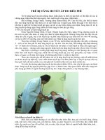
Figure 7.1 Patient anesthetized, temperature control assist Bair Hugger ® ,
Cardell ® monitor, Matrix ® anesthetic unit attached to adjustable height
hydraulic table (Canis Major, Midmark).
a
b
Figure 7.2 a. ECG evaluation software (DVM Solutions). b. Preanesthesia
blood pressure evaluation (DVM Solutions).
Anesthesia Protocols
Anesthesia protocols vary by patient age, condition,
morbidity factors, and length and type of procedure.
Local anesthetics are used on all dental surgical cases
where tissue is incised (see Table 7.1 at end of chapter).
Premedication
An intravenous catheter is placed and fl uids are admin-
istered in all patients undergoing anesthesia.
Premedication for healthy cats may include hydro-
morphone 0.1 mg/kg IM or SC or butorphanol 0.2 mg/
kg IM or SC combined with dexmedetomidine 0.0025 to
0.01 mg/kg IM (fi g. 7.3 ).
As cats become more debilitated or aged, butorphanol
0.2 mg/kg IM or SC or hydromorphone 0.05 – 0.2 mg/kg
IM or SC with 0.2 – 0.4 mg/kg midazolam IM can be
considered.
Anesthesia 171
Figure 7.3 Preanesthetic medication.
For fractious cats presenting without feline hyper-
trophic cardiomyopathy, a combined dose of mede-
tomidine HCl 0.01 – 0.02 mg/kg, plus buprenorphine
0.01 – 0.02 mg/kg, ketamine HCl 1 – 3 mg/kg, and butor-
phanol tartarate 0.1 mg/kg are mixed in one syringe and
administered IM; for fractious cats with feline hyper-
trophic cardiomyopathy, low - volume medetomidine
(0.005 mg/kg) plus butorphanol tartarate 0.1 – 0.2 mg/kg
plus or minus midazolam HCl 0.2 mg/kg may be
administered.
All patients should be individually assessed and a
patient - specifi c anesthetic premedication protocol devel-
oped, as the above are simply examples of typical pre-
medication protocols.
Induction
There are many feline anesthesia protocols for the
healthy young to middle - aged cat.
Chamber or Mask Induction
Chamber or mask induction should be avoided due to
catecholamine release during the excitement phase from
the struggle against restraint or as a reaction to the
pungent odor of the inhalant anesthetic agent.
Propofol
Propofol (2,6 - diisoproylphenol) (3 – 4 mg/kg IV; 3 mg/kg
if opioid is given as a premedicant), with half of the dose
given as a slow bolus over 40 – 60 seconds the rest to
effect, is a nonbarbiturate hypnotic. Slightly higher
doses are required for cats than dogs, and recoveries are
longer in cats than dogs when the infusion lasts more
than 30 minutes due to decrease in glucuronide conjuga-
tion. Propofol provides no analgesia in the cat. Propofol
is a direct myocardial depressant resulting in both
venous and arterial relaxation, thus creating hypoten-
sion. This hypotension is well recognized clinically and
must be considered when anesthetizing older or ill
patients.
Etomidate
Etomidate (0.5 – 1.5 mg/kg IV) is the induction drug of
choice for patients that have cardiovascular disease or
arrhythmias (except A - V dissociation) because cardiac
output and blood fl ow to the kidneys are maintained.
However, there have been reports of hemolysis in cats
after etomidate injection. A premedicant (e.g., butorpha-
nol IV, SC, IM; diazepam; or midazolam IV) should be
administered prior to etomidate administration.
Pain Control
Anesthesia protocols linked to pain control (in addition
to local anesthesia) include the following:
•
Expected mild to moderate pain – - buprenorphine
0.01 – 0.03 IM, IV sublingually mg/kg plus mid-
azolam 0.2 mg/kg, plus 0.0005 – 0.075 mg/kg dexme-
detomidine (0.0025 – 0.005 μ g/kg).
•
Expected moderate - to high - level pain – - hydromor-
phone 0.1 mg/kg plus midazolam 0.2 mg/kg, plus
dexmedetomidine 0.0005 – 0.075 mg/kg (0.5 – 7.5 μ g/
kg).
Intubation
All cats placed under anesthesia for oral assessment and
treatment must be intubated and the airway secured
with an infl atable cuff. Topical lidocaine may be applied
to the laryngeal mucosa to facilitate passage of the endo-
tracheal tube.
The endotracheal tube should be secured before the
cuff is infl ated. The cuff should be infl ated to a light seal.
Overinfl ation of the endotracheal cuff must be avoided.
Subcutaneous emphysema and pneumothorax have
occurred during or after anesthesia in cats anesthetized
for dental care. It is critical that anytime the head is
moved from side to side during assessment or dental
treatment, the endotracheal tube be disconnected from
the anesthesia machine and reconnected after the new
position is reached (fi gs. 7.4 a, b).
Maintenance
Anesthesia is generally maintained with isofl urane or
sevofl urane and oxygen. Little isofl urane or sevofl urane
172 Feline Dentistry
to support body temperature. It is much easier to prevent
hypothermia than to treat it (fi gs. 7.5 a, b).
Preparation of the Anesthetized
Dental Patient
The use of gauze sponges placed in the pharyngeal area
to absorb debris is controversial. Danger lies in gauze
entering the esophagus and either being vomited after
the surgical procedure is completed or ingestion leading
to gastrointestinal obstruction.
In the author ’ s opinion, packing the pharyngeal
area with gauze is not necessary because the seal pro-
vided by the endotracheal cuff is suffi cient to prevent
iatrogenic injury secondary to aspiration of surgical
debris.
Monitoring
Often, dental assessment results in a treatment plan that
requires multiple hours of anesthesia. Constant monitor-
ing of the cat ’ s physiological status is critical to a consis-
tent positive outcome.
Monitoring is accomplished through clinical appear-
ance as well as electrical monitoring systems. During
anesthesia the cat should have minimal jaw tone and
a
b
Figure 7.4 a. Lateral thoracic radiograph before anesthesia. b. Pneumotho-
rax, pneumomediastinum, and pneumoabdomen secondary to endotracheal
tube – induced tracheal tear.
a
b
Figure 7.5 a. Hot Dog ® patient warmer. b. Bair Hugger ® used below the
patient.
is metabolized. The insolubility of the inhalants allows
for a rapid induction and recovery.
Patient body temperature control is necessary. Long
anesthetic procedures coupled with the frequent use of
water and the ambient room temperature may create
hypothermia. In a risk determination study of 138 anes-
thetized cats, 71 (51%) had rectal body temperatures
≤ 35 ° C (95 ° F); the lowest recorded temperature was
28.8 ° C (83.8 ° F). Prolonged anesthesia dramatically
increased the risk of hypothermia. In addition to the
increased infection rate seen in hypothermic patients,
there is increased risk of fatal ventricular arrhythmias in
patients with body temperatures < 33 ° C. Bair Huggers ® ,
Therma - drapes ® , warm water circulating blankets, low -
fl ow anesthesia, a heat and moisture exchanger, plastic
wrap, leg wraps, radiant heating (Hot Dog ® patient
warming system), and warmed fl uids may all be used
Anesthesia 173
Figure 7.6 Wireless monitor with esophageal probe.
Figure 7.7 Monitor display (DVM Solutions).
palpebral refl ex. The pulse should be palpable and the
perfusion time should be two seconds or shorter. Breath-
ing during anesthesia should be even and regular.
Electronic monitoring includes electrocardiogram,
blood pressure, pulse oximetry, and end tidal CO
2
.
Apnea, temperature monitoring, and respiratory rate
are additionally helpful in assessing the cat ’ s response
to anesthesia (fi gs. 7.6 , 7.7 ).
Medical Conditions Requiring
Tailored Protocols
Renal Disease
Some cats with kidney disease may be dehydrated due
to their inability to concentrate urine. The dehydration
should be corrected before anesthesia if possible.
Mannitol is particularly useful in well - hydrated geri-
atric cats with chronic renal failure (with normal cardiac
function) to ensure diuresis. The dose of mannitol is
0.25 – 0.5 g/kg IV over 15 – 20 minutes.
Anesthesia may be induced with a combination of
propofol and diazepam. Ketamine and barbiturates
should be avoided. Maintenance with isofl urane is stan-
dard. Hypotension should be closely monitored and if
present adjusted with crystalloid administration.
Hyperthyroidism
Chronic unregulated hyperthyroidism can result in cats
that present thin, azotemic, with hypertrophic cardio-
myopathy, and with multiple oral issues, including
tooth resorption, oropharangyeal infl ammation, and
periodontal disease. If possible, patients should be
euthyroid before anesthesia. Patients with enlarged
hearts on thoracic radiographs should have echocardio-
grams performed before anesthesia.
Anesthesia protocol in the controlled hyperthyroid cat
should be tailored to prevent catecholamine release,
avoid arrhythmias, and promote normal blood pressure.
Premedication with an opioid is advised due to its
calming effect and minimal cardiovascular compromise.
Midazolam, etomidate, and diazepam are considered
safe to use for induction.
Ketamine should be avoided. Barbiturates may also
increase heart rate and should not be used. Propofol, a
generally accepted premedication in the healthy patient,
can also impair myocardial function in the ischemic
myocardium and should be used with caution.
Patient monitoring during anesthesia is critical, espe-
cially with regard to blood pressure measurement and
ECG. Hypotension can usually be managed with proper
intravenous fl uid administration without overload.
Patients with hypertrophic cardiomyopathy have
decreased compliance and ventricular volume. Dopa-
mine administration may be useful to increase blood
pressure to ensure adequate renal perfusion. Cardiac
tachyarrhythmias may be managed with propanolol.
Diabetes Mellitus
Diabetes mellitus requires special consideration in the
feline dental patient. Anesthesia should be scheduled in
the morning to avoid normal diurnal fl uctuations of
blood glucose levels. The client should be instructed to
give the patient only half the normal amount of insulin
the day of surgery.
Unless the cat has secondary organ compromise,
generally there are no specifi c medications to avoid
for induction or anesthetic maintenance. Diabetes can
lead to neutrophil dysfunction and impaired wound