Tài liệu Báo cáo khoa học: A novel coupled enzyme assay reveals an enzyme responsible for the deamination of a chemically unstable intermediate in the metabolic pathway of 4-amino-3-hydroxybenzoic acid inBordetellasp. strain 10d doc
Bạn đang xem bản rút gọn của tài liệu. Xem và tải ngay bản đầy đủ của tài liệu tại đây (242.5 KB, 7 trang )
A novel coupled enzyme assay reveals an enzyme responsible for the
deamination of a chemically unstable intermediate in the metabolic
pathway of 4-amino-3-hydroxybenzoic acid in
Bordetella
sp. strain 10d
Chika Orii
1
, Shinji Takenaka
2
, Shuichiro Murakami
2
and Kenji Aoki
2
1
Division of Science of Biological Resources, Graduate School of Science and Technology,
2
Department of Biofunctional Chemistry,
Faculty of Agriculture, Kobe University, Rokko, Kobe, Japan
2-Amino-5-carboxymuconic 6-semialdehyde is an unstable
intermediate in the meta-cleavage pathway of 4-amino-
3-hydroxybenzoic acid in Bordetella sp. strain 10d. In vitro,
this compound is nonenzymatically converted to 2,5-pyrid-
inedicarboxylic acid. Crude extracts of strain 10d grown on
4-amino-3-hydroxybenzoic acid converted 2-amino-5-car-
boxymuconic 6-semialdehyde formed from 4-amino-3-
hydroxybenzoic acid by the first enzyme in the pathway,
4-amino-3-hydroxybenzoate 2,3-dioxygenase, to a yellow
compound (e
max
¼ 375 nm). The enzyme in t he crude ex-
tract c arrying out the next step was purified to homogeneity.
The yellow compound formed from 4-amino-3-hydroxy-
benzoic acid by this purified enzyme and purified 4-amino-
3-hydroxybenzoate 2,3-dioxygenase in a coupled assay was
identified as 2-hydroxymuconic 6-semialdehyde by GC-MS
analysis. A mechanism for the formation of 2-hydroxy-
muconic 6-semialdehyde via enzymatic deamination and
nonenzymatic decarboxylation is proposed based on results
of spectrophotometric analyses. The purified enzyme, des-
ignated 2-amino-5-carboxymuconic 6-semialdehyde deami-
nase, is a new type of deaminase that differs from the
2-aminomuconate deaminases reported previously in that
it primarily and specifically attacks 2-amino-5-carboxymu-
conic 6-semialdehyde. The d eamination step in the p roposed
pathway differs from that in the pathways for 2-amino-
phenol and its derivatives.
Keywords: 4-amino-3-hydroxybenzoic acid; Bordetella sp.
strain 10d; 2-amino-5-carboxymuconic 6-semialdehyde;
2-hydroxymuconic 6-semialdehyde; 2-amino-5-carboxy-
muconic 6-semialdehyde d eaminase.
2-Aminophenol and its derivatives are intermediates in the
biodegradation of nitrobenzenes [1–4]. 2-Aminophenols
serve not only as a carbon source, but also as a nitrogen
source for g rowth of the assimilating bacteria. Deaminases,
which catalyze the release of ammonia, are a key enzyme in
the metabolic pathways of 2-amino phenol and its deriva-
tives. However, little is known about the metabolic steps
that lead to the release of ammonia and the properties of the
deaminase.
Pseudomonas sp. strain A P-3 and Pseudomonas pseudo-
alcaligenes strain JS45 convert 2 -aminophenol to 4-oxalo-
crotonic acid via 2-aminomuconic 6-semialdehyde and
2-aminomuconic acid in the modified meta-cleavage path-
way (Fig. 1B). The 2-aminomuconate deaminase from s train
AP-3 and that from strain JS45 have been purified and
characterized in detail [5,6]. The nucleotide sequence of the
gene encoding the deaminase from strain AP-3 is not similar
to any nucleotide sequences pr esent in the databases, other
than the recently reported nucleotide sequences of the gene
encoding 2-aminomuconate deaminase from Pseudomonas
putida HS12 and from Pseudomonas fluorescens strain KU-7
[6–8]. Although other deaminases have been detected in
crude extracts of nitrobenzene-assimilating bacteria, the
progress in the purification and characterization of the
enzymes is slow [2,4], p robably because the substrate for
the enzyme assay, 2-aminomuconic 6 -semialdehyde, which i s
formed by ring cleavage of 2-aminoph enol, is unstable and is
converted nonenzymatically to picolinic acid in vitro [9].
We have previously isolated Bordete lla sp. str ain 10d,
which grows on 4-amino-3-hydroxybenzoic acid, and puri-
fied and characterized the 4-amino-3-hydroxybenzoate 2,3-
dioxygenase involved i n t he initial step of t he m etabolism o f
this substrate [10]. The enzyme catalyzes the ring fission of
4-amino-3-hydroxybenzoic acid to form 2-amino-5-carb-
oxymuconic 6-semialdehyde (Fig. 1A). The cloning and
nucleotide sequence of the gene encoding the dioxygenase
(AhdA) have also been reported [11]. However, the
subsequent metabolism, including the deamination step,
have not been elucidated as 2-amino-5-carboxymuconic
6-semialdehyde is immediately converted nonenzymatically
to 2,5-pyridinedicarboxylic acid in vitro.
Here we report the purification and c haracterization of an
enzyme fromstrain 10d that uses 2-amino-5-carboxymuconic
Correspondence to K. Aoki, Department of Biofunctional Chemistry,
Faculty of A griculture, K o be University, R okko, Ko be 657–8501,
Japan. Fax: + 81 78 8820481, Tel.: + 81 78 8035891,
E-mail:
Enzymes: 2-amino-5-c arboxymucon ic 6-semialdehyde de aminase
(EC 3.5.99. – as proposed in this paper as a new subclass of deamin-
ases); 4-amino-3-hydroxybenzoate 2,3-dioxygenase (EC 1.13.1.–);
2-aminophenol 1,6-dioxygenase (EC 1.13.11.x); 2-aminomuconic
6-semialdehyde dehydrogenase (EC 1.2.1.32); 2-aminomuconate
deaminase (EC 3.5.99.5); catechol 2,3-dioxygenase (EC 1.13.11.2);
protocatechuate 2,3-dioxygenase (EC 1.13.11.x);
2,3-dihydroxybenzoate 3,4-dioxygenase (EC 1.13.11.14).
(Received 2 May 2004, revised 13 June 2004, accepted 18 June 2004)
Eur. J. Biochem. 271, 3248–3254 (2004) Ó FEBS 2004 doi:10.1111/j.1432-1033.2004.04258.x
6-semialdehyde as a substrate. Insights into the metabolic
fate of 4-amino-3-hydroxybenzoic acid in strain 10d are
revealed.
Materials and methods
Bacterial strain and growth conditions
Bordetella sp. strain 10d was isolated previously [10]. Strain
10d w as cultured in medium containing 0.12% (w/v)
4-amino-3-hydroxybenzoic acid and 1% (w/v) meat extract
[10].
Enzyme assay
2-Amino-5-carboxymuconic 6-semialdehyde was formed
from 4-amino-3-hydroxybenzoic acid in a coupled assay
by purified 4-amino-3-hydroxybenzoate 2,3-dioxygenase
provided in excess. The enzyme activity in the crude extract
and in the reaction mixture that used 2-amino-5-carboxy-
muconic 6-semialdehyde as substrate was measured by
monitoring the increase in t he absorbance of the reaction
product at 375 nm. The reaction mixture contained 2.9 mL
of 100 m
M
sodium/potassium p hosphate buffer ( pH 7.5),
0.1 mL of 5 m
M
4-amino-3-hydroxybenzoic acid, and
0.05 mL of crude extract. The reaction was started by
adding 0.1 mL of 4-amino-3-hydroxybenzoate 2,3-dioxy-
genase (0.8 UÆmL
)1
). After incubation for 10 min at 24 °C,
the absorbance at 375 nm was read. One unit of enzyme
activity was defined as t he amount o f enzyme t hat converted
1 lmol of 2-hydroxymuconic 6-semialdehyde per min. The
molar extinction coefficient of 4.4 · 10
4
for 2-hydroxy-
muconic 6-semialdehyde was used [12]. Specific activity was
defined a s units per mg protein. Protein concentration s were
measured by the method of Lowry et al. [13].
The substrate specificity of the purified enzyme was
examined with 2-aminomuconic 6 -semialdehyde and
2-aminomuconic acid using the same methods as described
previously [5,14,15].
Enzyme purification
All steps of the purification of the enzyme that used
2-amino-5-carboxymuconic 6-semialdehyde a s substrate
were carried out using modifications of methods described
previously [10]. Cells (14.8 g, wet weight) of strain 10d were
suspended in 2 0 m
M
Tris/HCl buffer (pH 8.0). Cell extract
(fraction 1, 150 mL) was prepared and treated with
streptomycin sulfate (fraction 2, 149 mL) as described
previously [9]. Fraction 2 was fractionated w ith ammonium
sulfate (38–60% saturation). After centrifugation (20 000 g
for 10 min), the pelleted precipitate was dissolved in 20 m
M
Tris/HCl buffer (pH 8.0). The solution was dialyzed against
buffer A [20 m
M
Tris/HCl buffer ( pH 8.0) containing 1 m
M
dithiothreitol and 0.5 m
ML
-ascorbic acid] (fraction 3,
46 mL). Fraction 3 was applied t o a DE52 cellulose column
(2.1 · 19 cm), and proteins were eluted with a linear
gradient (0–0.4
M
NaCl) at a flow rate of 40 mLÆh
)1
.The
active fractions were pooled (fraction 4, 30 mL). Fraction 4
was applied to a DEAE-Cellulofine A-800 column
(1.7 · 22 cm), and proteins were eluted with a linear
gradient (0–0.35
M
) of NaCl at a flow rate of 30 mLÆh
)1
.
The active fractions were pooled (fraction 5, 20 mL).
Fraction 5 was applied to a Phenyl-Cellulofine column
(1.6 · 13.7 cm), and proteins were eluted with a linear
gradient (0.5–0
M
) of ammonium sulfate at a flow rate of
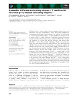
Fig. 1. Proposed pathway of 4-amino-3-
hydroxybenzoate metabolism in Bordetella sp.
strain 10d compared with the modified meta-
cleavage pathway of 2-aminophenol in Pseudo-
monas sp. strain AP-3. (A) Proposed pathway
of 4-amino-3-hydroxybenzoic acid in Borde-
tella sp. strain 10d (10). I, 4-amino-3-
hydroxybenzoic acid; II, 2-amino-5-
carboxymuconic 6-semialdehyde; III,2-hyd-
roxy-5-carboxymuconic 6-semialde hyde; IV,
2-hydroxymuconic 6-semialdehyde; V,2,5-
pyridinedicarboxylic acid; and VI,2-amino-
muconic 6-semialdehyde. (B) Pathway
of 2-aminophenol me tabolism in
Pseudomonas sp. strain AP-3 (6). I,2-amino-
phenol; II, 2-aminomuconic 6-semialdehyde;
III, 2-aminomuconic acid; IV, 4-oxalocrotonic
acid; and V, picolinic acid.
Ó FEBS 2004 2-Amino-5-carboxymuconic 6-semialdehyde deaminase (Eur. J. Biochem. 271) 3249
30 mLÆh
)1
. The active fractions were pooled (fraction 6,
24.5 mL). The enzyme purity was checked by SDS/PAGE
[16].
Production and isolation of enzymatic reaction products
in a coupled enzyme assay
The reaction mixture contained 107 mL of 50 m
M
sodium-
potassium phosphate buffer (pH 7.5), 9 mL of 5 m
M
4-amino-3-hydroxybenzoic acid, 5.1 mL of 4- amino-3-hy-
droxybenzoate 2,3-dioxygenase solutio n (8.8 lgÆmL
)1
), and
6 mL of purified enzyme solution (1.0 lgÆmL
)1
). After
incubation at 24 °C for 2.7 h with shaking at 100 r.p.m.,
the concentrations of 4-amino-3-hyd roxybenzoic acid,
2,5-pyridinedicarboxylic acid, ammonia, and 2 -hydroxymu-
conic 6 -semialdehyde i n t he reaction mixture were deter-
mined as described below. The reaction mixture was
concentrated to 10 mL with a rotary evaporator. The pH
of the concentrated solution was ad justed t o p H 3.0 with 5
M
metaphosphoric acid, and th e s olution was extracted with
ethyl acetate. The upper layer was collected and concentra-
ted to 10 mL. The extracted products were mixed with an
equimolar concentration of pentafluorophenylhydrazine at
24 °C for 30 min. The reaction mixture was then evapor-
ated to dryness. The hydrazone derivative was then mixed
with N,O-bis(trimethylsilyl)-trifluoroacetamide at 85 °Cfor
1.5 h. T he derivatized products were analyzed by GC-MS
as described below.
Analytical tests
UV-visible absorption spectra of reaction products and the
purified enzyme were recorded with a Beckman DU 650
spectrophotometer. Fluorescence spectra of the purified
enzyme and a cofactor released from the e nzyme were
recorded using a Hitachi F-2500 fluorescence spectropho-
tometer. The trimethyl-sililated or h ydrazone-derivatized
enzyme reaction products were analyzed with a Hitachi
M-2500 mass spectrometer at an ionization potential of
70 eV, coupled to a Hitachi G-3000 gas chromatograph. A
TC-1 fused silica capillary column (0.25 mm · 30 m; GL
Science, Tokyo, Japan) was used. A Hitachi L-6200 HPLC
system equipped with an Inertsil ODS-2 column
(4.6 · 150 mm, 5 lm; GL Science) was used for measuring
4-amino-3-hydroxybenzoic acid and 2,5-pyridinedicarboxy-
lic acid. The flow rate through the column at room
temperature was 0.4 mLÆmin
)1
. Samples were eluted with
a solvent of 0.05
M
phosphoric acid/methanol (65 : 35, v/v)
with monitoring at 278 nm. The cofactor from the purified
enzyme was detected by fluorescence ( F-1050) at an
excitation and emission wavelength of 450 and 530 nm,
respectively. Ammonia release was determined by measur-
ing the decrease in A
340
concomitant with NADPH
oxidation in the presence of glutamate dehydrogenase [18].
The N -terminal amino acid sequence was determined with a
Shimadzu PPSQ-10 protein sequencer using the method
reported previously [10]. The molecular mass of the native
enzyme was determined by gel filtration on Cellulofine
GCL-1000 sf using the method report ed previously [10 ]. The
molecular mass of the enzyme subunit was determined by
SDS/PAGE [16] using the LMW calibration kit (Amersham
Pharmacia Biotech) as size markers.
Chemicals
4-Amino-3-hydroxybenzoic acid and 2 ,5-pyridinedicarb-
oxylic acid were purchased from Tokyo Kasei Kogyo
(Tokyo, Japan); 2-aminophenol, catechol, metaphosphoric
acid, dithiothreitol,
L
-ascorbate, N,O-bis(trimethylsilyl)-tri-
fluoroacetoamide, NADPH, and glutamate dehydrogenase
were from Wako Pure Chemicals (Osaka, Japan); meat
extract (Extract Ehlrich) w as from Kyokuto Seiyaku Kogyo
(Osaka, Japan); and pentafluorophenylhydrazine was from
Pfaltz & Bauer. (Waterbury, CT, USA). DE52 cellulose was
from Whatman (Madison, WI, USA), and DEAE-Cellulo-
fine A-800, Phenyl-Cellulofine, and Cellulofine GCL-1000 sf
were from Seikagaku (Tokyo, Japan). 2-Aminophenol 1,6-
dioxygenase, 2-aminomuconic 6-semialdehyde d ehydrogen-
ase, and 4-amino-3-hydroxybenzoate 2,3-dioxygenase were
prepared as described p reviously [6,10,19]. 2-Amino-
muconic 6-semialdehyde was prepared enzymatically from
2-aminophenol using purified 2-aminophenol 1,6-dioxyge-
nase [6]. 2-Aminomuconic acid was synthesized by the
methods of He and Spain [5]. 2-Hydroxymuconic 6-semi-
aldehyde was prepared by incubating catechol with resting
cells of a mutant, strain Y-2, of the aniline-assimilating
Pseudomonas sp. strain AW-2 [20].
Results
Spectral changes during metabolism of 4-amino-3-
hydroxybenzoic acid by crude extracts of strain 10d
Strain 10d grows well in 4-amino-3-hydroxybenzoate
medium and completely degrades this substrate [10]. In
the culture broth, 2,5-pyridinedicarboxylic acid, which is
nonenzymatically converted v ia 2-amino-5-carboxymuconic
6-semialdehyde, cannot be detected by HPLC [10]. Cells of
strain 10d grown on 4 -amino-3-hydroxybenzoic acid were
washed and suspended in 50 m
M
sodium–potassium phos-
phate buffer (pH 6.8) containing 4-amino-3-hydroxy-
benzoic a cid. The substrate was also degraded without
accumulation of 2,5-pyridinedicarboxylic acid in the reac-
tion mixture. To reveal the subsequent metabolism in vivo,
including the deamination step the concentrated crude
extracts of strain 10d grown on 4-amino-3-hydroxybenzoic
acid were prepared by ammonia sulfate fractionation
(35–75% saturation). Figure 2A shows the changes in the
spectrum during the reaction in a coupled enzyme assay of
4-amino-3-hydroxybenzoic acid and the prepared crude
extracts. The absorption peaks at 263 and 294 nm charac-
teristic of 4-amino-3-hydroxybenzoic acid decreased as the
enzyme reaction proceeded and were almost completely
absent after 10 min of incubation. The maximum absorp-
tion peak shifted to 268 nm and the absorption peak at
375 nm derived from an intermediate increased during this
incubation time. The peak at 268 nm was assigned to
2,5-pyridinedicarboxylic acid based on the wavelength [10].
Purification and properties of the purified enzyme
The activity of the enzyme present in the crude extract of
strain 10d that used 2 -amino-5-carboxymuconic 6-semi-
aldehyde as substrate was measured by monitoring the
increase in the absorbance at 375 nm (Fig. 2A), but was not
3250 C. Orii et al. (Eur. J. Biochem. 271) Ó FEBS 2004
present in cell extracts of succinate/glucose-grown cells;
therefore, the s ynthesis of the e nzyme was indu cible. Table 1
shows a summary of a typical enzyme purification. The
enzyme was purified 103-fold with an overall yield of 2%.
The specific activity of the purified enzyme was 0.27
unitsÆmg protein
)1
. A fter electrophoresis, the purified
enzyme exhibited a single protein band on both n ative
and denaturing polyacrylamide gels Fig. 3A,B. The appar-
ent molecular mass w as determined to be 34 kDa by g el
filtration and 15 kDa by SDS/PAGE (Fig. 3B). Therefore,
the enzyme is a homodimer with 1 5-kDa subunits. The
N-terminal amino acid sequence o f the enzyme was
determined to be PKILVHSDAAPTTGFTNXHTP.
The purified enzyme was stable between pH 5.5 and 7.5
in 50 m
M
sodium/potassium phosphate buffer containing
1m
M
dithiothreitol and 0.5 m
ML
-ascorbate. The enzyme
maintained 80% activity up to 70 °C after 10-min incuba-
tion at pH 7.5. The enzyme activity decreased to 70% after
incubation at 75 °C for 10 min, and all activity was lost at
80 °C.
The two compounds tested, 2-aminomuconic 6-semi-
aldehyde and 2-aminomuconic acid, were shown not be
substrates of the p urified enzyme. The enzyme was i nhibited
(remaining activity indicated in parentheses) by the follow-
ing metal salts: 1 m
M
FeSO
4
(0%), 1 m
M
FeCl
3
(29%),
1m
M
MnSO
4
(0%), 1 m
M
CoCl
2
(0%), 1 m
M
NiSO
4
(0%),
and 1 m
M
ZnSO
4
(7%), K
3
Fe(CN)
6
and MgSO
4
did not
affect the enzyme activity. The addition of 1 m
M
iodoacetic
acid, p-chloromercuribenzoic acid, 5,5¢-dithiobis-(2-nitro-
benzoic acid) and 2,2¢-bipyridyl decreased the enzyme
activity to 95, 91, 86, and 95%, respectively.
Spectroscopic characterization of the purified enzyme
The c oncentrated enzyme solution (fraction 6) was yellow i n
color. The enzyme s olution showed one main absorption
peak at 266 nm and a broad absorption band in the visible
region (Fig. 4). The excitation spectrum of the heat-treated
enzyme with emission at 530 nm showed a m aximum at
367 nm and a s houlder around 4 49 nm (Fig. 4A). A peak at
514 nm was observed in the emission spectrum (Fig. 4B).
Authentic FAD in 50 m
M
sodium potassium phosphate
buffer (pH 7.0) showed maxima at 372 and 449 nm in the
excitation sp ectrum with emission at 530 nm. A peak at
527 nm was observed in the emission spectrum. These
results suggested that the e nzyme contains a flavin deriv-
ative. The flavin cofactor of the purified enzyme was
subsequently characterized using HPLC; a m ajor peak with
a retention time of 5.9 min was observed. In contrast,
authentic FAD and FMN showed a peak at 16.4 and
18.0 min, respectively.
Reaction products from 2-amino-5-carboxymuconic
6-semialdehyde
Figure 2B,C shows the changes in the absorption spectrum
during the coupled enzyme reaction of purified 4-amino-
3-hydroxybenzoate 2,3-dioxygenase and the enzyme puri-
fied here with 4-amino-3-hydroxybenzoic acid as substrate.
First the absorption around 350 nm increased, and t hen the
absorption peak at 375 nm appeared.
4-Amino-3-hydroxybenzoic acid (0.42 m
M
) was degraded
completely, 2,5-pyridinedicarboxylic acid (0.41 m
M
)and
2-hydroxymuconic 6-semialdehyde ( 0.028 m
M
) accumu-
lated, and ammonia (0.017 m
M
) was released during t he
enzyme reaction. Most of the 2-amino-5-carboxymuconic
6-semialdehyde formed by the a ction of 4-amino-3-hydroxy-
benzoate 2,3-dioxygenase was nonenzymatically converted
to 2,5-pyridinedicarboxylic acid [10], a nd the remainder was
converted (via two steps, one enzymatic and one nonenzy-
matic, see below) to 2-hydroxymuconic 6 -semialdehyde
and an almost equimolar concentration of ammonia. The
Fig. 2. Ab sorption spectra of the reaction products formed from
4-amino-3-hydroxybenzoic acid in an assay with crude extract and a
coupled assay with two purified enzymes. (A) The reaction mixture
consisted of 2.9 mL of 100 m
M
sodium/potassium phosphate buffer
(pH 7.5), 0.1 mL of 5 m
M
4-amino-3-hydroxybenzoic acid, and
0.05 mL of the crude extract (35–75% ammonia sulfate saturation)
(61 mgÆmL
)1
). The reaction was started by adding the enzyme solution.
After incubation at 24 °C, the sample was scanned with a spectro-
photometer and spectra were recorded every 2 min. (B) The reaction
mixture consisted of 2.9 mL of 100 m
M
sodium/potassium phosphate
buffer (pH 7.5), 0.1 mL o f 5 m
M
4-amino-3-hy droxy benzoic ac id,
0.1 mL of purified 4-amino-3-hydroxybenzoate 2,3-dioxygenase
solution (6 lgÆmL
)1
) and 0.1 mL of purified 2-amino-5-carboxy-
muconic 6-semialde hyde de aminase (7 1 lgÆml
)1
). The reaction was
started by a dding t he enzyme so lution. A fter i ncubation a t 2 4 °C, the
sample was scanned with a spectroph otometer and spectra were
recorded every 2 min. (C) E nlargement of the original plots shown
in (B).
Ó FEBS 2004 2-Amino-5-carboxymuconic 6-semialdehyde deaminase (Eur. J. Biochem. 271) 3251
proposed pathway is shown in Fig. 1A. Attempts to clarify
the stoichiometry by adding a small amount of the purified
dioxygenase to the reaction mixture with a large excess of
the purified enzyme reported here to avoid the formation
of 2,5-pyridinedicarboxylic acid from 2-amino-5-carboxy-
muconic 6-semialdehyde failed. The enzymatic reaction did
not proceed well because the dioxygenase is mo re unstable
than the purified enzyme reported here [10].
The enzyme reaction products were analyzed by GC and
GC-MS. Major ion peaks at 11.0 min (Fig. 1A, compound
IV) and 13.2 min (Fig. 1A, compound V) were o bserved.
The mass spectra (Table 2) and the GC retention times (R
t
)
of compound IV and compound V agreed with those of
trimethylsilylated pentafluorophenylhydrazone 2-hydroxy-
muconic 6-semialdehyde (R
t
¼ 11.0 min) and trimethyl-
silylated 2,5-pyridinedicarboxylic acid (R
t
¼ 13.2 min),
respectively.
Discussion
Although 2-amino-5-carboxymuconic 6-semialdehyde is
very labile, an enzyme able to use this compound as a
substrate was found in crude extracts of Bordetella sp. strain
10d. The enzyme w as purified to homogeneity and charac-
terized using a new coupled enzyme assay with 4-amino-
3-hydroxybenzoate 2,3-dioxygenase. A pathway for the
metabolism of 2-amino-5-carboxymuconic 6-semialdehyde
in strain 10d was proposed (Fig. 1A) based on results of
absorption spectra in a coupled enzyme assay, the enzyme
reaction product identified by GC-MS analysis, and the
determination of released ammonia. The coupled enzyme
assay revealed the mechanism of the deamination reaction
and the subsequent metabolism, including the deamination
step.
The product formed from 4-amino-3-hydroxybenzoic
acid by the action of purified 4-amino-3-hydroxybenzoate
2,3-dioxygenase and the purified enzyme reported here was
identified as 2-hydroxymuconic 6-semialdehyde (Fig. 1A,
compound IV). The accumulation of 2-hydroxymuconic
6-semialdehyde points to two possible deamination and
decarboxylation steps. The first possibility is that 2-amino-
5-carboxymuconic 6-semialdehyde (Fig. 1A, compound II)
is converted to 2-hydroxymuconic 6-semialdehyde via
2-aminomuconic 6-semialdehyde (Fig. 1A, compound VI).
In vitro, 2-aminomuconic 6-semialdehyde (Fig. 1B, com-
pound II; e
max
382 nm) is immediately converted to
picolinic acid (Fig. 1B, compound V, e
max
264 nm) [9].
The absorption peak at 382 nm increases rapidly and
reaches the maximum in 30 s, and then gradually decreases
Table 1. Purification of the 2-amino-5-carboxymuconic 6-sem ialdehyde deaminase f rom Bordetella sp. strain 10d. Fractions 1–6 refer t o the fractions
obtained at the en d of ste ps 1–6 of th e purification proc ed ure. See th e text fo r details.
Fraction Total activity (U) Total protein (mg) Specific activity (UÆmg
)1
) Recovery (%)
1. Cell extract 4.2 1600 2.6 · 10
)3
100
2. Streptomycin sulfate 4.1 1100 3.7 · 10
)3
98
3. Ammonium sulfate 2.8 290 9.7 · 10
)3
67
4. DE52 0.5 16 0.031 12
5. DEAE-Cellulofine A-800 0.25 5.0 0.050 6
6. Phenyl-Cellulofine 0.08 0.3 0.27 2
Fig. 3. PAGE and SDS/PAGE of the 2-amino-5-carboxymuconic
6-semialdehyde deaminase. (A) The purified enzyme (10 lg) was
electrophoresed o n a 12.5% (w/v) polyacrylamide disc gel (pH 8.0)
at 2 mA per tube for 2 h in a running buffer o f Tris/glycine
(pH 8.3) [30]. (B) The purified enzyme (10 lg) denatured with SDS
was electrophoresed on a 12.5% (w/v) polyacrylamide disc gel
containing 0.1% (w/v) SDS at 6 mA per tube for 3.5 h in a run-
ning buffer o f 0.1% (w/v) SDS/0.1
M
sodium phosphate (pH 7.2)
[16]. Standards were run separately. The gels were stained with
0.25% (w/v) Coomassie Brilliant Blue R-250 in a solvent of eth-
anol/acetic acid/H
2
O (9 : 2 : 9, v/v/v).
Fig. 4. U V-visible and fluorescence spectr a of the purified enzyme.
The main figure shows the UV-visible absorption spectrum of the
purified enzyme (1.1 mg) reco rded using 50 m
M
sodium-potassium
phosphate buffer (pH 7.0) as reference. The insets show (A) the
fluorescence excitation spe ctrum (detect ed at 530 nm) and ( B)
the emission spe ctrum (excited at 450 n m) of the supernatant of the
heat-treated enzyme (1.2 mg protein per mL). The cofactor derived
from the purified enzyme was relea sed by hea t treatme nt as
described previously [17].
3252 C. Orii et al. (Eur. J. Biochem. 271) Ó FEBS 2004
in 10 min [9]. I t cannot reasonably be assumed that
2-hydroxymuconic 6-semialdehyde accumulated via these
steps based on the changes in the absorption spectrum
(Fig. 2B,C). In addition, picolinic acid was not detected in
the reaction mixture after the coupled enzyme assay. The
other possibility is that 2-amino-5-carboxymuconic
6-semialdehyde is converted to 2-hydroxymuconic 6-semi-
aldehyde via 2-hydroxy-5-carboxymuconic 6-semialdehyde
(Fig. 1A, compound III). During a co upled assay with two
purified enzymes, a reaction product with an absorption
around 350 nm transiently accumulated (Fig. 2B,C). We
failed to isolate and identify s uch a compound; however, we
propose that the compound is 2-hydroxy-5-carboxymucon-
ic 6-semialdehyde and that t his compound is converted to
2-hydroxymuconic 6-semialdehyde by spontaneous decarb-
oxylation, based on electronic theory and previously
reported spectrophotometric data [21–23]. 3-Ketoacids
readily undergo decarboxylation under mild conditions,
and loss of C O
2
can occur readily only from the free
carboxylic acid [23]. Decarboxylation has a concerted
mechanism with an aromatic t ransition state. 2-hydroxy-5-
carboxymuconic 6 -semialdehyde has an aldehyde group
and a C-5 carboxyl group, which is a 3-ketoacid. As shown
in Fig. 1(A), compound III in the keto form possibly
releases CO
2
.Crawfordet al. and Nozaki et al.have
reported t hat p rotocatechuate 2,3-dioxygenase and c atechol
2,3-dioxygenase catalyze the ring fission of protocatechuic
acid (2,3-dihydroxybenzoic acid) to form 2-hydroxy-5-
carboxymuconic 6-semialdehyde (e
max
350 nm) [21,22].
The absorption peak at 350 nm derived from 2-hydroxy-
5-carboxymuconic 6-semialdehyde is observed and later an
absorption peak at 375 nm d erived from 2-hydroxymuconic
6-semialdehyde appears [22]. 2,3-Dihydroxybenzoate 3,4-
dioxygenase from Pseudomonas fluorescens 23D-1 catalyzes
the ring fission of 2,3-dihydroxybenzoic acid to form
2-hydroxymuconic 6-semialdehyde and CO
2
[24]. There-
fore, strain 10d converts 2-amino-5-carboxymuconic 6-semi-
aldehyde to 2-hydroxymuconic 6-semialdehyde in the
deamination and nonenzymatic decarboxylation s teps
(Fig. 1A). We named the enzyme r eported here 2-a mino-
5-carboxymuconic 6-semialdehyde deaminase.
2-Amino-5-carboxymuconic 6-semialdehyde deaminase
from strain 10d differs from previously reported 2-amino-
muconase deaminases in substrate specificity, thermo-
stability, subunit structure, a nd N-terminal amino acid
sequence [5,6]. The native enzyme of Pseudomona sp. strain
A-3 has a molecular mass o f 67 kDa and consists of four
identical subunits, w hile the e nzyme from P. pseudoalcalige-
nes strain JS45 has a molecular mass of 100 kDa and
consists of six identical subunits. The enzymes from strain
A-3 a nd strain JS45 maintain 80% a ctivity up to 50 °C. The
enzyme from strain JS45 is colorless and does not have an
absorbance peak at 300 nm [5]. A cofactor is not required
for t he enzyme activity. In contrast, the deaminase from
strain 10d contained an FAD-like cofactor, similar to
D
-amino acid oxidases [25–27], as indicated by the absorp-
tion peak of the purified enzyme at 266 nm. The typical
protein absorption p eak of 2 80 nm shifts to 265 nm if the
protein contains a flavin-type cofac tor [28]. We failed to
identify the cofactor of the deaminase from strain 10d
because the enzyme could not be purified in large enou gh
quantities. We previously reported the identification of the
enzyme involved in the initial step of the metabolism of
4-amino-3-hydroxybenzoic acid in Bordetella sp. 10d [10].
This first step, catalyzed by 4-amino-3-hydroxybenzoate
2,3-dioxygenase (Fig. 1A), is similar to the first step in the
modified meta-cleavage pathway for 2-aminophenol in
Pseudomonas sp. strain A P-3 catalyzed by 2-aminophenol
1,6-dioxygenase [10] (Fig. 1B). However, 4-amino-
3-hydroxybenzoate 2 ,3-dioxygenase differs from 2-amino-
phenol 1,6-dioxygenase in subunit structure and substrate
specificity [4,10]. The deamination steps in these pathways
differ from each other (Fig. 1A,B). Recently, Muraki et al.
reported that the carboxyl-group-substituted 2-aminophe-
nol, 3-hydroxyanthralinic acid (2-amino-3-hydroxybenzoic
acid), is metabolized to form 4-oxalocrotonate via 2 -amino-
3-carboxymuconic 6-semialdehyde and 2-aminomuconate
through an enzymatic decarboxylation step (2-amino-3-
hydroxymuconic 6-semialdehyde decarboxylase) and a
deamination step (2-aminomuconic 6-semialdehyde deami-
nase) in P. fluorescens strain KU -7 [7]. The de carboxylation
mechanism in t he metabolic pathways for 3-hydroxyanth-
ralinic acid differs from that in the pathway for 4-amino-
3-hydroxybenzoic acid.
The N-terminal amino acid sequence of the purified
enzyme d id not show significant levels of identity to
sequences of 2-aminomuconate deaminases [6,8,27] or to
any other sequences available in FASTA and BLAST
database programs at the DNA Data Bank of Jap an.
Recently, we reported the cloning and s equencing of the
gene encoding 4-amino-3-hydroxybenzoate 2 ,3-dioxygenase
from strain 10d [11]. Unfortunately, the cloned 4.2-kb
fragment does not contain the gene encoding the deaminase
reported here. In the cloned 5.2-kb fragment from P. pseudo-
alcaligenes JS45, there are no genes involved in the
2-aminophenol-metabolic pathway, except for am nBA,
which encodes 2-aminophenol 1,6-dioxygenase, and amnC,
which encodes 2-aminomuconic 6-semialdehyde d ehydro-
genase [29]. Analysis of the entire amino acid sequence of
2-amino-5-carboxymuconate 6-semialdehyde deaminase
Table 2. Mass spectra of the enzyme reaction products from 4-amino-3-hydroxybenzoic acid.
Compound Fragments of the derivatization product [m/z (assignment, relative intensity)]
IV: 2-hydroxymuconic
6-semialdehyde
a
466 (M
+
, 18.7%), 451 (M
+
-CH
3
, 100%), 436 (M
+
-CH
3
· 2, 0.53%), 421 (M
+
-CH
3
· 3, 0.53%),
377 [M
+
-OSi(CH
3
)
3
, 0.64%], 363 [M
+
-Si(CH
3
)
3
-CH
3
· 2, 4.8%], 299 (M
+
-C
6
F
5
, 65.1%),
195 ([C
6
F
5
N
2
]
+
, 8.7%), 147 {[(CH
3
)
2
¼O-OSi(CH
3
)
3
]
+
, 24.3%}, 73 {[Si(CH
3
)
3
]
+
, 98.4%}
V: 2,5-pyridine-
dicarboxylic acid
b
311 (M
+
, 30.6%), 296 (M
+
-CH
3
, 100%), 266 (M
+
-CH
3
· 3, 39.3%), 238 [M
+
-Si(CH
3
)
3
, 11.7%],
222 [M
+
, Si(CH
3
)
3
-O, 62.7%], 194 [M
+
-COOSi(CH
3
)
3
, 39.3%], 147 {[(CH
3
)
2
¼O-OSi(CH
3
)
3
}
+
,
100%), 77 [M
+
-COOSi(CH
3
)
3
-COOSi(CH
3
)
3
, 90.9%], 73 {[Si(CH
3
)
3
]
+
, 100%}
a
Pentafluorophenylhydrazine and trimethylsilylated product.
b
Trimethylsilylated product.
Ó FEBS 2004 2-Amino-5-carboxymuconic 6-semialdehyde deaminase (Eur. J. Biochem. 271) 3253
will reveal more information on t he narrow substrate
specificity and the cofactor.
References
1. Hasegawa, Y., Muraki, T., Tokuyama, T., Iwaki, H., Tatsuno, M.
& Lau, P.C. (2000) A novel degradative pathway of 2-nitoro-
benzoate via 3-hydro xyanthranilate in Pseudomonas fluorescens
strain KU-7. FEMS Microbiol. Lett. 190, 185–190.
2. Katsivela, E., Wray, V., Pieper, D.H. & Wittich, R F. (1999)
Initial reactions in the biodegradation of 1-chloro-4-nitrobenzene
by a newly isolated bacterium, strain LW1. Appl. Environ.
Microbiol. 65, 1405–1412.
3. Nishino, S.F. & Spain, J.C. (1993) D egradation of nitrobenzene by
a Pseudomonas pseudoalcaligenes. Appl. Environ. M icrobiol. 59,
2520–2525.
4. Spiess, T., Desiere, F., Fischer, P., Spain, J.C., Knackmuss, H.J. &
Lenke, H. ( 1998) A new 4 -nitrotoluene degradation p athway i n a
Mycobacterium strain. Appl. Environ. Microbiol. 64, 446–452.
5. He, Z. & Spain, J.C. (1998) A n ovel 2-aminomuconate d eaminase
in the nitrobenzene degradation pathway of Pseudomonas pseu-
doalcaligenes JS45. J. Bac teriol. 180 , 2502–2506.
6. Takenaka, S ., Murakami, S., Kim, Y J. & Aoki, K. (2000)
Complete nu cleotide sequence an d functional analysis of the genes
for 2-aminophenol m etabolism from Pseudomonas sp. AP-3. Arch.
Microbiol. 174, 265–272.
7. Muraki, T., Taki, M., Hasegawa, Y., Iwaki, H. & Lau, P.C. (2003)
Prokaryotic homologs of the euk aryotic 3-hydroxyanthranilate
3,4-diox ygena se and 2-amino-3-carboxymuconate-6-semialde
hyde decarboxylase in the 2-nitrobenzoate degradatio n pathw ay
of Pseudom ona s fluo res cen s strain K U-7. Appl. Environ. Microbiol.
69, 1564–1572.
8. Park, H S. & Kim, H S. (2001) Genetic and structural organi-
zation of the aminophenol catabolic operon and its implication for
evolutionary process. J. Bacteriol. 183, 5074–5081.
9. Aoki, K ., Takenaka, S., Murakami,S.&Shinke,R.(1997)Partial
purification and characterization of a bacterial dioxygenase that
catalyzes the ring fission of 2-aminophenol. Microbiol. Res. 152,
33–38.
10. Takenaka, S., Asami, T., Orii, C., Murakami, S. & Aoki, K.
(2002) A n ovel meta-cleavage dioxygenase t hat cleaves a carboxyl-
group-substituted 2-aminophenol: purification and characteriza-
tion of 4-amino-3-hydroxybe nzoate 2,3-dioxygenase from
Bordetella sp. stra in 1 0d. Eur . J. Biochem. 269, 5871–5877.
11.Murakami,S.,Sawami,Y.,Takenaka,S.&Aoki,K.(2004)
Cloning of a gene encoding 4-am ino-3-hyd roxyben zoate 2,3-
dioxygenase from Bordetella sp. 10d. Biochem. Biophys. Res.
Commun. 314, 489–494.
12. Nozaki, M. (1970) Metapyrocatechase (Pseudomonas). Methods
Enzymol. 17A, 522–525.
13. Lowry, O.H., Rosebrough, N.J., Farr, A.L. & Randall, R.J.
(1951) Protein measurement with the f oline phenol reage nt. J. Biol.
Chem. 193, 265–275.
14. He, Z., Davis, J.K. & Spain, J.C. (1998) Purification, character-
ization, and sequence analysis o f 2-aminomuconic d e hydrogenase
from Pseudomonas pseudoalcaligenes JS45. J. Bacteriol. 180, 4591–
4595.
15. Nishizuka, Y., I chiyama, A. & Hayaishi, O. (1970) Me tabolism of
the benzene ring of tryp tophan (mammals). Methods Enzymol.
17A, 463–491.
16. Weber, K. & Osborn, M. (1969) The reliability of m olecular
weight determin atio ns by dodecyl s ulfate–polyacrylamide gel
electrophoresis. J. Biol. Chem. 24 4, 4406–4412.
17. Klatt, P., Schmidt, K., Werner, E.R. & Mayer, B. (1996)
Determination of nitric oxide synthase co factors: heme, FAD,
FMN and tetrahydrobiopterin. Methods Enzymol. 268, 358–
365.
18. Koike, K., Hakamada, Y., Yoshimatsu, T., Kobayashi, T. & Ito,
S. (1996) NADP-specific glutamate dehydrogenase from alkalo-
philic Bacillus sp. KSM-635 purification and enzym atic properties.
Biosci. Biotechn B iochem. 60 , 1764–1767.
19. Takenaka. S., Murakami, S ., S hinke, R., Hatakeyama, K.,
Yukawa, H. & Aoki, K. (1997) Novel genes encoding 2-amino-
phenol 1,6-dioxygenase from Pseudomonas species AP-3 growing
on 2-aminophenol and catalytic propert ies of the purified enzyme.
J. Biol. Chem. 272, 14727–14732.
20. Aoki,K.,Kodama,N.,Murakami,S.&Shinke,R.(1997)Ahigh
level of accumulation of 2-hydroxymuconic 6-semialdehyde from
aniline b y t he transposi tional mutant Y-2 of Pseudomo nas species
AW-2. Microbiol. Res. 152, 129–135.
21. Crawford, R.L., Bromley, J.W. & Perkins-Olson, P.E. (1979)
Catabolism of protocatechuate by Bacillus macerans. Appl.
Environ. Mic robiol. 37 , 614–618.
22.Nozaki,M.,Kotani,S.,Ono,K.&Senoh,S.(1970)Meta-
pyrocatechase. 3. sub strate specificity and mode of ring fission.
Biochim. Biophys. Acta. 220, 213–223.
23. Vollhardt, K.P.C. & Schore, N.E. (1998) Organic Chemistry:
Structure and Function In (Vollhardt, K.P.C. & Schore, N.E., ed s),
pp. 1045–1046. W.H. Freeman, New York.
24. Ribbons, D.W. & Seinior, P.J. (1970) 2,3-Dihydroxybenzoate
3,4-oxygenase from Pseudomonas fl uorescens – oxidation of a
substrate analog. Arch. Biochem. Biophys. 138, 557–565.
25. Job, V., Marcone, G.L., Pilone, M., S. & Pollegioni, L. (2002)
Glycine oxidase from Bacillus subtilis: characterization of a new
flavoprotein. J. Biol. Chem. 277, 6985–6993.
26. Nishiya, Y. & Imanaka, T. (1998) Purification and characteriza-
tion of a n ovel glycine oxidase from Bacillus subtilis. FE BS Lett.
438, 263–266.
27. Pollegioni, L., Ceciliani, F., Curti, B., Ronchi, S. & Pilone, M.S.
(1995) Studies on th e structural an d funct ional aspec ts of Rhodo-
torula gracilis
D
-amino acid oxidase by limited trypsinolysis.
Biochem. J. 310, 577–583.
28. Cook, S.A. & Shiemke, A.K. (2002) Evidence that a type-2
NADH: quinone oxidoreductase mediates electron transfer to
particulate methane m onooxygenase in Methylococcus capsulatus.
Arch. Biochem. B iophys. 398, 3 2–40.
29. Davis, J.K ., He, Z., Somerville, C.C. & Spa in, J.C. (1999) Genetic
and biochemical comparison of 2-aminophenol 1,6-dioxygenase of
Pseudomonas pseudoalcaligenes JS45 to meta-cleavage dioxy-
genases: divergent evolution of 2-amin ophenol meta-cleavage
pathway. Arch. Microbiol. 172, 330–339.
30. Davis, B.J. (1964) Disc electrophoresis. II. Method and appli-
cation to human seru m proteins. Ann. NY Acad. Sci. 121,
404–427.
3254 C. Orii et al. (Eur. J. Biochem. 271) Ó FEBS 2004