Tài liệu Esophageal Reconstruction Authored by: Marta Strutyńska-Karpińska and Krzysztof docx
Bạn đang xem bản rút gọn của tài liệu. Xem và tải ngay bản đầy đủ của tài liệu tại đây (29.68 MB, 98 trang )
EsophagEal
REconstRuction
Authored by
Marta Strutyńska-Karpińska and
Krzysztof Grabowski
Esophageal Reconstruction
Authored by: Marta Strutyńska-Karpińska and Krzysztof Grabowski
Published by InTech
Janeza Trdine 9, 51000 Rijeka, Croatia
Copyright © 2012 InTech
All chapters are Open Access distributed under the Creative Commons Attribution 3.0 license, which allows users to
download, copy and build upon published articles even for commercial purposes, as long as the author and publisher are
properly credited, which ensures maximum dissemination and a wider impact of our publications. After this work has been
published by InTech, authors have the right to republish it, in whole or part, in any publication of which they are the author,
and to make other personal use of the work. Any republication, referencing or personal use of the work must explicitly
identify the original source.
As for readers, this license allows users to download, copy and build upon published chapters even for commercial
purposes, as long as the author and publisher are properly credited, which ensures maximum dissemination and a wider
impact of our publications.
Notice
Statements and opinions expressed in the chapters are these of the individual contributors and not necessarily those of the
editors or publisher. No responsibility is accepted for the accuracy of information contained in the published chapters. The
publisher assumes no responsibility for any damage or injury to persons or property arising out of the use of any materials,
instructions, methods or ideas contained in the book.
Publishing Process Manager Jelena Marušić
Typesetting InTech DTP Team
Cover Design InTech Design Team
First Published November, 2012
Printed in Croatia
A free online edition of this book is available at www.intechopen.com
Additional hard copies can be obtained from
Esophageal Reconstruction
Authored by: Marta Strutyńska-Karpińska and Krzysztof Grabowski
p. cm.
ISBN 978-953-51-0667-8
free online editions of InTech
Books and Journals can be found at
www.intechopen.com
Contents
Foreword 1
Chapter 1 Esophageal Reconstruction with Large Intestine
1. Vascular anatomy of the colon 3
2. Esophageal reconstructions using the colon 6
3. Esophageal reconstructions using the right colon 6
3.1. The technique of creation of an antiperistaltic graft
from the right colon on ileocolic vascular pedicle 6
3.2. The technique of construction of an isoperistaltic graft
from the right colon on middle colic vascular pedicle 13
3.3. The technique of construction of an isoperistaltic
graft from the right colon on left colic vascular pedicle 17
4. Esophageal reconstructions using the left colon 20
4.1. The technique of construction of an antiperistaltic graft
from the left colon on middle colic vascular pedicle 21
4.2. The technique of construction of an isoperistaltic graft
from the left colon on left colic vascular pedicle 23
4.3. The technique of construction of an antiperistaltic
graft from the left colon on left colic vascular pedicle 25
5. References 27
Chapter 2 Esophageal Reconstruction with Small Intestine
1. Vascular anatomy of the small intestine 33
2. Esophageal reconstructions using the jejunum 35
3. Esophageal reconstructions using the ileum 40
3.1. Esophageal reconstruction with the use of
the ileum alone 42
3.2.
Esophageal reconstruction using the ileum
and the caecum
44
3.3. Esophageal reconstruction using the ileum, the caecum
and part of the ascending colon 47
4. References 49
Chapter 3 Modifications and Complex Esophageal Reconstructions
1. Modifications of esophageal reconstructions 55
1.1. Resection of redundant intestine 55
2. Management of ischaemia in the cephalic portion of the
jejunal graft 57
2.1
Insertion from the ileum on middle colic vascular pedicle
59
2.2. Insertion from the colon on ileocolic vascular pedicle 60
2.3. Insertion from the colon on left colic vascular pedicle 64
2.4. Secondary mobilization of the graft 65
3. References 68
Chapter 4 Diagnosis and Treatment of Postoperative Complications After
Esophageal Reconstruction with Pedicled Intestinal Segments
1. Early complications after esophageal reconstruction 71
1.1. Necrosis of a part or a whole intestinal graft 71
1.2. Pneumothorax 74
1.3. Insufficiency of cervical anastomosis 74
1.4. Salivary fistula in the region of cervical anastomosis 75
1.5. Injury of the recurrent laryngeal nerve 76
2. Diagnosis and treatment of late complications after
esophageal reconstructions 76
2.1. Diagnosis of the esophageal substitute 77
2.2. Late complications in the region of cervical
anastomosis 79
2.2.1 Cicatrical stenosis of the cervical anastomosis 79
2.2.2. Diverticula in the region of cervical anastomosis 82
2.2.3. Pleural hernia of the esophageal substitute 83
2.2.4. Complications associated with reflux to the
esophageal substitute 87
2.2.5. Benign and malignant tumours of the
esophageal substitute 89
3. References 93
Esophageal Reconstruction
Marta Strutyńska-Karpińska
Krzysztof Grabowski
University of Medicine, Department and Clinic
of Gastrointestinal and General Surgery, Skłodowskiej-Curie str. 66,
Wrocław, Poland
Foreword
A signicant development of esophageal reconstructive surgery can be observed over the
years since 1907, when Cesar Roux rst succeeded in performing the esophageal reconstruc-
tion with a segment of the jejunum. Professional literature presents both, various modica-
tions of the surgical methods as well as original reconstructive procedures, which broaden
signicantly the range of surgical modalities and solutions in the surgical management of
this condition.
However all the achievements have not led to the development of one, universal and gener-
ally accepted surgical method. The main reason of the situation lies in diculty to standard-
ize reconstructive surgeries. Progress in this respect achieved over time consists mainly in
aempts to approximate optimally the function of the reconstructed esophagus to the func-
tion of a natural organ, and to minimize the number of both, early and late postsurgical com-
plications.
Success of every reconstructive surgery with the use of pedicled intestinal segment is condi-
tioned by ecient blood supply and adequate length of the pedicle that would enable free
from tension anastomosis of the gra with cervical esophagus or the pharynx. Selection of an
adequate segment of the intestine for esophageal graing is in every case closely associated
with the anatomical structure of the intestinal vasculature, what means that only the presence
of well developed and ecient main blood vessels and their branching arcades may author-
ize the surgeon to start mobilizing this or another intestinal segment as an esophageal gra.
Abandonment of this basic principle leads to severe postsurgical complications.
Authors
© 2012 Marta Strutyńska-Karpińska and Krzysztof Grabowski.; licensee InTech. This is an open access chapter
distributed under the terms of the Creative Commons Attribution License ( />licenses/by/2.0), which permits unrestricted use, distribution, and reproduction in any medium, provided the
original work is properly cited.
3Esophageal Reconstruction with Large Intestine
Esophageal Reconstruction with Large Intestine
1. Vascular anatomy of the colon
The colon is supplied with arterial blood from two main sources: the superior mesenteric artery
(arteria mesenterica superior) and the inferior mesenteric artery (arteria mesenterica inferior).
The right colon is supplied from the superior mesenteric artery through the following arteries:
ileocolic artery (arteria ileo-colica), right colic artery (arteria colica dextra) and middle colic ar-
tery (arteria colica media). The inferior mesenteric artery supplies arterial blood to the le colon
through the le colic artery (arteria colica sinistra). The ascending branch of the le colic artery
(ramus ascendens) is joined with the middle colic artery by the arc of Riolan, creating in this way
a connection between branches of inferior and superior mesenteric artery ( Fig. 1, 2).
Figure 1 Diagram of the arterial blood supply to the colon: 1 –art. mesenterica superior, 2 – art. mes-
enterica inferior, a – art. ileocolica, b – art. colica dextra, c – art. colica media, d – ramus ascendens art.
colicae sinistrae, e – ramus descendens art. colicae sinistrae, R – arcus Riolani
Individual vascular trunks of the right as well as of the le colon interconnect forming so-
called arcades. The arc of Riolan, the longest vascular arcade, also known as the marginal
artery, connects the blood vessels on the right and le part of the colon.
It is worth reminding that the ileocolic artery, the right colic artery and the le colic artery are situ-
ated behind the peritoneum, while the middle colic artery, arising from beneath the lower border
of the pancreas, passes between the layers of the transverse mesocolon and directs to the hepatic
exure of the colon. Recollection of these well known anatomical facts is inasmuch signicant as
during preliminary intraoperative evaluation of the type of colon vasculature, it turns out that only
Chapter 1
Esophageal Reconstruction4
mobilization of the ascending and the descending colon exposes clearly enough the topography of
main vascular trunks and anastomoses between them, what in turn aects signicantly the choice
of adequately supplied and long colon segment for esophageal reconstruction. Topography of the
venous drainage mirrors the arterial supply, i.e. arteries have respective vein equivalents.
Figure 2 Angiogram of the superior mesenteric artery and its branches: X – the iliac branch of the ileo-
colic artery, a – art. ileocolica, b – art. colica dextra, c – art. colica media, R- arcus Riolani
The above vascular anatomy of the colon is what we can read in human anatomy textbooks.
In clinical practice we meet certain deviations from the above presented topography. They
usually concern the right colon and the venous system (Fig. 3).
Own experimental studies as well as studies by other authors on intestinal preparations dem-
onstrate that 6% of population present with lack or hypoplasia of arcades anastomosing the
main venous trunks with simultaneously well developed arterial trunks and broad and well
developed arterial arcades within the right colon. In such cases the right colon cannot be used
for reconstruction due to inadequate venous system. Angiographic evaluations of the colonic
arterial system reveal that permanent arteries present in 100% of population include the ilecolic
artery, the middle colic artery and the le colic artery. Well developed arc of Riolan is met in
90% of population, and about 50% have good anastomoses between all colic arteries. The right
colic artery is absent in about 30-35% of population (Fig. 4). In 25% of population, its absence is
compensated for by 1 to 3 so-called additional middle colic arteries. However the arteries are
usually short, with a narrow lumen and the anastomosing arcades are also short and narrow.
5Esophageal Reconstruction with Large Intestine
Figure 3 Angiogram of the superior mesenteric vessels: 1 – art. mesenterica superior, A1 – vena mesenterica
superior, a – art. ileocolica, well developed, with ecient anastomosing arcades, a1 – vena ileocolica with
bushy branches, does not form anastomosing arcades
The above presented anatomical details of the arterial and venous vasculature of the colon are
very important, as they play a crucial role in the choice of an adequate, i.e. with good arterial
supply and ecient venous drainage, intestinal segment to form a pedicled gra of the colon.
Figure 4 Angiogram of the superior mesenteric artery and its branches: X – the iliac branch of the ileo-
colic artery, a – art. ileocolica, c – art. colica media; absent art. colica dextra, R – arcus Riolani
Esophageal Reconstruction6
2. Esophageal reconstructions using the colon
In order to organize and clarify descriptions of individual types of esophageal reconstruc-
tions, it should be remembered, as mentioned previously, that the main and at the same time
permanent vessels of the colon include the ileocolic, middle colic and le colic vessels.
So-called adequate vascular systems in the aspect of reconstruction include long vascular
trunks, which form wide arches passing into well developed and strong anastomosing ar-
cades. The connection between the branches of the inferior and superior mesentery arteries,
known as the arc of Riolan, is well developed. Such a situation gives the possibility of choos-
ing either right, or le part of the colon to form a pedicled gra. For esophageal reconstruc-
tion each of the above mentioned colic vessels may be used to mobilize gras in two positions:
the isoperistaltic and the antiperistaltic gras.
Selecting a segment of colon for esophageal reconstruction, one should always consider all
the possibilities of the mobilization of the gra, in every individual case choosing the most
suitable as far as blood supply and length is concerned pedicled segment of the colon. It
should be remembered that the gra’s length is strictly associated with the length of its vas-
cular pedicle.
3. Esophageal reconstructions using the right colon
Taking advantage of an adequate vascular system in the colon, and choosing the right colon,
the pedicled esophageal gra may be constructed using the following methods:
• from the right colon on ileocolic vascular pedicle in an antiperistaltic
position of the gra
• from the right colon on middle colic vascular pedicle in an isoperi-
staltic position of the gra
• from the right colon on le colic vascular pedicle in an isoperistaltic
position of the gra
3.1. The technique of creation of an antiperistaltic gra from the right colon on ileocolic
vascular pedicle
The surgical technique presented below was developed by Prof. Jezioro in 1961 and used sub-
sequently in the clinical practice in patients requiring esophageal reconstruction.
The abdominal cavity is approached from upper midline incision going several cm below and
passing by the umbilicus on the right side. Next the right colon and the terminal segment of the
ileum are mobilized. For this reason the small bowel loops are moved lewards and maintained
in this position with surgical towels. The parietal peritoneum is gradually transected starting
7Esophageal Reconstruction with Large Intestine
from the iloecolic region next to the large bowel and continuing until the right exure of the
colon. Next, slightly elevating the bowel, the caecum and the ascending colon are separated
together with blood vessels, and the ligaments of the right exure of the colon are exposed and
transected between ligatures. This maneuver allows to identify macroscopically and evaluate
the structure and ecacy of the vascular system in this part of the colon in the aspect of esopha-
geal reconstruction. The adequacy of circulation is ascertained when the arterial and venous
trunks of the iloecolic vessels are long and well developed and the arcades anastomosing them
to the right colon vessels, i.e. the right colic vessels and the laer and middle colic vessels are
long and broad. If the right colic vessels are missing, they can be replaced by middle colic ves-
sels. Next a biological trial is performed, i.e. the trunks of the right and middle colic vessels,
iliac branch of the ileocolic vessels and the vascular arch between the middle colic vessels and
the le colic vessels are clamped with vascular clamps, in order to create conditions resembling
those in a gra pedicled exclusively on iloecolic vessels. The trial is considered positive when
the separated intestinal segment maintains normal colour and reveals pulsation in the terminal
intestinal vessels close to the intestinal wall. The evaluation of blood supply to the isolated in-
testinal segment may be conrmed by intraoperative ultrasound examination.
Figure 5 Diagram illustrating mobilization of a graft from the right colon on ileocolic vessels pedicle in an
antiperistaltic position
Having evaluated the blood supply, with a positive outcome of the trial, the gra mobilization
may start (Fig. 5, 6). The greater omentum is mobilized from the transverse colon to the middle
of its length. Next the trunks of the right and middle colic vessels are ligated and transected.
The transverse colon should be transected in the middle its length. The eerent transverse colon
stump is sutured with a double-layer manual suture, or stapled. The vermiform appendix is
Esophageal Reconstruction8
excised in a routine manner. Subsequently, the iliac branch of the ileocolic vessels and terminal
vessels of the caecal segment of the ileum are ligated and transected. The ileum is transected
and the stumps are closed with a double-layer manual suture, or stapled. The caecal stump of
the transected ileum should be short in order not to create a diverticular excess, what may have
an unfavourable eect on subsequent function of the substitute esophagus.
Figure 6 Intraoperative picture of mobilization of the right colon graft on ileocolic pedicle in an anti-
peristaltic position. A –vascular pedicle, B – right colon
Cephalic stump of the gra, which is formed by the right transverse colon, is also closed with
a double-layer manual suture, or stapled. In this way the stage of mobilization of the gra
from the right colon on an ileocolic vascular pedicle is completed. Thus the reconstruction is
antiperistaltic – the cephalic segment of the gra from the right colon will be anastomosed
with the cervical esophagus, and the caecum – with the stomach.
The next stage includes construction of a retrosternal canal and passing the gra through a
canal created in the interior mediastinum to the neck. Construction of the retrosternal canal
requires special precision. This procedure should be initiated from the abdominal side in the
following way. The xiphoid process of the sternum is exposed. Next the parietal peritoneum
is separated from the diaphragm and the straight abdominal muscles and their origins are
detached from the xiphoid process and the region of costal angles. Sharp retractors are placed
onto the prepared costal arches and used to elevate the sternum (Fig. 7).
Preparing gently under visual control, and in close proximity to the posterior surface of the
sternum, the pericardium and pleural layers should be mobilized to the level of mid-sternum.
Then the canal should be widened to the sides and upwards to the neck, what requires special
precision and carefulness. The canal is widened with the use of a metal spatula with a round-
ed tip, 3 cm wide and 30 cm long. Maneuvering gently the spatula under visual control, in
close proximity to the posterior surface of the sternum, a wide retrosternal canal reaching the
jugular notch of the episternum is constructed. Having completed the retrosternal canal from
the abdominal side, the superior canal opening from the side of the neck should be formed.
9Esophageal Reconstruction with Large Intestine
Figure 7 Intraoperative picture of retrosternal canal – image from the abdominal side
In case of patients with post-burn cicatrical stenosis of the thoracic esophagus, the cervical stage
of the surgery is performed in the following way. The platysma muscle is exposed and tran-
sected with a skin incision on the le side of the neck, along and parallel to the anterior border
of the sternocleidomastoid muscle, which is continued to the episternum. Next the le middle
muscles of the neck (musculus sternothyreoideus et sternohyoideus) are exposed and transect-
ed at their sternal origin. In this way the le lobe of the thyroid gland is exposed. Further on, the
loose connective tissue of the jugular fossa is dissected and, preparing gently along the lateral
wall of the trachea, the anterior-medial border of the sternocleidomastoid muscle is exposed.
The upper belly of the omohyoid muscle is transected at the level of the carotid artery triangle
and the le superior and inferior thyroid vessels are mobilized. Ligature and transection of the
superior and inferior thyroid vessels exposes the le-side wall of the pharynx and the cervical
esophagus. When exposure of the pharynx is not necessary, it is enough to ligate and transect
only the inferior thyroid vessels. Preparing gently in the tracheoesophageal sulcus below the
larynx, the cervical esophagus is separated from the trachea. In order to facilitate the proce-
dure, a rubber drain is placed onto the mobilized esophageal segment and, pulling slightly the
drain, the whole cervical esophagus is exposed. Next traction sutures are placed in the lowest
point of the cervical esophagus and the esophagus is transected transversely above the sutures.
The distal stump of the esophagus supported on traction sutures is closed with a double-layer
manual suture, or the whole procedure of transection and closure of the distal esophagus may
be performed with the use of a surgical staple. The cervical esophagus, which is prepared for
being anastomosed to the gra, is le covered with a sterile towel.
Next the retrosternal canal is opened from the side of the neck. The mediastinal adipose tissue
should be dissected from the posterior surface of the sternum in the jugular fossa. Then, large
Esophageal Reconstruction10
cervical vessels and pleural laminae are carefully mobilized from the posterior sternum and
the sternoclavicular joints, especially on the le, thus creating a suciently wide opening to
the retrosternal canal from the side of the neck. Now a retractor is placed from the side of the
neck on the mobilized episternum, and the sternum is gently pulled upwards. At the same
time another retractor is placed on the xiphoid process from the side of the abdominal cavity.
Elevating gently the sternum upwards and moving from the side of the neck and the abdo-
men, the retrosternal canal is widened on the sides, producing a canal that is wide enough on
its whole length to hold the gra together with the vascular pedicle without tension.
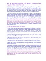
Figure 8 Picture of a patient after resection of thoracic oesophagus due to squamous cell oesophageal
cancer (condition prior to oesophageal reconstruction).
on the neck – salivary stula of the cervical esophagus;
on the right side of the chest – scar after thoracotomy;
in the epigastric midline – scar after laparotomy;
in the left hypochondrium – feeding gastric stula
The next stage consists in placing the gra, which was formed during the abdominal stage of the
operation, in the prepared retrosternal canal. For this reason, holding both previously placed retrac-
tors which elevate the sternum upwards, a thick long drain is inserted to the canal from the side of
the neck, one arm of the drain leading from the canal to the epigastrium, the other being maintained
on the neck. In order to shorten the gra’s route, a colon segment mobilized on a vascular pedicle is
passed beyond the stomach through an adequately wide slit, created in the hepatogastric ligament
before it is placed in the retrosternal canal. The cephalic segment of the gra is fastened to the drain
from the side of the abdomen. Gently pulling the drain’s arm protruding from the side of the neck,
the gra is pulled through the retrosternal canal and its cephalic part is exposed onto the neck in such
a way as to enable its tensionless anastomosis to the cervical esophagus. Now the sternum-elevating
retractors are removed. In this way the gra is positioned in the retrosternal canal. The part of the
11Esophageal Reconstruction with Large Intestine
right colon which forms the cephalic segment of the gra is situated on the neck, while the caecum
forming the caudal portion of the gra is anastomosed to the stomach. Thus constructed and placed
in the retrosternal canal gra is arranged antiperistaltically. When the gra is le in the retrosternal
canal, continuity of the gastrointestinal tract in the abdominal cavity should be restored, i.e. the ileum
should be anastomosed to the distal part of the transverse colon. During this procedure the blood
supply to the part of the gra emerging onto the neck should be monitored constantly. In case any
features of ischemia appear in the gra segment emerging onto the neck, the gra must be immedi-
ately evacuated from the retrosternal canal and the cause of ischaemia removed. Only eciently sup-
plied gra authorizes its anastomosis to the cervical esophagus. Anastomosis of the caudal segment
of the gra is performed in the prepyloric part of the stomach, what may prevent reux of the gastric
content to the replacement esophagus. Having performed all anastomoses within the abdominal cav-
ity, the last stage of the reconstructive surgery may be performed – end-to-side anastomosis of the cer-
vical esophagus with the lateral wall of the colon emerging onto the neck. The reconstructive surgery
is nished when the abdominal integuments and the cervical integuments are closed.
In case of patients aer resection of the thoracic esophagus due to cancer, the cervical stage of the
surgery is slightly dierent than in individuals with post-burn cicatrical stenosis. In some patients
resection of the thoracic portion of the esophagus due to cancer and reconstruction of the diges-
tive tract continuity is performed in a single-stage operation. The excised esophagus is then re-
placed with the whole stomach, or a tube formed from the greater curvature of the stomach local-
ized in the bed of the resected esophagus, i.e. in the posterior mediastinum. In patients, in whom
single-stage esophageal reconstruction by means of stomach is not possible for various reasons, a
reconstructive surgery with pedicled intestinal segment is considered. Due to a signicant extent
of the resection and reconstructive surgery, the procedure is performed in two stages. The rst
stage includes resection of the thoracic esophagus with lymphadenectomy, formation of a salivary
stula of the cervical esophagus, and a gastric or intestinal stula for feeding the patient (Fig. 8).
In the second stage, aer several weeks, the retrosternal replacement esophagus is constructed
with a pedicled colon segment. Mobilization of the pedicled colon segment, as well as creation
of a retrosternal canal from the side of the abdomen is performed in the above-described manner,
whereas opening of the retrosternal canal from the side of the neck is preceded by preparation of
the salivary stula and the cervical esophagus. The prepared segment of the cervical esophagus is
covered with a surgical towel and placed on the upper border of the surgical wound on the neck.
Opening of the retrosternal canal from the side of the neck is performed in the above-described
way. When the retrosternal canal is wide enough from the side of the neck, as well as from the
abdominal side, a mobilized pedicled colon segment is passed behind the sternum. Further proce-
dures, i.e. restoration of the continuity of the gastrointestinal tract in the abdominal cavity, anasto-
mosis of the caudal part of the gra with the stomach and anastomosis of the cephalic part of the
gra with the cervical esophagus is performed in the same way as described in patients with post-
burn cicatrical stenosis. In this group of patients the resection surgery is preceded by presurgical
chemotherapy, or chemo- and radiotherapy. In some patients chemotherapy is administered as
adjuvant therapy aer resection of the esophagus and prior to reconstructive surgery. As the aim
of the authors was to present only esophageal reconstructions with the use of pedicled intestinal
segments, treatment of esophageal carcinoma will not be discussed in details.
Esophageal Reconstruction12
Figure 9 Radiogram of a replacement retrosternal oesophagus from the right colon on ileocolic vascular
pedicle in an antiperistaltic position according to Jezioro
The above described modality of esophageal reconstruction is advantageous for a number of
reasons. The operation is fairly simple technically, provided the vascular system in this part of
the colon was evaluated accurately. Mobilization of the gra causes relatively small decit of
the intestine in the abdominal cavity. The gra, constructed according to the above-described
method, is long enough, and may be anastomosed to the pharynx, for example in patients
with obstructed cervical esophagus.
Theoretically, the only disadvantage of the reconstruction modality may be associated with
an antiperistaltic position of the gra, although control studies in patients with antiperistaltic
reconstructions did not conrm these fears (Fig. 9).
3.2. The technique of construction of an isoperistaltic gra from the right colon on middle
colic vascular pedicle
The right colon may be also used to isolate another kind – an isoperistaltic colon gra pedicled
on the middle colic artery. This type of gra is conditioned by the presence of well developed,
long main vascular trunks within the right colon, which are anastomosed with broad, well
developed and ecient arcades. The surgical modality presents as follows.
The abdominal cavity is opened through an upper midline incision passing by the umbilicus and go-
ing 2-3 cm below. The next stage is to mobilize the right colon and the terminal segment of the ileum
in a manner presented above, what enables macroscopic evaluation of the vascular system in this
part of the colon. On nding a positive vascular structure, a biological trial should be performed, in
which the trunks of the ileocolic and right colic vessels are clamped with vascular clamps, thus leav-
ing the selected part of the colon supplied only by the middle colic vessels. It should be remembered
that in some patients the right colic vessels are missing (see: Fig. 4), and the middle colic vessels
are shaped in the form of two or three additional colic vessels. In such cases, with narrow arcades
13Esophageal Reconstruction with Large Intestine
joining the middle colic vessels and the additional middle colic vessels, the use of both middle colic
trunks as the gra pedicle may be considered, provided the double pedicle does not shorten the
length of the mobilized colon gra. Any disturbances in the blood supply to the gra, arterial or
venous, observed during the biological trial, oblige to resign from this part of the colon and impose
selection of another, adequately supplied, segment of the large intestine.
In case the result of the biological trial is positive and no disturbances in the blood supply to the
isolated fragment of the colon are observed, mobilization of the gra may be initiated. First the
greater omentum is removed in the area of the mobilized colon segment, and next the vascular
trunks, which had been clamped in vascular clamps, are ligated and transected. In some cases
the arterial trunk should be ligated separately from the venous trunk, as they are oen distant
and their jointly ligation may contribute to shortening of the vascular pedicle in the mobilized
gra. Next the transverse colon should be transected in the middle of its length. The eerent
stump of the transverse colon is closed with a double-layer manual suture, or stapled. On the
other hand, the aerent stump, which forms the caudal segment of the mobilized gra, is closed
with a temporary suture until it is anastomosed with the stomach. Transection of the ileum in
the caecal region completes mobilization of the gra. The stumps of the transected ileum are
closed with a double-layer manual suture, or stapled, close to the caecum, without leaving di-
verticular excess. Appendectomy is performed in a routine manner. The isoperistaltic gra from
the right colon pedicled on the middle colic artery is thus constructed (Fig. 10).
Figure 10 Diagram of mobilization of a right colon graft together with the caecum on middle colic
vascular pedicle in an isoperistaltic position
Further steps are similar to those presented above. They include construction of the retroster-
nal canal, placement of the gra in the canal, restoration of gastrointestinal continuity and
anastomosing the gra with the stomach and the cervical esophagus.
Esophageal Reconstruction14
The above-presented modality of esophageal reconstruction has many advantages. At the same
time it is not free from certain disadvantages. The most signicant advantages include an isoperistal-
tic position of the gra, what undoubtedly has a positive eect on its functioning as an esophageal
replacement. Making a decision as to the choice of the above described reconstructive modality, it
should be remembered that it is conditioned by the presence of an exceptionally eective vascular
system within the right colon. In case additional middle colic vessels are present, they usually have
relatively short trunks, and shorter and narrower anastomosing arcades, and for these reasons the
mobilized gra may turn out to be too short to be anastomosed to the cervical esophagus. Another
disadvantage is a large mass of the cephalic segment of the gra, i.e. the caecum, what may hamper
safe passage of the gra through the superior opening of the retrosternal canal just beyond the le
sternoclavicular joint. Pressure present in the region of the superior opening of the canal leads to
irreversible ischemic changes and severe postsurgical complications, in form of necrosis of the ce-
phalic segment of the gra. In order to prevent such complications, it is necessary to perform partial
or complete removal of the joint, what is practiced in some surgical centres.
In cases, in which very long main vascular trunks in the right colon are found during the opera-
tion, and the anastomosing arcades are also long and wide, a similar gra may be constructed,
but without participation of the caecum. Then slightly longer segment of the transverse colon
should be mobilized. Such a variant of operation is possible when, apart from an adequate vascu-
lar system in the right colon, also the arc of Riolan is very well developed. In this way problems
associated with the presence of the caecum, which forms the cephalic portion of the gra, may be
avoided in some cases. Choosing this surgical modality, we obtain an isoperistaltic gra with a
straight shape and signicantly narrower diameter in the cephalic portion (Fig. 11).
Figure 11 Diagram of mobilization of a right colon graft on middle colic vascular pedicle in an isoperi-
staltic position without the caecum
15Esophageal Reconstruction with Large Intestine
As it was described in the previous section, the next stage of the operation includes construc-
tion of the retrosternal canal and passage of the gra through the canal created in the anterior
mediastinum. In order to obtain sucient mobility of the gra, it is worth considering reduc-
ing the caudal portion of the intestinal segment which will be anastomosed to the stomach.
The gra is then passed behind the stomach through a hole formed in the hepatogastric liga-
ment and placed in the created retrosternal canal. The proximal segment of the gra, i.e. the
caecum is exposed onto the neck, if the rst variant of the reconstructive surgery was chosen.
If mobilization of the gra was done according to the second variant, i.e. without participa-
tion of the caecum, the ascending colon is exposed on the neck. Anastomoses within the ab-
dominal cavity restoring the gastrointestinal continuity as well as anastomosis of the caudal
portion of the gra with the anterior wall of the prepyloric part of the stomach complete the
abdominal stage of the reconstructive surgery. The last stage includes anastomosing the cervi-
cal esophagus to the lateral wall of the colon exposed to the neck. Suturing of the abdominal
layers and the neck terminates the reconstructive surgery.
Remote follow up examinations in patients aer reconstructive surgery performed according to
the above described modality revealed ecient function of the replacement esophagus (Fig. 12, 13).
Figure 12 Radiogram of a replacement
retrosternal oesophagus from the right colon
on middle colic vascular pedicle in an
isoperistaltic position. A-P projection
Figure 13 Radiogram of a replacement retroster-
nal oesophagus from the right colon on middle
colic vascular pedicle in an isoperistaltic position.
Lateral projection
3.3. The technique of construction of an isoperistaltic gra from the right colon on le
colic vascular pedicle
Construction of a gra pedicled on the le colic artery is another possibility of using the right
colon for esophageal reconstruction. This variant of the reconstructive surgery is much more com-
Esophageal Reconstruction16
plicated and requires excellent surgical technique as well as careful intraoperative evaluation of
the colonic vasculature. This surgical modality may be used only when the right colonic as well
as the le colonic circulation is highly adequate and the arc of Riolan is very well developed. The
main advantage of this surgical modality is the possibility of obtaining a very long gra. The risks
however concern the vascular pedicle. The gra is supplied from the le colic vessels, but the only,
very long, route of blood supply to the whole gra is from the arc of Riolan. With an erroneous
evaluation of the ecacy of the gra vasculature, this situation may lead to peripheral ischaemia
of the mobilized segment of the colon. For this reason, when a decision is made to choose this
reconstructive modality, the intraoperative biological trial of vascular eciency should be meticu-
lously performed and accurately evaluated.
Figure 14 Diagram of mobilization of a right colon graft on left colic vascular pedicle in an
isoperistaltic position
Technically this modality is much more dicult than those previously described. Aer laparotomy
it is necessary to mobilize the right colon. The greater omentum is separated from the transverse
colon on its whole length. The result of the biological trial plays a decisive role. If, aer clamping the
trunks of the middle and right colic vessels and the arch joining the ileocolic vessels and the right
colic vessels, the observed colon does not reveal any signs of ischaemia, further mobilization of the
colonic segment may proceed. The le exure of the colon and the descending colon should be mo-
bilized. For this reason the small intestine loops are moved to the right side of the abdominal cavity
and maintained in this position with surgical towels. The peritoneum is transected longitudinally
on the external side of the descending colon as well as on the right side. Next, slightly elevating the
colon, the descending colon is carefully separated together with its vessels and the le exure of the
colon is exposed. Ligaments supporting the exure are cut between ligatures.
In the next stage of the surgery the vessels trunks which were previously clamped, i.e. the right
colic, middle colic and the arches between the right colic and ileocolic vessels are ligated and
transected. Also the ascending colon is transected at this level. Remembering that the gra has to
17Esophageal Reconstruction with Large Intestine
be long enough to reach the neck where it is anastomosed to the cervical esophagus, a longer or
shorter segment of the transverse colon is selected and cut at this level. Reduction performed in
the caudal portion of the gra permits to achieve its adequate mobility, and free from tension in
the vascular pedicle translocation of thus mobilized gra beyond the stomach, and next, through
the retrosternal canal, onto the neck (Fig. 14). Subsequently the continuity of the gastrointestinal
tract in the abdominal cavity should be restored and the caudal portion of the gra should be
anastomosed to the stomach. The reconstructive surgery is complete when the cephalic segment
of the gra is anastomosed to the cervical esophagus.
The advantageous points of this reconstructive modality include the possibility of obtaining
the longest gra of all previously described variants using the right colon provided the above
conditions concerning blood supply to the mobilized gra are fullled. Another advantage of
the presented modality is the isoperistaltic position of the gra, what has a benecial eect on
its further functioning as a replacement esophagus (Fig. 15, 16). The only disadvantage is theo-
retically much more dicult surgical technique in comparison to previously described variants.
Figure 15 Radiogram of a replacement
retrosternal oesophagus from the right colon
on left colic vascular pedicle in an isoperistaltic
position. A-P projection
Figure 16 Radiogram of a replacement
retrosternal oesophagus from the right colon
on left colic vascular pedicle in an isoperistaltic
position. Lateral projection
4. Esophageal reconstructions using the le colon
The presence of advantageous vasculature systems in the right and le halves of the colon as
well as ecient arc of Riolan provide opportunities of using the le colon to create a pedicled
esophageal gra in the following ways:
• from the le colon on the middle colic vascular pedicle in an antiperi-
staltic position of the gra