Tài liệu Endoscopy versus IVF: The Way to Go docx
Bạn đang xem bản rút gọn của tài liệu. Xem và tải ngay bản đầy đủ của tài liệu tại đây (492.71 KB, 26 trang )
Chapter 2
© 2012 Darwish, licensee InTech. This is an open access chapter distributed under the terms of the Creative
Commons Attribution License ( which permits unrestricted use,
distribution, and reproduction in any medium, provided the original work is properly cited.
Endoscopy versus IVF:
The Way to Go
Atef Darwish
Additional information is available at the end of the chapter
1. Introduction
1.1. Outlines
- Role of endoscopy in infertility. Microsurgical principles, reconstructive concept.
- Can endoscopy omit ART?
- Endoscopy Vs ART
- Advantages of ART over Endoscopy
- Fertility enhancing procedures:
Laparoscopic adnexal surgery.
Salpingoscopy
Hysteroscopy
- Endoscopy prior to ART (routine hysteroscopy, role of laparoscopy (tubes with or
withour hydrosalpnix and paratubal cysts, endometrioma).
- Endoscopically-assisted ART.
- Endoscopy for recurrent implantation failure.
- Future of endoscopy in the era of ART and keynote points.
2. Current approaches for infertility management
In modern practice, three schools are competitors for infertility management, namely
expectant, endoscopic and assisted reproductive techniques (ART) approaches. There are no
RCTs that compare the effectiveness of surgery againsteither IVF or expectant management.
The following table demonstrates pros and cons of each approach (1).
2.1. Rationale of expectant therapy
Any treatment should be compared to expectant therapy.
Enhancing Success of Assisted Reproduction
28
Table 1. Lines of infertility management
2.2. Drawbacks of the expectant therapy
No strict criteria on which to base management decisions.
Hence, the likelihood of spontaneous pregnancy for each individual couple must be
weighed against the potential benefits or risks of interventional treatment.
2.3. Is surgery better than IVF?
Logicstudies: Microsurgical reversal of sterilization is a highly cost-effective strategy when
compared with IVF for women aged 40 years and above (2).
Illogic studies: some over enthusiastic studies demonstrated that endoscopy is much
better than ART. In a bizarre study, Marana et al. (3) included 43 patientsand subjected
them to diagnostic or operative laparoscopy. Nine of themwith submucous-intramural or
multiple intramural fibroids underwent miomectomy by minilaparotomy following
hysteroscopy and chromopertubation. The mean length of follow- up was 49 months
(range: 11 to 118 months). They reported a very high pregnancy rate as 61 became
pregnant (40%).
ARTEndoscopic
management
Expectant
treatment
-Time saving
Excellent results
-Restores normal
anatomy.
-Enhances natural
pregnancy.
-Long-term results
Safe
cheap
Advantages
-Stress
-Expensive
-Risky
-OHSS
-Unpredictable
outcome
- Per trial result.
-expensive
-additional
specialist training
experience
-adverse effects
(including ectopic
pregnancies), and
operative risks.
-Unpredictable
outcome
Anxiety
Unpredictable
outcome
Disadvantages
Endoscopy versus IVF: The Way to Go
29
2.4. Advantages of laparoscopy over ART
Excellent results.
Long-lasting efficacy.
Reconstructive concept.
Physically and psychologically sound patient.
Tubal reconstructive surgery remains an important option for many couples and surgery
should be the first line approach for a correct diagnosis and treatment of tubal infertility (4).
2.5. Advantages of endoscopic management over conventional management
cosmetically most acceptable.
shorter hospital stay.
lower incidence of ileus.
faster recovery.
less post-operative pain and discomfort, and
earlier resumption of normal activities and employment.
reduced contamination of the surgical field with glove powder or lint.
bleeding is reduced due to tamponade of small vessels by the pneumoperitoneum
drying of tissues is minimal because surgery occurs in a closed environment
easy intraoperative access to the pouch of Douglas and the posterior aspects of the
genital organ.
2.6. Fertility-preserving reconstructive gynecologic surgery
Avoidance of serosal insults: tissue trauma, ischemia, hemorrhage, infection, foreign-
body reaction, and leaving raw surfaces.
minimizing tissue trauma: by using atruamatic techniques, meticulous hemostasis,
complete excision of abnormal tissues and precise alignment and approximation of
tissue planes .
Evidence of superiority of Laparoscopic reconstructive surgery: one study proved that
reconstructive surgery achieves a double pregnancy rate than non-reconstructive surgery
(5).
2.7. Is there a role for robotic surgery in improving pregnancy rate?
Among experienced endoscopists, it’s well known that it’s not the robot that does the
surgery, it’s the surgeon!
In a retrospective study, both robotically-assisted laparoscopic and standard laparoscopic
treatments of endometriosis had excellent outcomes. The robotic technique required
significantly longer surgical and anesthesia time, as well as larger trocars (6).
Enhancing Success of Assisted Reproduction
30
Facts Myth
no demonstration that it
increasess
p
eed or safet
y
Three-D vision
No RCT The sur
g
eon sees u
p
to 30% more endometriosis.
No RCT Less recurrence and slowl
y
No RCT Ra
p
id recover
y
and smooth
p
osto
p
course
this point is debated
among experts.
The dexterity (ability to bend at the “wrist”) of the robotic
instruments makes it possible to perform some surgeries
la
p
arosco
p
icall
y
that would otherwise re
q
uire la
p
arotom
y
.
Table 2. Pros and cons of Robotic surgery
Figure 1. Robotic surgery in Gynecology
2.8. Can IVF replace endoscopy?
Due to advances in the field of IVF/ICSI and stratification of management plans worldwide,
the overall pregnancy rate following IVF/ICSI overcame that following endoscopic surgery
in many centers. These encouraging results made some authors consider ART superior to
surgery and should be offered as a first-line treatment (7).
2.9. Which approach should we use: expectant, endoscopy or ART? (8)
The treatment choice depends upon:
Endoscopy versus IVF: The Way to Go
31
Severity of the tubal disease.
Duration of subfertility.
Maternal age.
Coexisting infertility factors.
Despite the widespread utilization of assisted reproductive techniques in recent years,
hysteroscopic as well as laparoscopic surgery should be firstly offered for patients with
adnexal and uterine lesions desiring fertility. Permanent correction of the patient’s problem
with frequent chances of pregnancy is a definite advantage of endoscopic surgery over
assisted reproductive techniques. Reconstructive endoscopic procedures could be
performed for fertile women as well e.g. hysteroscopic or laparoscopic myomectomy for
abnormal bleeding. The concept of reconstruction following microsurgical principles
coupled with refinement of instrumentation and techniques is would improve the results of
hysteroscopic and laparoscopic approaches. It is expected to expand to cover many
gynecologic aspects in the coming years particularly with the continuous advances in
technology of fine endoscopic surgery and the development of more suitable robotic
instrumentation.
2.10. Laparoscopy and IVF/ICSI are complementary since a long time (9)
The first in vitro fertilization (IVF) child ensued following the partnership by a scientist with
a focused ambition (Nobel laureate Robert Edwards) joining with the gynecologist who
introduced laparoscopy to Britain in the late 60's (Patrick Steptoe). Egg retrieval was done
laparoscopically. In modern practice, laparoscopic egg retrieval is still required whenever
inaccessible ovaries are encountered. A trial of transabdominal sonographic aspiration was
recently published with lower success rate of egg retrieval if compared to transvaginal
sonographic aspiration (10).
Laparoscopic GIFT: a blastocyst intrafallopian transfer was associated with an intrauterine
pregnancy; however, when the indication for blastocyst tubal transfer of an obstructed
cervix is associated with a foreshortened cervix requiring cervical cerclage, there can be
major health risks for infant and mother (11).
2.11. What’s the best approach?
Always try to use the appropriate approach for a suitable couple at the appropriate time. To
achieve the best results, try to stratify the lines of management according to pathology
putting in mind other circumstances. The following are examples of how to think in each
case separately.
3. Pelvic endometriosis: A good example of how to individualize
treatment
The optimal management of endometriotic ovarian cysts in infertile patients is less well
defined. Recent evidence of reduced responsiveness to gonadotrophins following
Enhancing Success of Assisted Reproduction
32
laparoscopic ovarian cystectomy has challenged the traditional surgical approach to
treatment (12). Indeed, it has been suggested that surgery should be undertaken only for the
treatment of large endometriomas or pain that is refractory to medical treatment, or to
exclude malignancy (13).
Laparoscopic surgery may be of benefit in treating subfertility associated with mild to
moderate endometriosis. However, additional studies in this field are needed before
definitive conclusions can be drawn (14). Laparoscopic excision of ovarian endometriomas
more than 3 cm in diameter may improve fertility. (level II evidence). The effect on fertility
of surgical treatment of deeply infiltrating endometriosis is controversial (level II evidence).
3.1. Is there a need to treat endometriosis in patients undergoing IVF?
In a meta-analysis (15)the chance of achieving pregnancy after IVF was significantly lower
for patients with endometriosis (odds ratio, 0.56; 95% confidence interval, 0.44-0.70), as
compared to those withtubal factor. They also reported decreased fertilization rates,
implantation rates and in the number of oocytes retrieved.
3.2. Mild endometriosis Vs severe endometriosis prior to IVF/ICSI
The same study (15) reported that the probability of pregnancy was reduced in women with
severe endometriosis as compared to those with mild disease.
Contrarily, a recent retrospective poorly designed study (16) demonstrated that ovarian
endometriosis does not reduce IVF outcome compared with tubal factor. Furthermore,
laparoscopic removal of endometriomas does not improve IVF results, but may cause a
decrease of ovarian responsiveness to gonadotropins. Nevertheless, they included a bizarre
group of patients with one or more endometrioma, unilateral or bilateral with a size of6 cm
and more importantly symptomatic as well as asymptomatic cases. In addition to being a
retrospective analysis, these heterogenous criteria would weaken this study. We believe that
stripping off cyst wall of a unilateral endometrioma wouldn’t be expected to affect ovarian
reserve or ovarian response to gonadotropins.
3.3. Advantages of laparoscopic surgery for endometriosis prior to IVF (ESHRE
Recommendations, 2005) (17)
confirms the diagnosis histologically
reduces the risk of infection
improvesaccess to follicles
Improves ovarian response.
3.4. More advantages include
Spontaneous pregnancy in mild and moderate disease.
Elimination of pelvic pain by destruction of the peritneal endometriotic lesions which
may be mistaken by OHSS if the patient is subjected to IUI.
Endoscopy versus IVF: The Way to Go
33
3.5. Precautions of laparoscopic surgery prior to IVF/ICSI
- The woman should be counseled regarding the risks of reduced ovarian function after
surgery and the loss of the ovary.
- The decision should be reconsidered if she has had previous ovarian surgery.
- RCTs showed that the excision technique is associated with a higher pregnancy rate and a
lower rate of recurrence although it may determine severe injury to the ovarian reserve.
- Improvements to this latter aspect may be represented by a combined excision-
vaporization technique or by replacing diathermy coagulation with surgical ovarian
suture.
4. Role of hysteroscopy prior to assisted reproduction
Failure of IVF treatment can be broadly attributed to embryonic, uterine or transfer factors,
but remains unexplained in most cases (18). A number of interventions have been proposed
to improve IVF outcome, many of which are not strictly evidence-based and their efficacy in
improving pregnancy rates remains controversial (19,20). One of the main causes of failure
of implantation after proper embryo transfer is intrauterine pathology. Whether to perform
hysteroscopic evaluation of the endometrial cavity prior to IVF/ICSI especially in patients
with repeated failures is a controversial issue that is open for criticism and deserves further
studies (21).
In a systematic review (Level Ia evidence), 5 reliable studies were included (22). Two RCT
showed a statistically significant improvement in the clinical pregnancy rate in the group
who had office hysteroscopy (pooled RR = 1.57, 95% CI 1.29–1.92, P < 0.00001). The
miscarriage rate was not statistically different between the office hysteoscopy and control
groups in either study (24% versus 29%, respectively, RR = 0.83, 95% CI 0.56–1.21, P = 0.33).
Three non-randomized controlled studies suggests that office hysteroscopy improves the
pregnancy rate in the subsequent IVF cycle (pooled RR = 2.01, 95% CI 1.60–2.52, P < 0.00001).
In addition to the well known diagnostic as well as therapeutic advantages of performing
hysteroscopy, even if the endometrial cavity was completely free, high pregnancy rate was
achieved after diagnostic hysteroscopy since uterine instrumentation during hysteroscopy
would inevitably cause a degree of endometrial injury and provokes a posttraumatic
reaction that involves release of cytokines and growth factors (23,24), which in turn may
influence the likelihood of implantation (25). Commencing IVF treatment soon after
hysteroscopy may take advantage of this immunological response (26). Performing
diagnostic hysteroscopy before assisted reproductive technologies
(ART) may be advisable
not only from the clinical but also
from the economic point of view (27). Enhanced clinical
pregnancy rates would be achieved on adding office hysteroscopy as a complementary step
prior to IVFspecially patients with recurrent IVF embryo transfer failures even after normal
hysterosalpingography findings. Some abnormal intrauterine findings that would affect the
prognosis of IVF/ICSI can be easily diagnosed by hysteroscopy like chronic endometritis,
Müllerian anomalies, retained fetal bones, or endocervical ossification. Moreover, contact
hysteroscopy may reveal addition valuable findings such as polyposis, strawberry pattern,
Enhancing Success of Assisted Reproduction
34
hypervascularisation, irregular endometrium with endometrial defects, or cystic
haemorrhagic lesion which are commonly seen with adenomyosis (28). Future high-quality
randomized trials are needed to confirm the favorable effect of standard hysteroscopy in
different IVF populations and examine whether newer and less invasive techniques of
uterine cavity evaluation such as mini-hysteroscopy (29) or hysterocontrast sonography (30)
would have an equally beneficial effect when compared with no intervention before IVF.
With the advent of technical refinements and advancement in hysteroscopic surgery, it is
expected that preoperative hysteroscopic evaluation of uteri prior to IVF/ICSI would be
widely performed. Unfortunately, many of studies on this topic focus on the central role of
hysteroscopic examination of the endometrial cavity in cases with recurrent failures
(28,31,32). This concept should be reviewed since office hysteroscopy or minihysteroscopy is
a simple outpatient conscious procedure (33-34) that provides excellent information on the
implantation site in the endometrial cavity in a very short time. Relying on
hysterosalpingography alone may be fallacious in some cases of fine intrauterine adhesions
that may be masked by dye especially oily dye. Likewise, transvaginal ultrasonography as
well as sonohysterograohy may miss some important fine intrautrerine lesions thatwould
simply contribute for failures (3). In one study, hysteroscopy succeeded to diagnose and
treat intrauterine lesions in 26% of patients prior to starting trials of assisted reproduction
(31). In a big sample sized study (36), intrauterine pathology was diagnosed in about 23% of
2500 cases prior to IVF trial. Another study diagnosed abnormalities in only 11 out of 678
cases. On reevaluation of DVD records of hysteroscopy by an experienced team, the same
team reported perfect diagnosis in 77.6% of cases (37).
Following recurrent IVF failure there is some evidence of benefit from hysteroscopy in
increasing the chance of pregnancy in the subsequent IVF cycle, both in those with abnormal
and normal hysteroscopic findings. Various possible mechanisms have been proposed for
this beneficial effect, but more randomized controlled trials are needed before its routine use
in the general subfertile population can be recommended (38).
4.1. What is the ideal approach prior to IVF?
In recent years, conflicting opinions on the role of hysteroscopy before any case of IVF/ICSI
or after failure once or more times. This conflict is due to different circumstances in different
parts of the world regarding:availability of free health insurance for IVF, experienced
hysteroscopists, availability of high-resolution 2D ultrasonography with or without SIS, use
of office versus conventional hysteroscopes, use of vaginoscopic approach or not and
socioeconomic level of the couple. Our opinion is summarized as follows:
In centers where health insurance is covering the cycles, experienced sonographers
performing high-resolution 2D ultrasonography with or without SIS, we believe that
they can proceed for IVF without prior hysteroscopy.
In centers where health insurance is not covering the cycles, experienced sonographers
performing high-resolution 2D ultrasonography with or without SIS are not available,
we believe that hysteroscopy specially office is very useful in such cases.
Endoscopy versus IVF: The Way to Go
35
In cases of failed IVF once, hysteroscopy is valuable and recommended.
In cases with recurrent implantation failure, hysteroscopy is mandatory.
4.2. Office hysteroscopy versus saline-infusion sonography (SIS)
In 1999, we published our first series of SIS for screening in infertile patients utilizing 0.9%
saline as an infusion solution and Nelaton catheters for injection (39). We reported satisfactory
results. One year later, we published a study (40) on the efficacy of SIS for the detection of
endometrial polyps in comparison to the conventional hysteroscopy. These studies compared
SIS versus conventional hysteroscopy with excellent results in favor of SIS. Later on, we
introduced office hysteroscopy (I use it since 2002 utilizing 2.6 mm telescope). With the advent
of vaginoscopic approach, the procedure gained more acceptability among our patients. Now,
after these years of experience we changed our mind and strongly say that office hysteroscopy
can easily replace indirect diagnostic tools like SIS or 4D ultrasonography. Moreover, more
detailed description of the endometrial cavity particularly the blood vessels would be obtained
only with office hysteroscopy as we recently published (41).
5. Role of hysteroscopy after embryo transfer
In a study evaluating the incidence of endometrial injury following embryo transfer, office
hysteroscopy was performed immediately following embryo transfer and demonstrated
marked endocervical and endometrial damage following rigid catheters more than soft
catheters (42). Even for cases of early abortion following IVF/ICSI, hysteroscopy was proved to
be very valuable. In one study (43), among 84 early abortion patients after IVF-ET, it succeeded
to diagnose intrauterine abnormalities in 58 (69.05%) of the patients, including intrauterine
adhesion in 32 (32/84, 38.10%), endometrial polyps in 12 (12/84, 14.29%), endometritis in 10
(10/84, 11.90%), submucous leiomyoma in 3 (3/84, 3.57%) and septa in 1 (1/84, 1.19%).
6. Hysteroscopic embryo transfer
As a trial of improving implantation rate following IVF/ICSI, some scattered papers
described hysteroscopically-guided embryo transfer. Principally, hysteroscopic approach
was selected in difficult cases of embryo transfer (44).
6.1. A new hysteroscopic tubal embryo transfer catheter was developed
Catheterization was performed in 60 patients at hysteroscopic insemination into tube, using
3 French catheters, in which the distal 3,4, and 5 cm tapered to 2 French. Hysteroscopic tubal
embryo transfer and conventional IVE-ET were performed in 30 patients with normal tubes,
who failed to achieve pregnancy after 2 IVF-ET trials. The success rate of complete insertion
with the catheter tapering at the distal 3 cm was significantly higher than that at the distal 5
cm. Since we obtained the highest success rate of insertion with the catheter tapering at the
distal 3 cm, we selected this catheter for the h-TEST. The rate of pregnancy in h-TEST was
significantly higher than that in conventional ET (45).
Enhancing Success of Assisted Reproduction
36
6.2. Hysteroscopic Endometrial Embryo Delivery (HEED) (46)
It refers to visually confirmed placement of the embryo(s) at a specific area on the surface of
the uterus. It is done in an office setting, using a special fiberoptic scope and camera plus
special tubing, and it takes approximately two minutes to perform. It uses nitrogen gas to
avoid deleterious effect of CO2 gas o n the embryos. HEED can also be used for earlier (day
2 or 3) embryos as well as the more advanced embryos. This is especially advantageous in
situations where the numbers of embryos are limited, or embryo quality is of concern. It is
particularly useful in patients with advanced reproductive age, or when egg production is
low, or in patients with poor sperm parameters. Patients will actually see the process on
video monitor. The entry into the uterus is not always easy, as the non-stirrable tip of the
catheter must usually go through different curvatures in the cervical canal and the uterine
cavity while minimizing injury to the lining of the uterus, before it reaches the final
destination. The flexible hysteroscope has a stirrable tip, helping guide the endoscope in a
gas expanded uterine cavity. The slightly expanded uterine cavity also helps avoid contact
between the hysteroscope and uterine surface. The final destination of the tip of the catheter
is visually confirmed. This more precise placement and lower volume of transfer fluid may
help reduce incidence of ectopic pregnancies even further. It may also reduce chances
placenta previa, where the after birth is lying over the uterine opening. Presence of uterine
contraction at the time of transfer that are otherwise not noticeable by using the “Blind”
embryo transfer technique, can be visually confirmed and embryo transfer deferred. Precise
and visually confirmed placement, may reduce percentage of multiple pregnancies, by
reducing number of embryos transferred because of the less uncertainty of the placement of
embryos with the “Blind” technique. Nevertheless, since the embryo(s) are laid on top of the
uterine surface, due to inherent uterine contractions over the next few days after the embryo
delivery and prior to their natural implantation in the uterine cavity, the embryo(s) may be
expelled either into the fallopian tube (causing ectopic pregnancy) or out of the uterus, as
they do with the current “blind” embryo transfer technique.
6.3. Subendometrial embryo delivery (SEED) (47)
Patients will actually see the process on video monitor. It will reduce the chances that the
embryo will fall out of the uterus, or that it will fall into the fallopian tube causing tubal
pregnancy. Post embryo implantation, the woman does NOT need to stay in bed for 2 days.
The main disadvantage includes a possible scratching of the lining of the uterus so that
pregnancy may not ensue. Candidates include any patient undergoing IVF, specially
patients with previously failed standard embryo transfers, patients with ectopic pregnancies
and tubal disease.
It is done in an office setting using a special fiberoptic scope and camera plus a special
tubing with a needlepoint, and it takes approximately two minutes to perform. It utilizes
flexible hysteroscope and an inert gas (nitrous gas) to avoid the deleterious effect of CO2 gas
on the embryos.
Endoscopy versus IVF: The Way to Go
37
6.4. Hysteroscopic cervical canal refashioning prior to difficult embryo transfer
(48)
In some cases, access to the endometrial cavity is extremely difficult or even impossible. In
some scarce studies. Sonographically-guided fine needle transmyometrial embryo transfer
was tried but this technique is not universally accepted. An attractive recent hysteroscopic
approach was described. The procedure is performed under general anesthesia. Patients are
taken into the theater with a full bladder in case ultrasound guidance is required to access
the uterine cavity. A Versapoint electrode (twizzle electrode) with a 1.9 mm Versascope
(Gynecare division, Johnson and Johnson) is used for the procedure. The Versapoint
electrode works on bipolar energy, so saline is used as the distension media. Versascope
sheath has a small diameter (3.5 mm) and it can be inserted into the cervical canal without
prior dilatation or with minimal dilatation. In two patients the canal is extremely tortuous
and fibrotic and it is not possible to negotiate with the delicate Versascope. Cervical
dilatation is achieved under ultrasound guidance in these women and the Versapoint
twizzle electrode is introduced through the operating channel of an operating hysteroscope
(Olympus).
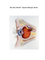
Figure 2. Hysteroscopic cervical canal refashoning
For women with a false passage and acute angulation of the uterus, the tissue between the
actual cervical canal and false passage is cut thus leaving a clean path which could be
negotiated with an ET catheter. For the problem of a severely fibrotic OS, 1 or 2 linear
releasing incisions are made with the Versapoint electrode, extending from the posterior
aspect of the internal OS towards the external OS for approximately 1 cm. In patients who
had a tortuous cervical canal, several projecting ridges are seen arising from the anterior,
posterior and/or lateral walls of the cervical canal. The hysteroscope is introduced into the
uterine cavity and then withdrawn towards the external OS. As the hysteroscope is moved
Enhancing Success of Assisted Reproduction
38
outwards the cervical canal projections distorting linearity of the canal are visualized. Linear
releasing incisions of approximately a centimeter are made into these projections and a
straightening of the canal is achieved. Subsequent to the procedure, dilatation is done to
further stretch the incised fibrous tissue, and it is now possible to dilate the cervix up to size
10/12 Hegar in even the most resistant cervix.
6.5. Hysteroscopic site-specific endometrial injury (49)
A site-specific hysteroscopic biopsy-induced injury of the endometrium during the
controlled ovarian hyperstimulation cycle has been shown to improve subsequent embryo
implantation in patients with repeated implantation failure. The procedure starts with
performing panoramic hysteroscopy. A flexible claw forceps is introduced through a 2.2 mm
working channel which is used to generate a local injury on the posterior endometrium at
midline 10-15 mm from the fundus on D4 to D7 of the stimulation cycle. The depth and
width of the injured site is 2 × 2 mm (i.e. a bite of the claw forceps). No antibiotic or
hemostatic drug is administered after the procedure.
Endometrial injury may have a beneficial role in implantation and improve the pregnancy
rate. However, there are still many unanswered question including patients selection,
timing, technique and number of endometrial biopsies needed (50).
7. Role of endoscopy in cases of hydrosalpnix
Tubal pathology, particularly hydrosalpinx, is associated with a low embryo
implantation rate in IVF as well as an increased risk for early pregnancy loss.
The role of surgery for tubal disease to improve IVF outcomes, in the absence of
hydrosalpinx, requires further evaluation.
In recent years, considerable attention has been given to the possible impact of the presence
of hydrosalpinx on implantation and ongoing pregnancy rates following IVF/ICSI (51,52).
The mechanism of disruption remains uncertain. However, proposed mechanisms may be
attributed to alteration in endometrial receptivity ordirect embryo toxic effect (53).
Furthermore, hydrosalpnix is liable be unintentionally punctured at the time of egg retrieval
or it may disturb the access to the ovary if it is too big. A systematic review of three RCTs
(54) showed that tubal surgery such as laparoscopic salpingectomy significantly increased
live birth rate (OR 2.13; 95% CI 1.24 to 3.65) and pregnancy rate (OR 1.75; 95% CI 1.07 to
2.86) in women with hydrosalpinges before IVF when compared with no treatment. There
are no significant differences in the odds of ectopicpregnancy (OR 0.42; 95% CI 0.08 to 2.14),
miscarriage (OR 0.49; 95% CI 0.16 to 1.52), treatment complication (OR 5.80; 95% CI 0.35 to
96.79) or implantation (OR 1.34; 95% CI 0.87to 2.05). Since hydrosalpinx reduces IVF
pregnancy rates (14,55), it is therefore suggested that women with hydrosalpinges should be
offered diagnostic/operative laparoscopy and a trial of salpingoneostomy. If failed or
inaccessable, salpingectomy could be offered prior to IVF/ICSI to improve the chance of a
live birth. Sometimes, laparoscopic access to the isthmic part of the tube is not feasible even
in experienced hands particularly in patients with history of repeated laparotomies,
Endoscopy versus IVF: The Way to Go
39
intestinal reanastomosis, or kidney transplantation. This situation may pave the way to
hysteroscopic occlusion of the fallopian tubes based on the reported success in hysteroscopic
tubal cannulation and sterilization techniques. The effectiveness of draining of
hydrosalpinges or performing salpingostomy on improving live birth rate prior to IVF/ICSI
needs further evaluation.
7.1. Methods of endoscopic proximal occlusion of functionless and harmful
hysrosalpnix
1. Laparoscopic: this can be easily performed using a bipolar grasping forceps or
monopolar grasping forceps. In either approach, take care to apply a little traction on
the tube medially to avoid scattered secondary coagulation towards the lateral pelvic
wall particularly when utilizing monopolar diathermy. By this way, the ureter would
be perfectly secured. Some center using clips.
2. Hysteroscopic: this approach can be performed whenever laparoscopic approach is
impossible or dangerous like cases with history of extensive abdominal surgery like
resection anastomosis of the intestine or previous colonic surgery, or patients with a
history of extensive or recurrent surgery for pelvic endometriosis. Practically,
endoscopists may face some cases without feasibility to perform laparoscopy from the
start. These cases deserve searching for an alternative approaches. Hysteroscopy comes
as an attractive valuable alternative. Some studies used Essure devices to
hysteroscopically occlude the proximal part of the fallopian tube. They reported some
case reports of successful pregnancy. Nevertheless, we believe that leaving a foreign
body in-utero would lead to decreasing implantation rate. Herein, I’ll discuss in details
our previous unique study on hysteroscopic tubal occlusion in cases with hydrosalpnix
(56). The in-vitro safety phase of this study is done on fresh uterine specimens removed
by abdominal or vaginal hysterectomy. In this phase the study, fresh hysterectomy
specimens are placed on the return electrode of diathermy, then the corneal ends of
both tubes are coagulated simulating the same manner as in the clinical phase.
Temperature study is done using digital thermometer over the uterine serosa at site of
the coagulation. Histopathologic sections are made to assess tissue effects and depth of
penetration using Nitro Blue Tetrazolium (NBT) to evaluate the extent of coagulation
on the tubal uterine junction. Computerized image analyzer (Leica Q 500 MB
Computerized Image Analyzer) is used to measure the depth of diathermy damage to
the surrounding myometrium. The clinical phase of this study is conducted at the out-
patient Infertility clinic of Women Health hospital, Assiut University, from April 2004
to October 2006 and included 27 patients with definite uni- or bilateral laparoscopically-
proved functionless hydrosalpinges scheduled for IVF/ICSI. All patients gave a written
consent and the study is approved by the institutional ethics committee. They were
randomly divided into 2 groups. Randomization is done using simple computer
generated randomization tables method. Group A comprised 14 patients who were
randomly allocated for laparoscopic occlusion. Laparoscopy is performed under
general endotracheal anesthesia using a standard double puncture technique. Once the
Enhancing Success of Assisted Reproduction
40
peritoneal cavity is entered, a panoramic evaluation of the pelvis is done. If the pelvis
looks frozen or if the access to the fallopian tubes is impossible, the patient is
considered failed laparoscopic approach. Those cases are subsequently treated by open
laparotomic or hysteroscopic approach but the results of these procedures are not
included in this study. If the procedure seems feasible, a third auxillary puncture is
done. Utilizing a bipolar forceps, the isthmic part of the fallopian tube is coagulated and
incised to ensure complete tubal occlusion as a case of tubal sterilization. The procedure
is completed after securing hemostasis. The patient is discharged after 3-4 hours under
antibiotic prophylaxis. Group B included 13 patients scheduled for hysteroscopic
approach. The cervix is primed in all cases using 200 Mg misoprostol 8 hours prior to
the procedure as previously described (57). The procedure is done immediately
postmenstrual without specific preparation. Local paracervical anesthesia is selected in
5 cases while spinal anesthesia in 6 cases, and general anesthesia in 2 cases. Selection of
the anesthestic technique is chosen according to patient preference after proper
explanation by the anesthiologist. The cervix is gently dilated till Hegar's 10 which is
followed by insertion of a rotatory continuous flow monopolar resectoscope. Once the
peritubal pulge (the proximal part of the intramural segment of the tubeis clearly seen,
a roller ball electrode of 3 mm size is bluged inside it and activated at 50 watts for about
8 seconds. A thorough comment on the fundus and the rest of the endometrial cavity is
reported. The patients are discharged immediately if the procedure is done under local
paracervical anesthesia, while the remaining cases are discharged few hours later. In
both groups, the procedure is preceded and done under prophylactic broad spectrum
antibiotic coverage to guard against any risk of flaring up of infection of the
functionless hydrosalpnix. In both groups, patients are instructed to come back the next
cycle postmenstually where hysterosalpingography (HSG) is done for most cases
especially those with unilateral functionless hydrosalpnix. If the patient refused and has
bilateral hydrosalpnix, sonohysterography (SHG) is done utilizing a simplified
technique as previously described (39). Tubal occlusion of the affected side is confirmed
if marked resistance is encountered on repeated injection of saline without evidence of
intraperitoneal leakage from the occluded side which is the main outcome measure.
Second-look office hysteroscopy is done for patients in group B whenever possible. The
in-vitro safety phase resulted in bilateral complete coagulation of the proximal part of
the tubes with secondary coagulation shown of up to 3 mm as shown in the
histopathologic sections. When the power of coagulation is 50-60 W and operating time
not prolonged more than 20 seconds , the thermal damage covered corneal end as
complete coagulation in addition to2mm -3 mm secondary coagulation of the adjacent
cornualendo- myometrium. Serosal temperature is not exceeding 41.9 Cº (range 39 Cº -
41.9 Cº) at any time during the procedure. No full thickness injuries are demonstrated
either histologically or suggested by the temperature studies. Hysteroscopic access is
achieved in 12 (85.7%)and occlusion is achieved in 9 (64.2%) cases. If the peritubal pulge
is not clearly visible, the case is considered as failed access to the proper site of
occlusion. In group B, diagnostic hysteroscopy showed fine marginal adhesions in 2
cases (15%) and a small polyp in one case (7.7%). Hysteroscopic tubal occlusion showed
Endoscopy versus IVF: The Way to Go
41
shorter operative time (9 ±2.76 min vs. 23.6 ±4.75 min, p= 0.0001) and hospital stay as
well (2 ±1.84 hours vs. 5 ±1.13 hours, p= 0.0001). No case of intraoperative complication
in either group is reported. There is no case of exaggerated postoperative pelvic pain or
fever in either group. HSG or SHG demonstrated complete tubal occlusion of the
affected side in all cases in both groups). Second-look office hysteroscopy is done in 8
cases of group B
which revealed no significant corneal lesions at the site of
hysteroscopic occlusion. Pregnancy is achieved in 4 (28.5%) and 4(30.7%) cases in both
groups respectively following IVF/ICSI without any significant difference between both
groups.
7.2. Comments on hysteroscopic tubal occlusion in hydrosalpnix
Hysteroscopic tubal occlusion of functionless hydrosdalpngies is a unioque one. It
demonstrates a valuable role of hysteroscopic approach that can be performed in difficult
cases with poor access to the isthmic part of the tubes via laparoscopy even with
experienced hands. The idea is attractive but further large-sample sized studies are required
to define the exact role of this approach.
One of the interesting additive items of this paper to the literature is the term "functionless"
hydrosalpnix. The proposed definition is very crucial to stratify cases suitable for
microsurgical salpingoneotomy and those cases suitable for occlusive procedures. By this
way, the place of reconstructive surgery is still preserved in modern practice even in the era
of IVF/ICSI. Ethically, every effort should be exerted to restore normal anatomy whenever
possible. This concept is of utmost importance particularly for the developing countries with
limited resources where no national programs to support assisted reproductive techniques.
Microsurgery to correct localized damage has the advantage of long-standing restoration of
fertility. A simple prognostic classification is lacking. The severity of the tubal damage and
the health of the mucosa is key in determining outcome. Proper selection of the tube for
either line of management requires expert knowledge with the principles of salpingoscopy.
Salpingoscopy during laparoscopy yields the best prognosis in patients with hydrosalpinx.
Performing salpingoscopy with laparoscopy could significantly increase accuracy in
predicting short-term fertility outcome. Whenever the mucosa is unhealthy, surgery is not
justified; early referral for IVF is indicated.
Hysteroscopic tubal occlusion of proximal part of the hydrosalpnix is feasible and
promising as a safe, effective, fast, and easy approach. It can be done as an out-patient
procedure under local paracervical block. It has the advantage of adding valuable
evaluation of the endometrial cavity prior to IVF/ICSI. Further large sample-sized studies
are required specially those utilizing bipolar resectoscope. The impact of hysteroscopic
tubal occlusion on subsequent implantation and pregnancy rates needs to be addressed in
another larger study. Since it is a preliminary study, the current role of hysteroscopic
occlusion should be limited to cases of failed laparoscopic approach. Further studies are
required before moving hysteroscopic occlusion to replace laparoscopic occlusion prior to
IVF/ICSI.
Enhancing Success of Assisted Reproduction
42
7.3. A suggested flowchart for management of functionless hydrosalpnix prior to
IVF/ICSI
Laparoscopic tubal surgery: tubal factors include proximal tubal occlusion, distal tubal
phimosis or occlusion or peritubal adhesions. Endoscopy (whether laparoscopy or
hysteroscopy) play a central role in the management of tubal disease.
Tubal pathology, particularly hydrosalpinx, is associated with a low embryo
implantation rate in IVF as well as an increased risk for early pregnancy loss.
The role of surgery for tubal disease to improve IVF outcomes, in the absence of
hydrosalpinx, requires further evaluation.
Figure 3. HSG: Hysterosalpingography. TVS: Transvaginal ultrasonography.
7.4. Laparoscopic management of distal tubal disease
Distal tubal occlusion may be due to hydrosalpnix, pyosalpnix or peritubal adhesions.
Obstruction of the distal fallopian tube is one of the most common causes of female infertility
(58). In cases of pyosalpnix, just tubal opening, drainage of pus and proper peritoneal toilet are
sufficient. Don't forget to take a tubal wall biopsy. Don't proceed for tubal occlusion at the
same setting for fear of disseminating infection and the possibility of tubal bilharziasis with
reported cases of spontaneous pregnancy after proper treatment. Nowadays, it is conceived
that the presence of hydrosalpinx is associated with a compromised outcome for IVF/ ICSI.
Hydrosalpinx is associated with lower implantation and fecundibility rates even if the
contralateral tube is sound which may be attributed to alteration in endometrial receptivity
Endoscopy versus IVF: The Way to Go
43
(59) or direct embryo toxic effect. Furthermore, it is liable to be unintentionally punctured at
the time of egg retrieval or it may disturb access to the ovary if it is too big. In a meta-analysis,
it has been demonstrated that there is a reduction by half in the probability of achieving a
pregnancy in the presence of hydrosalpinx, and an almost doubled rate of spontaneous
abortion (60). In an animal study, hydrosalpinx fluid is shown to contain toxins that are
potentially teratogenic (61). Proposed mechanisms of impaired implantation rate due to
hydrosalpinges are well addressed in the literature (62). Selected patients with unilateral
hydrosalpinges and a patent contralateral Fallopian tube may exhibit increased cycle fecundity
after salpingectomy or proximal tubal occlusion of the affected tube, and may conceive
without the need for IVF. In a retrospective case-control study, bilateral salpingectomy due to
hydrosalpinges restored a normal delivery as well as implantation rate after IVF treatment
compared to controls (63). Randomized controlled trials recommended performing
laparoscopic salpingectomy prior to IVF, especially inpatients with ultrasound-visible
hydrosalpinges (64). In a Cochrane review (65), it is concluded that further randomized trials
are required to assess other surgical treatments for hydrosalpinx, such as salpingostomy, tubal
occlusion or needle drainage of a hydrosalpinx at oocyte retrieval. Functionless hydrosalpinx
can be defined as a large blocked tube with lost major and minor folds, as seen at
salpingoscopy after laparoscopic salpingoneostomy.
Figure 4. Sonographic appearance of a typical hydrosalpnix.
On sonography, the dilated fallopian tube presents as a thin-
or thick-walled tubular fluid-
filled structure that may be elongated
or folded (figure). Longitudinal folds that are
present
in a normal fallopian tube may become thickened in the
presence of a hydrosalpinx. The
dilated fallopian tube may or
may not show longitudinal folds. These longitudinal folds are
pathognomonic of a hydrosalpinx .
If the elongated nature of these folds is not noted, they
may
be mistaken for mural nodules of an ovarian cystic mass. Identification of a separate
ovary helps
distinguish a hydrosalpinx from a cystic ovarian mass, an important
distinction
because malignancy is rare with an extraovarian
cystic adnexal mass. A significantly scarred
hydrosalpinx may
present as a multilocular cystic mass with multiple septa creating
multiple compartments. These septa
are generally incomplete, and the compartments can be
connected.
However, with more pronounced scarring, differentiation from
an ovarian mass
may not be possible (66). Potential pitfalls
in the diagnosis of hydrosalpinx include
Enhancing Success of Assisted Reproduction
44
paratubal, paraovarian,
or perineural cysts. In some cases, CT or
MRI may be helpful to
differentiate these conditions from a
hydrosalpinx (67).
7.5. Technical tricks of laparoscopic management of hydrosalpnix
a. Salpingoneostomy: One of the keys of success is to evaluate the tube externally and
internally. If peritubal adhesions exist, microsurgical adhesiolysis should be performed
at first. Be sure that the tube is freely mobile. Imagine the site of the new ostium before
dealing with the hydroslpnix. It should be directed towards the pouch of Douglas to
help ovum pick-up. Start by salpingoneostomy using a fine monopolar or bipolar
needle. The finest the needle, the better ostium. Incise the distended distal part of the
tube “ + shaped” (cruciate incision). Then, evaluate the tubal mucosa using a
salpingoscopy. Practically, use the diagnostic hysteroscopy which consists of a 4 mm
telescope and a 5 mm outer sheath. Connect it to a normal IV infusion set and use saline
as an irrigating fluid. Grasp the new ostium with an atruamatic grasping forceps and
insert the hysteroscope with comment on the major and minor folds till reaching the
narrowest part of the tube. If major and minor folds are lost this means that the
prognosis is poor even after proper refashioning. The next step is to grasp the tubal
lumen with atruamatic forcpes and to evert it outside. Lastly, fix the edges of the new
ostium either with monopolar spray coagulation just distal to the incised parts to evert
them or with the aid of fine sutures.
b. Salpingectomy: This procedure is indicated if a pathologic unilateral huge hydrosalpnix
is present to enhance spontaneous pregnancy or bilateral big hydrosalpnix before
IVF/ICSI. It is performed in the same manner as mentioned in the section of EP.
c. Tubal occlusion: Once the peritoneal cavity is entered, a panoramic evaluation of the
pelvis is performed. If the pelvis looked frozen or if access to the fallopian tubes is
impossible, the patient is considered a failed laparoscopic approach. Those cases are
subsequently treated by open laparotomic or hysteroscopic approach. If the procedure
seems feasible, a third auxiliary puncture is carried out. Utilizing a bipolar forceps, the
isthmic part of the fallopian tube is coagulated and incised to ensure complete tubal
occlusion, as a case of tubal sterilization. The procedure is completed after securing
hemostasis. The patient is discharged after 3-4 h under antibiotic prophylaxis.
Laparoscopic salpingectomy or bipolar proximal tubal occlusion yielded statistically
similar responses to controlled ovarian hyperstimulation and IVF-ET cycle outcome.
Proximal occlusion might be preferable in patients who present with dense pelvic
adhesions and easy access only to the proximal fallopian tube (68). Occlusion is
considered a minimally invasive procedure, requires less experience, feasible in most
cases, and has fewer burdens on the psychological status of those infertile women.
Hysteroscopic approach is recently described by our team at Assiut University
Institution (69). The cervix is primed in all cases using misoprostol (200 µg) 8 h prior to
the procedure. The procedure is carried out immediately postmenstrual without
specific preparation. Local paracervical, spinal or general anesthesia could be used.
Selection of the anaesthetic technique is chosen according to patient preference after
Endoscopy versus IVF: The Way to Go
45
proper explanation by the anaesthesiologist. The cervix is gently dilated with Hegar 10
and a rotatory continuous flow monopolar resectoscope is inserted. Once the peritubal
bulge (the proximal part of the intramural segment of the tubeis clearly seen, a roller
ball electrode (size: 3 mm) is introduced inside it and activated at 50 Watts for about 8
seconds. A thorough comment on the fundus and the rest of the endometrial cavity
should be reported. The patients are usually discharged immediately if the procedure is
carried out under local paracervical anesthesia, while the remaining cases are
discharged a few hours later.
8. Proximal tubal occlusion
Usually diagnosed by HSG and confirmed by laparoscopic chromopertubation test. The
most important job of the endoscopist is to find out contraindications for hysteroscopic tubal
cannulation procedure which include:
florid infection
genital tuberculosis
obliterative fibrosis and long tubal obliterations that are difficult to bypass with the
catheter.
severe tubal damage.
previously performed tubal surgery.
salpingitis isthmica nodosa.
isthmic occlusion with club-changed terminal, ampullar or fimbrial occlusion, and tubal
fibrosis
coaxial TO: combined distal and proximal tubal occlusion.
Don't try to cannulate the tube in such cases as failure would be expected and you would be
disappointed. In cases with isolated tubal occlusion, cannulation would be successful.
9. The following is our protocol for tubal disease management
Pathologic PTO: IVF
Isolated PTO: TC
Midsegment O: IVF
Small hydrosalpnix+ normal salpingoscope:OL
Functionless hydrosalpnix: PTO then IVF
Combined PTO and DTO: IVF
Peritubal adhesions: OL Vs IVF
10. Endoscopic uterine surgery
10.1. Endoscopic myomectomy prior to IVF/ICSI
The impact of uterine myoma on the outcome of IVF/ICSI is a very controversial topic.
Many centers are overdoing myomectomy for nearly all myomata regardless size and site
considerations. Contrary, other investigators have shown that fibroids don't exert a
Enhancing Success of Assisted Reproduction
46
deleterious effect. Nevertheless, many studies have provided evidence that uterine myomas
have a significant effect on IVF outcomes and there is a large body of evidence that
treatment of uterine myomas increases fertility and pregnancy rates, and decreases the rate
of pregnancy loss (70). There is no doubt that any cavity-distorting myoma should be
removed whether completely submucous or interstitial myoma with submucous
encroachment. This highlights the central role of prior hysteroscopy as well as saline
infusion solonhysterography (SIS) as previously described (39). Not only does sub mucous
myoma cause mechanicl interference with implantation, but it also alters endometrial
receptivity (71)
Controversy exists for interstitial and subserous myomata. The evidence supports treatment
of all very large myomas (>7 cm) (70). Subserosal myomas that are smaller than 7 cm in size
and intramural myomas of less than 4–5 cm in diameter appear to have little effect on IVF
outcomes. Larger intramural and subserosal myomas present a clinical dilemma and more
studies are needed to clarify a definitive plan for management (70). In a prospective
controlled study, the distance between the intramural myomas and the endometrial lining
did not appear to affect the IVF outcome. An insignificant tendency towards improvement
of IVF outcome is found in myomas at more than 5 mm from endometrial lining (72).
In a recent review of literature (73) on myoma and assisted reproduction technology and
spontaneous conception, hysteroscopic sub-mucous myoma resection is found to increase
pregnancy rates. Intramural fibroids appear to decrease fertility, but the myomectomy does
not improve assisted reproduction technology and spontaneous fertility. More high-quality
studies are needed to conclude toward the value of myomectomy for intramural fibroids.
Subserosal fibroids do not affect fertility outcomes, and removal does not confer benefit.
11. Keynote points
11.1. Tubal infertility
Endoscopy and ART arenot competitors but complementary.
First trial is the best trial for tubal surgery.
Performing laparoscopic surgery Forendometriosis prior to IVF isveryvaluable in
manycases.
There is NO adequate trialscomparing pregnancy rates with tubal surgeryversus IVF.
Per cycle pregnancy rate of IVF: higher
Tubal anastomosis for reversal ofsterilization has significantly higher cummulative
pregnancy rate than IVF and ismore cost effective even above 40 years.
Factors affecting counseling for tubal surgery or IVF:
- Age of the woman.
- Ovarian reserve.
- Number and quality of sperms/ejaculate.
Endoscopy versus IVF: The Way to Go
47
- Number of children desired.
- Site and extent of tubal disease.
- The presence of other infertility factors.
- The risk of ectopic pregnancy.
- The experience of the surgeon.
- The success rate of IVF program.
- The patient preference.
11.2. Uterine myoma and infertility
Uterine myoma may affect fertility according to its size, site and associated pathology.
Endoscopic approach has a definite role in its management. HM is the gold standard line of
management of submucous myoma of suitable size. LM doesn't seem to be superior to
conventional open myomectomy regarding fertility and is characterized by both short and
long term drawbacks. Uterine myomata would affect IVF/ICSI outcome whenever
disturbing the endometrial cavity or large sized. The impact of other types of myomata on
IVF/ICSI deserves further studies. Hysteroscopic myomectomy is indicated for intracavitary
myomas and submucous myomas having at least 50% of their volume within the uterine
cavity. The management of the subfertile women with small intramural fibroids (<5 cm) is
still a subject of debate (75,76).
Author details
Atef Darwish
Obstetrics and Gynecology, Assiut University, Assiut, Egypt
12. References
Pandian Z, Akande VA, Harrild K, Bhattacharya S. Surgery for tubal infertility.
[1]
Cochrane Database Syst Rev 2008; Issue 3. Art. No.: CD006415.
Petrucco OM, Silber SJ, Chamberlain SL, Warnes GM, Davies M. Live birth following
[2]
day surgery reversal of female sterilisation in women older than 40 years: a realistic
option in Australia? Med J Aust 2007; 187:271-3.
Marana R, Ferrari S, Merola A, Astorri AL, Pompa G, Milardi D, Giampietro A, Lecca A,
[3]
Marana E. Role of a mini-invasive approach in the diagnosis and treatment of tubo-
peritoneal infertility as an alternative to IVF . Minerva Ginecol. 2011 Feb;63(1):1-10.
Muzii L, Sereni MI, Battista C, Zullo MA, Tambone V, Angioli R Tubo-peritoneal factor
[4]
of infertility: diagnosis and treatment. Clin Ter. 2010;161(1):77-85.
Bateman BG, Nunley WC Jr, Kitchin JD 3rd. Surgical management of distal tubal
[5]
obstruction are we making progress? Fertil Steril. 1987 Oct;48(4):523-42
Nezhat C, Lewis M, Kotikela S, Veeraswamy A, Saadat L, Hajhosseini B, NezhatC.
[6]
Robotic versus standard laparoscopy for the treatment of endometriosis.Fertil Steril.
2010 Dec;94(7):2758-60.
Enhancing Success of Assisted Reproduction
48
Feinberg EC, Levens ED, DeCherney AH. Fertil Steril. Infertility surgery is dead: only
[7]
the obituary remains? 2008 Jan;89(1):232-6.
ASRM Practice Committee. The role of tubal reconstructive surgery in the era of
[8]
assisted reproductive technologies. Fertil Steril 2008; S250–S253.
Yovich JL. A clinician's personal view of assisted reproductive technology over 35
[9]
years. Reprod Biol. 2011 Dec;11 Suppl 3:31-42.
Barton SE, Politch JA, Benson CB, Ginsburg ES, GargiuloAR Transabdominal follicular
[10]
aspiration for oocyte retrieval in patients with ovaries inaccessible by transvaginal
ultrasound. Fertil Steril. 2011 Apr;95(5):1773-6.
Murray A, Hutton J.Successful tubal blastocyst transfer after laparoscopic cervical
[11]
cerclage: cesarean delivery of a live very low-birth-weight infant and later hysterectomy
for uterine rupture. Fertil Steril. 2011 Oct;96(4):895-7. Epub 2011 Jul 30.
Somigliana E, Vercellini P, Vigano P, Ragni G, Crosignani PG. Should endometriomas
[12]
be treated before IVF-ICSI cycles? Hum Reprod Update 2006;12:57-64
Garcia-Velasco JA, Somigliana E. Management of endometriomas in women requiring
[13]
IVF: to touch or not to touch. Hum Reprod 2009;24:496-501.).
Jacobsonet al., 2010 Cochrane
[14]
Barnhart K, Dunsmoor-Su R, Coutifaris C. Effect of endometriosis on in vitro
[15]
fertilization. Fertil Steril. 2002 Jun;77(6):1148-55.
Francesca Bongioanni,
1
Alberto Revelli,
2
Gianluca Gennarelli,
2
Daniela Guidetti,
1
Luisa
[16]
Delle Delle Piane,
2
and Jan HolteOvarian endometriomas and IVF: a retrospective case-
control study. prod Biol Endocrinol. 2011; 9: 81.
ESHRE guidelines for the diagnosis and treatment of endometriosis. 2005
[17]
www.endometriosis.org
Hamou J: Electroresection of fibroids. In Endoscopic Surgery for Gynaecologists. Ed by
[18]
Sutton C and Diamond M. London. Sauhders Co LTD, 1993, Ch41.
Margalioth EJ, Ben Chetrit A, Gal M, Eldar Geva T. Investigation and treatment of
[19]
repeated implantation failure following IVF-ET. Human Reproduction 2006;21, 3036–
3043.
Urman B, Yakin K, Balaban B. Recurrent implantation failure in assisted reproduction:
[20]
how to counsel and manage. A. General considerations and treatment options that may
benefit the couple. Reproductive BioMedicine Online 2005; 11, 371–381.
De Sutter P. Rational diagnosis and treatment in infertility. Best Practice and Research
[21]
in Clinical Obstetrics and Gynaecology 2006; 20, 647–664.
Tarek El-Toukhy, Sesh Kamal Sunkara, Arri Coomarasamy, Jan Grace, Yakoub Khalaf.
[22]
Outpatient hysteroscopy and subsequent IVF cycle outcome: a systematic review and
meta-analysis. Reprod Biomed Online. 2008;16 (5):712-9.
Sharkey A. Cytokines and implantation. Reviews of Reproduction 1998; 3, 52–61.
[23]
Basak S, Dubanchet S, Zourbas S. Expression of proinflammatory cytokines in mouse
[24]
blastocysts during implantation: modulation by steroids hormones. American Journal
of Reproductive Immunology2002;47, 2–11.
Endoscopy versus IVF: The Way to Go
49
Raziel A, Schachter M, Strassburger D. Favourable influence of local injury to the
[25]
endometrium in intracytoplasmic sperm injection patients with high-order implantation
failure. Fertility and Sterility 2007; 87, 198–201.
Lea RG, Sandra O. Immunoendocrine aspects of endometrial function and
[26]
implantation. Reproduction 2007; 134, 389–404.
La Sala, G.B., Montanari, R., Dessanti, L, Cigarini C, Sartori F. The role of diagnostic
[27]
hysteroscopy and endometrial biopsy in Assisted Reproductive Technologies. Fertil.
Steril. 1998; 70, 378–380.
Rama Raju GA, Shashi Kumari G, Krishna KM, Prakash GJ, Madan K. Assessment of
[28]
uterine cavity by hysteroscopy in assisted reproduction programme and its influence on
pregnancy outcome. Arch Gynecol Obstet. 2006;274(3):160-4.
De Placido G, Clarizia R, Cadente C. Compliance and diagnostic efficacy of mini-
[29]
hysteroscopy versus traditional hysteroscopy in infertility investigation. European
Journal of Obstetrics and Gynecology and Reproductive Biology 2007; 135, 83–87.
Tur-Kaspa I, Gal M, Hartman M. A prospective evaluation of uterine abnormalities by
[30]
saline infusion sonohysterography in 1,009 women with infertility or abnormal uterine
bleeding. Fertil Steril 2006; 86, 1731–1735.
Oliveira FG, Abdelmassih VG, Diamond MP, Dozortsev D, Nagy ZP, Abdelmassih R.
[31]
Uterine cavity findings and hysteroscopic interventions in patients undergoing in vitro
fertilization-embryo transfer who repeatedly cannot conceive. Fertil Steril.
2003;80(6):1371-5.
Demirol A, Gurgan T. Effect of treatment of intrauterine pathologies with office
[32]
hysteroscopy in patients with recurrent IVF failure. Reprod Biomed Online.
2004;8(6):726.
Cicinelli E, Parisi C, Galantino P, Pinto V, Barba B, Schonauer S.Cicinelli E Reliability,
[33]
feasibility, and safety of minihysteroscopy with a vaginoscopic approach: experience
with 6,000 cases. Fertil Steril. 2003 ;80(1):199-202. Cicinelli E . Diagnostic
minihysteroscopy with vaginoscopic approach: rationale and advantages. J Minim
Invasive Gynecol. 2005;12(5):396-400.)
Kamel HS, Darwish AM, Mohamed SA. Comparison of transvaginal ultrasonography
[34]
and vaginal sonohysterography in the detection of endometrial polyps. Acta Obstet
Gynecol Scand. 2000 ;79(1):60-4.
Karayalcin R, Ozcan S, Moraloglu O, Ozyer S, Mollamahmutoglu L, Batıoglu S.Results
[35]
of 2500 office-based diagnostic hysteroscopies before IVF. Reprod Biomed Online. 2010
May;20(5):689-93.
Fatemi HM, Kasius JC , Timmermans A, van Disseldorp J, FauserB C, P. Devroey P,
[36]
Broekmans FJ. Prevalence of unsuspected uterine cavity abnormalities diagnosed by
office hysteroscopy prior to in vitro fertilization. . Hum. Reprod. (2010) 25 (8): 1959-1965.
Kasius JC, Broekmans FJ, Veersema S, Eijkemans MJ, van Santbrink EJ, Devroey P,
[37]
Fauser BC, Fatemi HM. Observer agreement in the evaluation of the uterine cavity by
hysteroscopy prior to in vitro fertilization. Hum Reprod. 2011 Apr;26(4):801-7.
Pundir J, El Toukhy T. Uterine cavity assessment prior to IVF. Womens Health (Lond
[38]
Engl). 2010 Nov;6(6):841-7; quiz 847-8.
Enhancing Success of Assisted Reproduction
50
Darwish AM, Youssef AA. Screening sonohysterography in infertility.Gynecol Obstet
[39]
Invest. 48(1):43-7,1999.
Kamel HS, Darwish AM, Mohamed SA. Comparison of transvaginal ultrasonography
[40]
and vaginal sonohysterography in the detection of endometrial polyps. Acta Obstet
Gynecol Scand. 2000 Jan;79(1):60-4.
AtefM. Darwish, Ezzat H. Sayed , Safwat A. Mohammad,Ibraheem I. Mohammad,
[41]
Hoida I. Hassan. Reliability of out-patient hysteroscopy in one-stop clinic for abnormal
uterine bleeding. Gynecologic Surgery 2012;Volume 9, Issue 3, Page 289-295.
Poncelet C, Sifer C, Hequet D, Porcher R, Wolf JP, Uzan M, Ducarme G. Hysteroscopic
[42]
evaluation of endocervical and endometrial lesions observed after different procedures
of embryo transfer: a prospective comparative study. Eur J Obstet Gynecol Reprod Biol.
2009 Dec;147(2):183-6.
Wang XY, Li Z, A YN, Zou SH. Hysteroscopy for early abortion after IVF-ET: clinical
[43]
analysis of 84 cases. Zhonghua Nan Ke Xue. 2011 Jan;17(1):52-4.
Kilani Z, Shaban M, Hassan LH Live birth after hysteroscopic-guided embryo transfer:
[44]
a case report. Fertil Steril. 2009 Jun;91(6):2733.e1-2.
Kitamura
S, Sugiyama
T, Iida E, Miyazaki
T, Yoshimura Y. A New Hysteroscopic Tubal
[45]
Embryo Transfer Catheter: Development and Clinical Application. Journal of Obstetrics
and Gynaecology Research Volume 27, Issue 5, pages 281–284.
Kamrava MM, Tran L,Hall JL. Hysteroscopic endometrial embryo delivery (HEED). In:
[46]
Ectopic pregnancy- Modern Diagnosis and Management. Intech Pub Co, 2011.
Kamrava, M, Yin M. Hysteroscopic Subendometrial Embryo Delivery (SEED),
[47]
Mechanical Embryo Implantation. International Journal of Fertility and Sterility. Vol 4,
No 1, Apr-Jun 2010, Pages: 29-34.
Nalini Mahajan and Ila Gupta. Use of Versapoint to refashion the cervical canal to
[48]
overcome unusually difficult embryo transfers and improve in-vitro fertilization-
embryo transfer outcome: A case series. J Hum Reprod Sci. 2011; 4(1): 12–16.
Shang Yu Huang,Chin-Jung Wang, Yung-Kuei Soong, Hsin-Shih Wang, Mei Li Wang,
[49]
Chieh Yu Lin, and Chia Lin Chang. Site-specific endometrial injury improves
implantation and pregnancy in patients with repeated implantation failures. Reprod
Biol Endocrinol. 2011; 9: 140.
Almog B, Shalom-Paz E, Dufort D, Tulandi T.Promoting implantation by local injury to
[50]
the endometrium. Fertil Steril. 2010 Nov;94(6):2026-9.
Ahinko-Hakamaa K, Huhtala H, Tinkanen H. The validity of air and saline
[51]
hysterosalpingo-contrast sonography in tubal patency investigation before
insemination treatment. European Journal of Obstetrics and Gynecology and
Reproductive Biology 2007; 132, 83–87.
Strandell A. The influence of hydrosalpinx on IVF and embryo transfer: a review. Hum
[52]
Reprod Update 2000;6:387–95.
Nackley, A.C. and Muasher, S.H. The significance of hydrosalpinx in in vitro
[53]
fertilisation. Fertil Steril 1998; 69, 373–384.
Endoscopy versus IVF: The Way to Go
51
Camus E, Poncelet C, Goffinet F, Wainer B, Merlet F, Nisand I, et al. Pregnancy rates
[54]
after in-vitro fertilization in cases of tubal infertility with and without hydrosalpinx: a
meta-analysis of published comparative studies. Hum Reprod 1999;14:1243–9.
Strandell A, Waldenstrom U, Nilsson L, Hamberger L. Hydrosalpinx reduces in-vitro
[55]
fertilization/embryo transfer pregnancy rates. Hum Reprod 1994;9:861–3.
Darwish AM, ElSamam M. Is there a role for hysteroscopic tubal occlusion of
[56]
functionless hydrosalpinges prior to IVF/ICSI in modern practice. Acta Obstetricia et
Gynecologica. 2007; 86: 1484_1489.
A.M.Darwish, A.M.Ahmad and A.M.Mohammad. Cervical priming prior to operative
[57]
hysteroscopy: a randomized comparison of laminaria versus misoprostol. Human
Reproduction 2004; Vol.19, No.10 pp. 2391–2394.
Peacock LM, Rock JA. Distal tubal reconstructive surgery. In: Sanfilippo JS, Levine RL,
[58]
editors. Operative gynecologic endoscopy. New York: Springer-Verlag Inc; 1996. p.
182_91.
Seli E, Kayisli UA, Cakmak H, Bukulmez O, Bildirici I, Guzeloglu-Kayisli O. Removal of
[59]
hydrosalpinges increases endometrial leukaemia inhibitory factor (LIF) expression at
the time of the implantation window. Hum Reprod. 2005;20(11):3012-7.
Zeyneloglu HB, Arici A, Olive DL. Adverse effects of hydrosalpinx on pregnancy rates
[60]
after in vitro fertilization embryo transfer. Fertil Steril. 1998;70:492-9.
Chan LY, Chiu PY, Cheung LP, Haines CJ, Tung HF, Lau TK. A study of teratogenicity
[61]
of hydrosalpinx fluid using a whole rat embryo culture model. Hum Reprod.
2003;18(5):955-8.
Nackley AC, Muasher SH. The significance of hydrosalpinx in in vitro fertilisation.
[62]
Fertil Steril. 1998;69:373-84.
Sagoskin AW, Lessey BA, Mottla GL, Richter KS, Chetkowski RJ, Chang AS, et al.
[63]
Salpingectomy or proximal tubal occlusion of unilateral hydrosalpinx increases the
potential for spontaneous pregnancy. Hum Reprod. 2003; 18(12):2634-7.
Ejdrup H, Bredkjær, Ziebe S, Hamid B, Zhou Y, Loft A, et al. Delivery rates after in-
[64]
vitro fertilization following bilateral salpingectomy due to hydrosalpinges: a case
control study. Human Reprod. 1999;/14(1):/101-5.
Strandell A, Lindhard A, Waldenstro¨m U, Thorburn J, Janson PO, Hamberger L.
[65]
Hydrosalpinx and IVF outcome: a prospective, randomized multicentre trial in
Scandinavia on salpingectomy prior to IVF. Hum Reprod. 2001;/16(11): 2403-10.
Johnson NP Mak W Sowter MC. Surgical treatment for tubal disease in women due to
[66]
undergo in vitro fertilization (Cochrane Review). The Cochrane Library, Issue 2.
Chichester, UK: John Wiley & Sons, Ltd; 2005.
Atri M, Nazarnia S, Bret PM, Aldis AE, Kintzen G, Reinhold C. Endovaginal
[67]
sonographic appearance of benign ovarian masses. RadioGraphics1994; 14:747 -76.
Benjaminov
O, Atri
M. Sonography of the Abnormal Fallopian Tube AJR 2004; 183:737-
[68]
742.
Surrey ES, Schoolcraft WB. Laparoscopic management of hydrosalpinges before in vitro
[69]
fertilization-embryo transfer: salpingectomy versus proximal tubal occlusion. Fertil
Steril. 2001;/75(3):/612-7.