Therapeutic ultrasound as a potential male contraceptive: power, frequency and temperature required to deplete rat testes of meiotic cells and epididymides of sperm determined using a commercially available system doc
Bạn đang xem bản rút gọn của tài liệu. Xem và tải ngay bản đầy đủ của tài liệu tại đây (1.4 MB, 15 trang )
RESEARCH Open Access
Therapeutic ultrasound as a potential male
contraceptive: power, frequency and temperature
required to deplete rat testes of meiotic cells and
epididymides of sperm determined using a
commercially available system
James K Tsuruta
1*
, Paul A Dayton
3
, Caterina M Gallippi
3
, Michael G O’Rand
1,2
, Michael A Streicker
4
,
Ryan C Gessner
3
, Thomas S Gregory
3,6
, Erick JR Silva
1,2
, Katherine G Hamil
1,2
, Glenda J Moser
4
and David C Sokal
5
Abstract
Background: Studies published in the 1970s by Mostafa S. Fahim and colleagues showed that a short treatment
with ultrasound caused the depletion of germ cells and infertility. The goal of the current study was to determine
if a commercially available therapeutic ultrasound generator and transducer could be used as the basis for a male
contraceptive.
Methods: Sprague-Dawley rats were anesthetized and their testes were treated with 1 MHz or 3 MHz ultrasound
while varying power, duration and temperature of treatment.
Results: We found that 3 MHz ultrasound delivered with 2.2 Watt per square cm power for fifteen minutes was
necessary to deplete spermatocyte s and spermatids from the testis and that this treatment significantly reduced
epididymal sperm reserves. 3 MHz ultrasound treatment reduced total epididymal sperm count 10-fold lower than
the wet-heat control and decreased motile sperm counts 1,000-fold lower than wet-heat alone. The current
treatment regimen provided nominally more energy to the treatment chamb er than Fahim’s originally reported
conditions of 1 MHz ultrasound delivered at 1 Watt per square cm for ten minutes. However, the true spatial
average intensity, effective radiating area and power output of the transducers used by Fahim were not reported,
making a direct comparison impossible. We found that germ cell depletion was most uniform and effective when
we rotated the therapeutic transducer to mitiga te non-uniformity of the beam field. The lowest sperm count was
achieved when the coupling medium (3% saline) was held at 37 degrees C and two consecutive 15-minute
treatments of 3 MHz ultrasound at 2.2 Watt per square cm were separated by 2 days.
Conclusions: The non-invasive nature of ultrasound and its efficacy in reducing sperm count make therapeutic
ultrasound a promising candidate for a male contraceptive. However, further studies must be conducted to
confirm its efficacy in providing a contraceptive effect, to test the result of repeated use, to verify that the
contraceptive effect is reversible and to demonstrate that there are no detrimental, long-term effects from using
ultrasound as a method of male contraception.
Keywords: Male contraception, therapeutic ultrasound, testis, epididymis, wet-heat
* Correspondence:
1
The Laboratories for Reproductive Biology, Department of Pediatrics, 220
Taylor Hall, CB7500, The University of North Carolina at Chapel Hill, Chapel
Hill, North Carolina 27599, USA
Full list of author information is available at the end of the article
Tsuruta et al. Reproductive Biology and Endocrinology 2012, 10:7
/>© 2012 Tsuruta et al; licensee Bio Med Central Ltd. This is an Open Access article distributed under the terms of the Creative Commons
Attribution License (http://creativec ommons.org/licenses/by/2.0), which permits unrestricted use, distribution, and reproduction in
any medium, pro vided the original work is properly cited.
Background
An ideal male contraceptive would be inexpensive, reli-
able and reversible. Other desirable qualities include a
low incidence of side effects, prolonged duration of the
contraceptive effect a nd no need for invasive surgical
procedures or hormonal treatments. Men have not had
many options for non-invasive, side-effect-free, reliable
contraception without resorting to the use of condoms.
While the barrier method has proven to be a reliable
method to prevent the spread of sexually transmitted
diseases [1], it is not always accepted as a family plan-
ning method for committed, monogamous couples
[1,2].
Ultrasound’s potential as a male contraceptive was first
reported by Fahim et al. [3]. In a series of publications, it
was shown that a single application of ultrasound could
result in a dramatic loss of germ cells from testes and
that this loss of germ cells was reversible. No notable
side effects other than infertility were reported during
studies with rats, dogs and monkeys [4]. This method
was tested on several human subjects who were already
scheduled for orchiectomy to treat prostate cancer.
These men reported that the procedure was pain-free,
only creating a gentle feeling of warmth [4,5].
Fahim used frequenc ies, pow ers and a d uty cycle asso-
ciated with the therapeutic use of ultrasound rather than
parameters used for imaging tissue. In addition, Fahim
had an ultrasound generator and transducer built by
Whitewater Electronics (Helenville, WI) specifically for
use as a cont raceptive device [4,5]. Unfortunately, this
manufacturer is no longer in business and efforts to
locate Fahim’ s original instrumentation have proved
fruitless [personal communication, David S okal, Family
Health International].
Thus, the objective of this study was to determine if
commercially available therapeutic ultrasound generators
and transducers could replicate the loss of germ cells
demonstrated by Fahim. We report that a present-day
therapeutic ultrasound instrument was capable of inducing
a nearly complete loss of germ cells from rat testes only
when Fahim’s original treatment conditions were modified.
Methods
Animals
All animal work was approved by the Institutional Ani-
mal Care and Use Committee (IACUC) of Integrated
Laboratory Systems (ILS, Research Triangle Park, North
Carolina, USA) or by the IACUC of the University of
North Carolina (UNC, Chapel Hill, North Carolina,
USA). Pilot Studies and Study 1 were performed at ILS
while Study 2 was performed at UNC. Sprague Dawley
rats (retired male breeders and adult females) were
obtained from Charles Rivers Laboratories.
Male rats were anesthetized with isoflurane/oxygen (4%
for induction, 2 - 2.5% to maintain anesthesia) prior to
and during ultrasound treatment. A ligature was used to
prevent retraction of the testes into the abdomen by the
cremaster muscle during treatment.
Ultrasound
A therapeutic ultrasound generator (ME740, Mettler Elec-
tronics, Anaheim, CA) and two different transducers
(ME7413: 5 cm
2
surface area, 250 mm diameter; ME7410:
10 cm
2
surface area, 360 mm diameter; Mettler Electro-
nics, Anaheim, CA) were used to treat rat testes. This
instrument was capable of producing ultrasound of
1 or 3 MHz frequency with power up to a maximum of
2.2 W/cm
2
at a duty cycle of 100%. While the ME7413
transducer operated at both 1 MHz and 3 MHz, the larger
ME7410 transducer only produced 1 MHz ultrasound.
Treatment apparatus
A Plexiglas cylinder was used as the ultrasound chamber
(70 mm diameter, 25 mm tall). The bottom of this cham-
ber was a single layer of acoustically transparent latex. A
single layer of acoustically transparent polypropylene
mesh was held in place approximately 1 cm above the bot-
tom of the chamber to provide a reproducible distance
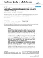
between the transducer and the scrotum. [Figure 1].
The ultrasound chamber was plumbed to allow input
of coupling medium across the bottom of the chamber
to dissipate any heat built up in the transducer. The
transducer was affixed to an offset cam to allow it to
rotate in a horizontal plane against the bottom of the
ultrasound chamber during treatment. Ultrasound gel
was used to coat the transducer face and the underside
of the latex sheet used as the bottom of the ultrasound
chamber to achieve acoustic coupling.
Beam field mapping
The spatial distribution of acoustic pressures delivered
by the ME7413 transducer to the testis was mapped as
follows: a needle hydrophone (Onda, Sunnyvale, CA)
was held vertically over the operating transducer and
raster scanned 1.5 cm from the transducer’ s face
(approximating the distance to the center of the testis)
in 0.5 mm increments using a computer controlled
motion stage (Newport, Irvine, CA). The beam field was
mapped at 1 MHz and at 3 MHz with the transducer
centered against the acoustically transparent latex sheet
used as the bottom of the treatment chamber. Distilled
water (DW), degassed distilled water and degassed 3%
(w/v) sodium chloride were tested as coupling media.
Both the ME7410 and ME7413 transducers were a lso
mapped at 1 MHz frequency at distances of 0.5 cm to
3.5 cm from the transducer face.
Tsuruta et al. Reproductive Biology and Endocrinology 2012, 10:7
/>Page 2 of 15
Determining the true effective radiating area (ERA) of our
transducers
Beam plots acquired with the transducer - hydrophone
separation set at 5 mm were used to determine the
actual effective radiating area of both transducers used
in our studies. Both transducers were driven at 1 MHz
frequency and 1 W/cm
2
intensity with the Mettler Soni-
cator 740 used in our studies. The beam area was
defined as the contiguous region with intensity greater
than 5% of the peak value.
Determining the true power output of our transducers
An Ultrasound Power Meter (model UPM-DT-1AV,
Ohmic Instrument Co., Easton, MD) was used to mea-
sure the power output of our transducers at 1 or 3 MHz
frequency, at intensities indicated by the Mettler
Son icator 740 to be 1 W/cm
2
and 2 W/cm
2
.Thetrans-
ducer face was centered 2 cm directly above the pres-
sure-sensing cone and the radiant force method was
used to determine the total output in Watts.
Temperature data
An implantable copper-constantan thermocouple (IT-21,
Physitemp Instruments, Clifton, NJ) was inserted down
the long axis of the testis at an oblique angle to avoid
piercing the epididymis to record testis temperature.
The bimetal probe was connected to an analog-to-digital
converter (Thermes USB, Physitemp Instruments, Clif-
ton, NJ) and data was collected using Labview software
(National Instruments, Austin, TX). Additional thermo-
couples were used to record the temperature of the cou-
pling medium and the surface of the scrotum.
Figure 1 Apparatus used to position rats for ultrasound treatment. Parts were cut from Plexiglas unless otherwise noted. A slanted section
supported most of the rat’s body above the level reached by re-circulating coupling medium. The rat’s scrotum was placed within the
ultrasound treatment chamber after using a ligature to retain the testes within the scrotum (not shown). The bottom of the treatment chamber
was formed of a single layer of latex, which was held in place against a rubber O-ring by an aluminum ring secured by machine screws. This
formed a liquid-tight seal, allowing coupling medium to be re-circulated through the treatment chamber and a holding vessel contained within
a temperature-regulated water bath (tubing, water bath, plumbing input and output have been omitted for clarity). A ring of ultrasound
absorbing material was suspended 1 cm from the bottom of the treatment chamber to aid positioning of the testes and to reduce reflection of
ultrasound energy. An ultrasound-transparent, nylon mesh was attached to the bottom of the ring to maintain a minimum distance of 1 cm
between the bottom of the ultrasound chamber and the proximal portion of the scrotum.
Tsuruta et al. Reproductive Biology and Endocrinology 2012, 10:7
/>Page 3 of 15
Ultrasound treatment
The t reatment frequency (1 MHz or 3 MHz), intensity
setting (1 W/cm
2
to 2.2 W/cm
2
), duty cycle (100% ) and
duration were selected on the ultrasound generator
[Tables1,2,and3].Ratswereanesthetizedandmain-
tained on 2 - 2.5% i soflurane/oxygen. A ligature to
retain t he test es was tied tightly enough only to prevent
the retraction of the testes from the scrotum during
treatment. If testis temperature was recorded, the ther-
mocouple was inserted at t his time. The rat was posi-
tioned so that his scrotum was centered on the mesh
layer of the ultrasound chamber. The appropriate cou-
pling medium was circulated through the ultrasound
chamber [Tables 1, 2, and 3]. The temperature of the
coupling medium was controlled by r e-circulating it
through a holding vessel contained wit hin a tempera-
ture-controlled bath. Temperature recording was
initiated one minute prior to the start of ultrasound
treatment and continued for one minute after the con-
clusion of ultrasound treatment to record pre- and post-
treatment baseline temperatures.
Sperm count and motility were assessed two weeks after
treatment
Preliminary Studies and Study 1: A testis and epididymis
were removed prior to whole-body cardiac perfusion with
Bouin’ s fixative. The cauda epididymis was carefully
remov ed and several cuts were made to allow the release
of sperm. The incised cauda epididymis was placed in
10 ml of M16 medium (Sigma, St. Louis, MO) for at least
one half hour to allow motile sperm to be released. For
determining sperm count, a dilution was made in distilled
water and counted on a hemocytometer. Sperm count was
expressed as millions of sperm per cauda epididymis. For
estimating sperm motility, a dilution was made in M16
medium. Motile and non-motile sperm were scored
visually using a hemocytometer.
Study 2: Sperm were collected from both cauda epidi-
dymides for determining sperm count, as described
above. The total sperm count was determined using a
hemocytometer by counting all sperm heads; the intact
sperm count was calculated after tallying the number of
sperm heads without an attached tail. Computer-aided
sperm analysis performed with a CEROS sperm analysis
system (software version 12.3; Hamilton Thorne Bios-
ciences, Beverly, MA) was used to determine sperm
motility.
Sperm count index
Sperm counts (10
6
per cauda epididymis) were assigned
to one of five arbitrary count ranges (< 11, 11-20, 21-40,
41-80, > 80). The count ranges were assigned values
(from low to high) of: 0, 1, 2, 4 and 10. The Sperm Count
Index was calculated as a weighted average using the
arbitrary values assigned to the count ranges and the per-
centage of counts that fell within each range. For exam-
ple, if 75% of a group’s sperm counts fell in the second
range of 11-20 × 10
6
and the remaining 25% of the
counts fell in the fourth range of 41-80 × 10
6
the count
index would be (0.75 × 1) + (0.25 × 4) = 1.75.
Fertility testing
For each mating trial, a single male was housed with a pair
of females for one week. In Pilot Study 2, the first mating
trial was initiated the day of the ultrasound treatment. A
second mating trial with a new pair of females occurred
during the second week after ultrasound treatment. Sperm
parameters were assessed at the conclusion of the second
mating trial. Females were held for at least four weeks
after the conclusion of their mating trial to complete preg-
nancies to term.
Untreated, sham-treated or wet-heat controls
Three different controls were used for comparison of
sperm counts and motilities. Untreated, retired breeders
served as untreated controls. Sham-treated animals
underwent all preparations for ultrasound treatment as
treated animals: anesthesia was administered and main-
tained at 2 - 2.5% isoflurane/oxygen, scrotal fur was
shaved, a ligature was used to retain the testes in the
scrotum, room temperature coupling medium was placed
in the treatment chamber, animal was placed on the
treatment apparatus and the scrotum was centered in the
treatment chamber. The temperature of the coupling
medium was not regulated, the coupling medium was
not re-circulated and the ultrasound generator was not
turned on for the sham-treated animals. The wet-heat
control animals were treated like the sham-treated con-
trols except that the temperature of the coupling medium
was held constant at 45°C while it was re-circulated
through the treatment chamber.
Table 1 Treatment parameters for preliminary studies
Parameter Preliminary
Study #1
Preliminary
Study #2
Coupling medium (°C) N.R. N.R.
Treatments 1 1
Duration (minutes) 10 10
Coupling medium DW or PBS dg-DW
Intensity (W/cm
2
) 1 2.2
Frequency (MHz) 1 1
Transducer (cm
2
)55
Fertility Trial No Yes
The temperature of the coupling medium was not regulated (N.R.) in these
studies. Coupling medium was distilled water (DW), phosphate buffered saline
(PBS), or degassed, distilled water (dg-DW). Fertility trial was conducted as
described in the Methods.
Tsuruta et al. Reproductive Biology and Endocrinology 2012, 10:7
/>Page 4 of 15
Histology
Pilot Studies and Study 1: Rats were anesthetized with iso-
flurane prior to cardiac perfusion with Bouin’s fixative.
One testis and one epididymis per animal were fixed for
histological examination. An additional 24 hours of
immersionfixationinBouin’ssolutionwasperformed
prior to 2 days of washing in 70% ethanol. Tissues were
processed into paraffin and 8 μm sections were stained
with hematoxylin and eosin using standard methods. Digi-
tal micrographs were assembled into larger montages
using the photomerge function in Photoshop CS (Adobe,
San Jose, CA).
Study 2: Testes and epididymides were drop-fixed in
Bouin’s fixative for 24 hours to prepare them for histol-
ogy. After an initial fixation of three hours, testes were
cut into 0.5 cm thick cross-sections to facilitate penetra-
tion of Bouin’s fixative. Fixed tissues were processed for
histology as described above for Study 1. Digital micro-
graphs were assembled into larger montages using an
Olympus BX51 microscope and motorized 2-dimen-
sional stage controlled by MetaMorph software (Mole-
cular Devices, Sunnyvale, CA).
Statistical analyses
One-way ANOVA analyses with post-tests were per-
formed using GraphPad Prism version 5.0 d, GraphPad
Software, San Diego California USA [6]. If data failed
Bartlett’s test for equal variances, significance was evalu-
ated usi ng the Kruskal-Wallis test and Dunn’s multiple
comparison post-test. In Study 1, sham-treated animals
Table 2 Treatment parameters for Study 1
Group name Sham Wet heat 1 MHz,
high power
3 MHz,
high power
3 MHz,
high power,
Na+
1 MHz,
low power
1 MHz,
low power,
Na+
Group number 1 2 3 4 5 6 7
Coupling medium (°C) NR 45 37 37 37 NR NR
Treatments 2 2 2 2 2 2 2
Duration (minutes) 15 15 15 15 15 15 15
Coupling medium dg-DW dg-DW dg-DW dg-DW dg-Na
+
dg-DW dg-Na
+
Intensity (W/cm
2
) - - 2.2 2.2 2.2 1 1
Frequency (MHz) - - 1 3 3 1 1
Transducer (cm
2
) n/a n/a 5 5 5 10 10
Rotation n/a n/a + + + - -
Animals 2 3 3 4 4 3 3
Degassed, 3% sodium chloride was used as the coupling medium (dg-Na
+
), otherwise degassed distilled water was used (dg-DW). Temperature of the coupling
medium was noted; otherwise there was no regulation (NR). Transducer ME7413 had a surface area of ~ 5 cm
2
(5) while model ME7410 had a surface area of ~
10 cm
2
(10). Transducers were stationary (-) or were rotated in a plane parallel to the bottom of the ultrasound chamber (+). All groups received two consecutive
treatments separated by two days.
Table 3 Treatment parameters for Study 2
Group name Untreated 37C,
2 × 15,
saline
37C,
2 × 10,
saline
37C,
1×10
35C,
2 × 15,
saline
35C,
2 × 15,
water
35C,
2 × 10,
saline,
2 W/cm
2
Group number 8 9 10 11 12 13 14
Coupling medium (°C) NR 37 37 37 35 35 35
Treatments - 22122 2
Duration (minutes) - 15 10 10 15 15 10
Coupling medium - dg-Na
+
dg-Na
+
dg-Na
+
or DW
dg-Na
+
dg-DW dg-Na
+
Intensity (W/cm
2
) - 2.2 2.2 2.2 2.2 2.2 2.0
Frequency (MHz) - 33333 3
Transducer (cm
2
) - 55555 5
Rotation - +++++ +
Animals 2 7 4 5 8 4 4
Degassed, 3% sodium chloride was used as the coupling medium (dg-Na
+
), otherwise distilled water (DW) or degassed, distilled water was used (dg-DW).
Temperature of the coupling medium was noted, otherwise there was no regulation (NR). Transducer ME7413 had a surface area of ~ 5 cm
2
(5). Transducers
were stationary (-) or were rotated in a plane parallel to the bottom of the ultrasound chamber (+). All groups received two consecutive treatments separated by
two days, except as noted for Groups 8 and 11.
Tsuruta et al. Reproductive Biology and Endocrinology 2012, 10:7
/>Page 5 of 15
(n = 2) were excluded from analysis but the remaining
treatment groups (n = 3 or 4) were analyzed fo r statisti-
cal differences.
Results
Field mapping and measuring the true ERA and power
output of our transducers
Field mapping of the output from the therapeutic transdu-
cer showed that there was a donut shaped “hotspot” in the
5-cm
2
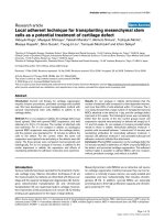
transducer’s output (ME7413) at 3 MHz [Figure 2].
The field map was the same regardless of the coupling
medium used (DW, degassed DW or 3% (w/v) saline). The
bea m field of the 5-cm
2
transducer changed when it was
mappedat1MHz:insteadofadonutshapedhotspot,
there was a discrete peak of energy near the center of the
transducer face [Additional file 1 Figure S1].
Beam plots from both transducers [Additional file 1
Figure S1] were used to determine the area of the beam
with energy equal to at least 5% of the peak beam energy
when the distance between the hydrophon e and transdu-
cer was set to 0.5 cm. The M E7413 transducer with a
nominalareaof5cm
2
had a true effective radiating area
of 4.4 cm
2
; the ME7410 transducer with a nominal area
of 10 cm
2
had a true effective radiating area of 9.3 cm
2
.
The power output of our transducers was determined at
intensities indicated by the Mettler Sonicator 740 to be
1W/cm
2
and 2 W/cm
2
. The 5 cm
2
transducer (ME7413)
atanominalintensitysettingof1W/cm
2
had an output
of 4.6 W at either 1 or 3 MHz; with a nominal intensity
setting of 2 W/cm
2
the output varied from 8.9 Watts at
1MHzto9.3Wattsat3MHz.The10-cm
2
transducer
(ME7410) was only m easured at 1 MHz and had an
output of 10.2 Watts at a nominal intensity setting of
1 W/cm
2
and an output of 20.0 Watts at a nominal inten-
sity setting of 2 W/cm
2
.
True spatially averaged intensities were determined for
our transducers
The 5-cm
2
transducer (ME7413) had an effective radiating
area of 4.4 cm
2
. At both 1 and 3 MHz frequency the actual
intensity for this transducer at an indicated 1 W/cm
2
was
1.05 W/cm
2
. The actual intensity for this transducer at an
indicat ed 2 W/cm
2
varied from 2.02 W/cm
2
at 1 MHz to
2.11 W/cm
2
at 3 MHz. The spatially averaged intensities
determined for this transducer were all wi thin 6% of the
values indicated by the Mettler Sonicator 740.
The 10-cm
2
transducer (ME7410) was only capable of
operating at 1 MHz frequency and had an effective radiat-
ing area o f 9.3 cm
2
. The actual intensity determined for
this transducer at an indicated 1 W/cm
2
was 1.1 W/cm
2
and at an indicated 2 W/cm
2
the actual value was 2.15 W/
cm
2
. The spatially averaged intensities determined for this
transducer were within 10% of the values indicated by the
Mettler Sonicator 740.
Mitigating thermal bio-effects
In order to create a more even field of ultrasound at
both frequencies, we devised a method to rotate the
transducer in a horizontal plane coincident with the
bottom surface of the ultra sound cha mber with the cen-
ter of rotation offset 8 mm from the center of the trans-
ducer face. The movement of the transducer mimics its
use as a therapeutic device and results in an averaging
of the field output over time.
The distance between the transducer and the scrotum
was initially set to 3 cm. In an attempt to increase the
energy delivered to the testes, the distance between the
scrotum and the transducer was successively decreased.
Some rats’ testes actually rested on the bottom of the
ultrasound chamber, separated from the transducer only
byalayeroflatex.Thismayhavebeenresponsiblefor
some localized heat damage to the scrotum; these rats
woul d occasionally develop a small circular disco lorati on
on their scrotum.
Constructing a mesh support provided a reproducible
offset of 1 cm between the bottom of the treatment
chamber and the scrotum; recirculating the coupling
medium e liminated any thermal bio-effects localized to
the scrotum.
Pilot study 1: published treatment parameters did not
alter testis histology
Attempts to cause germ cell loss using a single ten min-
ute dose of ultrasou nd at 100% duty cycle, 1 MHz and
1W/cm
2
(Pilot Study 1) did not alter testis histology.
These were the original parameters that were reported
by Fahim to cause the loss of almo st all germ cells from
Figure 2 Beam field map of the Model ME7413 therapeutic
ultrasound transducer acquired at 3 MHz. Normalized acoustic
pressure is plotted on the Y-axis. The X and Y-axes represent the
coordinates used to measure acoustic pressure delivered by the
ultrasound transducer.
Tsuruta et al. Reproductive Biology and Endocrinology 2012, 10:7
/>Page 6 of 15
the testis [4]. Pilot study 1 used phosphate buffered
saline or distilled water as the coupling medium filling
the ultrasound chamber. The coupling medium sur-
rounded the scrotum and allowed ultrasound to be effi-
ciently transmitted from the transducer to the scrotum;
ultrasound passed through the scrotum and was
absorbed by the testes.
Pilot study 2: increased power and degassed coupling
medium
An experiment using a single treatment of 1 MHz at
2.2 W/cm
2
and 100% duty cycle through degassed water
was performed (Pilot Study 2). Treating with 2.2 W/cm
2
was more successful than treating with 1 W/cm
2
.Two
weeks after ultrasound treatment, the testis was depleted
of developing germ cells a nd sperm cou nt was reduced
to 200 × 10
3
sperm per cauda epididymis. T hese sperm
were not motile when analyzed in M16 medium.
Fahim reported that his ultrasound conditions caused
rats to immediately lose their fertility [4]. When we treated
with low frequency and high power (Pilot Study 2), pups
were sired during the first and second weeks after treat-
ment. However, there were no motile sperm at the end of
this pair of one-week mating trials. Hypothetically, if
another mating trial had been performed during the third
week after treatment, the rat would have been infertile.
This demonstrated that even though motile sperm were
not detected at the end of the second mating trial, there
were sufficient motile sperm during the initial two-week
period after treatment for fertility.
Study 1: two consecutive treatments
In an attempt to bring post-treatment sperm counts closer
to zero, the effect of two consecutive treatments separated
by two days were tested [Study 1, Table 2]. Two weeks
after treatment, total sperm count in the cauda epididymis
dropped below 2 × 10
6
total sperm with essentially no
motility when 3 MHz ultrasound was applied at 2.2 W/
cm
2
through 37°C distilled water at 100% du ty cycle
[Table 4 Group 4]. Using coupling medium heated to 45°
C allowed us to achieve internal testis temperatures com-
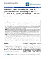
parable to the ultrasound treated testes [Figure 3]. Inter-
estingly, heat alone [Table 4 Group 2] was more effective
at reducing epididymal sperm count than the use of
1 MHz ultrasound either when the temperature of the
coupling medium was held constant at 37°C [Table 4
Group 3, Tukey’ s post-test, p < 0.001] or when the tem-
perature of the coupling medium was not regulated [Table
4 Group 6, Tukey’s post-test, p < 0.001], however when 1
MHz ultrasound was applied through 3% saline at low
power, sperm count was reduced sufficiently so that there
was no significant difference from wet heat.
In contrast, the use of 3 MHz ultrasound resulted in a
total epididymal sperm count ~10-fold lower than wet
heat alone but with almost 1,000 times fewer motile
sperm recovered from the epididymis: 3 MHz treated
animals [Table 4 Group 4] had ~ 6 × 10
3
motile sperm
per cauda epididymis while wet heat treated animals
[Table 4 Group 2] had ~5 × 10
6
motile sperm per cauda
epididymis (derived from data presented in Table 4;
motile sperm = total sperm × % motile).
Study 1: combining heat and ultrasound more effective
than heat alone
The normal testis [Figur e 4, A-D] had a complex epithe-
lium consisting of many spermatogenic cells in various
stages of spermatogenesis. Two weeks after using wet heat
to elevate testis temperature there was a significant loss of
spermatogenic cells although most seminiferous tubules
still retained some spermatogenic cells [Figure 4, E-H].
In contrast, combining elevated temperature and 3 MHz
ultrasound [Table 4 Group 4 or 5] caused testis-wide
depletion of germ cells [Figure 5]. The loss of developing
spermatocytes and spermatids from the seminiferous
epithelium was extensive; almost all tubules examined
were effectively depleted by this treatment [Additional
file 2 Figure S2]. The loss of spermatogenic output was
reflected by sperm counts in these animals below 2 × 10
6
sperm per cauda epididymis, two weeks after ultrasound
treatment [Table 4 Groups 4 and 5].
Table 4 Testis temperatures and sperm parameters from Study 1
Group Treatment n Testis temperature (°C) Sperm count
(10
6
)
Motility
(%)
1 Sham 2 30.1 ± 0.8 380 ± 33 45 ± 3
2 Wet heat 3 42.6 ± 0.1 23 ± 4 22 ± 5.8
3 Low freq., high power 3 40.5 ± 1.2 84 ± 3 § 54 ± 2
4 High freq., high power 3 41.8 ± 0.6 1.9 ± 0.9 † 0.3 ± 0.3
5 High freq., high power, Na+ 4 nd 1.5 ± 0.8 † nd
6 Low freq., low power 3 42.1 ± 2.8 96 ± 17 § 39 ± 2
7 Low freq., low power, Na
+
3 35.4 ± 1.9 51 ± 5 40 ± 2
Sperm analyses were performed two weeks after ultrasound treatment. Average sperm count represents millions of sperm per cauda epididymis ± SEM. %
Motility represents the percentage of recovered sperm with forward motility ± SEM. The average maximum testis temperature during treatment is listedin
degrees Celsius ± SEM. Not determine d (nd). §, statistically greater than the wet-heat control (Group 2) by Tukey’s post-test (p < 0.001). †, statistically lower than
1 MHz, high power (Group 3) and 1 MHz, low power (Group 6) by Tukey’s post-test (p < 0.001).
Tsuruta et al. Reproductive Biology and Endocrinology 2012, 10:7
/>Page 7 of 15
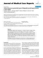
Study 2: varying 3 MHz ultrasound treatments
All animals in Study 2 were treated with 3 MHz ultra-
sound. We varied the temperature of the coupling med-
ium (35 or 37°C), its composition (DW or saline), the
number (1 or 2) or duration of treatments (10 or 15 min-
utes) to determine the effect of these changes in treat-
ment on mean motile sperm count per cauda epididymis
[Figure 6]. Except for the group treated through degassed
distilled water at 35°C (Group 13), all treatments resulted
in a significantly lower mean motile sperm count than
the untreated group (Group 8) according to Dunnett ’ s
multiple comparison test (p < 0.001).
The most effective treatment in Study 2 (Group 9: treat-
ing twice for 15 minutes at 3 MHz and 2.2 W/cm
2
inten-
sity through degassed 3% saline held at 37°C) resulted in 3
± 1 million motile sperm per cauda epididymis and a
Sperm Count Index equal to 0. The next three lowest
sperm counts were in Groups 10 - 12; all of these treat-
ments resulted in mean motile sperm counts greater than
50 million sperm per cauda epididymis which was signifi-
cantly higher than observed for Group 9 [Figure 6, Krus-
kal-Wallis with Dunn’ s post-test, refer to figure for
p-values]. Group 12 had a Sperm Count Index equal to 3.9
and approximately one third of this group’s sperm counts
fell into the range of 41 - 80 million sperm per c auda epidi-
dymis. Group 10 had a Sperm Count Index of 6.0 wit h a
mean sperm count of 67 ± 7 million motile sperm per
cauda epididymis. As the higher Sperm Count Index indi-
cated, a much larger pro portion (7/8) o f this group’ssperm
counts fell into the range of 41 - 80 million sperm per
cauda epididymis.
Study 2: saline was a more effective coupling medium
than distilled water at 35°C
When animals were treated once at 37°C for 10 minutes at
2.2 W/cm
2
there was not a significant difference in sperm
count as a function of coupling medium (degassed distilled
wat er versus degassed 3% saline) so this data was pooled
(Group 11). However, when animals were treated twice at
35°C for 15 minutes at 2.2 W/cm
2
the composition of the
coupling medium did make a significant difference in
sperm count (Tukey’s post-test, p < 0.01): degassed 3% sal-
ine (Group 12) resulted in a sperm count 50% lower than
degassed distilled water (Group 13). The use of saline
resulted in about half of the sperm counts for Group 12 to
be lower than 41 × 10
6
per cauda epididymis (Sperm
Count Index = 3.9) while the use of distilled water (Group
13) resulted in only about 12% of counts below that
threshold [Figure 6, Sperm Count Index = 8.1]. In addi-
tion, the number of intact sperm was significantly lower
(Tukey’s post-test, p < 0.05) when treating at 35°C through
3% saline [Figure 7, Group 12] than through degassed dis-
tilled water [Figure 7, Group 13].
Most effective treatment
When the four treatments groups (Groups 9 - 12) with the
lowest mean sperm counts in Study 2 were compared by
one-wayANOVA,Group9wasfoundtohaveasignifi-
cantly lower mean motile sperm count than the other
three groups (Kruskal-Wallis with Dunn’s post-test, refer
to Figure 6 for p-values). In addition, the percentage of
intact sperm in Group 9 [Figure 7] was significantly lower
(Tukey’s post-test, p < 0.01) than the untreated control
[Figure 7, Group 8]. Thus, the treatment that reduced
cauda epididymis sperm count two weeks after treatment
to t h e lowest levels was the same in Study 1 (Group 5) and
in Study 2 (Group 9): two 15- minute treatments with 3
MHz ultrasound at 2.2 W/cm
2
through degassed 3% saline
maintained at 37°C.
Discussion
Rat as a model system
Rats are reported to retain fertility even with extremely
low sperm counts [7]. In contrast to rats, the World
Health Organization has defined oligospermia in men as
less than 20 million sperm/ml in the ejaculate and men
32
34
36
38
40
42
05101
5
wet heat
1 MHz
min
u
t
es
temperature
(C)
Figure 3 Representativ e tempera ture curves during ultrasound
or wet heat. A thermal couple was inserted down the long axis of the
testis and another was placed in the coupling medium. Coupling
medium was re-circulated at 37°C during ultrasound treatments and at
45°C for the wet heat control. The rotation frequency of the transducer
correlated with temperature fluctuations at the site of the thermal
couple. The wet heat control yielded a testis temperature profile
similar to an ultrasound treated testis.
Tsuruta et al. Reproductive Biology and Endocrinology 2012, 10:7
/>Page 8 of 15
are generally considered sub-fertile when their sperm
concentration drops below 10 million sperm/ml [8].
Thus, we anticipate that decreasing sperm count suffi-
ciently to cause infertility in rats would also cause infer-
tility in men. However, sperm counts or concentrations
that would represent infertility in men could allow rats
to retain their fertility. Our second pilot study showed
that the absence of motile sperm at the end of a mating
trial did not rule out the ability to sire pups. With the
mating scheme used in our study, it appeared that
sperm count was changing rapidly and that the count
on the day of conception could be higher than the
count determined at necropsy. Consequently, in lieu of
testing fertility we decided to assay epididymal sperm
reserves to monitor the efficacy of our treatment
conditions.
Our results clearly show that therapeutic ultrasound
treatment depleted developing germ cells from the testis
and subsequently decreased the size of sperm reserves in
the epididymis when rats were treated with two consecu-
tive ultrasound treatments separated by two days [Table 4
Figure 6]. This differs from reports in the 1970s by Fahim
et al. [3,4], which reported t hat a single treatment of
1 MHz ultrasound wa s sufficient t o induce a contraceptive
effect of approximately six months duration. No mention
of controlling the temperature of the coupling medium
appeared in those original reports. In contrast, we found
that combining elevated temperature, high power and
high frequency was the most effective method for reducing
sperm count.
Variation between ultrasound transducers
A direct compa rison between our treatments and those
of Fahim are not possible without measuring the true
effective radiating area (ERA, cm
2
) and power output
(Watts) for all of the transducers used i n these studies in
order to calculate the true spatial average intensity (SAI,
W/cm
2
) delivered during treatment. The SAI reported by
clinical therapeutic ultrasound systems is not directly
regulated in the United States by the Food and Drug
Administration (FDA) even though this is the parameter
most often used clinically to determine dosing during
treatment. The FDA does require the true power output
to be within ± 20% of the value reported by the manufac-
turer however no specific guideline was presented for the
accuracy in reporting ERA [9]; most manufacturers
report ERA with an error of ± 20 - 25%. Therefore, the
true SAI for a transducer could vary by up to 150% from
the displayed value while still satisfying FDA guidelines
for ERA and power output. A study of sixty-six therapeu-
tic ultrasound transducers showed that their true SAI
varied from -43% to +63% of the displayed value [10].
The effects of ultrasound are dose-dependent, thus
reproducible clinical dosing of therapeutic ultrasound
Figure 4 Represe ntative histology of no rmal or wet-heat-treated testes and seminiferous tubules. A-D: hematoxylin and eosin stained
cross-sections of untreated testis. The tall seminiferous epithelium contains many spermatocytes (sp), round spermatids (rs) and condensing
spermatids (cs). Tails (t) of condensing spermatids and newly released testicular sperm are seen in the lumen (Lu) of some tubules. E-H: testis
cross-section stained two weeks after wet heat treatment. Almost all tubules have enlarged luminal diameters after treatment with heat alone.
The seminiferous epithelium (e) is reduced in height due to the loss of many spermatocytes and spermatids. Some tubules have disorganized
epithelium (*).
Tsuruta et al. Reproductive Biology and Endocrinology 2012, 10:7
/>Page 9 of 15
requires determining the actual ERA, power output and
SAI of the generator and transducers being used for
treatment.
In some cases, more advanced monitoring techniques
such as quantitative Schlieren assessment may be
required to discern differences in output of transducers
operated under identical nominal parameters [11]. This
method can measure the power distribut ion in discrete
portion s of the ultrasound beam that are not captured by
measurements mandated by the FDA such as beam non-
uniformity ratio (BNR) and the aforementioned total
power and ERA. Differences in the distribution of power
within an ultrasound field may account for the ability of
nominally identical transducers to heat t issue at signifi-
cantly different rates [11].
We determined the actual effective radiating areas and
power output of the transducers used in our stud ies. The
true SAI of our transducers were determined to be within
10% of the values reported by our therapeutic ultrasound
generator. In addition to determining the true ERA, power
output and SAI for our transducers, we have also provided
beam plots [Additional file 1 Figure S1] to facilitate
comparison of our study results with future studies and to
begin to standardize the clinical dosing of therapeutic
ultrasound when used as a male contraceptive.
Since Fahim’s custom-built generator and transducer
were not available for testing, we cannot rule out the pos-
sibility that his system delivered more ultrasound energy
to the testes than our therapeutic ultrasound instrument.
Accordingly, we modified our coupling medium and treat-
ment parameters to increase the delivery of ultrasound
energy to the testes. While attempting to maximize energy
delivery, we also took steps to mitigate any thermal bio-
effects observed on the scrotal epithelium. The transducer
face became quite hot to the touch by the end of each
treatment so we reasoned that conductive transfer of heat
caused occasional circular discolorations when the scro-
tum was pressed against the bottom of the treatment
chamber. We modified the interior of our chamber to pro-
vide a reproducible offset between the scrotum and the
chamber bottom/transducer. This also provided a space to
re-circulate coupling medium between the scrotum and
chamber bottom/transducer to dissipate any localized
buildup of heat. Irregularities in the beam field prompted
Figure 5 Testis histology two weeks after 3 MHz ultrasound (Group 4). (A) The loss of spermatogenic cells after this treatment was more
complete than after the wet heat treatment. This resulted in a shorter epithelium and a larger diameter lumen. (B) An isolated cluster of tubules
in this particular animal showed evidence of thermal damage (td) in addition to the loss of spermatogenic cells. (C) Most tubules had a very
short epithelial layer and increased lumen diameter due to the loss of all spermatocytes and spermatids. (D) Tubules that appear to have a larger
epithelial layer and smaller diameter lumen were still missing spermatocytes and spermatids.
Tsuruta et al. Reproductive Biology and Endocrinology 2012, 10:7
/>Page 10 of 15
us to rotate the transducer to achieve a time averaging of
the beam field energy. These modifications to the origin-
ally published protocol, especially the rotation of the trans-
ducer, may have caused a decrease in energy delivered to
the testes. Rotating the beam field with an 8 mm offset
cam caused a central area of continuous ultrasound expo-
sure, surrounded by an area of lower, time-averaged ultra-
sound exposure. Time-averaging the beam field may
account for the increased power, duration and number of
treatments that we required to replicate Fahim’s origi nal
0
50
100
150
200
250
300
>80
40-80
20-40
11-20
<11
**
*
***
***
Figure 6 Average and distribution of motile sperm counts from Study 2. Motile sperm count was determined two weeks after treatment
and was plotted as the mean ± SEM (10
6
per cauda epididymis). The stacked bars represent the proportion of sperm counts that fell into the
following ranges of sperm counts (10
6
per cauda epididymis): < 11, 11 - 20, 21 - 40, 41 - 80, and > 80. Sperm Count Index was calculated as
described in the Methods and is reported above each bar. Groups 8 - 12 failed Bartlett’s test and were analyzed by the Kruskal-Wallis test with
Dunn’s post-test, symbols represent groups statistically different from Group 9: *, p < 0.05; **, p < 0.01; ***, p < 0.005. Groups 12 - 14 passed
Bartlett’s test: symbols represent groups statistically different from Group 12 by Tukey’s post-test: §, p < 0.01.
Tsuruta et al. Reproductive Biology and Endocrinology 2012, 10:7
/>Page 11 of 15
result; the central area of continuous ultrasound exposure
may account for the occasional thermal damage observed
in some seminiferous tubules.
Coupling medium
Our attempts to deplete germ cells using 1 MHz ultra-
sound at 1 W/cm
2
without controlling the temperature of
the coupling medium were only partially successful [Study
1, Table 4 Group 6 and 7]. The use of 1 MHz ultrasound
at either low or high power was less effective than the use
of wet heat alone (p < 0.001) [Table 4; Group 2 versus
Groups 3 or 6]. However, 1 MHz ultrasound decreased
sperm count almost two-fold when the coupling medium
was switched from degassed distilled water to 3% (w/v)
sodium chloride in degassed distilled water [Table 4;
Group 6 vs. Group 7]. The use of 3% sodium chloride and
1 MHz ultrasound [Table 4; Group 7] decreased sperm
count to levels that were not statistically different from
that achieved with wet heat alone [Table 4; Group 2].
When the temperature of the coupling medium was
held at 37°C, 3 MHz ultrasound at 2.2 W/cm
2
decreased
sperm count below 2 × 10
6
sperm per cauda in the pre-
sence or absence of saline [Study 1, Table 4 Group 4 and
5]. Attempting to reduce sperm c ount with the coupling
medium held at 35°C was only partially successful [Study
2, Figure 6, Groups 12, 13, and 14]. However, the use of
degassed 3% saline again caused a two-fold decrease in
sperm count compared to the use of degassed distilled
water [Figure 6, Group 12 vs. 13]; this drop in sperm
count was statist ically signi ficant (p < 0.01). The number
of intact sperm also decreased significantly (p < 0.05)
when degassed 3% saline was used [Figure 7, Group 12
vs. 13]. When treatment conditions were less effective at
reducing sperm count (combinations of degassed distilled
water, lower temperature, lower power, or lower fre-
quency) it appears that the addition of 3% saline to the
coupling medium may cause a statistically significant
drop in sperm count.
*
Figure 7 Percentage of intact sperm recovered in Study 2. Sperm counts ta llied both in tact sperm and sperm heads not attached to a tail.
The number of intact sperm was expressed as a percentage of the total number of sperm recovered. *, Group 9 was statistically lower than
Groups 8, 13 and 14 by Tukey’s post-test (p < 0.01). §, Group 12 was statistically lower than Group 13 by Tukey’s post-test (p < 0.05).
Tsuruta et al. Reproductive Biology and Endocrinology 2012, 10:7
/>Page 12 of 15
This corroborates a report in the literature that this
coupling medium was more effective than distilled water
alone when attempting to deplete germ cells from Maca-
que testes [4]. The biophysical basis for this phenomenon
is currently unknown since theacousticpressuredeliv-
ered by our ultrasound transducers was not affected by
either including 3% saline or by degassing our distilled
water. However, 3% saline could have a biological effect
on the scrotal epithelia or the dartos fascia (mu scular tis-
sue under the skin) that enhances the transmission of
ultrasound energy. The dartos fascia is responsible for
the furrowing of the scrotal skin, an adaptation related to
thermal regulation of the testes. Although 3% saline
failed to enhance the effect of 3 MHz ultrasound at 37°C,
additional studies exploring different coupling media an d
the effect of their temperature are warranted.
Conclusions
Potential applications
Depleting spermatocytes and spermatids from testes non-
invasively with therapeutic ultrasound has multiple appli-
cations. If the method proves to be reversible, it would
provide a new tool for investigating spermatogonial expan-
sion and differentiation. By creating testes depleted of dif-
ferentiated spermatogenic or meiotic cells, investigators
could test directly the effect of compounds proposed to
regulate spermatogonia. In addition, spermatogonial stem
cells are assayed by colony formation after transplantation
into recipient testes depleted of germ cells by chemical
treatment [12]. Rat spermatogonia can develop within the
mouse seminiferous epithelium into spermatids that are
morphologically distinct from those of the mouse [13,14].
Therefore, ultrasound-treated, syngeneic testes could serve
as an alternatively prepared host for assaying spermatogo-
nial stem cell numbers.
If the method can be made permanent, a non-invasive
method for controlling various domestic pet populations
could be developed. Leoci [15] has successfully used thera-
peutic ultrasound as a non-invasive method for canine
sterilization. Fahim reported that his treatment method
did not affect testosterone production by Leydig cells [4].
Thus, ultrasound treatment could be adopted as part of a
larger strategy to control nuisance animal populations
using the trap-neuter-return model [16,17]. Introducing
sterile males into a population was effective in controlling
insect populations [18] and was proposed to be effective in
species where a dominant male breeds with a harem of
fem ales in a restricted territory such as white-tailed deer
(Odocoileus virginianus) [19] or feral horses [20-22]. Con-
trolling deer populations in urban or suburban areas
would accrue many public health benefits since white-
tailed deer carry ticks that transmit disease [23-25], are at
risk for tuberculosis [26,27], and in the United States there
are about 247 thousand collisions each year between deer
and automobiles that damage approximately 1 billion dol-
lars in property and kill approximately 200 people [28,29].
Ultimately, the most significant application of treating
testes with ultrasound will be to address the global health
issue of unintended pregnancies. The World Health Orga-
nization estimates that 228 million of the 600 million
women of reproductive age worldwide are at risk for
mistimed or unwanted pregnancy [30-32]. Yearly these
unintended pregnancies result in almost 50 million abor-
tions; almost half of these abortions are classed as unsafe,
resulting in 47 thousand maternal deaths [33,34]. In the
United States alone there are at least 3 million unintended
pregnancies each year representing about 50% of all preg-
nancies [35,36]. Clearly, developing another safe, efficient
and inexpensive method for contraception would only
help to lower the rate of unwanted pregnancies and abor-
tions. A permanent or reversible method of contraception
based on therapeutic ultrasound tre atment could encou-
rage more men to share greater responsibility for family
planning.
Future studies
Future studies will determine if our ultrasound treatment
parameters result in a reversible loss of fertility as pre-
viously reported by Fahim. The treatment that was most
effective at reducing epididymal sperm count (3 MHz,
2.2 W/cm
2
, two 15-minute treatments separated by two
days with coupling medium temperature maintained at
37°C) represents an upper limit for applying ultrasound to
the testes since thermal bio-effects were noted in some
treated tubules. Results from Study 2 showed that rela-
tively small changes in treatment conditions caused statis-
tically significant changes in sperm count when assessed
two weeks after treatment. Longer-term studies will be
required to determine if those treatment conditions cause
a progressive loss of spermatogenic cells that ultimately
results in the depletion of epididymal sperm reserves. A
major goal of our future studies will be to determine the
“minimum effective dose” of ultrasound that induces a
reversible loss of fertility.
In conclusion, our results demonstrate that a short
exposure to therapeutic ultrasound is an effective method
for depleting testes of spermatogenic cells and reducing
epididymal sperm reserves within two weeks of treat-
ment. The odds of conceiving decrease linearly when
sperm concentrations are below 40 million sperm/ml
[37] and effective contraception occurs when hormonal
treatment or vasectomy cause sperm concentration to
fall below 3 million sperm/ml [38,39]. Our ability to use
a widely available therapeutic ultrasound system to
reduce motile sperm count below 5 million sperm per
cauda epididymis just two weeks after treatment shows
that therapeutic ultrasound holds great promise as the
basis for a male contraceptive. Optimizing the treatment
Tsuruta et al. Reproductive Biology and Endocrinology 2012, 10:7
/>Page 13 of 15
conditions, studying the safety of repeated use, the dura-
tion of the contraceptive effect and it s reversibility and
are the next required steps to establish whether therapeu-
tic ultrasound can serve as the basis for a new, long term,
reversible male contraceptive.
Additional material
Additional file 1: Figure S1: Beam field maps of the 5 cm
2
and 10 cm
2
transducers. The ME7413 (5 cm
2
) and ME7410 (10 cm
2
) transducers were
mapped at 1 MHz frequency and 1 W/cm
2
power at a variety of
distances from the transducer face. Beam field maps were identical
regardless of the coupling medium used (DW, degassed DW or 3%
saline).
Additional file 2: Figure S2: Two treatments with 3 MHz ultrasound
uniformly depleted the testis of spermatocytes & spermatids. This is the
same testis depicted in Figure 5. Two consecutive fifteen minute
treatments of 3 MHz ultrasound at 2.2 W/cm
2
were applied through
degassed, distilled water held at 37°C. This magnification emphasizes the
uniformity of the ultrasound effect. Only 5% of the seminiferous tubules
were observed to have thermal damage while the remaining tubules
were depleted of spermatocytes and spermatids. This treatment would
have provided at least two months of infertility since spermatogonia
require that much time to enter the epididymis as sperm.
Acknowledgements
The authors thank DA O’Brien (University of North Carolina) and E Lissner
(Parsemus Foundation) for thoughtful discussions.
Funding
This research was supported by a grant from the Parsemus Foundation and
by a Grand Challenges Explorations Grant from the Bill & Melinda Gates
Foundation (JKT & PAD). Funding for preparation of the manuscript was
provided by the Bill & Melinda Gates Foundation with additional funding
from the Parsemus Foundation for publication of the manuscript. The
Parsemus Foundation participated in discussions about experimental design.
Neither foundation participated in the collection, analysis or interpre tation of
data, the writing of the manuscript or the decision to submit the manuscript
for publication.
Author details
1
The Laboratories for Reproductive Biology, Department of Pediatrics, 220
Taylor Hall, CB7500, The University of North Carolina at Chapel Hill, Chapel
Hill, North Carolina 27599, USA.
2
Department of Cell & Developmental
Biology, CB7090, The University of North Carolina at Chapel Hill, Chapel Hill,
North Carolina 27599, USA.
3
Department of Biomedical Engineering, 152
MacNider Hall, CB7575. School of Medicine, The University of North Carolina
at Chapel Hill, Chapel Hill, North Carolina 27599, USA.
4
Integrated Laboratory
Systems, 601 Keystone Park Drive, Suite 100, Durham, North Carolina 27713,
USA.
5
FHI360, 2224 E. NC Highway 54, Durham, North Carolina 27713, USA.
6
Virginia Polytechnic Institute and State University, School of Biomedical
Engineering and Sciences, Center for Injury Biomechanics, 440 ICTAS
Building, Stanger Street, Mail Code 0194, Blacksburg, Virginia 24061, USA.
Authors’ contributions
JKT directed research for Study 2, treated animals, analyzed histology,
analyzed sperm parameters, and wrote the manuscript; PAD, CMG, RCG, and
TSG designed, performed or analyzed in vitro ultrasound experiments, MGO,
PAD, GJM and DCS analyzed data and directed Pilot Studies and Study 1;
MAS treated animals, performed necropsies and supervised all animal
experiments performed at ILS; EJRS and KGH treated animals, performed
necropsies and analyzed sperm. All authors participated in experimental
design and read and approved the final manuscript. The authors declare
that no actual or potential conflict of interest exists that could
inappropriately influence, or be perceived to influence, this work. All authors
read and approved the final manuscript.
Competing interests
The authors declare that they have no competing interests.
Received: 13 July 2011 Accepted: 30 January 2012
Published: 30 January 2012
References
1. Wellings K, Collumbien M, Slaymaker E, Singh S, Hodges Z, Patel D, Bajos N:
Sexual behaviour in context: a global perspective. Lancet 2006,
368:1706-1728.
2. Williams JK, Wyatt GE, Wingood G: The Four Cs of HIV Prevention with
African Americans: Crisis, Condoms, Culture, and Community. Curr HIV/
AIDS Rep 2010.
3. Fahim MS, Fahim Z, Der R, Hall DG, Harman J: Heat in male contraception
(hot water 60 degrees C, infrared, microwave, and ultrasound).
Contraception 1975, 11:549-562.
4. Fahim MS, Fahim Z, Harman J, Thompson I, Montie J, Hall DG: Ultrasound
as a new method of male contraception. Fertil Steril 1977, 28:823-831.
5. Fahim MS: Apparatus useful in suppression of spermatogenesis. US
Patent No. 4,078,556 1978.
6. Graphpad Software. [].
7. Amann RP: Detection of alterations in testicular and epididymal function
in laboratory animals. Environ Health Perspect 1986, 70:149-158.
8. Cooper TG, Noonan E, von Eckardstein S, Auger J, Baker HW, Behre HM,
Haugen TB, Kruger T, Wang C, Mbizvo MT, Vogelsong KM: World Health
Organization reference values for human semen characteristics. Hum
Reprod Update 2010, 16:231-245.
9. Performance standards for sonic, infrasonic, and ultrasonic radiation
emitting products. Food and Drug Administration, Code of Federal
Regulations, Title 21 2011, 8(1050).
10. Straub SJ, Johns LD, Howard SM: Variability in effective radiating area at 1
MHz affects ultrasound treatment intensity. Physical therapy 2008,
88:50-57.
11. Johns LD, Demchak TJ, Straub SJ, Howard SM: The role of quantitative
Schlieren assessment of physiotherapy ultrasound fields in describing
variations between tissue heating rates of different transducers.
Ultrasound in medicine & biology 2007, 33:1911-1917.
12. Brinster RL, Avarbock MR: Germline transmission of donor haplotype
following spermatogonial transplantation. Proc Natl Acad Sci USA 1994,
91:11303-11307.
13. Clouthier DE, Avarbock MR, Maika SD, Hammer RE, Brinster RL: Rat
spermatogenesis in mouse testis. Nature 1996, 381:418-421.
14. Franca LR, Ogawa T, Avarbock MR, Brinster RL, Russell LD: Germ cell
genotype controls cell cycle during spermatogenesis in the rat. Biol
Reprod 1998, 59:1371-1377.
15. Leoci R, Aiudi G, De Sandro Salvati A, Silvestre F, Binetti F, Lacalandra GM:
Ultrasound as a mechanical method for male dog contraception.
Reproduction in domestic animals = Zuchthygiene 2009, 44(Suppl 2):326-328.
16. Hughes KL, Slater MR:
Implementation of a feral cat management
program
on a university campus. J Appl Anim Welf Sci 2002, 5:15-28.
17. Levy JK, Gale DW, Gale LA: Evaluation of the effect of a long-term trap-
neuter-return and adoption program on a free-roaming cat population.
J Am Vet Med Assoc 2003, 222:42-46.
18. Knipling E: Sterile-Male Method of Population Control. Science 1959,
130:902-904.
19. Miller BF, DeYoung RW, Campbell TA, Laseter BR, Ford WM, Miller KV: Fine-
scale genetic and social structuring in a central Appalachian white-tailed
deer herd. Journal of Mammology 2010, 91:681-689.
20. Klingel H: Social organization of feral horses. J Reprod Fertil Suppl 1982,
32:89-95.
21. Turner JW Jr, Kirkpatrick JF: Androgens, behaviour and fertility control in
feral stallions. J Reprod Fertil Suppl 1982, 32:79-87.
22. Dell’Omo G, Palmery M: Fertility control in vertebrate pest species.
Contraception 2002, 65:273-275.
23. Magnarelli LA, Anderson JF, Burgdorfer W: Rocky mountain spotted fever
in Connecticut: human cases, spotted-fever group rickettsiae in ticks,
and antibodies in mammals. Am J Epidemiol 1979, 110:148-155.
24. Pound JM, Miller JA, George JE, Fish D, Carroll JF, Schulze TL, Daniels TJ,
Falco RC, Stafford KC, Mather TN: The United States Department of
Agriculture’s Northeast Area-wide Tick Control Project: summary and
conclusions. Vector Borne Zoonotic Dis 2009, 9:439-448.
Tsuruta et al. Reproductive Biology and Endocrinology 2012, 10:7
/>Page 14 of 15
25. Yabsley MJ: Natural history of Ehrlichia chaffeensis: vertebrate hosts and
tick vectors from the United States and evidence for endemic
transmission in other countries. Vet Parasitol 2010, 167:136-148.
26. Palmer MV, Whipple DL, Payeur JB, Alt DP, Esch KJ, Bruning-Fann CS,
Kaneene JB: Naturally occurring tuberculosis in white-tailed deer. JAm
Vet Med Assoc 2000, 216:1921-1924.
27. Schmitt SM, O’Brien DJ, Bruning-Fann CS, Fitzgerald SD: Bovine
tuberculosis in Michigan wildlife and livestock. Ann N Y Acad Sci 2002,
969:262-268.
28. Allan WF, Wells JK: Characteristics of Vehicle-Animal Crashes in Which
Vehicle Occupants Are Killed. Traffic Injury Prevention 2005, 6:56-59.
29. Nonfatal Motor-Vehicle Animal Crash-Related Injuries - United States,
2001-2002. Morbidity and Mortality Weekly Report 2004, 53:675-681.
30. Ahman EL, Shah IH: Contraceptive use, fertility, and unsafe abortion in
developing countries. Eur J Contracept Reprod Health Care 2010, 15(Suppl
2):S77-82.
31. Ross J, Winfrey W: Unmet need for contraception in the developing
world and the former Soviet Union: an updated estimate. International
Family Planning Perspectives 2002, 28:138-143.
32. Lerberghe W, Manuel A, Matthews Z, Wolfeim C: The World Health Report
2005, Overview. Make every mother and child count. Geneva: The World
Health Organization; 2005.
33. Shah I, Ahman E: Unsafe abortion in 2008: global and regional levels and
trends. Reprod Health Matters 2010, 18:90-101.
34. Ahman E, Shah I: Unsafe abortion. Global and regional estimates of the
incidence of unsafe abortion and associated mortality in 2008. Geneva:
World Health Organization;, sixth 2008.
35. Finer LB, Henshaw SK: Disparities in rates of unintended pregnancy in
the United States, 1994 and 2001. Perspect Sex Reprod Health 2006,
38:90-96.
36. Henshaw SK: Unintended pregnancy in the United States. Fam Plann
Perspect 1998, 30:24-29, 46.
37. Bonde JP, Ernst E, Jensen TK, Hjollund NH, Kolstad H, Henriksen TB,
Scheike T, Giwercman A, Olsen J, Skakkebaek NE: Relation between semen
quality and fertility: a population-based study of 430 first-pregnancy
planners. Lancet 1998, 352:1172-1177.
38. World Health Organization Task Force on Methods for the Regulation of
Male Fertility: Contraceptive efficacy of testosterone-induced
azoospermia and oligozoospermia in normal men. Fertil Steril 1996,
65:821-829.
39. Gu YQ, Wang XH, Xu D, Peng L, Cheng LF, Huang MK, Huang ZJ,
Zhang GY: A multicenter contraceptive efficacy study of injectable
testosterone undecanoate in healthy Chinese men. J Clin Endocrinol
Metab 2003, 88:562-568.
doi:10.1186/1477-7827-10-7
Cite this article as: Tsuruta et al.: Therapeutic ultrasound as a potential
male contraceptive: power, frequency and temperature required to
deplete rat testes of meiotic cells and epididymides of sperm
determined using a commercially available system. Reproductive Biology
and Endocrinology 2012 10:7.
Submit your next manuscript to BioMed Central
and take full advantage of:
• Convenient online submission
• Thorough peer review
• No space constraints or color figure charges
• Immediate publication on acceptance
• Inclusion in PubMed, CAS, Scopus and Google Scholar
• Research which is freely available for redistribution
Submit your manuscript at
www.biomedcentral.com/submit
Tsuruta et al. Reproductive Biology and Endocrinology 2012, 10:7
/>Page 15 of 15