Aging of the Respiratory System: Impact on Pulmonary Function Tests and Adaptation to Exertion pdf
Bạn đang xem bản rút gọn của tài liệu. Xem và tải ngay bản đầy đủ của tài liệu tại đây (266.59 KB, 16 trang )
Aging of the Respiratory System: Impact on Pulmonary
Function Tests and Adaptation to Exertion
Jean-Paul Janssens, MD
Outpatient Section of the Division of Pulmonary Diseases, Geneva University Hospital, 1211 Geneva 14, Switzerland
Life expectancy has risen sharply during the past
century and is expected to continue to rise in virtually
all populations throughout the world. In the United
States population, life expectancy has risen from
47 years in 1900 to 77 in 2001 (74.4 for the male and
79.8 for the female population) [1]. The proportion of
the population over 65 years of age currently is more
than 15% in most developed countries and is ex-
pected to reach 20% by the year 2020. Healthy life
expectancy, at the age of 60, is at present 15.3 years
for the male population and 17.9 years for the female
population [2]. These demographic changes have a
major impact on health care, financially and clini-
cally. Awareness of the basic changes in respiratory
physiology associated with aging and their clinical
implication is important for clinicians. Indeed, age-
associated alterations of the respiratory system tend
to diminish subjects’ reserve in cases of common
clinical diseases, such as lower respiratory tract in-
fection or heart failure [3,4].
This review explores age-related physiologic
changes in the respiratory system and their conse-
quences in respiratory mechanics, gas exchange, and
respiratory adaptation to exertion.
Structural changes in the respiratory system
related to aging
Most of the age-related functional changes in the
respiratory system resul t from three physiologic
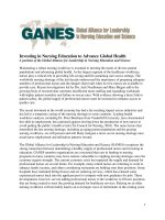
events: progressive decrease in compliance of the
chest wall, in static elastic recoil of the lung (Fig. 1),
and in strength of respiratory muscles.
Age-associated changes in the chest wall
Estenne and colleagues measured age-related
changes in chest wall compliance in 50 healthy
subjects ages 24 to 75: aging was associated with a
significant decrease (À31%) in chest wall compli-
ance, involving rib cage (upper thorax) compliance
and compliance of the diaphragm-abdomen compart-
ment (lower thorax) [5]. Calcifications of the costal
cartilages and chondrosternal junctions and degenera-
tive joint disease of the dorsal spine are common
radiologic observations in older subjects and contrib-
ute to chest wall stiffening [6]. Changes in the shape
of the thorax modify chest wall mechanics; age-
related osteoporosis results in partial (wedge) or
complete (crush) vertebral fractures, leading to
increased dorsal kyphosis and anteroposteriordi-
ameter (barrel chest). Indeed, prevalence of vertebral
fractures in the elderly population is high and
increases with age; in Europe, in female subjects
over 60, the prevalence of vertebral fractures is
16.8% in the 60 to 64 age group, increasing to 34.8%
in the 75 to 79 age group [7]. Men also show an
increase in vertebral fractures with age, but rates are
approximately half those of the female population
[8]. A study of 100 chest radiographs of subjects ages
75 to 93 years, without cardiac or pulmonary dis-
orders, illustrates the frequency of dorsal kyphosis
in this age group: 25% had severe kyphosis as a
consequence of vertebral wedge or crush fractures
(>50°), 43% had moderate kyphosis (35°–50°), and
only 23% had a normal curvature of the spine [6].
0272-5231/05/$ – see front matter D 2005 Elsevier Inc. All rights reserved.
doi:10.1016/j.ccm.2005.05.004 chestmed.theclinics.com
E-mail address:
Clin Chest Med 26 (2005) 469 – 484
Respiratory muscle function
Respiratory muscle performance is impaired con-
comitantly by the age-related geometric modi-
fications of the rib cage, decreased chest-wall
compliance, and increase in functional residual
capacity (FRC) resulting from decreased elastic recoil
of the lung (Fig. 2) [9]. The kyphotic curvature of the
spine and the anteroposterior diameter of the chest
increase with aging, thereby decreasing the curvature
of the diaphragm and thus its force-generating
capacity [6]. Changes in chest wall compliance lead
to a greater contribution to breathing from the dia-
phragm and abdominal muscles and a lesser contri-
bution from thoracic muscles. The age-related
reduction in chest-wall compliance is somewhat
greater than the increase in lung compliance; thus,
compliance of the respiratory system is 20% less in a
60-year-old subject compared with a 20-year-old (see
Fig. 1) [9]. As such, during normal resting tidal
breathing, the increase in breathing-related energy
expenditure (elastic work) in a 60-year-old man is
estimated at 20% compared with that of a 20-year-
old, placing an additional burden on the respiratory
muscles [8].
Respiratory muscle strength decreases with age
(Table 1). Polkey and colleagues report a significant,
although modest, decrease in the strength of the
diaphragm in elderly subjects (n = 15; mean age 73,
range 67–81 years) compared with a younger control
group (n = 15; mean age 29, range 21 – 40 years):
À13% for transdiaphragmatic pressure during a
maximal sniff (sniff transdia phragmatic pressure
[Pdi]: 119 versus 136 cm H
2
O) and À23% during cer-
vical magnetic stimulation (twitch Pdi: 26.8 versus
35.2 cm H
2
O) [10]. There was, however, a consid-
erable overlap between groups, and the magnitude
of the difference in this study was relatively small.
010203040-10-20-30
100
80
60
40
20
0
-40
100
80
60
40
20
0
FRC
RV
lung
Chest wall
rs
0102030
40-10
-20-30
100
80
60
40
20
0
-40
100
80
60
40
20
0
FRC
RV
lung
Chest wall
rs
Pressure (cm H2O)
Pressure (cm H2O)
% TLC% TLC
A
B
Fig. 1. Static pressure-volume curves showing changes in the compliance of the chest wall, the lung, and the respiratory system
between an ‘‘ideal’’ 20-year-old (A) and a 60-year-old subject (B). Note increase in RV and FRC and decrease in slope of
pressure-volume curve for the respiratory system (rs) in the older subject, illustrating decreased compliance of the respiratory
system. (Data from Turner J, Mead J, Wohl M. Elasticity of human lungs in relation to age. J Appl Physiol 1968;25:664–71.)
janssens470
Similarly, Tolep and cowor ker s report maxim al
Pdi values in healthy elderly subjects (n = 10; ages
65–75, 128 ± 9 cm H
2
O), which were 25% lower
than values obtained in young adults (n = 9; ages
19–28, 171 ± 8 cm H
2
O) [11]. Although one cross-
sectional study fails to demonstrate any relationship
between age and maximal static respiratory pressures
in 104 subjects over 55 [12], larger studies—also
based on noninvasive measurements (maximal inspir-
atory and expiratory pressures [MIP and MEP] at the
mouth and sniff nasal inspiratory pressure [SNIP])—
document an age-related decrease in respiratory
muscle performance [13–16].
Respiratory muscle strength is related to nutri-
tional status, often deficient in the elderly. Enright
and colleagues demonstrate significant correlations
between MIP or MEP pressures and lean body mass
(measured by bioelectric impedance), body weight, or
body mass index [14]. Arora and Rochester show the
deleterious impact of undernourishment on respira-
tory muscle strength or maximal voluntary ventila-
tion: the decrease in respiratory muscle strength and
maximal voluntary ventilation was highly significant
in undernourished subjects (71 ± 6% of ideal body
weight) compared with control subjects (104 ± 10%
of ideal body weight) [17]. Necropsy studies confirm
the correlation between body weight and diaphragm
muscle mass further [18].
Age-associated alterations in skeletal muscles also
affect respiratory muscle function [19]. MIP and
MEP in elderly subjects are correlated strongly and
independently with peripheral muscle strength (hand-
grip) [13]. Peripheral muscle strength declines with
aging. Bassey and Harries report a 2% annual
decrease in handgrip strength in 620 healthy subjects
over age 65 [20]. Decrease in muscle strength results
from a decrease in cross-sectional muscle fiber area
(process referred to as sarcopenia), a decrease in the
number of muscle fibers (especially type II fast-
twitch fibers and motor units), alterations in neuro-
muscular junctions, and loss of peripheral motor
neurons with selective denervation of type II muscle
fibers [21–26]. Other proposed mechanisms of age-
related muscular dysfunction include impairment of
the sarcoplasmic reticulum Ca
++
pump resulting from
uncoupling of ATP hydrolysis from Ca
++
transport
(which may reduce maximal shortening velocity and
relaxation), loss of muscle proteins resulting from
decreased synthesis (ie, decreased ‘‘repair’’ ability
and protein turnover), and a decline in mitochondrial
oxidative capacity [27–31].
Respiratory muscle function also is dependent on
energy availability (ie, blood flow, oxygen content,
and carbohydrate or lipid levels) [32]. Decreased
respiratory muscle strength is described in patients
who have chronic heart failure (CHF). Mancini and
colleagues show that CHF has a highly significant
impact on respiratory muscle strength and on the
tension-time index [33]. The tension-time index de-
scribes the relationship between force of contraction
(Pdi/Pdi
max
) and duration of contraction (ratio of
inspiratory time to total respiratory cycle duration
[TI/TTOT]) and is related inversely to respiratory
muscle endurance. In elderly subjects who have heart
Table 1
Maximal inspiratory and expiratory pressures measured at
the mouth in older subjects, by age group and sex
Age group (y)
MIP (cm H
2
O)
n = 5201
MEP (cm H
2
O)
n = 756
Women
65 –69 59 125
70 –74 56 121
75 –79 49 102
80 –84 45 84
>85 40 94
Men
65 –69 84 188
70 –74 81 179
75 –79 74 161
80 –84 64 142
>85 56 131
Data from Enright PL, Kronmal RA, Manolio TA, et al.
Respiratory muscle strength in the elderly: correlates and
reference values. Am J Respir Crit Care Med 1994;149:
430 – 8.
10 20 30 40 50 60 70
10
20
30
40
50
60
70
0
Age (years)
% TLC
RV
FRC
Fig. 2. Progressive and linear increase in RVand FRC
between the ages of 20 and 60 years. Gray zones represent
± 1 SD. (Data from Turner J, Mead J, Wohl M. Elasticity of
human lungs in relation to age. J Appl Physiol 1968;25:
664–71.)
aging of the respiratory system 471
failure, the tension-time index increases, primarily
because of an increase in Pdi/Pdi
max
and, during
exercise, approaches values shown to generate fatigue
[34]. Evans and coworkers show a significant corre-
lation between cardiac index and sniff Pdi [35].
Nishimura and coworkers make a similar observation
in subjects who have CHF, showing significant cor-
relations between MIP and cardiac index or maximal
oxygen consumption (Vo
2max
) per body weight, as
an index of cardiovascular performance [36].
Other frequent clinical situations that produce
diminished respiratory muscle function in the elderly
include Parkinson’s disease and sequelae of cerebral
vascular disease [37,38]. Myasthenia gravis is an-
other cause of respiratory muscle weakness, although
encountered less commonly.
Changes in the lung parenchyma and peripheral
airways
The human respiratory system is exposed con-
tinuously to air and a variety of inhaled pollutants.
This creates a challenge for physiologists and cli-
nicians, namely to differentiate—in studies of human
lungs—the true impact of normal aging (ie, physio-
logic aging) from that of environmental exposure.
Environmental tobacco smoke and particulate air
pollution have measurable and well-documented ef-
fects on respiratory symptoms and disease in the
elderly [39 – 41]. Appropriate animal models, there-
fore, are needed to study pathologic changes that
occur with aging per se. Senescence-accelerated mice
(SAM; a murine model of accelerated senescence) is
proposed as such a model, permitting investigation of
the differences between the aging lung and cigarette
smoke–related airspace enlargement [42,43]. Mor-
phometric studies of SAM show a notable homoge-
neous enlargement of alveolar duct size with aging.
Cellular infiltrates in the alveoli rarely are seen, sug-
gesting that the airspace enlargement does not result
from inflammation, as opposed to what is seen in
emphysema. The ratio of lung weight to body weight
does not decrease with aging, suggesting little or no
lung destruction [42]. Elastic fibers of the lung in
SAM have a reduced recoil pressure, causing
distention of the alveolar spaces and increased lung
volume [43]. Age-related changes in the pressure-
volume curves show a shift leftwards and upwards
(ie, loss of elastic recoil of the lung) (see Fig. 1).
These changes are similar to those described in senile
hyperinflation of the lung in humans [9,44].
As noted in SAM during the course of aging,
alveolar ducts in humans increase in diameter and
alveoli become wider and shallower [45].This
enlargement is remarkably homogeneous as opposed
to the irregular distribution of airspace enlargement in
emphysema. Morphometric studies consistently find
an increase in the average distance between airspace
walls (mean linear intercept) and a decrease in the
surface area of airspace wall per unit of lung volume
beginning in the third decade of life. The decrease in
surface area of airspace wall per unit of lung volume
approximately is linear and continues throughout life,
resulting in a 25% to 30% decrease in nonagenarians
[46,47]. Although these changes are histologically
different from emphysema (no destruction of alveolar
walls), they result in similar changes in lung
compliance. Thus, as described by Turner coworkers
in subjects ages 20 to 60, static elastic recoil pressure
of the lung decreases as a part of normal aging (0.1–
0.2 cm H
2
O Á year
À1
), and the static pressure-volume
curve for the lung is shifted to the left and has a
steeper slope [9,48]. Verbeken and coworkers pro-
pose that the changes in structural and functional
characteristics caused by isolated airspace enlarge-
ment that are seen in the elderly be differentiated
from emphysema by the absence of alveolar wall de-
struction and inflammation and designated as senile
lung [45].
In a postmortem study, mean bronchiolar diame-
ter also decreased significantly after age 40 [49].
Bronchiolar narrowing and increased resistance were
independent of any emphysematous changes or of
previous bronchiolar injury. This decline in small
airway diameter may contribute to the decrement in
expiratory flow noted with aging [49]. Reduction in
supporting tissues around the airways further in-
creases the tendency for the small airways (<2 mm)
to collapse.
Pulmonary function tests
Specifics of pulmonary function testing in an older
population
The application of conventional quality control
standards to objective assessment of pulmonary
function in older subjects may prove difficult because
of mood alterations, fatigability, lack of cooperation,
or cognitive impairment. Indeed, prevalence of
dementia increases with aging, reaching 5.6% after
age 75, 22% after age 80, and 30% as of age 90 [50].
The relationship between ability to perform spirome-
try and cognitive function in the elderly is reported by
several investigators [51–54]. The feasibility of high-
quality spirometry in elderly subjects who do not
have cognitive impairment is confirmed in a large
janssens472
Italian study of 1612 ambulatory subjects ages 65 and
older who did nor did not have chronic airflow
limitation: tests with at least three acceptable curves
were obtained in 82% of normal subjects and in 84%
of patients who have chronic airflow limitation [55].
Cognitive impairment, however, lower educational
level, and shorter 6-minute walking distance levels
were found to be independent predictors of a poor
acceptability rate [55]. Pezzoli and colleagues per-
formed spirometric testi ng in 715 subjects w ho
had respiratory symptoms and reported a feasibility
rate (according to ATS criteria) of 82%; low Mini–
Mental State Examination and activities of daily
living scores were associated with poor spirometric
performance [56]. Lower feasibility rates for spirome-
try are reported in elderly patients who were in-
stitutionalized (41%) and hospitalized (50%), with a
clear relationship between the degree of cognitive
impairment and feasibility of testing [53,54]. The
prevalence of delirium in older people on hospital
admission ranges from 10% to 24%, whereas de-
lirium develops in 5% to 32% of older patients after
admission [57]. Underdiagnosis, therefore undertreat-
ment, of chronic obstructive pulmonary di sease
(COPD) in older subjects may be related to difficul-
ties encountered in performing spirometry adequately
in this population.
Alternative tests for the measurment of COPD in
the elderly have been explored to find methods that
may be less cooperation dependent for test subjects.
Measurement of airway resistance using the forced
oscillation technique (FOT) is applied more easily
than spirometry in older patients who have cogni-
tive disorders [53,54]. In elderly patients who are
hospitalized or institutionalized, measurement of
airway resistance by FOT was successful in 74% to
76% of patients tested. The reported sensitivity and
specificity for the detection of COPD in older sub-
jects were 76% and 78%, respectively; thus, FOT is
useful in this population [54]. Conversely, assessment
of airway resistance using the interrupter technique,
widely used in epidemiologic and pediatric studies, in
spite of its attractive simplicity, performed poorly in
the detection of COPD in older subjects compared
with FOT or spirometry, with a higher coefficient
of variation than FOT [58]. The negative expiratory
pressure technique (NEP), which does not require a
forced expiratory maneuver, is useful to detect flow
limitation [59]. The test involves applying negative
pressure at the mouth during a tidal expiration. When
the NEP elicits an increase in flow throughout the
expiration, patients are not flow limited. In contrast,
when patients do not have an increase in flow during
most or part of the tidal expiration on application of
NEP, they are considered flow limited. This technique
has significant limitations, as it underestimated the
presence of COPD without resting flow limitation in
a study of 26 adults ages 42 to 87 (mean 65 ±
10 years) and, therefore, cannot be considered a sub-
stitute for spirometric screening for COPD [59].
For assessment of respiratory muscle perfor-
mance, SNIP and MIP and MEP are feasible in older
subjects, although SNIP tends to be easier to perform
and better tolerated than MIP; these tests show an
important learning effect and must be repeated at least
five (MIP and MEP) to 10 (SNIP) times [60,61].
Reported coefficients of variation for MIP and MEP
in healthy elderly subjects are, respectively, 10.2%
and 12.8% [62].
Plethysmographic measurement of lung volumes
seldom is required in this age group and, to the
author’s knowledge, no specific reference values are
available for subjects over age 70.
Lung volumes
The major determinants of static lung volumes are
the elastic recoil of the chest wall and that of the lung
parenchyma. Loss of elastic recoil of the lung pa-
renchyma and, to a lesser degree, decrease in re-
spiratory muscle performance result in an increase in
residual volume (RV): RV increases (air-trapping)
by approximately 50% between ages 20 and 70 (see
Fig. 2). Conversely, there is a progressive decrease in
vital capacity to approximately 75% of best values.
Because of the increased stiffness of the chest wall,
the age-related diminished elastic recoil of the lungs
is counterbalanced by an increased elastic load
from the chest wall; total lung capacity (TLC)
thus remains fairly constant throughout life [63].
Increased elastic recoil of the chest wall and dimin-
ished elastic recoil of the lung parenchyma also
explain the increase in FRC (ie, elderly subjects
breathe at higher lung volumes than younger sub-
jects) (see Figs. 1 and 2) [63].
The closing volume (ie, the volume at which small
airways in dependent regions of the lung begin to
close during expiration) increases with age. Prema-
ture closure of terminal airways is related to a loss of
supporting tissues around the airways. The closing
volume begins to exceed the supine FRC at approxi-
mately 44 years of age and to exceed the sitting
FRC at approximately 65 years of age [64]. Closing
volume may reach 55% to 60% of TLC and equal
FRC; as such, normal tidal breathing may occur
with a significant proportion of peripheral airways
not contributing to gas exchange (low ventilation-
perfusion ratio [V/Q] zones). Although this is sug-
aging of the respiratory system 473
gested as an important mechanism for the age-related
decrease in Pao
2
, increase in alveolar-arterial differ-
ence in partial pressure of oxygen (Pao
2
À Pao
2
),
and decrease in carbon monoxide transfer, measure-
ment of V/Q inequality using the multiple inert gas
eli mination technique (MIGET) fails to show a
significant increase in low V/Q areas with aging in
64 subjects ages 18 to 71 [65].
Spirometry
Forced expiratory volumes increase with growth
up to the age of approximately 18. According to
European Community for Coal and Steel data, no
significant changes occur in forced expiratory volume
in 1 second (FEV
1
) or forced vital capacity (FVC)
between the ages of 18 and 25 [66]. After this pla-
teau, FEV
1
and FVC start to decrease, although more
recent studies excluding smokers suggest a later start
of FEV
1
and FVC decline in nonsmokers [67]. Cross-
sectional and longitudinal studies show an acceler-
ated decline in FEV
1
and FVC with age; the rate of
decline is greater in cross-sectional versus longitudi-
nal studies and in men versus women and more rapid
in patients who have increased airway reactivity [63].
The age-related decrease in FEV
1
and FVC initially
was considered linear, but more recent studies—
including subjects ages 18 to 74—suggest that the
decline may be nonlinear and accelerates with ag-
ing [68 – 71].
Regression equations, based on extrapolations
from groups of younger subjects, tend to overestimate
predicted values for FEV
1
, FVC, and FEV
1
/FVC in
elderly subjects [67]. Few studies report results ob-
tained in large samples of elderly subjects. Ericsson
and Irnell, for example, report measurements per-
formed on 264 normal ‘‘elderly’’ subjects, none of
whom was older than 71 years of age [72]. Fowler
and colleagues studied 182 Londoners over age 60,
but only 44 subjects were over age 75 and 23 were
over 80 [73]. The three largest studies (all cross-
sectional) reporting spirometric data from healthy
elderly subjects were published by Milne and Wil-
liamson, Enright and colleagues, and DuWayne
Schmidt and colleagues[52,74,75]. D uWayne
Schmidt and colleagues included patients ages 20 to
94 and found that decline in FEV
1
and FVC with age
was linear (À31 mL/year in men and À27 mL/year in
women). Values for FEV
1
/FVC were stable in young
adults and decreased in women over age 55 and in
men over age 60 to 70 to 75% range [75]. The study
by Milne and Williamson includes a large number of
active or former smokers, and 20% of subjects had
regular cough and phlegm; thus, it is unreliable [52].
Enright and colleagues selected 777 healthy non-
obese, never-smokers ages 65 to 85 who had no
history of lung disease from 5201 ambulatory elderly
participa nts of the Car diovascular Health Study;
estimation of annual decline for FEV
1
was 32 mL/
year in women and 27 mL/year for men and, for
FVC, 33 mL/year in women and 20 mL/year in men
(Box 1) [13,74,108,113,117]. Regression equations
suggest a linear relationship between age and decline
in FEV
1
and FVC in this study [73]. In summary, the
average yearly decline of FEV
1
and FVC is approx-
imately 30 mL/year, although it may be overesti-
mated by cross-sectional studies. Whether or not
decline of forced expiratory volumes with age is
linear remains controversial, and longitudinal studies
of older nonsmoking subjects are required to clarify
this issue.
According to publi she d referen ce values for
FEV
1
/FVC, using a threshold value of FEV
1
/FVC
less than 70% for defining the presence of airway
obstruction, as suggested by the Global Initiative for
Chronic Lung Diseases (GOLD) Workshop Sum-
mary, may lead to overdiagnosis of COPD. This is
illustrated by a Norwegian study of forced expiratory
volumes in 71 asymptomatic never-smokers, ages 70
or older; according to GOLD criteria, 25% had stage I
COPD and 10% stage II; for subjects older than 80,
results were, respectively, 32% and 18% [76]. Using
the regression equations published by Enright and
colleagues, normal values for FEV
1
/FVC are less
than 70% for men ages 80 and older and women ages
92 and older (see Box 1) [74].
Flow-volume curves and peak expiratory flow
Fowler and colleagues r eport characteristic modi-
fications in the expiratory flow-volume curve with
aging ( Fig. 3 ) [73]. The changes in e xpiratory flow-
volume s uggested alterations in the s mall peripheral
airways, with an obstructive pattern present even in
lifetime nonsmokers, s uggesting that this p attern may be
normal in old age. Similar results are reported by Babb
and Rodarte, who compared e xpiratory flow rates i n 1 7
younger adults (ages 35–45) with those of 1 9 o ld er
adults (ages 65–75) ; i n this s tudy, decline i n p eak
expiratory flow (PEF) in the older group is proportional
to loss of lung elastic r ecoil [77]. Changes i n peripheral
airways and loss o f supporting tissue around the airways
(‘‘senile l ung’’) (discussed previously) a re plausible
explanations for these findings.
Although PEF rates tend to decrease with age, the
variability in predicted peak flow values is large, and
prediction equations are, therefore, not reliable
[78,79]. PEF lability (maximal difference in PEF
janssens474
per mean PEF) is shown to correlate with a diagnosis
of asthma in younger subjects. Although middle-aged
and older persons seem to be successful in providing
a measure of PEF reliably at home, older age per se
was a factor of increased variability in longitudinal
monitoring of ambulatory PEF (independent predic-
tor of higher PEF lability) [80]. In a study of 1223
subjects (mean age 66, range 43–80), Enright and
colleagues report an upper limit of normal of 16%
for PEF lability in older patients [80]. Another study
Box 1. Regression equations for pulmonary function test variables in older subjects
Spirometry: men
FEV
1
(liters) = (0.0378
Â
height
cm
) À (0.0271
Â
age
years
) À 1.73; LLN = À0.84
FVC = (0.0567
Â
height
cm
) À (0.0206
Â
age
years
) À 4.37; LLN = À1.12
FEV
1
/FVC% = (À0.294
Â
age
years
) + 93.8; LLN = À11.7
Spirometry: women
FEV
1
(liters) = (0.0281
Â
height
cm
) À (0.0325
Â
age
years
) À 0.09; LLN = À0.48
FVC = (0.0365
Â
height
cm
) À (0.0330
Â
age
years
) À 0.70; LLN = À0.64
FEV
1
/FVC% = (À0.242
Â
age
years
) + 92.3; LLN = À9.3
Maximal mouth inspiratory and maximal mouth expiratory pressures: men
MIP (cm H
2
O) = (0.131
Â
weight
lb
) À (1.27
Â
age
years
) + 153; LLN = À41
MEP (cm H
2
O) = (0.25
Â
weight
lb
) À (2.95
Â
age
years
)+347;LLN=À71
Maximal mouth inspiratory and maximal mouth expiratory pressures: women
MIP (cm H
2
O) = (0.133
Â
weight
lb
) À (0.805
Â
age
years
)+96;LLN=À32
MIP (cm H
2
O) = (0.344
Â
weight
lb
) À (2.12
Â
age
years
) + 219; LLN = À52
6-minute walk test: men (n = 117; ages 40 to 80)
6MWD
meters
= (7.57
Â
height
cm
) À (5.02
Â
age
years
) À 309 m; LLN = À153 m
6-minute walk test: women (n = 173, ages 40 to 80)
6MWD
meters
= (2.11
Â
height
cm
) À (2.29
Â
weight
kg
) À (5.78
Â
age
years
) + 667 m;
LLN = À139 m
Maximal heart rate (n = 18712)
Maximal heart rate = 208 À (0.7
Â
age)
Maximal oxygen consumption (n = 100; ages 15 to 71)
Vo
2max
(L/min) = (0.046
Â
height
cm
) À (0.021
Â
age
years
) À 0.62 (0: male; 1: femal e) À 4.31 L;
LLN = À.89 L
Abbreviations: LLN, lower limit of normal (mean À 1.96 SD); 6MWD, distance walked during a
6-minute test.
aging of the respiratory system 475
by the same group, based on a larger community
sample of 4581 persons ages 65 and older, reports an
upper limit of normal of 29% for PEF lability. A cut-
off value of 30% for PEF lability, th erefore, is
recommended in older subjects for the diagnosis of
asthma [78].
No specific changes are noted regarding the
inspiratory flow curves, although maximal inspiratory
flow values decrease with aging. Because lung
deposition of inhaled drugs is flow dependent with
available powder-inhaling devices, determination of
maximal inspiratory flow in older subjects may be
relevant when considering topical bronchodilator or
anti-inflammatory treatment with a powder inhaler
[81,82]. Some powder-inhaling devices require min-
imal inspiratory flows (through the device) of up to
60 L Á min
À1
and these values may not be attained in
very elderly patients. With the Turbuhaler, for
instance, lung deposition at an inspirator flow of
30 L Á min
À1
is approximately half that obtained at
60 L Á min
À1
, although equivalent to that obtained
with a metered-dose inhaler [82].
Airway resistance and conductance
When adjusted for lung volume, age has no sig-
nificant effect on airway resistance. Peripheral air-
ways contribute marginally to the total resistance of
the airways and, therefore, changes in the peripheral
airways are not reflected by changes in airway re-
sistance [63]. Using the FOT, Pasker and colleagues
find a weak impact of age on resistance and re-
actance, with opposite effects according to sex; the
investigators consider the relationship between FOT
measurements and age clinically irrelevant [83].
Respiratory muscle testing
Respiratory muscle weakness may lead to short-
ness of breath, reduced exercise tolerance, and, in
more severe cases, alveolar hypoventilation and re-
spiratory failure. The overall strength of respiratory
muscles can be measured noninvasively by recording
MIP and MEP or by measuring SNIP [61,84]. These
measurements can be performed easily at bedside
[61,84]. Inspiratory pressures are measured at FRC or
at RV. Expiratory pressures usually are measured at
TLC. As discussed previously, the learning effect for
MIP, MEP, and SNIP measurements is important,
with significant increases over at least five consecu-
tive maneuvers [13,61]. Values greater than or equal
to 80 cm H
2
O (in men) or 70 cm H
2
O (in women) for
MIP or greater than or equal to 70 cm H
2
O in men
and 60 cm H
2
O in women for SNIP exclude clinically
relevant respiratory muscle weakness [85].
Available reference values for these measurements
show a decrease with age of respiratory muscle
strength (see Table 1 and Box 1) [13–16]. Enright
and colleagues measured MIP and MEP in ambula-
tory subjects ages greater than or equal to 65; normal
values for women ages greater than or equal to 65 and
males ages greater than or equal to 75 are below the
aforementioned threshold for clinically relevant res-
piratory muscle dysfunction [13]. Nutritional status
(body weight, bioelectric impedance, and body mass
index) and peripheral muscle strength (handgrip)
correlate significantly with MIP and MEP values
[13]. Other investigators find values in the same
range for MIP, MEP, or SNIP [15,16,86]. The de-
crease in respiratory muscle strength likely is relevant
in elderly patients in clinical situations where an
additional load is placed on the respiratory muscles,
such as pneumonia or left ventricular failure [35,36].
The effects of poor nutritional status and CHF on
respiratory muscle strength are discussed previously.
Gas exchange
Changes in arterial oxygen tension and
ventilation-perfusion relationships
Wagner and coworkers, using the MIGET, report
an increase, with aging, in V/Q imbalance, with a rise
VolumeTLC RV
Flow (L/sec)
12
8
4
0
-4
-8
Fig. 3. Changes in the expiratory flow-volume curve with
aging, suggesting obstruction to airflow. Curves from an
older (dashed line) and a younger subject (solid line),
normalized to percentage of vital capacity. (Data from Babb
TG, Rodarte JR. Mechanism of reduced maximal expiratory
flow with aging. J Appl Physiol 2000;89:505–11; and
Fowler RW, Pluck RA, Hetzel MR. Maximal expiratory
flow-volume curves in Londeners aged 60 years and over.
Thorax 1987;42:173–82.)
janssens476
in units with a high V/Q (wasted ventilation or
physiologic dead space) and in units with a low V/Q
(shunt or venous admixture) [87,88]. The decrease in
Pao
2
with age is described a consequence of this
increased heterogeneity of V/Q and, in particular, of
the increase in units with a low V/Q (dependent parts
of the lung, poorly ventilated during tidal breathing,
as reflected by an increased closing volume) [87].
These conclusions are based, however, on a small
number of observations. More recently, Cardus and
coworkers described the age-related changes in V/Q
distribution in 64 healthy subjects ages 18 to 71 [65].
Although there was a slight increase in V/Q mismatch
in older patients, shunt and low V/Q areas did not
exceed 3% of total cardiac output, and decrease in
Pao
2
with age was minimal (6 mm Hg). Most of the
variance of V/Q mismatch was not a result of aging
and remained unexplained; the role of an increase in
closing volume with aging was not supported by
these data. This elegant study included, unfortunately,
a small number of older patients (only 4 were older
than 60 ) and may n ot reflect chan ges in V/Q
distribution occurring in the very old. Indeed, closing
volume increases with age but may equal FRC only
when subjects reach approximately 65 years of age
(according to Leblanc and coworkers [89], FRC À
closing volume = 1.95 À [0.03
Â
age]).
Regressions proposed for the computing of Pao
2
as a function of age vary widely, mainly in relation to
the coefficient attributed to age [90]. Indeed, for an
82-year-old man, predicted values for Pao
2
range
from 8.4 to 11.3 kPa (63–84 mm Hg). Delclaux and
colleagues measured arterial blood gases in 274 sub-
jects ages 65 to 100 (mean 82 years) with and without
airway obstruction; mean Pa o
2
was 10 ± 1.4 kPa
(75 ± 11 mm Hg) [90]. These investigators suggest
accepting as normal a Pao
2
of 10.6 to 11.3 kPa
(80–85 mm Hg) for subjects 65 years of age and
older [90]. Guenard and coworkers find no significant
correlation between Pao
2
and age in 74 subjects ages
69 to 104; mean values reported were 11.2 ± 1.0 kPa
(84 ± 7.5 mm Hg) [91]. Conversely, Sorbini and
colleagues showed that there was a linear, but re-
ciprocal, relationship between age and Pao
2
in non-
smoking healthy subjects, with the following
regression equation: Pao
2
= 109 À (0.43
Â
age)
(the fact that patients were supine during arterial
sampling probably explains lower Pao
2
values ob-
tained from this regression) [92]. More recently,
Cerveri and coworkers suggest that the decrease in
Pao
2
with aging is not linear [93]. In their study,
arterial blood gas tests were analyzed in 194 non-
smoking subjects ages 40 to 90. Stratifying the results
by 5-year age intervals, the investigators found a clear
decline in Pao
2
up to 70 to 74 years of age, followed
by a slight rise in Pao
2
from ages 75 to 90. For
healthy patients o lder than 75, Pao
2
was not
correlated with age; m ean values reported were
83 ± 9 mm Hg (11.1 ± 1.2 kPa), and fifth percentile
was at 68.4 mm Hg (9.2 kPa) [93].
A modest increase in the Pao
2
À Pao
2
with age is
expected because of the previously described increase
in V/Q heterogeneity. Acco rding to Sorbini and
coworkers [92], the highest normal value for the
Pao
2
À Pao
2
at a certain age is given by the
equation: Pao
2
À Pao
2
(mm Hg) 1.4 ± 0.43
Â
age
(years). High values obtained by this equation (ie,
4.8 kPa [36 mm Hg] for 80 years of age) also may
result from the supine position of subjects at time
of sampling. More recent studies find no significant
relationship between age and Pao
2
À Pao
2
; however,
values reported are well above normal values for
younger adults (ie, 3.2 ± 1.4 kPa [24 ± 10 mm Hg]
[90] and 4.4 ± 0.6 kPa [33 ± 4.5 mm Hg] [91]).
Carbon monoxide transfer factor
Flattening of the internal surface of the alveoli
(ductectasia) in the elderly is associated with a re-
duction in alveolar surface (75 m
2
at the age of
30 years versus 60 m
2
at age 70 years, a reduction of
0.27 m
2
Á year
À1
) [63]. Because of loss of alveolar
surface area, decreased density of lung capillaries,
decline in pulmonary capillary blood volume, and
increased V/Q heterogeneity, it is estimated that, even
in healthy nonsmokers, there is a yearly decline in
the diffusing capacity of the lung for carbon mon-
oxide (Dlco) of 0.2 to 0.32 mL Á min
À1
Á mmHg
À1
from middle ages and onward in men and a decrease
of 0.06 to 0.18 mL Á min
À1
Á mmHg
À1
in women
[91,94]. Gue´nard and Marthan determined, in a popu-
lation of 74 healthy subjects aged 69 to 104, the
following regression equation for transfer capacity of
the lung for carbon monoxide (Tlco) versus age
(age explaining 29% of the variance of Tlco): Tlco
(mL Á min
À1
Á kPa
À1
) = 126 – 0.9
Â
age (years;
r = 0.54, P < 0.001) [91].
Regulation of breathing
Aging and ventilatory responses
Aging is associated with a marked attenuation
in ventilatory responses to hypoxia and hypercap-
nia [95–97]. Kronenberg and Drage compared the
aging of the respiratory system 477
responses to hypercapnia and hypoxia in eight
healthy young men (22 – 30 years old) with those of
eight older men (64–73 years old) [95]. In the older
subjects, ventilatory response to hypoxia was four
times less than that of the younger group; response to
hypercapnia was decreased by 58%. Mouth occlusion
pressure (P
0.1
), an index of respiratory drive, is the
inspiratory pressure generated at the mouth when
occluding the airway 0.1 second after the beginning
of inspiration. Peterson and colleagues describe, in
subjects ages 65 to 79, a 50% reduction in the re-
sponse to isocapnic hypoxia and a 60% reduction in
that to hyperoxic hypercapnia measured by P
0.1
com-
pared with younger subjects [97]. More recently,
however, two studies cast doubt on the age-related
decrease in hypoxic ventilatory response. Smith and
colleagues studied two groups of nonsmoking male
subjects, ages 30 ± 7 and 73 ± 3, who were submitted
to 20 minutes of acute isocapnic hypoxia; ventilatory
responses and increment in neuromuscular drive were
similar in both groups [98]. Similarly, Pokorski and
Marczak compare the ventilatory response to iso-
capnic h ypoxia in 19 wome n ages 7 1 ± 1 to
16 younger women and find no significant difference
between groups in slopes of the DVe (ventilation)
to DSao
2
(arterial oxygen saturation) ratio and DP
0.1
/
DSao
2
[99].
The importance of the decrease in ventilatory re-
sponse to hypercapnia in older subjects al so is
unsettled: as in the st udy by Kronenberg and
Drage, Brischetto and colleagues report a reduction
in the slope of the ventilatory response to hyper-
capnia in older subjects (À67%) versus a younger
control group [95,96]. Rubin and coworkers, how-
ever, in a comparative study of ventilatory response
and P
0.1
response to hypercapnia, fail to disclose
significant differences between older (n = 10, ages
over 60) versus younger adults (n = 18, ages
under 30) [100].
Thus, although some studies suggest that there is
an age-related decline in the ability to integrate
information received from sensors (peripheral and
central chemoreceptors and mechanoreceptors) and
generate appropriate neural activity, further inves-
tigations are needed to clarify this issue.
Agingalsoisassociatedwithadecreasedpercep-
tion of a dded resistive or elastic loads [57 ,101,102] .
Older subjects h ave a lower perception of m ethacholine-
induced bronchoconstriction th an younger subjects.
Although available evidence yields conflicting results,
blunting o f the re sponse to hypoxia and hy percapnia a nd
lower ability to perceive bronchoconstriction may rep-
resent a part ial loss o f importan t prote ctive me chani sms
(alarm signals).
During sleep
The prevalence of slee p-disordered breathing
increases in elderly subjects. In middle-aged popula-
tions, the prevalence of the obstructive sleep apnea
syndrome (OSAS), using an apnea/hypopnea index
(AHI) of 15 events Á h
À1
as a cut-off value, is ap-
proximately 4% in women and 9% in men [103].In
older subjects, however, 13% to 62% of elderly
subjects suffer from OSAS with an AHI greater than
10 events per hour [104]. Sleep-disordered breathing
may be associated with impairment in cognitive
function and is reported to be more frequent in Alz-
heimer’s disease [105]. As discussed previously,
aging is associated with a diminished perception of
added resistive loads, such as that generated by
bronchoconstriction or upper airway collapse. Indeed,
respiratory effort in response to upper airway oc-
clusion in elderly patients is decreased compared with
younger subjects. Krieger and coworkers recorded
esophageal pressure during sleep in 116 patients who
had OSAS (AHI > 20) and showed that indexes of
respiratory effort were reduced significantly in older
compared with younger patients (inspiratory effort at
end of apnea: maximal esophageal pressure 40 ± 2
versus 56 ± 3 cm H
2
O) [106]. The lesser increase in
respiratory effort in older patients may result from a
decrease in respiratory drive and respiratory muscle
performance. In spite of the fact that indexes of
respiratory effort during apneic episodes were lower
in older individuals, mean apnea duration was not
prolonged significantly in older patients (28.3 ± 0.7 s
versus 30.4 ± 0.9 s), and postapneic Sao
2
was higher
in older individuals.
Ventilatory response to exercise
Performance during the 6-minute walk test
In subjects who do not have significant osteo-
articular or neuromuscular limitation, the 6-minute
walk test is a widely used standardized measurement
for evaluating physical function; results of a 6-minute
walk test are useful to quantify physical limitation
and monitor progressi on of disease in chronic
obstructive or restrictive disorders, CHF, or pulmo-
nary vascular diseases; performance is correlated with
health-related quality-of-life scores and predictive of
morbidity and mortality in disorders, such as pulmo-
nary hypertension or CHF [107]. There is a 15%
learning effect when tests are performed on two
successive days. Coefficient of variation is 8%.
Enright and Sherrill established reference equations
janssens478
for the 6-minute walk test from results collected in
290 healthy subjects ages 40 to 80 (see Box 1)
[108]. Predicted values for distance walked decrease
linearly with age, with a difference of approximately
200 meters between the ages of 40 and 80 years.
Mean baseline Sao
2
was stable at 96%. The 6-minute
walk test is a submaximal exercise test (peak Vo
2max
during a 6-minute walk is approximately 80% of
Vo
2max
during maximal exercise testing); thus,
potentially it is less sensitive for the detection of
cardiac or pulmonary disorders.
Maximal oxygen consumption and aging
The ability to perform physical tasks declines with
advancing age. Vo
2max
, expressed in L Á min
À1
,
reaches a peak between 20 and 30 years of age.
Longitudinal and cross-sectional studies thereafter
show a decrease in Vo
2max
at an estimated rate of 9%
to 10% per decade or À0.37 to À1.32 mL/kg/min/
year (see Box 1) [109 – 114]. The ventilatory thresh-
old also decreases with age, although less rapidly
than Vo
2max
[115]. The decrease in Vo
2max
is more
pronounced in sedentary subjects than in those
remaining physically active [109]. In fact, loss in
Vo
2max
is attenuated in fit elderly individuals and
may not be significant clinically. Vo
2max
of older
trained athletes is shown to be higher than that of
middle-aged untrained men [116]. Thigh muscular
mass also has a positive impact on Vo
2max
[113]. The
Fick equation gives the relationship between cardiac
output, peripheral oxygen extraction ([Ca À Cv]o
2
),
and oxygen consumption per unit time (Vo
2
): Vo
2
=
cardiac output
Â
[Ca À Cv]o
2
. Maximal heart rate
(HR) in healthy adults decreases with age: Tanaka
and colleagues, in a recent a meta-analysis compiling
data from 18,712 subjects, show that maximal HR is
independent of sex and level of physical activity and
is predicted mainly by age alone; they computed the
equation, maximal HR = 208 À 0.7
Â
age (r = À 0.90
versus age), which gives slightly higher values than
the commonly used predictive equation, maximal
HR = 220 À age [117]. Factors limiting Vo
2
in older
subjects are reduced maximal HR (resulting from a
decrease in sensitivity of cardiac b-adrenergic recep-
tors), decreased lef t ventricular ejection fraction,
reduced maximal cardiac output, and reduced periph-
eral muscle mass. Fleg and Lakatta measured 24-hour
urinary creatinine excretion, an index of muscle mass,
in 184 healthy nonobese volunteers, aged 22 to 87,
who performed a maximal treadmill exercise [118].
Vo
2max
showed a strong negative linear relationship
with age. When Vo
2max
was normalized for creati-
nine excretion, a large portion of the age-associated
decline in Vo
2max
was explained by the loss of
muscle mass [118].
Ventilation and exercise
Ventilation during exercise in the elderly is asso-
ciated with more abdominal contribution than in
young adults and a concomitant change in respiratory
pattern (higher rate and lower tidal volume), which
may result from increased stiffness of the tho-
racic cage.
When compared with younger subjects, initial
ventilatory (and circulatory) responses to exercise are
slowed in the elderly. Although respiratory frequency
increases rapi dly, ri se in t idal volume and total
ventilation is delayed [119].
In contrast to the previously discussed decreased
response to hypercapnia at rest, elderly subjects seem
more responsive than younger subjects to carbon
dioxide during exercise. Poulin and colleagues
demonstrate, in a sample of 224 subjects aged 56 to
85, that, for a given carbon dioxide production
(Vco
2
), the ventilatory response (Ve/Vco
2
)in-
creases with aging [120]. Similarly, Inbar and co-
workers find a 14% increase in Ve/Vco
2
and a 13%
increase in Ve/Vo
2
between the ages of 20 and 70, in
a large cross-sectional study of 1424 healthy subjects
[114]. This also was reported by Brischetto and
colleagues [95]. In both of these studies, this response
was related neither to oxygen desaturation nor to
increased metabolic acidosis; Prioux and colleagues,
however, show that, above the anaerobic threshold,
older subjects had, for a given carbon dioxide pro-
duction, higher lactate concentrations [121]. A higher
dead space – to – tidal volume ratio in elderly subjects
most probably is contributive to the higher Ve/Vco
2
ratio. In agreement with this hypothesis is the
observation of a higher difference between end-tidal
and arterial carbon dioxide tensions (Paco
2
) in older
subjects and the increase in V/Q heterogeneity with
aging (described previously) [65,114]. In itself, this
may increase dyspnea for a given workload. Indeed,
for a given Ve, the oxygen cost of breathing is higher
in elderly subjects.
Response to exercise training
Pulmonary rehabilitation programs are shown to
improve exercise capacity in older patients who have
COPD. A retrospective study by Couser and co-
workers compares the impact of a 2-month rehabili-
tation program in 28 subjects ages 75 years and
older versus 56 subjects aged less than 75. Improve-
ment in 12-minute walking distance was significantly
aging of the respiratory system 479
higher in the older patient group (+167 m; 38%
increase) than in the younger group (+107 m; 23%
increase) [122].
Aerobic training also is feasible in older healthy
individuals and results in significant, although often
modest, improvements in Vo
2
peak in older subjects.
For insta nce, Malbut and colleagues studied the
effects of a 6-month aerobic training program on
maximal aerobic power of 26 healthy elderly people
(79 to 91 years) [123]. After training, Vo
2max
in-
creased by 15% in women but not in men. Another
study of 22 sedentary subjects (aged 80 to 92) shows,
after 6 months of moderate-intensity aerobic exercise
training, an improvement in exercise test duration
(+33%) and peak Vo
2
(+9%) [124].Training
programs of 4 to 12 months, in older individuals,
show average increases in Vo
2max
of 8.5% to 25%
[125–128].
Summary
Compliance of the chest wall and the respira-
tory system and lung elastic recoil decrease with
aging, resulting in static air trapping (increased RV),
increased FRC, and increased work of breathing.
Respiratory muscle function also is affected by aging,
either as a consequence of geometric changes in the
rib cage, nutritional status (lean body mass, body
weight), cardiac function, or through the age-related
reduction in peripheral muscle mass and function,
referred to as sarcopenia. In subjects 80 years of age
and older, values of MIP may reach critically low
values; this may result in alveolar hypoventilation or
respiratory failure in clinical situations such as left-
sided heart failure or pneumonia. Expiratory flow
rates also decrease with aging, with characteristic
changes in the flow-volume curves suggesting in-
creased collapsibility of peripheral airways.
Gas exchange is remarkably well preserved at rest
and during exertion in spite of a reduced alveolar
surface area and increased ventilation-perfusion het-
erogeneity. In fact, in older athletes who have regular
physical training, the respiratory system remains
capable of adapting to high levels of exercise. In
sedentary individuals, however, Vo
2max
decreases
regularly with aging, whereas work of breathing, at a
given level of ventilation, increases. Decreased sen-
sitivity of respiratory centers to hypoxia or hypercap-
nia may result in a diminished ventilatory response in
case of acute disease, such as heart failure, infection,
or aggravated airway obstruction, although published
data as to the ventilatory response to hypoxia in
the elderly are inconclusive. Furthermore, blunted
perception of added resistive loads (ie, broncho-
constriction) and diminished physical activity may
result in a lesser awareness of respiratory disease and
delay diagnosis.
References
[1] Arias E, Anderson R, Kung H, et al. National vital
statistics reports. Hyattsville (MD)7 National Center
for Health Statistics; 2003.
[2] World Health Organization. W orld health report 2004.
Available at: />en/annex_4_en.pdf.
[3] Janssens JP, Krause KH. Pneumonia in the very old.
Lancet Infect Dis 2004;4:112–24.
[4] Janssens JP, Pache JC, Nicod LP. Physiological
changes in respiratory function associated with
ageing. Eur Respir J 1999;13:197 – 205.
[5] Estenne M, Yernault JC, De Troyer A. Rib cage and
diaphragm-abdomen compliance in humans: effects
of age and posture. J Appl Physiol 1985;59:1842–8.
[6] Edge JR, Millard FJ, Reid L, et al. The radiographic
appearances of the chest in persons of advanced age.
Br J Radiol 1964;37:769 – 74.
[7] Cummings SR, Melton LJ. Epidemiology and out-
comes of osteoporotic fractures. Lancet 2002;359:
1761 –7.
[8] Gunby MC, Morley JE. Epidemiology of bone loss
with aging. Clin Geriatr Med 1994;10:557 – 71.
[9] Turner J, Mead J, Wohl M. Elasticity of human lungs
in relation to age. J Appl Physiol 1968;25:664 –71.
[10] Polkey MI, Harris ML, Hughes PD, et al. The
contractile properties of the elderly human dia-
phragm. Am J Respir Crit Care Med 1997;155:
1560 –4.
[11] Tolep K, Higgins N, Muza S, et al. Comparison of
diaphragm strength between healthy adult elderly and
young men. Am J Respir Crit Care Med 1995;152:
677 – 82.
[12] McElvaney G, Blackie S, Morrison NJ, et al.
Maximal static respiratory pressures in the normal
elderly. Am Rev Respir Dis 1989;139:277 –81.
[13] Enright PL, Kronmal RA, Manolio TA, et al.
Respiratory muscle strength in the elderly: correlates
and reference values. Am J Respir Crit Care Med
1994;149:430–8.
[14] Enright PL, Adams AB, Boyle PJR, et al. Spirometry
and maximal respiratory pressure references from
healthy Minnesota 65 to 85 year old women and men.
Chest 1995;108:663–9.
[15] Uldry C, Fitting J-W. Maximal values of sniff nasal
inspiratory pressure in healthy subjects. Thorax 1995;
50:371 –5.
[16] Black LF, Hyatt RE. Maximal respiratory pressures:
normal values and relationship to age and sex. Am
Rev Respir Dis 1969;99:696 – 702.
[17] Arora NS, Rochester DF. Respiratory muscle strength
janssens480
and maximal voluntary ventilation in undernourished
patients. Am Rev Respir Dis 1982;126:5 – 8.
[18] Arora NS, Rochester DF. Effect of body weight and
muscularity on human diaphragm muscle mass,
thickness, and area. J Appl Phsiol 1982;52:64 – 70.
[19] Tolep K, Kelsen S. Effect of aging on respiratory
skeletal muscles. Clin Chest Med 1993;14:363 – 78.
[20] Bassey EJ, Harries UJ. Normal values for handgrip
strength in 920 men and women aged over 65 years,
and l ongitudinal changes over 4 years in 620
survivors. Clin Sci 1993;84:331 –7.
[21] Evans WJ. What is sarcopenia? J Gerontol A Biol Sci
Med Sci 1995;50(Spec no):5 – 8.
[22] Carmeli E, Reznick AZ. The physiology and bio-
chemistry of skeletal muscle atrophy as a function of
age. Proc Soc Exp Biol Med 1994;206:103 – 13.
[23] Carmeli E, Coleman R, Reznick AZ. The biochem-
istry of aging muscle. Exp Gerontol 2002;37:477 –89.
[24] Booth FW, Weeden SH, Tseng BS. Effect of aging
on human skeletal muscle and motor function. Med
Sci Sports Exerc 1994;26:556 – 60.
[25] Baumgartner RN, Stauber PM, McHugh D, et al.
Cross-sectional age differences in body composition
in persons 60 + years of age. J Gerontol A Biol Sci
Med Sci 1995;50:M307 –16.
[26] Newman AB, Haggerty CL, Goodpaster B, et al.
Strength and muscle quality in a well-functioning
cohort of older adults: the Health, Aging and Body
Composition Study . J A m Geriatr So c 2003;51:323 –30.
[27] Narayanan N, Jones D, Xu A, et al. Effects of aging
on sarcoplasmic reticulum function and contraction
duration in skeletal muscles of the rat. Am J Physiol
1996;271:1032–40.
[28] Balagopal P, Rooyackers O, Adey D, et al. Effects of
aging on in vivo synthesis of skeletal muscle myosin
heavy-chain and sarcoplasmic protein in humans. Am
J Physiol 1997;273:790 –800.
[29] Taylor D, Kemp G, Thompson C, et al. Ageing:
effects on oxidative function of skeletal muscle in
vivo. Moll Cell Biochem 1997;174:321 – 4.
[30] Trounce I, Byrne E, Marzuki S. Decline in skeletal
muscle mitochondrial respiratory chain function:
possible factor in ageing. Lancet 1989;1:637 – 9.
[31] Brierley E, Johnson M, James O, et al. Effects of
physical activity and age on mitochondrial function.
Q J Med 1996;89:251 – 8.
[32] Wilson DO, Rogers RM, Hoffman RM. Nutrition and
chronic lung disease. Am Rev Respir Dis 1985;132:
1347 –65.
[33] Mancini D, Henson D, LaManca J, et al. Respiratory
muscle f unctio n and dyspno ea in patients with
chronic heart failure. Circulation 1992;86:909 – 18.
[34] Stassijns G, Lysens R, Decramer M. Peripheral and
respiratory muscles in chronic heart failure. Eur
Respir J 1996;9:2161 – 7.
[35] Evans S, Watson L, Hawkins M, et al. Respiratory
muscle strength in chronic heart failure. Thorax 1995;
50:625 –8.
[36] Nishimura Y, Maeda H, Tanaka K, et al. Respiratory
muscle strength and hemodynamics in chronic heart
failure. Chest 1994;105:355 –9.
[37] Brown LK. Respiratory dysfunction in Parkinson’s
disease. Clin Chest Med 1994;15:715 – 27.
[38] Vingerhoets F, Bogousslavsky J. Respiratory dys-
function in stroke. Clin Chest Med 1994;15:729– 37.
[39] Anderson HR, Atkinson RW, Bremner SA, et al.
Particulate air pollution and hospital admissions for
cardiorespiratory diseases: are the elderly at greater
risk? Eur Respir J Suppl 2003;40:39s – 46s.
[40] Aga E, Samoli E, Touloumi G, et al. Short-term
effects of ambient particle s on mortality in the
elderly: results from 28 cities in the APHEA2 project.
Eur Respir J Suppl 2003;40:28s – 33s.
[41] Jaakkola MS. Environmental tobacco smoke and
health in the elderly. Eur Respir J 2002;19:172 – 81.
[42] Teramoto S, Fukuchi Y, Uejima Y, et al. A novel model
of senile lung: senescence accelerated mouse (SAM).
Am J Respir Crit Care Med 1994;150:238 –44.
[43] Kurozumi M, Matsushita T, Hosokawa M, et al.
Age-related changes in lung structure and function in
the senescence-accelerated mouse (SAM): SAM-P/1
as a new murine model of senile hyperinflation
of Lung. Am J Respir Crit Care Med 1994;149:
776–82.
[44] Bode FR, Dosman J, Martin RR, et al. Age and sex
differences in lung elasticity, and in closing capacity
in nonsmokers. J Appl Physiol 1976;41:129 – 35.
[45] Verbeken E, Cauberghs M, Mertens I. The senile
lung. Comparison with normal and emphysematous
lungs. I: structural aspects. Chest 1992;101:793 – 9.
[46] Thurlbeck W. The internal surface area of nonemphy-
sematous lungs. Am Rev Respir Dis 196 7;95:765–73.
[47] Gillooly M, Lamb D. Airspace size in lungs of life-
long nonsmokers: effect of age and sex. Thorax 1993;
48:39–43.
[48] Niewohner D, Kleinerman J, Liotta L. Elastic
behaviour of post-mortem human lungs: effects of
aging and mild emphysema. J Appl Physiol 1975;38:
943 – 9.
[49] Niewoenner D, Kleinerman J. Morphologic basis of
pulmonary resistance in human lung and effects of
aging. J Appl Physiol 1974;36:412 – 8.
[50] Nussbaum RL, Ellis CE. Alzheimer’s disease and Par-
kinson’s disease. N Engl J Med 2003;348:1356 – 64.
[51] Sherman CB, Kern D, Richardson ER, et al. Cogni-
tive function and spirometry performance in the
elderly. Am Rev Respir Dis 1993;148:123 –6.
[52] Milne JS, Williamson J. Respiratory function tests in
older people. Clin Sci 1972;42:371 – 81.
[53] Carvalhaes-Neto N, Lorino H, Gallinari C, et al.
Cognitive assessment of lung function in the elderly.
Am J Respir Crit Care Med 1995;152:1611–5.
[54] Janssens JP, Nguyen M, Herrmann F, et al. Diagnostic
value of respiratory impedance measurements in
elderly subjects. Respir Med 2001;95:415 – 22.
[55] Bellia V, Pistelli R, Catalano F, et al. Quality control
of spirometry in the elderly. The S.A.R.A. study.
SAlute Respiration nell’Anziano = Respiratory
aging of the respiratory system 481
Health in the Elderly. Am J Respir Crit Care Med
2000;161(4 Pt 1):1094 – 100.
[56] Pezzoli L, Giardini G, Consonni S, et al. Quality of
spirometric performance in older people. Age Ageing
2003;32(1):43–6.
[57] Manning H, Mahler D, Harver A. Dyspnea in the
elderly. In: Mahler DA, editor. Pulmonary disease in
the elderly patient. New York7 Marcel Dekker; 1993.
p. 81 – 111.
[58] Guo YF, Herrmann F, Ghezal S, et al. Comparison of
airway resistance measurements by the forced oscil-
lation technique and the interrupter technique for
detecting obstructive lung disease in elderly patients.
Swiss Med Wkly 2004;134(Suppl 139):S18.
[59] Koulouris NG, Valta P, Lavoie A, et al. A simple
method to detect expiratory flow limitation during
spontaneous breathing. Eur Respir J 1995;8:306 –13.
[60] Larson JL, Covey MK, Vitalo CA, et al. Maximal
inspiratory pressure. Learning effect and test-retest
reliability in patients with chronic obstructive pulmo-
nary disease. Chest 1993;104:448 –53.
[61] He´ritier F, Rahm F, Pasche P, et al. Sniff nasal
inspiratory pressure: a noninvasive assessment of
inspiratory muscle strength. Am J Respir Crit Care
Med 1994;150:1678–83.
[62] McConnell AK, Copestake AJ. Maximum stati c
respiratory pressures in healthy elderly men and
women: issues of reproducibility and interpretation.
Respiration (Herrlisheim) 1999;66:251–8.
[63] Crapo RO. The aging lung. In: Mahler DA, editor.
Pulmonary disease in the elderly patient. New York7
Marcel Dekker; 1993. p. 1 – 21.
[64] Tockman M. Aging of the respiratory system. In:
Hazzard W, Bierman E, Blass J, et al, editors.
Principles of geriatric medicine and gerontology.
New York7 McGraw-Hill; 1994. p. 555 – 64.
[65] Cardus J, Burgos F, Diaz O, et al. Increase in
pulmonary ventilation-perfusion inequality with age
in healthy individuals. Am J Respir Crit Care Med
1997;156(2 Pt 1):648 – 53.
[66] Quanjer P. Standardized lung function testing: report
working party ‘‘Standardization of lung function
tests’’. European Community for Coal and Steel,
Luxembourg. Bull Eur Physiopathol Respir 1983;
19(Suppl 5):22 – 7.
[67] Kerstjens HA, Rijcken B, Schouten JP, et al. Decline
of FEV
1
by age and smoking status: facts, figures,
and fallacies. Thorax 1997;52:820 –7.
[68] Dockery DW, Ware JH, Ferris Jr BG, et al. Distribu-
tion of forced expiratory volume in one second and
forced vital capacity in healthy, white, adult never-
smokers in six US cities. Am Rev Respir Dis 1985;
131:511 –20.
[69] Glindmeyer HW, Lefante JJ, McColloster C, et al.
Blue-collar normative spirometric values for Cauca-
sian and African-American men and women aged 18
to 65. Am J Respir Crit Care Med 1995;151(2 Pt 1):
412 – 22.
[70] Brandli O, Schindler C, Kunzli N, et al. Lung
function in healthy never smoking adults: reference
values and lower limits of normal of a Swiss
population. Thorax 1996;51:277–83.
[71] Burrows B, Lebowitz MD, Camilli AE, et al.
Longitudinal changes in forced expiratory volume
in one second in adults. Methodologic considerations
and findings in healthy nonsmokers. Am Rev Respir
Dis 1986;133:974 –80.
[72] Ericcson P, Irnell L. Spirometric studies of ventilatory
capacity in elderly people. Acta Med Scand 1969;
185:179–84.
[73] Fowler RW, Pluck RA, Hetzel MR. Maximal expira-
tory flow-volume curves in Londeners aged 60 years
and over. Thorax 1987;42:173–82.
[74] Enright PL, Kronmal RA, Higgins M, et al. Spi-
rometry reference values for women and men 65
to 85 years of age. Am Rev Respir Dis 1993;147:
125 –33.
[75] DuWayne Schmidt C, Dickman ML, Gardner RM,
et al. Spirometric standards for healthy elderly men
and women: 532 subjects, ages 55 through 94 years.
Am Rev Respir Dis 1973;108:933–9.
[76] Hardie JA, Buist AS, Vollmer WM, et al. Risk of
over-diagnosis of COPD in asymptomatic elderly
never-smokers. Eur Respir J 2002;20:1117 – 22.
[77] Babb TG, Rodarte JR. Mechanism of reduced
maximal expiratory flow with aging. J Appl Physiol
2000;89:505 –11.
[78] Enright PL, McClelland RL, Buist AS, et al. Corre-
lates of peak expiratory flow lability in elderly
persons. Chest 2001;120:1861–8.
[79] Bellia V, Pistelli F, Giannini D, et al. Questionnaires,
spirometry and PEF monitoring in epidemiological
studies on elderly respiratory patients. Eur Respir J
Suppl 2003;40:21s –7s.
[80] Enright PL, Burchette R, Peters J, et al. Peak flow
lability: association with asthma and spirometry in an
older cohort. Chest 1997;112:895–901.
[81] Pauwels R, Newman S, Borgstro¨m L. Airway de-
position and airway effects of antiasthma drugs de-
livered from metered-dose inhalers. Eur Respir J
1997;10:2127–38.
[82] Borgstro¨m L, Bondesson E, More´n F, et al. Lung
deposition of budesonide inhaled via Turbuhaler
1
:
a comparison with terbutaline sulphate in normal
subjects. Eur Respir J 1994;7:69 – 73.
[83] Pasker H, Mertens I, Clement J, et al. Normal values
of total respiratory input resistance and reactance for
adult men and women. Eur Respir Rev 1994;4:134 – 7.
[84] Hamnegard C-H, Wragg S, Kyroussis D, et al.
Portable measurement of maximum mouth pressures.
Eur Respir J 1994;7:398 – 401.
[85] Polkey MI, Green M, Moxham J. Measurement of
respiratory muscle strength. Thorax 1995;50:1131–5.
[86] Wijkstra PJ, Van der Mark TW, Boezen M, et al. Peak
inspiratory mouth pressure in healthy subjects and in
patients with COPD. Chest 1995;107:652 – 6.
[87] Wagner PD, Laravuso RB, Uhl RR, et al. Continuous
distributions of ventilation-perfusion ratios in normal
janssens482
subjects breathing air and 100 per cent O2. J Clin
Invest 1974;54:54 –68.
[88] Wagner PD, Saltzman HA, West JB. Measurement of
continuous distributions of ventilation-perfusion
ratios: theory. J Appl Physiol 1974;36:588 –99.
[89] Leblanc P, Ruff F, Milic-Emili J. Effects of age and
body position on ‘‘airway closure’’ in man. J Appl
Physiol 1970;28:448–51.
[90] Delclaux B, Orcel B, Housset B, et al. Arterial blood
gases in elderly persons with chronic obstructive pul-
monary disease (COPD). Eur Respir J 1994;7:856–61.
[91] Gue´nard H, Marthan R. Pulmonary gas exchange in
elderly subjects. Eur Respir J 1996;9:2573 – 7.
[92] Sorbini CA, Grassi V, Solinas E, et al. Arterial
oxygen tension in relation to age in healthy subjects.
Respiration (Herrlisheim) 1968;25:3 –13.
[93] Cerveri I, Zoia MC, Fanfulla F, et al. Reference val-
ues of arterial oxygen tension in the middle-aged and
elderly. Am J Respir Crit Care Med 1995;152:934–41.
[94] Butler C, Kleinerman J. Capillary density: alveolar
diameter, a morphometric approach to ventilation and
perfusion. Am Rev Respir Dis 1970;102:886 – 94.
[95] Brischetto M, Millman D, Peterson D, et al. Effect of
aging on ventilatory response to exercise and CO2.
J Appl Physiol 1984;56:1143–50.
[96] Kronenberg R, Drage G. Attenuation of the ventila-
tory and heart rate responses to hypoxia and hyper-
capnia with aging in normal man. J Clin Invest 1973;
52:1812–9.
[97] Peterson D, Pack A, Silage D, et al. Effects of aging
on ventilatory and occlusion pressure responses to
hypoxia and hypercapnia. Am Rev Respir Dis 1981;
124:387–91.
[98] Smith WD, Cunningham DA, Poulin MJ, et al.
Ventilatory responses to isocapnic hypoxia in the
eighth decade. Adv Exp Med Biol 1995;393:267–70.
[99] Pokorski M, Marczak M. Ventilatory response to
hypoxia in elderly women. Ann Hum Biol 2003;30:
53 –64.
[100] Rubin S, Tack M, Cherniack NS. Effect of aging on
respiratory responses to CO2 and inspiratory resistive
loads. J Gerontol 1982;37:306 –12.
[101] Tack M, Altose M, Cherniack N. Effects of ageing
on respiratory sensation produced by elastic loads.
J Appl Physiol 1981;50:844 – 50.
[102] Tack M, Altose M, Cherniack N. Effect of aging on
the perception of resistive ventilatory loads. Am Rev
Respir Dis 1982;126:463 –7.
[103] Young T, Palta M, Dempsey J, et al. The occurence of
sleep-disordered breathing among middle-aged
adults. N Engl J Med 1993;328:1230 – 5.
[104] Janssens JP, Pautex S, Hilleret H, et al. Sleep
disordered breathing in the elderly. Aging Clin Exp
Res 2000;12:417 –29.
[105] Dealberto M, Pajot N, Courbon D, et al. Breathing
disorders during sleep and cognitive performance in
an older community sample: the EVA study. J Am
Geriatr Soc 1996;44:1287–94.
[106] Krieger J, Sforza E, Boudewijns A, et al. Respiratory
effort during obstructive sleep apnea: role of age and
sleep state. Chest 1997;112:875–84.
[107] American Thoracic Society. ATS statement: guide-
lines for the six-minute walk test. Am J Respir Crit
Care Med 2002;166:111 – 7.
[108] Enright PL, Sherrill DL. Reference equations for the
six-minute walk in healthy adults. Am J Respir Crit
Care Med 1998;158(5 Pt 1):1384 – 7.
[109] Dehn MM, Bruce RA. Longitudinal variations in
maximal oxygen intake with age and activity. J Appl
Physiol 1972;33:805–7.
[110] Astrand I, Astrand PO, Hallback I, et al. Reduction in
maximal oxygen uptake with age. J Appl Physiol
1973;35:649–54.
[111] Mahler DA, Cunningham LN, Curfman GD. Aging
and exercise performance. Clin Geriatr Med 1986;2:
433–52.
[112] Robinson S, Dill DB, Tzankoff SP, et al. Longitudi-
nal studies of aging in 37 men. J Appl Physiol 1975;
38:263 –7.
[113] Jones NL, Makrides L, Hitchcock C, et al. Normal
standards for an incremental progressive cycle er-
gometer test. Am Rev Respir Dis 1985;131:700–8.
[114] Inbar O, Oren A, Scheinowitz M, et al. Normal
cardiopulmonary responses during incremental exer-
cise in 20- to 70-yr-old men. Med Sci Sports Exerc
1994;26:538–46.
[115] Posner JD, Gorman KM, Klein HS, et al. Ventilatory
threshold: measurement and variation with age. J Appl
Physiol 1987;63:1519–25.
[116] Heath GW, Hagberg JM, Ehsani AA, et al. A
physiological comparison of young and older endur-
ance athletes. J Appl Physiol 1981;51:634 – 40.
[117] Tanaka H, Monahan KD, Seals DR. Age-predicted
maximal heart rate revisited. J Am Coll Cardiol 2001;
37:153 –6.
[118] Fleg JL, Lakatta EG. Role of muscle loss in the age-
associated reduction in VO2 max. J Appl Physiol
1988;65:1147–51.
[119] Ishida K, Sato Y, Katayama K, et al. Initial ventilatory
and circulatory responses to dynamic exercise are
slowed in the elderly. J Appl Physiol 2000;89:1771 – 7.
[120] Poulin MJ, Cunningham DA, Paterson DH, et al.
Ventilatory response to exercise in men and women
55 to 86 years of age. Am J Respir Crit Care Med
1994;149:408–15.
[121] Prioux J, Ramonatxo M, Hayot M, et al. Effect of
ageing on the ventilatory response and lactate kinetics
during incremental exercise in man. Eur J Appl
Physiol 2000;81:100–7.
[122] Couser JI, Guthmann R, Abdulgany Hamadeh M,
et al. Pulmonary rehabilitation improves exercise
capacity in older elderly patients with COPD. Chest
1995;107:730–4.
[123] Malbut KE, Dinan S, Young A. Aerobic training in
the ‘oldest old’: the effect of 24 weeks of training.
Age Ageing 2002;31:255 – 60.
[124] Vaitkevicius PV, Ebersold C, Shah MS, et al. Effects
of aerobic exercise training in commun ity-based
aging of the respiratory system 483
subjects aged 80 and older: a pilot study. J Am Geriatr
Soc 2002;50:2009–13.
[125] Yerg 2nd JE, Seals DR, Hagberg JM, et al. Effect of
endurance exercise training on ventilatory function in
older individuals. J Appl Physiol 1985;58:791 – 4.
[126] Vincent KR, Braith RW, Feldman RA, et al. Improved
cardiorespiratory endurance following 6 months of
resistance exercise in elderly men and women. Arch
Intern Med 2002;162:673–8.
[127] Seals DR, Hurley BF, Schultz J, et al. Endurance
training in older men and women II. Blood lactate
response to submaximal exercise. J Appl Physiol
1984;57:1030–3.
[128] Posner JD, Gorman KM, Windsor-Landsberg L, et al.
Low to moderate intensity endurance training in
healthy older adults: physiological responses after
four months. J Am Geriatr Soc 1992;40:1 – 7.
janssens484