PERIPHERAL BLOOD BASED C-PCR ASSAY FOR DIAGNOSING EXTRA-PULMONARY TUBERCULOSIS pdf
Bạn đang xem bản rút gọn của tài liệu. Xem và tải ngay bản đầy đủ của tài liệu tại đây (463.99 KB, 7 trang )
Indian Journal of Experimental Biology
Vol. 47, June 2009, pp. 447-453
Peripheral blood based C-PCR assay for diagnosing extra−pulmonary tuberculosis
Rajiv Khosla
1a
, Alka Dwivedi
1b
, B C Sarin
2
& P K Sehajpal
1*
1
Department of Molecular Biology and Biochemistry, Guru Nanak Dev University, Amritsar 143 005, India
2
Department of Tuberculosis and Chest Diseases,
Sri Guru Ram Das Institute of Medical Sciences and Research, Amritsar 143 005, India
Received 8 February 2009
Extra pulmonary tuberculosis (EPTB) constitutes around 20% of all tuberculosis cases in India. Conventional methods
are of limited use in diagnosing this form of the disease. Polymerase chain reaction (PCR) has emerged as a sensitive and
specific tool for documenting the presence of Mycobacterium tuberculosis in clinical samples but lacks quantitative ability.
The present study evaluates peripheral blood as an alternative clinical specimen for diagnosing EPTB. Peripheral blood
samples from 38 EPTB and 89 non tuberculous subjects were analyzed for the presence of tubercle bacilli by MPB 64 gene
based PCR method. The assay gave an overall sensitivity of 60.53% with negative predictive value of 76.92% which is
superior to present gold standard of mycobacterial culture (10.53 and 72.36%). Additionally, 43.82% of non tuberculous
subjects gave positive results with the PCR, thus mitigating the clinical utility of this test. An in-house Competitive PCR
(C-PCR) assay was used to determine the mycobacterial load in peripheral blood from culture positive, culture negative
EPTB patients and non tuberculous controls which ranged from 7498 – 12498, 602 – 4797 and 101 – 800 genome
equivalent (ge)/mL, respectively. The data clearly demonstrated that C-PCR assay can furnish insightful information in
diagnosing extra pulmonary disease.
Keywords: Competitive PCR, Extra-pulmonary tuberculosis, Mycobacterium tuberculosis, PCR
Incidence of extra pulmonary tuberculosis (EPTB) is
on the increase world over and the same is higher in
Asians than Caucasian populations
1,2
. Rapid diagnosis
followed by immediate initiation of treatment is
essential for arresting the progression of this fatal
disease not only at individual level but also within the
community. The conventional approaches to diagnose
pulmonary tuberculosis (TB) either lack sensitivity or
are time consuming and these limitations are further
accentuated in patients with extra pulmonary
presentations. Sputum is the most frequently used
specimen for revealing the presence of tubercle bacilli
in TB. However, its clinical significance in EPTB is
very discouraging
3
. The diagnosis in such cases
posses great challenge and depends upon procuring
relevant clinical material from the site of infection
that often requires invasive procedures. In view of the
mentioned difficulties, the institution of appropriate
anti tuberculosis therapy (ATT) is by and large
subjective and depends on clinical acumen of the
physician
4
.
Polymerase chain reaction (PCR) has emerged as a
promising alternative tool with a high degree of
sensitivity and specificity over the conventional
methods
5
. Standard PCR, a qualitative test, fails to
differentiate individuals with clinically active disease
from the infected ones. Quantitative differentiation is
therefore warranted in Indian scenario where
approximately 40% of the total adult population is
infected with M. tuberculosis bacilli
6
. Competitive-
PCR (CPCR) assay is a sensitive quantitative method
for enumerating mycobacterial load in clinical
specimens
7
. Since earlier reports document
hematogenous dissemination of M. tuberculosis in TB
patients
8,9
, the present study evaluates the clinical
utility of an in-house newly developed MPB 64 gene
based C-PCR assay for detection and identification of
M. tuberculosis in peripheral blood of EPTB patients.
Materials and Methods
Clinical specimens
Peripheral blood samples (38), along with pleural
effusion specimens, were collected before the start of
_
______________
*Correspondent author
Telephone: +91 92 162 18220; Fax: 0183-2258820
E-mail:
Present address
a
Department of Biotechnology, Doaba College,
Jalandhar, 144 001, India
b
Greenwood Genetic Centre, Greenwood, South Carolina, USA
INDIAN J EXP BIOL, JUNE 2009
448
ATT from extra pulmonary TB patients visiting
DOTS centers at Sri Guru Ram Das Institute of
Medical Sciences and Research, Amritsar, India and
TB and Chest Hospital, Govt. Medical College,
Amritsar, India. All patients were HIV negative with
no history of immunosuppressive conditions such as
renal transplantation, diabetes, radiotherapy and
cancer. Name, age, sex, history of ATT, family
history of ATT and AFB status were recorded of each
patient. Additionally, 89 peripheral blood samples
were collected as non tuberculous controls. Informed
consent was obtained in writing from all the
participants and the study was approved by the
research degree board of the Guru Nanak Dev
University, Amritsar, India.
Processing of clinical specimens
Pleural effusion sample—Pleural effusion samples
collected in the presence of sodium fluoride (10
mg/mL), as an anticoagulant and preservative were
centrifuged at 10,000 rpm for 15 min. The pellet
obtained was used for microscopic analysis and
culture of mycobacteria using Lowenstein – Jensen
(L-J) slants following standard mycobacterial
procedures.
Peripheral blood—Red blood cells were selectively
removed by lyses of peripheral blood samples
collected from TB patients and control subjects. The
remaining leucocytes were pelleted and subjected to
mycobacterial DNA isolation employing modified
freezing and thawing protocol
10
for PCR analysis.
PCR analysis—PCR amplification was performed
on isolated DNA samples using specific primers for
MPB 64 gene of M. tuberculosis. The sequence of the
primers used to amplify the 240bp region was:
Forward primer (FW) 5-
TCCGCTGCCAGTCGTCTTCC-3 and
Reverse primer (RW) 5-
GTCCTCGCGAGTCTAGGCCA – 3.
Amplification reaction was performed in 25 l of
master cocktail containing 10 mM Tris (pH 9.0), 50
mM KCl, 0.01% gelatin, 1.5 mM MgCl
2
, 50 M of
each dNTP ( dATP, dGTP, dCTP and dTTP), 200 nM
of each primer, 25 g/mL of 8-Methoxypsoralen
(Sigma-Aldrich Inc., MO, USA). The content was
exposed to UV radiations for 4 min followed by the
addition of 0.5 U of Taq polymerase (Bangalore
Genei, Bangalore, India). The reaction mixture was
subjected to initial denaturation at 94°C for 3 min and
then cycled through 35 cycles of denaturation at 94°C
for 30s, annealing at 60°C for 30s and extension at
72°C for 30s followed by holding at 72°C for 3 min.
PCR products were analyzed on 2% agarose gel
stained with 0.5 g/mL of ethidium bromide.
C-PCR assay
Development of competitor—Strategy for
generating a competitor of MPB 64 gene is shown in
Figs 1 and 2. A 30bp modified FW (MFW) primer
was designed to have its 5’ flanking region similar to
the FW primer, and an additional 10bp region (from
nt 522 to 531) appended to the 3’ end. The MFW and
RW primer pair was used to amplify a DNA fragment
(competitor construct) of 198bp, which was resolved
in agarose gel. Subsequently, it was eluted and
purified using gel extraction kit (Bangalore Genei,
Bangalore, India) as per the manufacturer’s
instructions. The competitor (198bp) and the target
(240bp sequence of MPB 64 gene) were initially
amplified separately and then co-amplified with the
same primer pair (FW and RW) at an optimized
annealing temperature of 55°C using the same
reaction conditions and cycling parameters as
described above.
Determination of mycobacterial load—The
bacillary load was determined in the peripheral blood
samples from EPTB patients and non tuberculous
subjects. Constant amount of mycobacterial DNA was
coamplified with known amount of competitor
constructs and the absolute absorbance of amplified
products (240bp and 198bp) were compared. The
Fig. 1— Strategy to develop competitor of MPB 64 gene of
M. tuberculosis genome
KHOSLA et al.: BLOOD C-PCR ASSAY FOR PULMONARY TUBERCULOSIS
449
point of equivalence was determined by plotting log
of the ratio of target and competitor (Log T/C) against
log of competitor (Log C)
7
and the number of tubercle
bacilli were calculated
11
.
Statistical analysis—Analysis was carried out
using SPSS ver. 10 for windows software (SPSS Inc.,
Chicago, IL, USA). Sensitivity, specificity, positive
predictive value (PPV) and negative predictive values
(NPV) were determined
12
.
Results
A total of 127 individuals participated in the
present study, of which 38 were extra pulmonary TB
patients (tuberculous pleural effusion) while the rest
89 donors were asymptomatic for TB. The
distribution of patient and control subjects based on
age, gender, ATT history and family history of ATT
is summarized in Table 1.
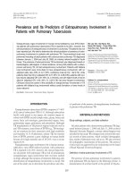
Microbiological analysis—No extra pulmonary
specimen yielded positive results with AFB staining,
while only 10.53% of them gave positive culture results.
PCR analysis—PCR amplification readily detected
MPB 64 gene sequence of M. tuberculosis in all the
peripheral blood samples of culture positive extra
pulmonary TB cases, whereas 55.88% of
microbiologically negative clinically diagnosed extra
pulmonary TB patients revealed positive
amplification results from blood (Table 2).
Additionally, 43.82% of the control subjects
amplified M. tuberculosis specific PCR products with
peripheral blood. Overall sensitivity and NPV for
peripheral blood based PCR assay in extra pulmonary
disease was 60.53% and 76.92% when compared to
culture isolation of mycobacteria (10.53% and
72.36%). However, its specificity and PPV were
56.18% and 37.09% as against that of culture (100%)
(Table 3). Interestingly, the intensity of PCR products
in non tuberculous cases was significantly lower than
that obtained from patient population (Fig. 3).
Table 1—Demographic features of subjects
[Values in parentheses are mean ± SD]
Group of patients Extra pulmonary TB
(38)
Non tuberculous
(89)
Age in year
02-75
(31.66 ± 15.94)
16-42
(25.42 ± 07.87)
Sex (Male/Female) 21/17 59/30
History of ATT (Yes/No) 00/38 00/89
Family history of ATT
(Yes/No)
05/33 03/86
Total number of subjects are indicated in parenthesis
Table 2—Amplification of MPB 64 gene of M. tuberculosis in the
peripheral blood samples of patient population
PCR Status Patient group (n)
PCR positive
(%)
PCR negative
(%)
Smear negative culture positive
Extra pulmonary TB patients (04)
04 (100) 00 (0.00)
Smear negative culture negative
Extra pulmonary TB patients (34)
19 (55.88) 15 (44.12)
Non tuberculous (89) 39 (43.82) 50 (56.18)
Fig. 2—Generation and co-amplification of 198bp competitor
with M. tuberculosis target DNA. [Lane M-100bp DNA ladder;
Lane 1- Purified M. tuberculosis DNA; Lane 2-198bp competitor
generated using MFW and RW primers; Lane 3- Purified 198bp
competitor amplified using FW and RW primers and Lane 4-
Target and competitor co amplified using FW and RW primers in
the same tube]
Fig. 3—Representative agarose gel electrophoresis of PCR
products using MBP-64 gene specific primer pair from EPTB
patients and controls. [Lane M- 100bp DNA ladder; Lane 1-
negative control; Lane 2:-smear negative, culture positive EPTB
patients; Lanes 3,4-smear negative, culture negative EPTB
patients; and lanes 5,6- non tuberculous controls]
INDIAN J EXP BIOL, JUNE 2009
450
C-PCR assay
Validation of cPCR assay—Constant amount of
DNA was taken and its 10-fold dilution was titrated
against serially diluted competitor with its
concentration ranging from 10 fg to 1000 fg.
Densitometric and computational analyses revealed the
point of equivalence to be 195.706 and 20.214 fg,
respectively (Figs 4, 5). The bacillary load thereby
calculated was 9.02 × 10
5
and 9.3 × 10
4
copies in the
two dilutions.
Determination of mycobacterial load—In order to
quantify mycobacterial load in culture positive and
culture negative EPTB patients, the dilution range of
competitor varying from 10 to 1.25 fg and 2.5 to
0.3125 fg, respectively was titrated with constant
amount of DNA (Fig. 6A). Densitometric scanning
followed by computational analysis revealed the point
of equivalence to be 2.485 and 0.629 fg (Fig. 6B)
which corresponds to 11,431 and 2,893 copies of
M. tuberculosis, respectively. Similarly, in non
tuberculous subjects, the dilutions of competitor varied
from 0.1 to 0.0125 fg (Fig. 6C). The point of
equivalence was revealed to be 0.025 fg which
corresponds to 115 copies of M. tuberculosis
organisms.
Apparently, the mycobacterial load determined by
MPB 64 gene based C-PCR assay in peripheral blood
samples from smear negative culture positive extra
pulmonary TB patients ranged from 1.630 – 2.717 fg
which corresponds to 7,498
– 12,498 M. tuberculosis
organisms, whereas in culture negative patients, the
point of equivalence varied between 0.131 – 1.043 fg
which is equivalent to 602–4,797 TB bacilli. In non
tuberculous controls the point of equivalence ranged
from 0.022-0.174 fg which reflected that in
asymptomatic patients the detectable TB bacilli by
C-PCR varied from 101 – 800 (Table 4).
Table 3—Comparison of sensitivity, specificity and predictive
values between culture and peripheral blood based PCR in EPTB
patients
Extra pulmonary TB patients (n=38) %
Variables tested
Culture PCR
Sensitivity 10.53 60.53
Specificity 100 56.18
PPV 100 37.09
NPV 72.36 76.92
Fig. 4—Top panel showing a representative agarose gel
electrophoretic resolution of co-amplified products of unknown
concentration of M. tuberculosis DNA. [Lane T- mycobacterial
DNA Target; Lane C- competitor. The lower panel shows the
determination of point of equivalence by computational analysis
following densitometric scanning of agarose gel picture]
Table 4—Determination of mycobacterial load in peripheral blood
samples of patient groups employing C-PCR assay
Patient Group (n) Point of equivalence
1.630 – 2.717 fg Extra pulmonary TB patients (38)
Smear negative culture positive (04)
Smear negative culture negative (34)
0.131 – 1.043 fg
Non tuberculous (39) 0.022 – 0.174 fg
Fig. 5—Top panel showing a representative agarose gel
electrophoretic resolution of co-amplified products of 1:10 diluted
M. tuberculosis DNA used in Fig. 4. [Lane T- mycobacterial
DNA Target; Lane C- competitor. The lower panel shows the
determination of point of equivalence by computational analysis
following densitometric scanning of agarose gel picture]
KHOSLA et al.: BLOOD C-PCR ASSAY FOR PULMONARY TUBERCULOSIS
451
Discussion
The conventional approaches to diagnose extra
pulmonary TB either lack sensitivity or are time
consuming, which is an important impediment to
global TB control. The same is apparent from the
present investigation, as none of the patients with
extra pulmonary presentation was found to be smear
positive. Moreover, only 10.53% EPTB specimens
could grow on L-J slants. The lower sensitivity of
culture in extra pulmonary disease is well accepted
and explained by the fact that mycobacteria might be
inactivated by immune response of the host
13
. The
average time for detection of M. tuberculosis in extra-
pulmonary samples was 48.16 ± 13.37 days. (Data not
shown).
PCR has been shown to be a promising alternative
for establishing rapid diagnosis of tuberculosis with a
high degree of sensitivity and specificity. Extra
pulmonary TB is usually a paucibacillary disease and
patients often present with atypical symptoms as it
may involve almost any organ of the body.
Appropriate biological sample from such patients is
collected employing invasive procedures and in some
cases it’s virtually impossible to collect the specimen.
These problems warrant less perilous and more
accessible clinical specimen. M. tuberculosis
disseminates into the peripheral blood of TB patients,
with or without compromised immune function
8,9,14
.
Therefore, peripheral blood is a good alternative
clinical material in patients with EPTB for detecting
M. tuberculosis by PCR.
PCR yielded high sensitivity as well as NPV but
low specificity and PPV when compared to culture
isolation of M. tuberculosis in extra pulmonary
disease (Table 3). High NPV of peripheral blood
based PCR test in EPTB patients strongly indicates
that the test could help in excluding the presence of
TB disease, which is in disagreement to a recent
report
15
. Therefore, this remarkable ability of blood
based PCR test to detect EPTB cases can replace the
need for more invasive diagnostic approaches.
Interestingly, ours is the first investigation where a
single copy target (MPB 64 gene) based PCR has
been utilized for detecting genome of M. tuberculosis
in peripheral blood. Other studies on EPTB employed
multicopy target, IS 6110, for peripheral blood based
PCR assay
16-18
. However, IS 6110 based assay has a
big disadvantage in Indian scenario where a sizable
proportion of M. tuberculosis isolates are known to
lack these elements
19,20
.
The specificity of PCR assay in present
investigation was found to be lower (56.18%) than
culture (100%). The low specificity was evidently
influenced by positive PCR results (43.82%) among
non-TB subjects, which in turn undermines the
clinical relevance of this test in diagnosing TB. It is
important to mention that the intensity of the PCR
products from non tuberculous controls was much
lower as compared to their diseased counterparts and
was possibly due to lower mycobacterial burden in
control population. This finds support from the
observation that highest intensity of amplified
products was observed among smear negative culture
positive patients (Fig. 1). The possibility of
contamination was ruled out by assessing the
amplification results in the presence of 8-
methoxypsoralen; the latter in the presence of UV
radiations, is known to intercalate into double
stranded nucleic acid thereby forming a covalent
interstrand cross-link, which is inhibitory for their
amplification
21
. Additionally, due precautions were
taken to avoid contamination by separating the areas
where blood samples were processed for DNA
isolation, from areas of PCR amplification and
analysis of amplified products. The mycobacterial
presence in peripheral blood of controls can be
explained by the fact that around 40% Indian adults
are reported to be infected by M. tuberculosis and do
not manifest the symptoms of active disease
6
. Clearly
the standard PCR failed to differentiate asymptomatic
controls from the paucibacillary EPTB patients.
To address this concern, standard PCR was
modified to enable quantification of mycobacterial
Fig. 6—Representative agarose gel electrophoresis picture(s) of
C-PCR amplified products for the calculation of mycobacterial load
from peripheral blood specimens of (A) smear negative, culture
positive EPTB patients; (B) smear negative, culture negative EBTP
patients and (C) non-tuberculous control. [Lanes T and C represent
controls amplified for only mycobacterial target and competitor,
respectively. The amount of competitor used in femtogram (fg) for
co-amplification with constant mycobacterial target is indicated
above the respective lanes]
INDIAN J EXP BIOL, JUNE 2009
452
load by C-PCR assay and thereby differentiate
asymptomatic controls from their active counterparts.
C-PCR technique is based on the assumption that
amplified product ratio of target and competitor
reliably reveals the ratio of their initial copy number.
Equimolar concentration of the target and competitor
in the reaction resulted in amplification of PCR
targets of equal intensity. Given the amount of
competitor is known at the point of equivalence; the
amount of the target could be determined
11
.
Prerequisite for the C-PCR assay is a competitor
which differs in size from the mycobacterial target. A
difference, of 42bp, in size of the target and the
competitor was created using a simple PCR based
strategy (Figs 2, 3). The underline principle of
C-PCR assay was validated using an unknown
amount of M. tuberculosis DNA from culture biomass
and also assaying its 10-fold dilution. Computational
analysis of the densitometric scanning of the
amplified products revealed the bacterial DNA load of
195.706 fg and 20.214 fg, respectively (Figs. 4, 5).
These determinations revealed a very good fit thus
verifying the reliability of this technique in
determining bacillary load.
C-PCR analysis of DNA samples isolated from
peripheral blood of culture positive EPTB patients
revealed point of equivalence as 2.485 fg which was
equivalent to 11,431 ge/mL bacilli (Fig 6A).
Similarly, among culture negative patients, the cPCR
assay (Fig. 6B) reflected the mycobacterial burden to
be equivalent to 2,893 M. tuberculosis organisms,
which was almost one fourth of culture positive
individuals. Furthermore, the point of equivalence for
asymptomatic controls (Fig. 6C) was 0.025 fg which
corresponds to 115 ge/mL of M. tuberculosis.
Based on C-PCR assay in peripheral blood of
EPTB patients, the mycobacterial load varied from
7498-12,498 ge/mL in smear negative/culture positive
to 602-4797 ge/mL in smear negative/culture negative
patients. However, among non tuberculous controls,
the mycobacterial load ranged between 101-800
ge/mL. These observations suggested that individuals
with bacterial load of <800 ge/mL should be treated
as carrying clinically irrelevant number of bacilli,
where as those with a threshold value of >7498
bacilli/mL should indicate an active disease (Table 4).
Additionally, all those individuals harboring
mycobacterial load between these values need to be
considered as presumptive TB cases. Keeping in view
the enormity of TB burden in India, more detailed
investigations are needed to ascertain the significance
of mycobacterial load during various clinical stages of
M. tuberculosis infection, especially in different
Indian populations where such data is totally lacking.
In conclusion, the data generated in the present
study clearly exhibits extraordinary sensitivity of
C-PCR assay in differentiating between clinically
irrelevant and relevant mycobacterial load. This study
also points out that the dissemination of M.
tuberculosis in peripheral blood is more common than
previously thought
12
. This novel armamentarium, in
fight against tuberculosis, could help in understanding
the dissemination dynamics of tubercle bacilli in
circulation. Moreover, such an approach could bring a
new dimension in the early detection of
M. tuberculosis, in EPTB patients, from a readily
accessible clinical specimen and would help in the
better management of this ancient scourge.
Acknowledgement
Financial assistance from University Grants
Commission (UGC), New Delhi, in the form of major
research project no. F.3-101/2003 (SR) is gratefully
acknowledged. Thanks are due to Mr Ajay Kumar for
his help in preparing the manuscript.
References
1 Kant L, Improving detection of infectious cases, Indian J
Tuber, 48 (2001)115.
2 Pahwa R, Hedau S, Jain S, Jain N, Arora V M, Kumar N &
Das B C, Assessment of possible tuberculous
lymphadenopathy by PCR compared to non-molecular
methods, J Med Microbiol, 54 (2005) 873.
3 Richter C, Kox L F, Van Leeuwen J, Mtoni I & Kolk A H J,
Peripheral blood based PCR assay for Mycobacterium
tuberculosis in 158 Tanzanian patients with extra pulmonary
tuberculosis, Eur J Clin Microbiol Infect Dis, 15 (1996) 813.
4 Moudgil H & Leitch A G, Extra-pulmonary tuberculosis in
Lothian 1980-1989: ethnic status and delay from onset of
symptoms to diagnosis, Respir Med, 88 (1994) 507.
5 Su W J, Recent advances in the molecular diagnosis of
tuberculosis, J Microbiol Immunol Infect, 35 (2002) 209.
6 RNTCP status report. TB India (2007) Central TB division,
Directorate General of health services, Ministry of Health and
Family Welfare, New Delhi, India. pp. 10.
7 Dwivedi A & Sehajpal P K, Development of a competitor
DNA template of the 38 kDa gene for molecular quantification
of M. tuberculosis, Int J Lung Dis, 9 (2005) 1412.
8 Kolk A H, Kox L F, Kuijper S & Richter C, Detection of
Mycobacterium tuberculosis in peripheral blood, Lancet 344
(1994) 694.
9 Rolfs A, Beige J, Finckh U, Kohler B, Schaberg T, Lokies J &
Lode H, Amplification of Mycobacterium tuberculosis from
peripheral blood, J Clin Microbiol, 33 (1995) 3312.
KHOSLA et al.: BLOOD C-PCR ASSAY FOR PULMONARY TUBERCULOSIS
453
10 Dwivedi A, Chaubey B, Sarin B C, Mittar D & Sehajpal P K,
A new rapid method for the isolation of mycobacterial DNA,
Indian J Vet Path, 24 (2000) 87.
11 Raeymaekers L, Quantitative PCR: Theoretical
considerations with practical implications, Annal Biochem,
214 (1993) 582.
12 Knapp R G & Miller III M.C, Clinical epidemiology and
biostatistics (Harwal publishing company, Malven,
Pennsylvania) 1992.
13 Ferrer J, Pleural tuberculosis, Eur Respir J, 10 (1997) 942.
14 Khan M A, Mirza S H, Abbasi S A, Butt T & Anwar M,
Peripheral blood based polymerase chain reaction in
diagnosis of pulmonary tuberculosis, J Ayub Med Coll
Abbottabad, 18 (2006) 25.
15 Pai M, Flores L L, Hubbard A, Riley L W & Colford J M Jr.,
Nucleic acid amplification testsin the diagnosis of
tuberculous pleuritis. A systematic review and meta-analysis,
BMC Infect Dis, 4 (2004) 6.
16 Honore S, Vincensini J P, Hocqueloux, L, Noguera M E,
Farge D, Lagrange P & Herrmann J L, Diagnostic value of a
nested polymerase chain reaction assay on peripheral blood
mononuclear cells from patients with pulmonary and extra-
pulmonary tuberculosis, Int J Tuberc Lung Dis, 5 (2001) 754.
17 Ritis K, Tzoanopoulos D, Speletas M, Papadopoulos E &
Arvanitidis L, Amplification of IS 6110 sequence for
detection of Mycobacterium tuberculosis complex in HIV –
negative patients with fever of unknown origin (FUO) and
evidence of extra pulmonary disease, J Int Med, 248 (2000)
415.
18 Mirza S, Restrepo B I, McCormick J B & Fischer-Hoch S P,
Diagnosis of tuberculous lymphadenitis using a polymerase
chain reaction on peripheral blood mononuclear cells, Am J
Trop Med Hyg, 69 (2003) 465.
19 Radhakrishnan I, Manju Y K, Kumar R A and Mundayoor S
(2001) Implications of Low Frequency of IS6110 in
Fingerprinting Field Isolates of Mycobacterium tuberculosis
from Kerala, India, J Clin Microbiol, 39 (2001) 1683.
20 Chauhan D S, Sharma VD, Parashar D, Chauhan A, Singh D,
Singh H B, Das R, Aggarwal B M, Malhotra B, Jain A,
Sharma M, Kataria V K, Aggarwal J K, Hanif M, Shahani A
& Katoch V M, Molecular typing of Mycobacterium
tuberculosis isolates from different parts of India based on
IS6110 element polymorphism using RFLP analysis, Indian
J Med Res, 125 (2007) 577.
21 Meier A, Persing D H, Finken M & Bottger E C, Elimination
of contaminating DNA within polymerase chain reaction
reagents: Implications for a general approach to detection of
uncultured pathogens, J Clin Microbiol, 31 (1993) 646.