Báo cáo khoa học: Specific cleavage of the DNase-I binding loop dramatically decreases the thermal stability of actin pot
Bạn đang xem bản rút gọn của tài liệu. Xem và tải ngay bản đầy đủ của tài liệu tại đây (546.44 KB, 11 trang )
Specific cleavage of the DNase-I binding loop dramatically
decreases the thermal stability of actin
Anastasia V. Pivovarova
1
, Sofia Yu. Khaitlina
2
and Dmitrii I. Levitsky
1,3
1 A. N. Bach Institute of Biochemistry, Russian Academy of Sciences, Moscow, Russia
2 Institute of Cytology, Russian Academy of Sciences, St Petersburg, Russia
3 A. N. Belozersky Institute of Physico-Chemical Biology, Moscow State University, Moscow, Russia
Introduction
Actin is one of the most abundant and highly conserved
cell proteins. It is involved in many different cellular
processes that are essential for growth, differentiation
and motility. Actin is found in two main states: as
monomers (G-actin) and as a helical polymer (F-actin).
Polymerization of G-actin into F-actin is accompanied
by hydrolysis of tightly bound ATP followed by a
slower release of P
i
; as a result, protomers of F-actin
contain tightly bound ATP, ADP or ADP-P
i
.
The atomic resolution structures of G-actin revealed
that it is divided into two easily distinguishable
domains by a deep cleft containing the tightly bound
nucleotide and cation [1]. The nucleotide-binding cleft
was suggested to exist in two main states, closed and
open [2,3], and solution studies on nucleotide exchange
and susceptibility of the cleft to limited proteolysis
appear to be consistent with the opening of the cleft
upon the transition from the ATP- to ADP-G-actin
Keywords
actin; differential scanning calorimetry;
DNase-I binding loop; proteolytic cleavage;
thermal unfolding
Correspondence
D. I. Levitsky, A. N. Bach Institute of
Biochemistry, Russian Academy of
Sciences, Leninsky Prospect 33, 119071
Moscow, Russia
Fax: +7 495 954 2732;
Tel: +7 495 952 1384
E-mail:
(Received 10 June 2010, revised 14 July
2010, accepted 16 July 2010)
doi:10.1111/j.1742-4658.2010.07782.x
Differential scanning calorimetry was used to investigate the thermal
unfolding of actin specifically cleaved within the DNaseI-binding loop
between residues Met47-Gly48 or Gly42-Val43 by two bacterial proteases,
subtilisin or ECP32 ⁄ grimelysin (ECP), respectively. The results obtained
show that both cleavages strongly decreased the thermal stability of mono-
meric actin with either ATP or ADP as a bound nucleotide. An even more
pronounced difference in the thermal stability between the cleaved and
intact actin was observed when both actins were polymerized into fila-
ments. Similar to intact F-actin, both cleaved F-actins were significantly
stabilized by phalloidin and aluminum fluoride; however, in all cases, the
thermal stability of the cleaved F-actins was much lower than that of intact
F-actin, and the stability of ECP-cleaved F-actin was lower than that of
subtilisin-cleaved F-actin. These results confirm that the DNaseI-binding
loop is involved in the stabilization of the actin structure, both in mono-
mers and in the filament subunits, and suggest that the thermal stability of
actin depends, at least partially, on the conformation of the nucleotide-
binding cleft. Moreover, an additional destabilization of the unstable
cleaved actin upon ATP ⁄ ADP replacement provides experimental evidence
for the highly dynamic actin structure that cannot be simply open or
closed, but rather should be considered as being able to adopt multiple
conformations.
Structured digital abstract
l
MINT-7980274: Actin (uniprotkb: P68135) and Actin (uniprotkb:P68135) bind (MI:0407 )by
biophysical (
MI:0013)
Abbreviations
D-loop, DNase I-binding loop; DSC, differential scanning calorimetry; DH
cal,
calorimetric enthalpy; T
m,
thermal transition temperature.
3812 FEBS Journal 277 (2010) 3812–3822 ª 2010 The Authors Journal compilation ª 2010 FEBS
[4–6]. By contrast, in the numerous crystal structures
published to date, including those of ADP-G-actin
[7,8], the open nucleotide-binding cleft has been
observed only in profilin-bound actin crystals [9]. Only
the closed conformation was also revealed by molecu-
lar dynamics simulations of the crystal actin structures
[10–12], whereas metadynamics simulations have dem-
onstrated that the closed conformation of the nucleo-
tide-binding cleft is the most stable state only when
ATP is bound, and the ADP-bound state favors a
more open conformation of the cleft [13]. It is possible
therefore that crystallization favors a closed state for
G-actin even though the state of the cleft in solution
may vary [14,15].
The other nucleotide-dependent conformational
transitions in actin crystals involve so-called DNase
I-binding loop (D-loop) in subdomain 2 of the actin
molecule [7]. In tetramethylrhodamine-modified ADP-
G-actin, the D-loop was found to be in an a-helical
conformation, whereas this region was disordered in
the ATP-bound actin, suggesting that the D-loop folds
on ATP hydrolysis [7,16]. This transition within the
D-loop was supported by the results of metadynamic
simulations demonstrating a distinct allosteric relation-
ship between the conformation of the D-loop (ordered
or disordered) and the state of the nucleotide-binding
cleft (open or closed) [13]. These data are consistent
with the results of the biochemical observations indi-
cating that proteolytic modifications of the D-loop
affect the state of the interdomain cleft [5,17].
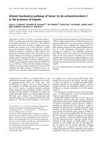
The D-loop of actin can be specifically cleaved with
two bacterial proteases. One of them is subtilisin,
which cleaves the D-loop between Met47 and Gly48
[18]. The other protease, which specifically cleaves
actin at the only site between Gly42 and Val43
(Fig. 1), was initially isolated and characterized as a
minor protein of lactose-negative Escherichia coli A2
strain and referred to as protease ECP32 [19,20]. More
recently, it was found that the A2 strain producing
protease ECP32 is identical to Serratia grimesii, and
therefore this enzyme was named grimelysin [21].
Although ECP32 and grimelysin were suggested to be
identical enzymes [21], both names are used in the lit-
erature. In accordance with previous studies [5,17], we
refer to this protease as ECP in the present study. The
nucleotide-binding cleft in both subtilisin-cleaved and
ECP-cleaved G-actin is clearly in a more open confor-
mation compared to intact actin, as demonstrated by
the increased nucleotide exchange rate in solution
[17,22] and their higher susceptibility to limited prote-
olysis [5,17]. By contrast, the crystal structure of ECP-
cleaved G-actin showed the nucleotide-binding cleft to
be in a typical closed conformation, probably as a
result of crystallization preferentially trapping actin in
only one of its possible conformations [14].
Taking into account the ambiguity of the nucleo-
tide-binding cleft conformation and its relationship
with the D-loop, the present study aimed to determine
whether the specific cleavage of the D-loop affects the
structural properties of the entire actin molecule and,
in particular, conformational transitions of the nucleo-
tide-binding cleft. For this purpose, we studied the
effects of the D-loop cleavage on the thermal unfold-
ing of G- and F-actin. Previously, the thermal unfold-
ing of G-actin containing different nucleotides was
indirectly studied with the DNase-I inhibition assay
[23] and by monitoring the change in absorbance of
tetramethylrhodamine-actin [24]. The results obtained
showed that replacement of the tightly bound ATP by
ADP led to a significant decrease in the thermal stabil-
ity of G-actin [23,24]. In the present study, we applied
differential scanning calorimetry (DSC), which is the
most direct and effective method for studying the ther-
mal unfolding of proteins. Previous studies have shown
that DSC can be successfully used to reveal the
changes in the thermal unfolding of actin induced by
interaction of G-actin with actin-binding proteins [25–
27], G–F transformation of actin, and stabilization of
F-actin by phalloidin and P
i
analogs [28]. Moreover,
the effects of nucleotides on the thermal unfolding of
F-actin have been studied by this method and a ‘disso-
ciative’ mechanism for the thermal denaturation of
F-actin has been proposed [28,29].
In the present study, we used DSC to characterize
the thermal unfolding of actin specifically cleaved
within the D-loop by ECP or subtilisin. The results
obtained show for the first time that the cleavage
strongly decreases the thermal stability of G-actin and
especially that of F-actin, both in the absence and the
Fig. 1. 3D atomic structure of G-actin. The four subdomains are
indicated by the encircled numbers. Arrows show the cleavage
sites in the D-loop, between Gly42 and Val43 by ECP32 ⁄ grimelysin,
and between Met47 and Gly48 by subtilisin.
A. V. Pivovarova et al. Thermal unfolding of cleaved actin
FEBS Journal 277 (2010) 3812–3822 ª 2010 The Authors Journal compilation ª 2010 FEBS 3813
presence of phalloidin and P
i
analogs. These results
are discussed with regard to a more open conforma-
tion of the interdomain cleft in the cleaved actin com-
pared to intact actin, both in the monomers and in the
filament subunits.
Results
Effects of the D-loop cleavage on the thermal
unfolding of G-actin
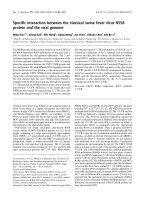
The excess heat capacity curves obtained for intact,
ECP-cleaved, and subtilisin-cleaved ATP-Ca-G-actins
are presented in Fig. 2. It is seen that the G-actin spe-
cies cleaved within the D-loop are clearly less thermo-
stable than noncleaved G-actin. The thermal
transitions of both ECP-cleaved and subtilisin-cleaved
G-actin are shifted to a lower temperature, by 4–5 °C,
compared to that of intact G-actin, and the values
of calorimetric enthalpy, DH
cal
, determined for the
cleaved G-actins are $ 57–63% of those for nonc-
leaved G-actin (Table 1). Thus, both cleavages within
the D-loop strongly decrease the thermal stability of
ATP-Ca-G-actin, with no significant difference
between the effects of ECP and subtilisin. It is
important to note that heating cleaved G-actins in the
calorimeter cell did not lead to any further proteolysis
of the proteins (Fig. 2, inset).
We also compared the thermal unfolding of intact
and ECP-cleaved G-actins in the different states, with
the tightly bound Ca
2+
replaced by Mg
2+
and with
the tightly bound ATP replaced by ADP (Fig. 3). The
replacement of Ca
2+
by Mg
2+
in ATP-G-actin had no
appreciable effect on the thermal unfolding of either
intact or ECP-cleaved G-actin: in both cases, it only
slightly decreased the maximum thermal transition
temperature (T
m
), by 1–2 °C, with no effect on the
DH
cal
value (Table 1).
By contrast, the replacement of bound ATP by
ADP caused a dramatic decrease in the thermal stabil-
ity of G-actin. Intact ADP-Mg-G-actin demonstrated
the thermal transition with T
m
of 48.8 °C (Fig. 3A)
(i.e. 11 °C less than that of ATP-Mg-G-actin) and its
calorimetric enthalpy (340 kJÆmol
)1
) was much
less than that of ATP-Mg-G-actin (570 kJÆmol
)1
)
(Table 1). Figure 3A shows that the sample contains
only ADP-actin because no peak at 60 °C (correspond-
ing to the thermal transition of ATP-Mg-actin) was
seen on the thermogram. A similar effect was observed
on ECP-cleaved Mg-G-actin with ATP replaced by
ADP (Fig. 3B). In this case, the nucleotide replace-
ment decreased the T
m
by 9 °C and led to a more than
two-fold decrease in the DH
cal
value (Table 1).
Thermal unfolding of F-actin with the cleaved
D-loop
Previous studies have shown that ECP-cleaved actin is
unable to polymerize unless its tightly bound Ca
2+
is
replaced with Mg
2+
, and that the Mg
2+
-bound form
has higher critical concentration and polymerizes more
slowly than Mg-G-actin cleaved with subtilisin
[17,20,30]. In agreement with these data, in the present
study, ECP-cleaved Mg-G-actin polymerized more
Fig. 2. Temperature dependences of the excess heat capacity (C
p
)
of intact (curve 1), ECP-cleaved (curve 2) and subtilisin-cleaved
(curve 3) ATP-Ca-G-actins. The actin concentration was 24 l
M.
Other conditions: 2 m
M Hepes (pH 7.6), 0.2 mM CaCl
2
and 0.2 mM
ATP. The inset shows representative SDS ⁄ PAGE patterns of intact
(lanes 1 and 1¢), ECP-cleaved (lanes 2 and 2¢) and subtilisin-cleaved
(lanes 3 and 3¢) G-actin before (lanes 1, 2 and 3) and after heating
in the calorimetric cell up to 80 °C (lanes 1¢,2¢ and 3¢). Note that
the positions of actin (lanes 1 and 1¢) and its C-terminal fragments
produced by ECP (36 kDa) (lanes 2, 2¢) or by subtilisin (35 kDa)
(lanes 3 and 3¢) remain unchanged after the heating–cooling
procedure.
Table 1. Calorimetric parameters obtained from the DSC data for
intact, ECP-cleaved and subtilisin-cleaved G-actins. The parameters
were extracted from Figs 2 and 3. The error of the given values of
T
m
did not exceed ±0.2 °C. The relative error of the given values of
DH
cal
did not exceed ±10%.
G-actin Nucleotide Cation T
m
(°C) DH
cal
(kJÆmol
)1
)
Intact ATP Ca
2+
61.2 585
Intact ATP Mg
2+
59.9 570
Intact ADP Mg
2+
48.8 340
ECP-cleaved ATP Ca
2+
55.9 370
ECP-cleaved ATP Mg
2+
53.6 390
ECP-cleaved ADP Mg
2+
44.5 145
Subtilisin-cleaved ATP Ca
2+
57.0 335
Thermal unfolding of cleaved actin A. V. Pivovarova et al.
3814 FEBS Journal 277 (2010) 3812–3822 ª 2010 The Authors Journal compilation ª 2010 FEBS
slowly than subtilisin-cleaved Mg-G-actin, which, in
turn, demonstrated slower polymerization than intact,
noncleaved Mg-G-actin. Nevertheless, light-scattering
measurements showed complete polymerization of all
the Mg-G-actin species to Mg-F-actin after 1.5 h of
incubation with 100 mm KCl and 1 mm MgCl
2
in the
presence of 1 mm ATP (Fig. 4, inset).
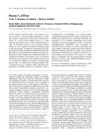
Figure 4 shows that Mg-F-actin obtained from the
cleaved Mg-G-actin is much less thermostable than
noncleaved Mg-F-actin, and a decrease in the thermal
stability is even more pronounced than in the case of
G-actin. The thermal transitions of ECP-cleaved and
subtilisin-cleaved F-actin are shifted to a lower temper-
ature, by 11.3 and 8.8 °C, respectively, compared to
that of intact F-actin (Table 2). Importantly, a pro-
nounced difference is observed between the thermal
transitions of ECP-cleaved and subtilisin-cleaved
F-actin (Fig. 4). ECP-cleaved F-actin unfolds not only
at lower temperature (58.6 versus 61.1 °C), but also
with a much lower cooperativity. The width at the
half-height of the thermal transition, which can serve
as a relative measure for cooperativity of the transi-
tion, was equal to 8.5 °C for ECP-cleaved Mg-F-actin
and 4.3 °C for subtilisin-cleaved Mg-F-actin. Thus,
Fig. 4. DSC curves of Mg-F-actin assembled from intact (curve 1),
ECP-cleaved (curve 2) and subtilisin-cleaved (curve 3) ATP-Mg-G-
actin. The actin concentration was 24 l
M. Other conditions: 20 mM
Hepes (pH 7.3), 0.1 M KCl, 1 mM MgCl
2
and 0.7 mM ADP. The
inset shows time courses of polymerization of intact (curve 1),
ECP-cleaved (curve 2) and subtilisin-cleaved (curve 3) actins. Poly-
merization was monitored by recording light-scattering intensity at
350 nm upon the addition of 0.1
M KCl and 1 mM MgCl
2
to ATP-
Mg-G-actins.
Fig. 3. DSC curves of intact G-actin (A) and ECP-cleaved G-actin
(B) with different tightly bound nucleotide and cation: ATP-Ca-G-
actin, ATP-Mg-G-actin and ADP-Mg-G-actin. The actin concentration
was 24 l
M. Other conditions: 2 mM Hepes (pH 7.6), 0.2 mM CaCl
2
or MgCl
2
, and 0.2 mM ATP or ADP.
Table 2. Calorimetric parameters obtained from the DSC data for
Mg-F-actin assembled from intact, ECP-cleaved and subtilisin-
cleaved Mg-G-actin. The parameters were extracted from Figs 4
and 5. The error of the given values of T
m
did not exceed ± 0.2 °C.
The relative error of the given values of DH
cal
did not exceed
±10%.
Mg-F-actin Stabilizer T
m
(°C) DH
cal
(kJÆmol
)1
)
Intact – 69.9 650
Intact Phalloidin 82.5 1065
Intact AlF
4
À
83.4 800
Intact Phalloidin + AlF
4
À
90.8 1080
ECP-cleaved – 58.6 525
ECP-cleaved Phalloidin 68.5 655
ECP-cleaved AlF
4
À
70.3 415
ECP-cleaved Phalloidin + AlF
4
À
81.7 690
Subtilisin-cleaved – 61.1 415
Subtilisin-cleaved Phalloidin 76.5 635
Subtilisin-cleaved AlF
4
À
76.4 720
Subtilisin-cleaved Phalloidin + AlF
4
À
84.4 780
A. V. Pivovarova et al. Thermal unfolding of cleaved actin
FEBS Journal 277 (2010) 3812–3822 ª 2010 The Authors Journal compilation ª 2010 FEBS 3815
although both cleaved G-actins unfold similarly
(Fig. 2), a pronounced difference in the thermal
unfolding between ECP-cleaved and subtilisin-cleaved
actin is revealed when these actins are polymerized into
filaments.
Stabilization of the cleaved F-actin by phalloidin
and aluminum fluoride
It is well known that cyclic heptapeptide phalloidin
binds to F-actin with very high affinity at the interface
of three adjacent actin protomers [31,32] and stabilizes
actin filaments (i.e. it significantly increases the thermal
stability of F-actin) [25,28,33–35]. A very similar stabi-
lizing effect was observed in the presence of P
i
analogs,
aluminum fluoride (AlF
À
4
) or beryllium fluoride (BeF
x
)
[25,28,34,36], which form complexes with F-actin
subunits that mimic their ADP-P
i
state. The stabilizing
effects of phalloidin and AlF
4
À
(or BeF
x
) were similar
but independent of each other because simultaneous
addition of both stabilizers caused an additional
increase in the thermal stability of F-actin [28,34].
The subsequent experiments were designed to inves-
tigate the effects of the two F-actin stabilizers, phalloi-
din and AlF
4
À
, on the thermal unfolding of F-actin
specifically cleaved within the D-loop. In agreement
with previous studies [25,28,29,34], the binding of
phalloidin or AlF
4
À
significantly increased the thermal
stability of Mg-F-actin. Both stabilizers shifted the
maximum of the F-actin thermal transition from
69.9 °C to 82–83 °C (Table 2), and their simultaneous
addition increased the T
m
up to $ 91 °C(Fig. 5A).
Similar to intact F-actin, both cleaved F-actin species
are significantly stabilized by phalloidin and AlF
4
À
(Fig. 5B,C). Each of these stabilizers increased the T
m
of the cleaved F-actin, by 10–12 °C for ECP-cleaved
F-actin and by $ 15 °C for subtilisin-cleaved F-actin
(Table 2), and their simultaneous addition resulted in
an additive effect that is expressed in the further
increase of the T
m
value by $ 12–13 °C (Fig. 5B) or
8 °C (Fig. 5C). However, in all these stabilized states,
the T
m
value for the cleaved F-actin was significantly
lower than that of intact F-actin, by 9–14 °C for ECP-
cleaved F-actin and by 6–7 °C for subtilisin-cleaved
F-actin (Table 2). This means that ECP-cleaved F-actin
is less thermostable than subtilisin-cleaved F-actin not
only in the absence of stabilizers (Fig. 4), but also in
the presence of phalloidin and AlF
4
À
(Fig. 5B,C).
There are also other distinct differences between
ECP-cleaved F-actin and subtilisin-cleaved F-actin,
whose thermal denaturation is more similar to that of
intact F-actin. First, along with the main transition at
68.5 °C, the DSC profile of the phalloidin-stabilized
ECP-cleaved F-actin demonstrated a pronounced
shoulder at $ 60 °C (Fig. 5B). Second, in the presence
of AlF
4
À
, this cleaved F-actin demonstrated, along with
the main thermal transition at 70 °C, a clear peak at
$ 57 °C corresponding to the thermal unfolding of this
protein in the absence of AlF
4
À
(Fig. 5B). This suggests
a much lower affinity of ECP-cleaved F-actin for
phalloidin and AlF
4
À
than that in intact and subtilisin-
cleaved F-actin. To test this assumption, we investi-
gated the thermal unfolding of ECP-cleaved F-actin in
the presence of different concentrations of phalloidin
and AlF
4
À
(Fig. 6).
At relatively low phalloidin ⁄ actin molar ratio of
1 : 4, two peaks are observed on the DSC profile
Fig. 5. DSC curves of intact Mg-F-actin (A), ECP-cleaved Mg-F-
actin (B) and subtilisin-cleaved Mg-F-actin (C) stabilized by phalloidin
or AlF
4
À
, or simultaneously by both stabilizers. Concentrations of
stabilizers: 24 l
M phalloidin and 1 mM AlF
4
À
(5 mM NaF and 1 mM
AlCl
3
). Other conditions were as described in Fig. 4.
Thermal unfolding of cleaved actin A. V. Pivovarova et al.
3816 FEBS Journal 277 (2010) 3812–3822 ª 2010 The Authors Journal compilation ª 2010 FEBS
(Fig. 6A), and the large peak with T
m
at 58.6 °C corre-
sponds to the nonstabilized ECP-cleaved F-actin (i.e. it
reflects the thermal unfolding of those actin protomers,
which are not affected by phalloidin). This means that
effect of phalloidin on the thermal stability of ECP-
cleaved F-actin is much less cooperative than in the
case of intact F-actin, when one bound phalloidin was
shown to stabilize up to seven neighboring protomers
in the actin filament [37]. The peak of nonstabilized
actin disappeared with an increase in the phalloi-
din ⁄ actin molar ratio (Figs 5B and 6A). However, the
pronounced shoulder at $ 61–65 °C was observed on
the DSC profile of ECP-cleaved F-actin even in the
presence of a three-fold molar excess of phalloidin
(Fig. 6A), thus suggesting that protomers of phalloi-
din-stabilized F-actin exist in two structural states with
different thermal stability.
At a low concentration of AlF
4
À
(0.1 mm), we again
observed a pronounced peak at 58.6 °C corresponding
to the nonstabilized ECP-cleaved F-actin (Fig. 6B).
Thus, a much higher concentration of AlF
4
À
(more
than 1 mm) is required to achieve complete thermal
stabilization of ECP-cleaved F-actin compared to
intact F-actin, for which full stabilization was observed
even in the presence of 50 lm AlF
4
À
[36]. This reflects
at least an order of magnitude lower affinity of the
cleaved F-actin to AlF
4
À
.
Importantly, upon simultaneous addition of AlF
4
À
and phalloidin, we observed neither the peak of
nonstabilized actin protomers, nor the shoulder char-
acteristic of phalloidin-stabilized ECP-cleaved F-actin
(Fig. 6B). These results suggest that the binding of
AlF
4
À
to ADP-F-actin substantially modifies the struc-
tural state of cleaved actin subunits stabilized by phal-
loidin or phalloidin increases the affinity of the cleaved
actin subunits to AlF
4
À
.
Discussion
The data reported in the present study show that
cleavage of actin between Gly42-Val43 or Met47-Gly48
within the D-loop strongly decreases the thermal
stability both of monomers and polymers. According to
previous studies, these cleavages increased the rate of
the nucleotide exchange on the cleaved G-actin and its
susceptibility to limited proteolysis, probably as a result
of the transition of the nucleotide-binding cleft to a
more open conformation [5,17,22]. The relationship
between the conformation of the D-loop and the nucleo-
tide-binding cleft was recently demonstrated in metady-
namic simulations experiments [13]. We assume
therefore that the decrease in the thermal stability
observed by DSC on actin species cleaved within the
D-loop is associated with opening of the cleft.
Does the thermal stability of G-actin reflect the
conformational state of the nucleotide-binding
cleft?
An intact actin structure is maintained by the presence
of high-affinity cation and nucleotide tightly bound in
the interdomain cleft; removal of the nucleotide or cat-
ion results in actin denaturation. Therefore, the stabil-
ity of actin depends on the affinity of the tightly
bound cation and nucleotide that involves both pro-
tein–ligand interaction and conformation of the inter-
domain cleft. Upon heating, irreversible unfolding of
G-actin is preceded by reversible loss of the nucleo-
tide–cation complex [23]. Obviously, the more tightly
nucleotide and cation are bound in the interdomain
cleft and the more ‘closed’ is the cleft, the higher the
temperature needed to remove them from the cleft and
to induce thermal unfolding of G-actin. The relative
Fig. 6. DSC curves for ECP-cleaved Mg-F-actin (24 lM) either in
the presence of phalloidin (Ph) at different concentrations (6, 12 or
72 l
M) (A), or in the presence of 0.1 mM AlF
4
À
in the absence or in
the presence of 24 l
M Ph, and in the presence of 0.5 mM
AlF
4
À
+24lM Ph (B). Other conditions were as described in Fig. 4.
A. V. Pivovarova et al. Thermal unfolding of cleaved actin
FEBS Journal 277 (2010) 3812–3822 ª 2010 The Authors Journal compilation ª 2010 FEBS 3817
affinity of G-actin for ATP is much higher than for
ADP [4], and the interdomain cleft is suggested to be
in a more open conformation in the ADP-bound state
than in the ATP-bound state [5,12,13]. In agreement
with this and with previous studies [23,24], the results
obtained in the present study show that ADP-G-actin
is much less thermostable than ATP-G-actin (Fig. 3A
and Table 1). ATP-G-actin is less thermostable with
bound Mg
2+
than with Ca
2+
[23] (Table 1), and this
reduction in stability may be explained by the lower
affinity of ATP-G-actin for Mg
2+
than for Ca
2+
[4,38]. Thus, the ligand-dependent thermal stability of
actin monomer can be accounted for by the different
affinity of these ligands to actin. However, the thermal
stability of the cleaved actins is lower than the corre-
sponding stability of non-modified actin both in the
ATP- and ADP-states. This cannot be explained by
the different affinity but suggests that the thermal sta-
bility of G-actin may depend on the conformation of
the nucleotide-binding cleft. This suggestion is sup-
ported by the studies on the effects of actin-binding
proteins on actin structure.
Actin-binding proteins profilin and cofilin, when
bound to G-actin between subdomains 1 and 3, have
antagonistic effects on the conformation of the nucleo-
tide-binding cleft. Profilin stabilizes the ‘open’ confor-
mation of the cleft [7,39,40], whereas cofilin appears to
lock the cleft in its ‘closed’ conformation [39–42]. Pre-
vious studies on the thermal unfolding of G-actin
showed that profilin binding decreased the actin ther-
mal stability [23], whereas significant stabilization of
G-actin was observed in its complexes with cofilin
[25,26]. Stabilization of G-actin was also observed in
the complexes of G-actin with thymosin b
4
[27] and
gelsolin segment 1 [24], which appear to induce confor-
mational transitions closing the nucleotide-binding
cleft [6,27,43,44].
Thus, the increased thermal stability of G-actin
appears to correspond to the closed conformation of
the nucleotide-binding cleft, whereas the decreased
thermal stability is a feature of the actin with the open
cleft conformation. The cleavage within the D-loop
enhances the nucleotide exchange [17] and increases
accessibility of the cleft to limited proteolysis [5], which
characterizes the cleft opening. It is therefore likely
that the decreased thermal stability of G-actin cleaved
within the D-loop also results from the opening of the
nucleotide-binding cleft in these actin species.
It is noteworthy that the replacement of tightly
bound ATP by ADP in ECP-cleaved G-actin induces
an additional decrease in the thermal stability of this
actin species already destabilized by the cleavage
within the D-loop (Fig. 3B and Table 1). This suggests
that the nucleotide-binding cleft is highly dynamic and
cannot be simply open or closed but rather should be
considered as being more open or more closed. In
these terms, by analogy with the ‘superclosed’ state
recently revealed in ATP-G-actin by molecular dynam-
ics simulations [12], the nucleotide-binding cleft of
ADP-G-actin cleaved within the D-loop appears to
adopt the extra open conformation.
Comparison of the effects produced by the
cleavage of the D-loop with ECP and subtilisin
Although the cleavages of the D-loop between Gly42-
Val43 and Met47-Gly48 decreased the thermal stability
of G-actin to a similar extent (Fig. 2), the effects of
the cleavages became quite different when the cleaved
actins were polymerized into filaments. The thermal
stability of F-actin assembled from ECP-cleaved actin
was noticeably less than that of subtilisin-cleaved
F-actin (Fig. 4). These results are consistent with the
earlier observed effects of these cleavages on the sus-
ceptibility of the nucleotide-binding cleft to limited
proteolysis with trypsin [5,17]. In the cleaved G-actins,
susceptibility of trypsin cleavage sites at Arg62 and
Lys68 in the nucleotide-binding cleft was increased
similarly [17]. After polymerization, these sites became
almost inaccessible for trypsin in intact F-actin and
only slightly accessible for trypsin in subtilisin-cleaved
F-actin. By contrast, F-actin assembled from ECP-
cleaved G-actin was easily fragmented by trypsin.
These observations indicate that the open conforma-
tion of ECP-cleaved actin was preserved upon poly-
merization, whereas F-actin assembled from subtilisin-
cleaved monomers more closely resembled intact
F-actin than ECP-cleaved F-actin [17]. Thus, the lower
thermal stability of ECP-cleaved versus subtilisin-
cleaved F-actin corresponds to a more open nucleo-
tide-binding cleft.
According to the recent model of actin filament [45],
the N-terminal part of the D-loop is located at the
inter-monomer interface, participating both in the
intra-strand contacts between actin subunits along
the filament and in the lateral contacts stabilizing the
inter-strand interaction, whereas the C-terminal part of
the loop is not involved in the inter-strand contacts.
Recently, this structural difference was supported in
mutational cross-linking experiments showing that the
N-terminal part of the D-loop (residues 41–45) is in
close proximity to residue 265 of the actin subunit in
the opposite strand and can be easily cross-linked to
this residue, whereas the rate and extent of the cross-
linking reaction strongly declined for the C-terminal
residues of the D-loop [46]. Therefore, the inter-strand
Thermal unfolding of cleaved actin A. V. Pivovarova et al.
3818 FEBS Journal 277 (2010) 3812–3822 ª 2010 The Authors Journal compilation ª 2010 FEBS
contacts of the N-terminal part of the D-loop appear
to play a crucial role in stabilization of the actin fila-
ment [17,30,47]. The cleavage of the D-loop between
Gly42 and Val43 impairs these contacts [47], and this
may explain why the cleavage of the D-loop in its
N-terminal part with ECP more strongly destabilizes
F-actin than cleavage by subtilisin between Met47 and
Gly48 in the C-terminal part of the loop. It is also
important that the cleavage with ECP did not affect
the filament length but more strongly enhanced the
turnover rate of polymer subunits than the cleavage
with subtilisin [17,30]. Thus, the low thermal stability
of F-actin assembled from ECP-cleaved monomers
strongly correlates with the high dynamics of this actin
species [17], supporting the idea of the monomer disso-
ciation being the first step of thermal inactivation of
F-actin [28,29].
Although both cleaved actins are stabilized with
phalloidin and AlF
4
À
, stabilization of ECP-cleaved
F-actin demonstrates specific features that are not
characteristic of subtilisin-cleaved or intact actin. The
most interesting features are the extremely low affinity
of ECP-cleaved F-actin to AlF
4
À
and the pronounced
shoulder observed on the DSC profile of this actin spe-
cies even in the presence of a three-fold molar excess
of phalloidin (Figs 5B and 6). This suggests that pro-
tomers of phalloidin-stabilized cleaved F-actin exist in
two different structural states. Phalloidin binds to
F-actin at the interface of three adjacent actin protom-
ers [31] and appears to stabilize actin filament in two
inter-related ways: by stabilizing lateral interactions
between the two filament strands and by inducing con-
formational changes in actin subunits resulting in the
state of the nucleotide-binding cleft being similar to
that in ATP-actin filaments without phalloidin [32]. It
is plausible that the shoulder on the DSC profile of
phalloidin-stabilized ECP-cleaved F-actin belongs to a
population of the protomers in which the conforma-
tional effect of phalloidin is not completed. This expla-
nation, although requiring further examination with
independent approaches, is supported by the disap-
pearance of the shoulder after the addition of AlF
4
À
(Fig. 6B). This P
i
analog (as well as another analog,
BeF
x
) is known to bind to P
i
site in the nucleotide-
binding cleft and mimic ADP-P
i
or ATP actin fila-
ments [48], thus stabilizing the filament by closing the
cleft in actin subunits [49,50]. Hence, the increase in
the thermal stability of ECP-cleaved F-actin and the
disappearance of the shoulder on the DSC profile can
be accounted for by the combined effect of phalloidin
and AlF
4
À
on the nucleotide-containing cleft. Accord-
ingly, the phalloidin-induced effect may increase the
affinity of AlF
4
À
to actin, whereas AlF
4
À
-induced clo-
sure of the cleft diminishes the population of the su-
bunits remaining nonstabilized by phalloidin via its
effect on the cleft conformation. This interpretation is
consistent with recently published DSC data showing
that cooperative effect of phalloidin on the thermal
stability of F-actin becomes noncooperative in the
presence of AlF
4
À
[51].
Phalloidin can stabilize F-actin with a very high coo-
perativity, with the half-maximal effect being observed
at a phalloidin ⁄ actin molar ratio of 1 : 20 [52]. In the
DSC experiments on intact actin [37], only 10–15% of
actin protomers remained unaffected by phalloidin at a
phalloidin ⁄ actin molar ratio of 1 : 4. By contrast, more
than half of subunits of ECP-cleaved F-actin remained
nonstabilized by phalloidin under the same conditions
(Fig. 6A), consistent with a reduced cooperativity in
the effect of phalloidin on the steady-state ATPase
activity of ECP-cleaved actin [17]. Taken together with
the evidence concerning the critical role of the lateral
contacts for stabilization of filaments assembled from
ECP-actin monomers [47], these data allow us to
assume that only the effect of phalloidin on the con-
formation of the nucleotide-binding cleft is coopera-
tive; it is propagated along the filament by allosteric
interactions between phalloidin-bound and free pro-
tomers. By contrast, the stabilizing effect of phalloidin
on the lateral inter-strand interactions is noncoopera-
tive; it requires direct binding of phalloidin to actin
protomers.
According to this interpretation, an explanation for
the appearance of the pronounced shoulder on the
DSC profile of the ECP-cleaved F-actin stabilized by
phalloidin (Fig. 6A) can be proposed. This shoulder
appears to reflect the thermal unfolding of the actin
protomers whose cleft remains open, and therefore
they are stabilized only by lateral inter-strand interac-
tions induced by the direct binding of phalloidin. On
the other hand, the main transition at 68.5 °C
(Fig. 6A) most likely corresponds to the thermal
unfolding of actin subunits that are stabilized not only
by the inter-strand interactions, but also by phalloidin-
induced closing of the nucleotide-binding cleft.
In conclusion, the results obtained in the present
study suggest that the thermal stability of actin, regard-
less of whether it is modified by limited proteolysis or
by stabilizers, depends on the conformation of the
interdomain nucleotide-binding cleft. Accordingly, the
lower thermal stability of subtilisin- or ECP-cleaved
actin compared to intact actin supports the idea [5,13]
and also provides additional experimental evidence for
a distinct allosteric relationship between conformation
of the D-loop and the state of the nucleotide-binding
cleft.
A. V. Pivovarova et al. Thermal unfolding of cleaved actin
FEBS Journal 277 (2010) 3812–3822 ª 2010 The Authors Journal compilation ª 2010 FEBS 3819
Experimental procedures
Reagents
Subtilisin (type VIII bacterial protease), ATP, ADP,
EGTA, Hepes, phenylmethylsulfonyl fluoride, KCl, CaCl
2
,
MgCl
2
, AlCl
3
, NaF and phalloidin were purchased from
Sigma Chemical Co. (St Louis, MO, USA); hexokinase was
kindly provided by Dr N. Yu. Goncharova (Department of
Biochemistry, School of Biology, Moscow State University,
Russia).
Protein preparations
Rabbit skeletal muscle actin was prepared from acetone-
dried muscle powder according to the method of Spudich
and Watt [53]. G-actin was stored in buffer containing
2mm Tris-HCl (pH 8.0), 0.2 mm ATP, 0.2 mm CaCl
2
,
0.5 mm b-mercaptoethanol and 0.03% NaN
3
(buffer G).
The actin molar concentration was determined by monitor-
ing A
290
using an E
1%
of 6.3 cm
)1
[54] and a molecular
mass of 42.3 kDa. ECP-cleaved G-actin was obtained as
described previously [17,30]. Ca-G-actin (3.0 mgÆmL
)1
) was
digested at an enzyme ⁄ protein mass ratio of $ 1 : 100 for
4 h at 25 °C and then overnight at 4 °C. Because actin
cleaved with ECP between Gly42 and Val43 is fairly resis-
tant to further proteolysis by this protease, it was not nec-
essary to use any protease inhibitor. The cleaved actin was
used within 8–10 h. Subtilisin-cleaved actin was prepared
essentially as described by Schwyter et al. [18]. Ca-G-actin
(3 mgÆmL
)1
) was digested for 1 h at an enzyme ⁄ protein
mass ratio of 1 : 500 at 25 °C, and the proteolysis was
stopped with 2 mm phenylmethylsulfonyl fluoride. The
cleaved actin preparations were analyzed by SDS ⁄ PAGE
[55]. Usually, more than 85% of actin was cleaved. It is
important that the main part of the noncleaved actin
appears to correspond to small aggregates of unfolded (so-
called ‘inactivated’) G-actin [56], in which the D-loop
becomes almost inaccessible to proteolytic cleavage [57].
ATP-Ca-G-actin was transformed into ATP-Mg-G-actin
by a 3–5 min of incubation with 0.2 mm EGTA ⁄ 0.1 mm
MgCl
2
at 25 °C. To obtain ADP-Mg-G-actin, the actin-
bound ATP was converted into ADP by incubation of
ATP-Mg-G-actin with 0.8 mm ADP, 1 mm glucose and
hexokinase (8 UÆmL
)1
) for 2 h at 4 °C [5]. It is known that,
under similar conditions, only $ 0.4% of ATP was deter-
mined in the actin samples after 1 h of incubation with glu-
cose and hexokinase [58].
Intact, ECP-cleaved and subtilisin-cleaved Mg-G-actins
(3 mgÆmL
)1
) were polymerized by the addition of 100 mm
KCl and 1 mm MgCl
2
in the presence of 1 mm ATP. Poly-
merization was monitored by an increase in intensity of
light scattering at 90° measured at 350 nm on a Cary
Eclipse fluorescence spectrophotometer (Varian Australia
Pty Ltd, Mulgrave, Victoria, Australia).
Stabilization of F-actin (24 lm) by phalloidin or by alu-
minum fluoride (AlF
4
À
) was performed as described previ-
ously [25,28], by the addition of 6–72 lm phalloidin or 0.1–
1.0 mm AlCl
3
in the presence of 5 mm NaF and 0.7 mm
ADP.
DSC
DSC experiments were performed on a DASM-4M differen-
tial scanning microcalorimeter (Institute for Biological
Instrumentation, Pushchino, Russia) as described previously
[25,28,29,36]. All measurements were carried out at a scan-
ning rate of 1 KÆmin
)1
. The experiments with G-actin were
performed in 2 mm Hepes, pH 7.6, containing 0.2 mm CaCl
2
or MgCl
2
and 0.2 mm ATP (or 0.2 mm ADP in the case of
ADP-Mg-G-actin), whereas the thermal unfolding of F-actin
was studied in 20 mm Hepes (pH 7.3), 0.1 m KCl, 1 mm
MgCl
2
and 0.7 mm ADP. The final concentration of actin
was 24 lm. The reversibility of the thermal transitions was
assessed by reheating of the sample immediately after cool-
ing from the previous scan. The thermal denaturation of all
actin samples was fully irreversible. Calorimetric traces were
corrected for instrumental background and possible aggre-
gation artifacts by subtracting the scans obtained from the
reheating of the samples. The temperature dependence of
the excess heat capacity was further analyzed and plotted
using Origin software (MicroCal, Northampton, MA,
USA). The thermal stability of actin was described by the
T
m
, and DH
cal
was calculated as the area under the excess
heat capacity function. DSC experiments with different actin
species were performed at least twice with very good repro-
ducibility, and the representative curves are shown.
Acknowledgements
We are grateful to Dr Alevtina Morozova for provid-
ing us with protease ECP32 ⁄ grimelysin. This work was
supported by the Russian Foundation for Basic
Research (grants 09-04-00266 to D.I.L. and 08-04-
00408 to S.Yu.Kh), the Program ‘Molecular and Cell
Biology’ of the Russian Academy of Sciences, and by
the grant from the President of Russian Federation
(grant MK 2965.2009.4 to A.V.P.).
References
1 Kabsch W & Holmes KC (1995) The actin fold. FASEB
J 9, 167–174.
2 Tirion MM & ben-Avraham D (1993) Normal mode
analysis of G-actin. J Mol Biol 230, 186–195.
3 Page R, Lindberg U & Schtt CE (1998) Domain
motions in actin. J Mol Biol 280, 463–474.
4 Kinosian HJ, Selden LA, Estes JE & Gershman LC
(1993) Nucleotide binding to actin. Cation dependence
Thermal unfolding of cleaved actin A. V. Pivovarova et al.
3820 FEBS Journal 277 (2010) 3812–3822 ª 2010 The Authors Journal compilation ª 2010 FEBS
of nucleotide dissociation and exchange rates. J Biol
Chem 268, 8683–8691.
5 Strzelecka-Golaszewska H, Moraczewska J, Khaitlina
SYu & Mossakowska M (1993) Localization of the
tightly bound divalent-cation-dependent and nucleotide-
dependent conformational changes in G-actin using lim-
ited proteolytic digestion. Eur J Biochem 211, 731–742.
6 Kudryashov DS & Reisler E (2003) Solution properties
of tetramethylrhodamine-modified G-actin. Biophys J
85, 2466–2475.
7 Otterbein LR, Graceffa P & Dominguez R (2001) The
crystal structure of uncomplexed actin in the ADP state.
Science 293, 708–711.
8 Rould MA, Wan Q, Joel PB, Lowey S & Trybus KM
(2006) Crystal structures of expressed non-polymeriz-
able monomeric actin in the ADP and ATP states.
J Biol Chem 281, 31909–31919.
9 Chik JK, Lindberg U & Schutt CE (1996) The structure
of an open state of b-actin at 2.65 A
˚
resolution. J Mol
Biol 263, 607–623.
10 Zheng X, Diraviyam K & Sept D (2007) Nucleotide
effects on the structure and dynamics of actin. Biophys
J 93, 1277–1283.
11 Dalhaimer P, Pollard TD & Nolen BJ (2008) Nucleo-
tide-mediated conformational changes of monomeric
actin and Arp3 studied by molecular dynamics simula-
tions. J Mol Biol 376, 166–183.
12 Splettstoesser T, Noe
´
F, Oda T & Smith JC (2009)
Nucleotide-dependence of G-actin conformation from
multiple molecular dynamics simulations and observa-
tion of a putatively polymerization-competent super-
closed state. Proteins 76, 353–364.
13 Pfaendtner J, Branduardi D, Parrinello M, Pollard TD
& Voth GA (2009) Nucleotide-dependent conforma-
tional states of actin. Proc Natl Acad Sci USA 106,
12723–12728.
14 Klenchin VA, Khaitlina SYu & Rayment I (2006) Crys-
tal structure of polymerization-competent actin. J Mol
Biol 362, 140–150.
15 Reisler E & Egelman EH (2007) Actin structure and
function: what we still do not understand. J Biol Chem
282, 36133–36137.
16 Graceffa P & Dominguez R (2003) Crystal structure of
monomeric actin in the ATP state. Structural basis of
nucleotide-dependent actin dynamics. J Biol Chem 278,
34172–34180.
17 Khaitlina SYu & Strzelecka-Goaszewska H (2002) Role
of the DNase-I-binding loop in dynamic properties of
actin filament. Biophys J 82, 321–334.
18 Schwyter D, Phillips M & Reisler E (1989) Subtilisin-
cleaved actin: polymerization and interaction with myo-
sin subfragment 1. Biochemistry 28, 5889–5895.
19 Khaitlina SYu, Smirnova TD & Usmanova AM (1988)
Limited proteolysis of actin by a specific bacterial prote-
ase. FEBS Lett 228, 172–174.
20 Khaitlina SYu, Collins JH, Kuznetsova IM, Pershina
VP, Synakevich IG, Turoverov KK & Usmanova AM
(1991) Physicochemical properties of actin cleaved with
bacterial protease from E. coli A2 strain. FEBS Lett
279, 49–51.
21 Bozhokina E, Khaitlina S & Adam T (2008) Grimely-
sin, a novel metalloprotease from Serratia grimesii,is
similar to ECP32. Biochem Biophys Res Commun 367,
888–892.
22 Ooi A & Mihashi K (1996) Effects of subtilisin cleavage
of monomeric actin on its nucleotide binding. J Bio-
chem 120, 1104–1110.
23 Schuler H, Lindberg U, Schutt CE & Karlsson R
(2000) Thermal unfolding of G-actin monitored with
the DNase I-inhibition assay stabilities of actin
isoforms. Eur J Biochem 267, 476–486.
24 Perieteanu AA & Dawson JF (2008) The real-time mon-
itoring of the thermal unfolding of tetramethylrhod-
amine-labeled actin. Biochemistry 47, 9688–9696.
25 Dedova IV, Nikolaeva OP, Mikhailova VV, dos
Remedios CG & Levitsky DI (2004) Two opposite
effects of cofilin on the thermal unfolding of F-actin: a
differential scanning calorimetric study. Biophys Chem
110, 119–128.
26 Bobkov AA, Muhlrad A, Pavlov DA, Kokabi K,
Yilmaz A & Reisler E (2006) Cooperative effects of
cofilin (ADF) on actin structure suggest allosteric mech-
anism of cofilin function. J Mol Biol 356, 325–334.
27 Dedova IV, Nikolaeva OP, Safer D, De La Cruz EM &
dos Remedios CG (2006) Thymosin b4 induces a con-
formational change in actin monomers. Biophys J 90,
985–992.
28 Levitsky DI, Pivovarova AV, Mikhailova VV &
Nikolaeva OP (2008) Thermal unfolding and aggre-
gation of actin. Stabilization and destabilization of actin
filaments. FEBS J 275, 4280–4295.
29 Mikhailova VV, Kurganov BI, Pivovarova AV &
Levitsky DI (2006) Dissociative mechanism of F-actin
thermal denaturation. Biochemistry (Mosc) 71,
1261–1269.
30 Khaitlina SYu, Moraczewska J & Strzelecka-Goas-
zewska H (1993) The actin ⁄ actin interactions involving
the N-terminus of the DNase-I binding loop are crucial
for stabilization of the actin filament. Eur J Biochem
218, 911–920.
31 Oda T, Namba K & Maeda Y (2005) Position and ori-
entation of phalloidin in F-actin determined by X-ray
fiber diffraction analysis. Biophys J 88, 2727–2736.
32 Pfaendtner J, Lyman E, Pollard TD & Voth GA (2010)
Structure and dynamics of the actin filament. J Mol
Biol 396, 252–263.
33 Le Bihan T & Gicquaud C (1991) Stabilization of actin
by phalloidin: a differential scanning calorimetric study.
Biochem Biophys Res Commun 181, 542–547.
A. V. Pivovarova et al. Thermal unfolding of cleaved actin
FEBS Journal 277 (2010) 3812–3822 ª 2010 The Authors Journal compilation ª 2010 FEBS 3821
34 Levitsky DI, Nikolaeva OP, Orlov VN, Pavlov DA,
Ponomarev MA & Rostkova EV (1998) Differential
scanning calorimetric studies on myosin and actin. Bio-
chemistry (Mosc) 63, 322–333.
35 Visegrady B, Lo
¨
rinczy D, Hild G, Somogyi B & Nyitrai
M (2004) The effect of phalloidin and jasplakinolide on
the flexibility and thermal stability of actin filaments.
FEBS Lett 565, 163–166.
36 Nikolaeva OP, Dedova IV, Khvorova IS & Levitsky DI
(1994) Interaction of F-actin with phosphate analogues
studied by differential scanning calorimetry. FEBS Lett
351, 15–18.
37 Visegrady B, Lo
¨
rinczy D, Hild G, Somogyi B & Nyitrai
M (2005) A simple model for the cooperative stabiliza-
tion of actin filaments by phalloidin and jasplakinolide.
FEBS Lett 579, 6–10.
38 Selden LA, Gershman LC, Kinosian HJ & Estes JE
(1987) Conversion of ATP-actin to ADP-actin reverses
the affinity of monomeric actin for Ca
2+
vs Mg
2+
.
FEBS Lett 217, 89–93.
39 Kardos R, Pozsonyi K, Nevalainen E, Lappalainen P,
Nyitrai M & Hild G (2009) The effects of ADF ⁄
cofilin and profilin on the conformation of the ATP-
binding cleft of monomeric actin. Biophys J 96, 2335–
2343.
40 Paavilainen VO, Oksanen E, Goldman V & Lappalai-
nen P (2008) Structure of the actin-depolymerizing fac-
tor homology domain in complex with actin. J Cell Biol
182, 51–59.
41 Blondin L, Sapountzi V, Maciver SK, Renoult C,
Benyamin Y & Roustan C (2001) The second
ADF ⁄ cofilin actin-binding site exists in F-actin, the
cofilin–G-actin complex, but not in G-actin. Eur
J Biochem 268, 6426–6434.
42 Kamal JKA, Benchaar SA, Takamoto K, Reisler E &
Chance V (2007) Three-dimensional structure of cofilin
bound to monomeric actin derived by structural mass
spectrometry data. Proc Natl Acad Sci USA 104, 7910–
7915.
43 De La Cruz EM, Ostap EM, Brundage RA, Reddy KS,
Sweeney HL & Safer D (2000) Thymosin-b4 changes
the conformation and dynamics of actin monomers.
Biophys J 78, 2516–2527.
44 Tellam RL (1986) Gelsolin inhibits nucleotide exchange
from actin. Biochemistry 25, 5799–5804.
45 Oda T, Iwasa M, Aihara T, Maeda Y & Narita A
(2009) The nature of the globular- to fibrous-actin tran-
sition. Nature 457, 441–445.
46 Oztug Durer ZA, Diraviyam K, Sept D, Kudryashov
DS & Reisler E (2010) F-actin structure destabilization
and DNase I binding loop fluctuations. Mutational
cross-linking and electron microscopy analysis of loop
states and effects on F-actin. J Mol Biol 395, 544–557.
47 Wawro B, Khaitlina SYu, Galinska-Rakoczy A &
Strzelecka-Golaszewska H (2005) Role of actin DNase-
I-binding loop in myosin subfragment 1-induced poly-
merization of G-actin: implications for the mechanism
of polymerization. Biophys J 88, 2883–2896.
48 Combeau C & Carlier M-F (1988) Probing the mecha-
nism of ATP hydrolysis on F-actin using vanadate and
the structural analogs of phosphate BeF
3
–
and AlF
4
–
.
J Biol Chem 263, 17429–17436.
49 Muhlrad A, Cheung P, Phan BC, Miller C & Reisler E
(1994) Dynamic properties of actin. Structural changes
induced by beryllium fluoride. J Biol Chem 269, 11852–
11858.
50 Orlova A & Egelman EH (1993) Structural basis for the
destabilization of F-actin by phosphate release follow-
ing ATP hydrolysis. J Mol Biol 232, 334–341.
51 Orban J, Lo
¨
rinczy D, Hild G & Nyitray M (2008) Non-
cooperative stabilization effect of phalloidin on
ADP.BeF
x
- and ADP.AlF
4
-actin filaments. Biochemistry
47, 4530–4534.
52 Drewes G & Faulstich H (1993) Cooperative effects on
filament stability in actin modified at the C-terminus by
substitution or truncation. Eur J Biochem 212, 247–253.
53 Spudich JA & Watt S (1971) The regulation of rabbit
skeletal muscle contraction. I. Biochemical studies of
the interaction of the tropomyosin-troponin complex
with actin and the proteolytic fragments of myosin.
J Biol Chem 246, 4866–4871.
54 Houk WT & Ue K (1974) The measurement of actin
concentration in solution: a comparison of methods.
Anal Biochem 62, 66–74.
55 Laemmli UK (1970) Cleavage of structural proteins
during the assembly of the head of bacteriophage T4.
Nature 227, 680–685.
56 Kuznetsova IM, Biktashev AG, Khaitlina SY, Vass-
ilenko KS, Turoverov KK & Uversky VN (1999) Effect
of self-association on the structural organization of par-
tially folded proteins: inactivated actin. Biophys J 77,
2788–2800.
57 Matveyev VV, Usmanova AM, Morozova AV, Collins
JH & Khaitlina SY (1996) Purification and character-
ization of the proteinase ECP 32 from Escherichia coli
A2 strain. Biochim Biophys Acta 1296, 55–62.
58 Gershman LC, Selden LA, Kinosian HJ & Estes JE
(1989) Preparation and polymerization properties of
monomeric ADP-actin. Biochim Biophys Acta 995,
109–115.
Thermal unfolding of cleaved actin A. V. Pivovarova et al.
3822 FEBS Journal 277 (2010) 3812–3822 ª 2010 The Authors Journal compilation ª 2010 FEBS