Báo cáo khoa học: Post-ischemic brain damage: pathophysiology and role of inflammatory mediators ppt
Bạn đang xem bản rút gọn của tài liệu. Xem và tải ngay bản đầy đủ của tài liệu tại đây (378.41 KB, 14 trang )
MINIREVIEW
Post-ischemic brain damage: pathophysiology and role of
inflammatory mediators
Diana Amantea
1
, Giuseppe Nappi
2
, Giorgio Bernardi
3
, Giacinto Bagetta
1,4
and Maria T. Corasaniti
5
1 Department of Pharmacobiology, University of Calabria, Rende (CS), Italy
2 IRCCS ‘‘C. Mondino Institute of Neurology’’ Foundation, Pavia, Italy and Department of Clinical Neurology and Otorhinolaryngology,
‘La Sapienza’ University, Rome, Italy
3 IRCCS-Santa Lucia Foundation, Centre of Excellence in Brain Research and Department of Neuroscience, ‘‘Tor Vergata’’ University, Rome,
Italy
4 University Centre for Adaptive Disorders and Headache, Section of Neuropharmacology of Normal and Pathological Neuronal Plasticity,
University of Calabria, Rende (CS), Italy
5 Department of Pharmacobiological Sciences, ‘‘Magna Graecia’’ University, Catanzaro, Italy and Experimental Neuropharmacology Center
‘‘Mondino-Tor Vergata’’, IRCCS-C. Mondino Foundation, Rome, Italy
Stroke is a major cause of death and long-term disabil-
ity worldwide and is associated with significant clinical
and socioeconomical implications, emphasizing the
need for effective therapies. In fact, current therapeutic
approaches, including antiplatelet and thrombolytic
drugs, only partially ameliorate the clinical outcome of
stroke patients because such drugs are aimed at
preserving or restoring cerebral blood flow rather than
at preventing the actual mechanisms associated with
neuronal cell death [1,2].
Keywords
brain ischemia; cytokines; matrix
metalloproteinases; microglia;
neuroinflammation
Correspondence
D. Amantea, Department of
Pharmacobiology, University of Calabria, via
P. Bucci, Ed. Polifunzionale, 87036
Arcavacata di Rende (CS), Italy
Fax: +39 0984 493271
Tel: +39 0984 493270
E-mail:
(Received 28 June 2008, revised 7 October
2008, accepted 21 October 2008)
doi:10.1111/j.1742-4658.2008.06766.x
Neuroinflammatory mediators play a crucial role in the pathophysiology of
brain ischemia, exerting either deleterious effects on the progression of tis-
sue damage or beneficial roles during recovery and repair. Within hours
after the ischemic insult, increased levels of cytokines and chemokines
enhance the expression of adhesion molecules on cerebral endothelial cells,
facilitating the adhesion and transendothelial migration of circulating
neutrophils and monocytes. These cells may accumulate in the capillaries,
further impairing cerebral blood flow, or extravasate into the brain paren-
chyma. Infiltrating leukocytes, as well as resident brain cells, including
neurons and glia, may release pro-inflammatory mediators, such as cyto-
kines, chemokines and oxygen ⁄ nitrogen free radicals that contribute to the
evolution of tissue damage. Moreover, recent studies have highlighted the
involvement of matrix metalloproteinases in the propagation and regula-
tion of neuroinflammatory responses to ischemic brain injury. These
enzymes cleave protein components of the extracellular matrix such as
collagen, proteoglycan and laminin, but also process a number of cell-sur-
face and soluble proteins, including receptors and cytokines such as inter-
leukin-1b. The present work reviewed the role of neuroinflammatory
mediators in the pathophysiology of ischemic brain damage and their poten-
tial exploitation as drug targets for the treatment of cerebral ischemia.
Abbreviations
BBB, blood–brain barrier; COX-2, cyclooxygenase-2; ICAM-1, intercellular adhesion molecule 1; ICE, interleukin-1b-converting enzyme; IL,
interleukin; IL-1ra, interleukin-1 receptor antagonist; iNOS, inducible nitric oxide synthase; MCAO, middle cerebral artery occlusion; MCP-1,
monocyte chemotactic protein-1; MMP, matrix metalloproteinase; NO, nitric oxide; TNF, tumor necrosis factor.
FEBS Journal 276 (2009) 13–26 ª 2008 The Authors Journal compilation ª 2008 FEBS 13
The development of tissue damage after an ischemic
insult occurs over time, evolving within hours or sev-
eral days and is dependent on both the intensity and
the duration of the flow reduction, but also on flow-
independent mechanisms, especially in the peri-infarct
brain regions [3].
A few minutes after the onset of ischemia, tissue
damage occurs in the centre of ischemic injury, where
cerebral blood flow is reduced by more than 80%. In
this core region, cell death rapidly develops as a conse-
quence of the acute energy failure and loss of ionic
gradients associated with permanent and anoxic depo-
larization [4,5]. A few hours later, the infarct expands
into the penumbra, an area of partially preserved
energy metabolism, as a result of peri-infarct spreading
depression and molecular injury pathways that are
activated in the cellular and extracellular compart-
ments. At this stage, cellular damage is mainly trig-
gered by excitotoxicity, mitochondrial disturbances,
reactive oxygen species production and programmed
cell death [6]. The evolution of tissue damage further
perpetuates for days or even weeks as a result of sec-
ondary phenomena such as vasogenic edema and
delayed inflammatory processes [3].
There is increasing evidence demonstrating that
neuroinflammatory processes play a pivotal role in the
pathophysiology of brain ischemia. The inflammatory
cascade is characterized by an immediate phase, which
is initiated a few hours after stroke and may last for
days and weeks as a delayed tissue reaction to injury
[5,7]. In addition to their deleterious contribution to
ischemic tissue damage, inflammatory mediators may
also exert beneficial effects on stroke recovery [8–10].
Mechanisms of post-ischemic
inflammation
Cellular response to injury
Post-ischemic inflammation is characterized by a rapid
activation of resident microglial cells and by infiltra-
tion of neutrophils and macrophages in the injured
parenchyma, as demonstrated both in animal models
[11,12] and in stroke patients [13–15]. Within hours
after the ischemic insult, increased levels of cytokines
and chemokines enhance the expression of adhesion
molecules, such as intercellular adhesion molecule 1
(ICAM-1), on cerebral endothelial cells, facilitating the
adhesion and transendothelial migration of circulating
neutrophils and monocytes. These cells may accumu-
late in the capillaries, further impairing cerebral blood
flow, or may extravasate into the brain parenchyma
where they release neurotoxic substances, including
pro-inflammatory cytokines, chemokines and oxy-
gen ⁄ nitrogen free radicals [16]. Four to six hours after
ischemia, astrocytes become hypertrophic, followed by
activation of microglial cells that evolve into an ame-
boid type with an enlarged cell body and shortened
cellular processes. Twenty-four hours after focal ische-
mia, an intense microglial reaction develops in the
ischemic tissue, particularly in the penumbra, and
within days most microglial cells transform into
phagocytes [7,17,18]. Activation of microglial cells
enhances the inflammatory process and contributes to
tissue injury, as demonstrated by the evidence that
minocycline or other immunosuppressant drugs reduce
infarct damage by preventing microglial activation
induced by stroke [19,20]. In addition to their deleteri-
ous role, macrophages and microglial cells also con-
tribute to tissue recovery by scavenging necrotic debris
and by facilitating plasticity [16]. Indeed, selective
ablation of proliferating microglial cells exacerbates
brain injury produced by transient middle cerebral
artery occlusion (MCAO) in mice [21]. Therefore,
depending on the pathophysiologic context, the contri-
bution of inflammatory cells to tissue damage may be
different.
Adhesion molecules
The recruitment and infiltration of leukocytes into the
brain is promoted by the expression of receptors and
adhesion molecules induced by neuroinflammatory
mediators that are rapidly released from injured tissue
following ischemic insult. Indeed, focal ischemia is
associated with significantly elevated levels of cyto-
kines, such as tumor necrosis factor (TNF)-a, inter-
leukin (IL)-1b and IL-6 [22,23], and chemokines, such
as monocyte chemotactic protein-1 (MCP-1) and mac-
rophage inflammatory protein-1 alpha [24–26]. These
mediators induce the expression of the adhesion mole-
cules ICAM-1 [27–30], P-selectin and E-selectin [31,32]
and integrins [33,34] on endothelial cells and leukocytes,
which promote the adhesion and transendothelial
migration of leukocytes [13,35,36]. By this mechanism,
activated neutrophils and platelets accumulate in
cerebral capillaries and further impair blood perfusion
of the injured tissue [37,38]. ICAM-1-deficient or
P-selectin-deficient mice show smaller infarct volumes
and less neutrophil infiltration following acute stroke
compared with wild-type mice [39–41]. However,
although there was initial enthusiasm concerning the
neuroprotective effect of antibodies raised against
adhesion molecules in preclinical studies [32,40,42],
administration an antibody against ICAM-1 in
humans failed to improve stroke outcome [43,44].
Neuroinflammatory mediators in brain ischemia D. Amantea et al.
14 FEBS Journal 276 (2009) 13–26 ª 2008 The Authors Journal compilation ª 2008 FEBS
Transcription factors
In rodent models of transient MCAO, inflammatory
genes (including cytokines, chemokines, adhesion mol-
ecules and pro-inflammatory enzymes) are upregulated
a few hours after the insult and remain elevated for
days [45–49]. The expression of these pro-inflammatory
genes is regulated by transcription factors that are
strongly stimulated by the ischemic insult and may
exert opposing effects on the evolution of tissue dam-
age [50]. Some transcription factors, such as cyclic
AMP response element-binding protein, hypoxia
inducible factor-1, nuclear factor-E2-like factor 2,
c-fos, p53 and peroxisome proliferator-activated recep-
tors alpha and gamma, are known to prevent ischemic
brain damage [51–57]. By contrast, nuclear factor-
kappaB, activating transcription factor-3, CCAAT-
enhancer binding protein-beta, interferon regulatory
factor-1, signal transduction and activator of transcrip-
tion-3, and early growth response-1 have been demon-
strated to mediate post-ischemic neuronal damage
[49,58–63]. Many transcription factors, including
nuclear factor-kappaB, interferon regulatory factor-1,
early growth response-1 and CCAAT-enhancer binding
protein-beta promote pro-inflammatory gene expres-
sion that, in turn, contributes to secondary neuronal
death [50,63]. Recent evidence suggests that the high-
mobility-group box 1 protein prompts the induction of
pro-inflammatory mediators, including the inducible
form of nitric oxide synthase (iNOS), cyclooxygenase-2
(COX-2), IL-1b and TNF-a, contributing to post-
ischemic brain damage [64–66].
Enzymes
Both in human stroke and in animal models, neu-
trophils, vascular cells and, most notably, neurons,
show increased expression of COX-2, an enzyme impli-
cated in post-ischemic inflammation through the
production of toxic prostanoids and superoxide
[59,67–69]. COX-2-deficient mice develop less inflam-
mation after stroke [69], and post-ischemic treatment
with COX-2 inhibitors reduces blood–brain barrier
(BBB) damage and leukocyte infiltration induced by
transient focal cerebral ischemia in rat [70]. Moreover,
it has been recently suggested that COX-2-derived
prostaglandin E2 may contribute to ischemic cell dam-
age by disrupting Ca
2+
homeostasis in neurons via
activation of prostaglandin E2 EP1 receptors [71].
Infiltrating neutrophils, microglia ⁄ macrophages and
endothelial cells may release toxic amounts of nitric
oxide (NO) via the iNOS isoform, which is strongly
induced following the ischemic insult both in animal
models [72,73] and in stroke patients [74]. Immediately
after brain ischemia, NO produced by endothelial
NOS exerts beneficial effects by promoting vasodilata-
tion, whereas NO produced during later stages of
injury by overactivation of neuronal NOS and de novo
expression of iNOS contributes to ischemic tissue
injury [75]. Despite substantial evidence underlying the
deleterious role of iNOS-derived NO in ischemic path-
ophysiology [73,75–77], by using chimeric iNOS-defi-
cient mice, a recent study has suggested that this
enzyme may not be implicated in the development
of brain damage induced by transient focal ischemia
[78], but further evidence is needed to confirm this
hypothesis.
Excessive production of NO by iNOS is responsible
for cytotoxicity by inhibiting ATP-producing enzymes,
by producing peroxynitrite and by stimulating other
pro-inflammatory enzymes such as COX-2 [79]. More-
over, NO has been suggested to promote ischemic cell
death via S-nitrosylation and, thereby, activation of
matrix metalloproteinase (MMP)-9 [80].
Recent studies have highlighted the involvement of
MMPs in ischemic pathophysiology. MMPs cleave
protein components of the extracellular matrix, such as
collagen, proteoglycan and laminin, but also process a
number of cell-surface and soluble proteins, including
receptors, cytokines and chemokines [81]. Thus, in
addition to their physiological roles, such as extra-
cellular matrix remodelling, MMPs contribute to the
propagation and regulation of neuroinflammatory
responses to injury [82,83]. Two members of this class
of proteases, the gelatinases MMP-2 and MMP-9, have
been strongly implicated in ischemic pathophysiology
because they contribute to the disruption of the BBB
and hemorrhagic transformation following injury both
in animal models [84–87] and in stroke patients [88–
90]. Previous studies have described increased expres-
sion and activity of gelatinases in the brain following
transient focal ischemia [85,91–94]. Moreover, in a rat
model of transient MCAO, we have recently demon-
strated that gelatinolytic activity increases very early
after the start of reperfusion in the regions supplied by
the middle cerebral artery. Enzyme activity was mainly
detected in neuronal nuclei during the early stages
after the insult, but also appeared in the cytosolic com-
partment and in non-neuronal, presumably glial, cells
at later reperfusion times [95].
Treatment with MMP inhibitors or MMP neutraliz-
ing antibodies has been reported to decrease infarct
volume and to prevent BBB disruption after perma-
nent or transient MCAO in rodents [84,87,96]. We
have previously demonstrated that systemic adminis-
tration of the MMP inhibitor, GM6001, at a dose that
D. Amantea et al. Neuroinflammatory mediators in brain ischemia
FEBS Journal 276 (2009) 13–26 ª 2008 The Authors Journal compilation ª 2008 FEBS 15
significantly prevents the increase of MMP-2 and
MMP-9 in the ischemic hemisphere, results in reduced
infarct volume in rats subjected to transient MCAO
[97].
MMP-9, but not MMP-2 [98], gene knockout is
associated with reduced infarct size and less BBB dam-
age in mouse models of ischemic stroke [87,99,100].
The mechanisms of brain damage involve gelatinase-
mediated disruption of the BBB integrity, of edema
and hemorrhagic transformation, as well as of white
matter myelin degradation [83,99]. Recent work has
also emphasized the role of MMPs and their endoge-
nous inhibitors (tissue inhibitor of matrix metallopro-
teinases) in the regulation of neuronal cell death
through the modulation of excitotoxicity [101], anoikis
[80], calpain activity [102], death receptor activation
[103], neurotrophic factor bioavailability [104] and pro-
duction of neurotoxic products [80,105]. Moreover,
these proteases may regulate inflammatory processes
because they have been involved in the processing of
pro-inflammatory cytokines, such as IL-1b, into its
biologically active form both in vitro [106] and under
ischemic conditions in vivo [97]. Indeed, we have dem-
onstrated that systemic administration of a neuropro-
tective dose of GM6001 prevents the early increase of
IL-1b in the cortex of rats subjected to transient
MCAO. This suggests that, in addition to extracellular
matrix degradation, MMPs might elicit some direct,
pathogenic effects that contribute to brain tissue dam-
age under various neuropathological conditions,
including brain ischemia.
A recent study has also demonstrated that the extra-
cellular MMP inducer is strongly upregulated in endo-
thelial cells and astrocytes of peri-focal regions
2–7 days after permanent MCAO in mice. The expres-
sion of the extracellular MMP inducer has been
spatially and temporally associated with the delayed
increase of MMP-9, suggesting its involvement in
neurovascular remodelling after stroke [107]. Accord-
ingly, inhibition of MMP-9 between 7 and 14 days after
stroke results in a substantial reduction in the number
of neurons and new vessels implicated in neurovascular
remodelling [108]. This was associated with reduced
vascular endothelial growth factor signalling resulting
from MMP inhibition [108]. These findings underscore
the complexity of MMP activity during tissue injury,
ranging from detrimental effects during the early phases
after stroke to beneficial roles at later stages [109].
Cytokines
After an ischemic insult, several cytokines are upregu-
lated in cells of the immune system, but also in resi-
dent brain cells, including neurons and glia [110].
While some cytokines, such as IL-1, appear to exacer-
bate cerebral injury, others (e.g. IL-6, IL-10 and trans-
forming growth factor-beta) seem to provide
neuroprotection [111].
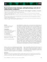
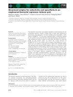
The pro-inflammatory cytokine IL-1b represents a
crucial mediator of neurodegeneration induced by excit-
atory or traumatic brain injury and, most notably, by
experimental cerebral ischemia in rodents [112] (Fig. 1).
Neurons, other cells
Endothelium
Fig. 1. Putative mechanisms implicated in
IL-1b-induced neuroinflammation after
stroke injury. CNS, central nervous system.
Neuroinflammatory mediators in brain ischemia D. Amantea et al.
16 FEBS Journal 276 (2009) 13–26 ª 2008 The Authors Journal compilation ª 2008 FEBS
Focal brain ischemia produced by either permanent or
transient MCAO in rats results in a significant induc-
tion of IL-1b mRNA [23,113,114]. Accordingly, IL-1b
protein levels increase very early following permanent
MCAO [115,116] and peak within hours of reperfusion
in transient focal ischemic models in rodents
[97,117,118]. The main source of the cytokine after cere-
bral ischemia are endothelial cells, microglia and macro-
phages, although it may also be expressed by neurons
and astrocytes [119,120]. Activation of p38 mitogen-
activated protein kinase has been suggested to underlie
IL-1b production by astrocytes and microglia during
ischemic injury in rats [121–123]. Moreover, there is evi-
dence suggesting that activation of the Toll-like recep-
tor-4 may be responsible for (pro-)IL-1b production
following cerebral ischemia [124].
Intracerebral injection of IL-1b neutralizing anti-
body to rats reduces ischemic brain damage [125], and
both intracerebroventricular and systemic administra-
tion of IL-1 receptor antagonist (IL-1ra) markedly
reduces brain damage induced by focal stroke, further
implicating IL-1b in ischemic pathophysiology [126–
129]. IL-1b expression is closely associated with an
upregulation of ICAM and endothelial leucocyte adhe-
sion molecule, which reach a peak between 6 and 12 h
after the onset of ischemia [130]. ICAM-1-deficient
mice suffer smaller infarcts after transient MCAO,
suggesting that part of the IL-1b-dependent injury is
mediated by the activation of ICAM-1 [41].
IL-1b is synthesized as a precursor molecule, pro-
IL-1b, which is cleaved and converted into the mature,
biologically active form of the cytokine by caspase-1,
formerly referred to as interleukin-1b-converting
enzyme (ICE) [131–133]. Inhibition of caspase-1 by
Ac-YVAD.cmk affords neuroprotection in rodent
models of permanent [134] or transient [117] MCAO,
and evidence from knockout mice indicates that cas-
pase-1 is important in the development of cerebral
ischemic damage [135,136]. However, to date, it is not
clear whether neuroprotection yielded by caspase-1-
preferring inhibitors is mediated by reduced IL-1b
production or by interference with the cell-death
process [137]. Although most studies have clearly
established the role of ICE in the maturation of IL-1b,
evidence from ICE-deficient mice and from in vitro
studies suggests that cytokine activation might also
involve other mechanisms [138–140]. Interestingly,
in vitro studies have described the involvement of
MMPs in cytokine processing. The conversion of
recombinant pro-IL-1b into mature IL-1b has been
demonstrated to occur after co-incubation with recom-
binant MMP-2 or MMP-9, the latter operating a more
effective and rapid cleavage [106].
We have recently demonstrated that the early
increase of IL-1b detected in the ischemic cortex of
rats subjected to transient MCAO is not associated
with increased activity of caspase-1 [97]. By contrast,
as discussed above, cytokine production during ische-
mia-reperfusion injury appears to be dependent on
MMP activity because systemic administration of the
MMP inhibitor, GM6001, prevents the early increase
of mature IL-1b in the ischemic cortex [97] (Fig. 1). As
cytokines, such as IL-1b, regulate the expression and
the activation of MMPs, a complex cross-regulation
does occur between these neuroinflammatory media-
tors, and further studies are needed to understand their
spatio-temporal occurrence during stroke injury.
Despite being structurally and functionally corre-
lated with IL-1, results from animal studies suggest
that IL-18 is not involved in stroke pathophysiology
[141]. However, blood levels of the cytokine increase in
acute stroke patients and appear to be predictive of
unfavourable clinical outcome [142,143].
In addition to IL-1b, brain injury induced by focal
ischemia is characterized by a significant and rapid
upregulation of TNF-a, as demonstrated both in ani-
mal models and in stroke patients. Increased expres-
sion of TNF-a has been described in neurones,
especially during the first hours after the ischemic
insult, and at later stages in microglia ⁄ macrophages
and in cells of the peripheral immune system [22,144–
147]. A focal ischemic insult has also been shown to
upregulate expression of the TNF-a receptor, p75, in
resident microglia and infiltrating macrophages of the
injured hemisphere [145,148].
Administration of neutralizing antibodies raised
against TNF-a or soluble TNF receptor 1 results in
reduced infarct size in rats subjected to permanent
MCAO, suggesting that the cytokine exacerbates ische-
mic injury [28,149–151]. However, to date, the role of
TNF-a has not been fully clarified because neuronal
damage caused by focal brain ischemia is exacerbated
in mice genetically deficient in p55 TNF receptors
[152]. The pleiotropic activities of TNF are mediated
by two structurally related, but functionally distinct,
receptors, namely p55 and p75. Selective deletion of
the p55
gene results in increased brain damage, as
compared with wild-type and p75-deficient mice fol-
lowing transient focal ischemia [153]. Moreover, ische-
mic preconditioning by TNF-a has been suggested to
occur via p55 receptor upregulation in neurons [154].
Thus, the roles of p55 and p75 in modulating cell
death ⁄ survival remain unclear, as both receptors may
activate intracellular mechanisms contributing either to
the induction of cell-death mechanisms or to anti-
inflammatory and anti-apoptotic functions [155].
D. Amantea et al. Neuroinflammatory mediators in brain ischemia
FEBS Journal 276 (2009) 13–26 ª 2008 The Authors Journal compilation ª 2008 FEBS 17
IL-6 expression significantly increases in the acute
phase of cerebral ischemia [156,157] and remains ele-
vated in neurons and reactive microglia of the ischemic
penumbra up to 14 days after the ischemic insult
[158,159]. In patients with acute brain ischemia,
plasma concentrations of IL-6 are strongly associated
with stroke severity and long-term clinical outcome
[160–162]. In a double-blind clinical trial on patients
with acute stroke, intravenous administration of
human recombinant IL-1ra ameliorates clinical out-
come and reduces blood concentrations of IL-6 [163].
This is in contrast to the results from animal studies
suggesting that IL-6 may exert a neuroprotective role
during stroke. In fact, intracerebroventricular injection
of recombinant IL-6 reduces ischemic brain damage
induced by permanent MCAO in rat [164]. It has been
suggested that increased levels of the endogenous
cytokine prevent damaged neurons from undergoing
apoptosis via signal transduction and activator of
transcription-3 activation [165].
Among other cytokines involved in stroke patho-
physiology, IL-10 and transforming growth factor-beta
have been demonstrated to have anti-inflammatory
effects, providing significant protection against ische-
mic brain damage [166].
Chemokines
Chemokines are regulatory polypeptides that mediate
cellular communication and leukocyte recruitment in
inflammatory and immune responses. Increased
mRNA expression for MCP-1 and macrophage
inflammatory protein-1 alpha has been described in
the rat brain after focal cerebral ischemia, and both
chemokines have been suggested to contribute to tis-
sue damage via recruitment of inflammatory cells
[25,167,168]. Expression of MCP-1 has been described
in neurons 12 h after focal brain ischemia, but also in
astrocytes and microglia at later stages following the
insult [26,169]. The MCP-1 levels are also increased
in the cerebrospinal fluid of stroke patients [170].
MCP-1 is a major factor driving leukocyte infiltration
in the brain parenchyma [171]. Mice deficient in
MCP-1 develop less infarct volume as a consequence
of focal brain ischemia [172]. Similarly, in mice defi-
cient in the gene for the MCP-1 receptor, CCR2,
transient focal ischemia results in reduced infarct size,
edema, leukocyte infiltration and expression of inflam-
matory mediators [173]. Moreover, MCP-1, as well as
stromal cell-derived factor-1a, have been shown to
trigger migration of newly formed neuroblasts from
neurogenic regions to ischemic damaged areas
[169,174].
Stromal cell-derived factor-1a expression is increased
in the ischemic penumbra, particularly in perivascular
astrocytes [175]. This chemokine has been suggested to
promote neuroprotection by increasing bone marrow-
derived cell targeting to the ischemic brain and by
improving local cerebral blood flow [176,177]. The cru-
cial involvement of chemokines in regulating cell
migration, promoting the interaction of stem cells with
ischemia-damaged host tissue, might be useful for
improving the clinical application of stem cell therapy.
Another chemokine implicated in ischemic patho-
physiology is fractalkine, whose expression is increased
in neurons and in some endothelial cells after a focal
ischemic insult. Interestingly, expression of its receptor,
CX3CR1, was observed only in microglia ⁄ macrophages,
suggesting that fractalkine is involved in neuron–
microglia signalling [178]. In fact, this chemokine
participates in leukocyte migration and in the activa-
tion and chemoattraction of microglia into the infract-
ed tissue [178]. Indeed, fractalkine-deficient mice
exhibit a smaller infarct size and lower mortality after
transient focal cerebral ischemia, further underlying
the detrimental effect of this chemokine on stroke
outcome [179].
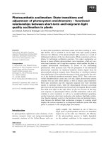
Conclusions
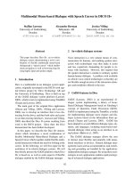
Neuroinflammatory mechanisms activated following an
ischemic insult play a complex role in the pathophysi-
ology of cerebral ischemia (Fig. 2). The induction of
pro-inflammatory genes may occur very early after the
insult and commonly aggravate tissue damage. Thus,
early inflammatory responses appear to contribute to
ischemic injury, whereas late responses may represent
endogenous mechanisms of recovery and repair. The
switch from detrimental to beneficial effects seems to
depend on the strength and the duration of the insult
and is crucial for determining the time-window for an
effective pharmacotherapy.
Given its pivotal role in stroke pathophysiology, the
IL-1 system represents an attractive therapeutic target
(Fig. 1). Indeed, IL-1ra reduces brain injury in animal
models of cerebral ischemia and, in a recent random-
ized clinical trial, intravenous administration of
recombinant human IL-1ra in patients with acute
stroke provided evidence for safety and for effective
reduction of peripheral inflammatory markers [163].
Recombinant human IL-1ra administered intrave-
nously has also been shown to penetrate the human
brain at experimentally therapeutic concentrations
[180], although its slow penetration into cerebrospinal
fluid [181] will probably result in subtherapeutic con-
centrations during the crucial early hours of an acute
Neuroinflammatory mediators in brain ischemia D. Amantea et al.
18 FEBS Journal 276 (2009) 13–26 ª 2008 The Authors Journal compilation ª 2008 FEBS
stroke. Further work is necessary to identify a suitable
therapeutic regime prior to phase II ⁄ III clinical trials.
Acknowledgements
Financial support from the Italian Ministry of Univer-
sity and Research (PRIN prot. 2006059200_002) is
gratefully acknowledged.
References
1 Gladstone DJ, Black SE, Hakim AM & Heart and
Stroke Foundation of Ontario Centre of Excellence in
Stroke Recovery (2002) Toward wisdom from failure:
lessons from neuroprotective stroke trials and new
therapeutic directions. Stroke 33, 2123–2136.
2 Lo EH, Dalkara T & Moskowitz MA (2003) Mecha-
nisms, challenges and opportunities in stroke. Nat Rev
Neurosci 4, 2123–2126.
3 Hossmann KA (2006) Pathophysiology and therapy
of experimental stroke. Cell Mol Neurobiol 26,
1057–1084.
4 Hossmann KA (1994) Viability thresholds and the
penumbra of focal ischemia. Ann Neurol 36, 557–565.
5 Dirnagl U, Iadecola C & Moskowitz MA (1999)
Pathobiology of ischaemic stroke: an integrated view.
Trends Neurosci 22, 391–397.
6 Lo EH, Moskowitz MA & Jacobs TP (2005) Exciting,
radical, sucidal: how brain cells die after stroke. Stroke
36, 189–192.
7 Stoll G, Jander S & Schroeter M (1998) Inflammation
and glial responses in ischemic brain lesions. Prog
Neurobiol 56, 149–171.
8 Del Zoppo GJ, Becker KJ & Hallenbeck JM (2001)
Inflammation after stroke: is it harmful? Arch Neurol
58, 669–672.
9 Kriz J (2006) Inflammation in ischemic brain injury:
timing is important. Crit Rev Neurobiol 18, 145–157.
10 Denes A, Vidyasagar R, Feng J, Narvainen J, McColl
BW, Kauppinen RA & Allan SM (2007) Proliferating
resident microglia after focal cerebral ischaemia in
mice. J Cereb Blood Flow Metab 27, 1941–1953.
11 Schilling M, Besselmann M, Leonhard C, Mueller M,
Ringelstein EB & Kiefer R (2003) Microglial activation
precedes and predominates over macrophage infiltra-
tion in transient focal cerebral ischemia: a study in
green fluorescent protein transgenic bone marrow
chimeric mice. Exp Neurol 183, 25–33.
12 Tanaka R, Komine-Kobayashi M, Mochizuki H,
Yamada M, Furuya T, Migita M, Shimada T, Mizuno
Y & Urabe T (2003) Migration of enhanced green
fluorescent protein expressing bone marrow-derived
microglia ⁄ macrophage into the mouse brain following
permanent focal ischemia. Neuroscience 117, 531–539.
13 Lindsberg PJ, Carpe
´
n O, Paetau A, Karjalainen-Linds-
berg ML & Kaste M (1996) Endothelial ICAM-1
expression associated with inflammatory cell response
in human ischemic stroke. Circulation 94, 939–945.
14 Gerhard A, Neumaier B, Elitok E, Glatting G, Ries V,
Tomczak R, Ludolph AC & Reske SN (2000) In vivo
imaging of activated microglia using [11C]PK11195
and positron emission tomography in patients after
ischemic stroke. Neuroreport 11, 2957–2960.
15 Price CJ, Menon DK, Peters AM, Ballinger JR, Barber
RW, Balan KK, Lynch A, Xuereb JH, Fryer T, Guad-
agno JV et al. (2004) Cerebral neutrophil recruitment,
Fig. 2. Main pathways implicated in the
neuroinflammatory response to ischemic
injury. CBF, cerebral blood flow; MIP-1a,
macrophage inflammatory protein-1 alpha;
ROS, reactive oxygen species.
D. Amantea et al. Neuroinflammatory mediators in brain ischemia
FEBS Journal 276 (2009) 13–26 ª 2008 The Authors Journal compilation ª 2008 FEBS 19
histology, and outcome in acute ischemic stroke: an
imaging-based study. Stroke 35, 1659–1664.
16 Danton GH & Dietrich WD (2003) Inflammatory
mechanisms after ischemia and stroke. J Neuropathol
Exp Neurol 62, 127–136.
17 Zhang ZG, Chopp M & Powers C (1997) Temporal
profile of microglial response following transient (2 h)
middle cerebral artery occlusion. Brain Res 774, 189–
198.
18 Schilling M, Besselman M, Muller M, Strecker JK,
Ringelstein EB & Kiefer R (2005) Predominant phago-
cytic activity of resident microglia over hematogenous
macrophages following transient cerebral brain ische-
mia: an investigation using green fluorescent protein
transgenic bone marrow chimeric mice. Exp Neurol
196, 290–297.
19 Yrjanheikki J, Tikka T, Keinanen R, Goldsteins G,
Chan PH & Koistinaho J (1999) A tetracycline
derivative, minocycline, reduces inflammation and
protects against focal cerebral ischemia with a wide
therapeutic window. Proc Natl Acad Sci USA 96,
13496–13500.
20 Hailer NP (2008) Immunosuppression after traumatic
or ischemic CNS damage: it is neuroprotective and
illuminates the role of microglial cells. Prog Neurobiol
84, 211–233.
21 Lalancette-He
´
bert M, Gowing G, Simard A, Weng YC
& Kriz J (2007) Selective ablation of proliferating
microglial cells exacerbates ischemic injury in the
brain. J Neurosci 27, 2596–2605.
22 Liu T, Clark RK, McDonnell PC, Young PR, White
RF, Barone FC & Feuerstein GZ (1994) Tumor necro-
sis factor-alpha expression in ischemic neurons. Stroke
25, 1481–1488.
23 Wang X, Yue TL, Barone FC, White RF, Gagnon RC
& Feuerstein GZ (1994) Concomitant cortical expres-
sion of TNF-alpha and IL-1 beta mRNAs follows
early response gene expression in transient focal ische-
mia. Mol Chem Neuropathol 23, 103–114.
24 Kim JS, Gautam SC, Chopp M, Zaloga C, Jones ML,
Ward PA & Welch KM (1995b) Expression of mono-
cyte chemoattractant protein-1 and macrophage
inflammatory protein-1 after focal cerebral ischemia in
the rat. J Neuroimmunol 56, 127–134.
25 Wang X, Yue TL, Barone FC & Feuerstein GZ (1995)
Monocyte chemoattractant protein-1 messenger
RNA expression in rat ischemic cortex. Stroke 26,
661–665.
26 Che X, Ye W, Panga L, Wu DC & Yang GY (2001)
Monocyte chemoattractant protein-1 expressed in neu-
rons and astrocytes during focal ischemia in mice.
Brain Res 902, 171–177.
27 Stanimirovic DB, Wong J, Shapiro A & Durkin JP
(1997) Increase in surface expression of ICAM-1,
VCAM-1 and E-selectin in human cerebromicrovascu-
lar endothelial cells subjected to ischemia-like insults.
Acta Neurochir Suppl 70, 12–16.
28 Yang GY, Gong C, Qin Z, Ye W, Mao Y & Bertz AL
(1998) Inhibition of TNFalpha attenuates infarct vol-
ume and ICAM-1 expression in ischemic mouse brain.
Neuroreport 9, 2131–2134.
29 Yang GY, Schielke GP, Gong C, Mao Y, Ge HL, Liu
XH & Betz AL (1999) Expression of tumor necrosis
factor-alpha and intercellular adhesion molecule-1 after
focal cerebral ischemia in interleukin-1beta converting
enzyme deficient mice. J Cereb Blood Flow Metab 19,
1109–1117.
30 Scho
¨
ning B, Elepfandt P, Daberkow N, Rupprecht S,
Stockhammer F, Stoltenburg G, Volk HD & Woicie-
chowsky C (2002) Differences in immune cell invasion
into the cerebrospinal fluid and brain parenchyma dur-
ing cerebral infusion of interleukin-1beta. Neurol Sci
23, 211–218.
31 Zhang R, Chopp M, Zhang Z, Jiang N & Powers C
(1998b) The expression of P- and E-selectins in three
models of middle cerebral artery occlusion. Brain Res
785, 207–214.
32 Huang J, Choudhri TF, Winfree CJ, McTaggart RA,
Kiss S, Mocco J, Kim LJ, Protopsaltis TS, Zhang Y,
Pinsky DJ et al. (2000) Postischemic cerebrovascular
E-selectin expression mediates tissue injury in murine
stroke. Stroke 31, 3047–3053.
33 Kim JS, Chopp M, Chen H, Levine SR, Carey JL &
Welch KM (1995a) Adhesive glycoproteins CD11a and
CD18 are upregulated in the leukocytes from patients
with ischemic stroke and transient ischemic attacks.
J Neurol Sci 128, 45–50.
34 Fiszer U, Korczak-Kowalska G, Palasik W, Korlak J,
Go
´
rski A & Czonkowska A (1998) Increased expres-
sion of adhesion molecule CD18 (LFA-1beta) on the
leukocytes of peripheral blood in patients with acute
ischemic stroke. Acta Neurol Scand 97, 221–224.
35 Matsuo Y, Onodera H, Shiga Y, Shozuhara H, Nin-
omiya M, Kihara T, Tamatani T, Miyasaka M & Kog-
ure K (1994) Role of cell adhesion molecules in brain
injury after transient middle cerebral artery occlusion
in the rat. Brain Res 656, 344–352.
36 Okada Y, Copeland BR, Mori E, Tung MM, Thomas
WS & del Zoppo GJ (1994) P-selectin and intercellular
adhesion molecule-1 expression after focal brain ische-
mia and reperfusion. Stroke 25, 202–211.
37 Del Zoppo GJ, Schmid-Scho
¨
nbein GW, Mori E, Cope-
land BR & Chang CM (1991) Polymorphonuclear
leukocytes occlude capillaries following middle cerebral
artery occlusion and reperfusion in baboons. Stroke
22, 1276–1283.
38 Mori E, del Zoppo GJ, Chambers JD, Copeland BR &
Arfors KE (1992) Inhibition of polymorphonuclear
leukocyte adherence suppresses no-reflow after focal
cerebral ischemia in baboons. Stroke 23, 712–718.
Neuroinflammatory mediators in brain ischemia D. Amantea et al.
20 FEBS Journal 276 (2009) 13–26 ª 2008 The Authors Journal compilation ª 2008 FEBS
39 Connolly ES Jr, Winfree CJ, Springer TA, Naka Y,
Liao H, Yan SD, Stern DM, Solomon RA, Gutierrez-
Ramos JC & Pinsky DJ (1996) Cerebral protection
in homozygous null ICAM-1 mice after middle
cerebral artery occlusion. Role of neutrophil adhesion
in the pathogenesis of stroke. J Clin Invest 97, 209–
216.
40 Connolly ES, Winfree CJ, Prestigiacomo CJ, Kim SC,
Choudhri TF, Hoh BL, Naka Y, Solomon RA & Pin-
sky DJ (1997) Exacerbation of cerebral injury in mice
that express the P-selectin gene: identification of
P-selectin blockade as a new target for the treatment
of stroke. Circ Res 81, 304–310.
41 Soriano SG, Lipton SA, Wang YMF, Xiao M,
Springer TA, Gutierrezramo JC & Hickey PR (1996)
Intercellular adhesion molecule-1-deficient mice are less
susceptible to cerebral ischemia-reperfusion injury. Ann
Neurol 39, 618–624.
42 Chopp M, Li Y, Jiang N, Zhang RL & Prostak J
(1996) Antibodies against adhesion molecules reduce
apoptosis after transient middle cerebral artery occlu-
sion in rat brain. J Cereb Blood Flow Metab 16, 578–
584.
43 Enlimomab Acute Stroke Trial Investigators (2001)
Use of anti-ICAM-1 therapy in ischemic stroke: results
of the enlimomab acute stroke trial. Neurology 57,
1428–1434.
44 Furuya K, Takeda H, Azhar S, McCarron RM, Chen
Y, Ruetzler CA, Wolcott KM, DeGraba TJ, Rothlein
R, Hugli TE et al. (2001) Examination of several
potential mechanisms for the negative outcome in a
clinical stroke trial of enlimomab, a murine anti-
human intercellular adhesion molecule-1 antibody: a
bedside-to-bench study. Stroke 32, 2665–2674.
45 Soriano MA, Tessier M, Certa U & Gil R (2000) Par-
allel gene expression monitoring using oligonucleotide
probe arrays of multiple transcripts with an animal
model of focal ischemia. J Cereb Blood Flow Metab
20, 1045–1055.
46 Vemuganti R, Bowen KK, Dhodda VK, Song G,
Franklin JL, Gavva NR & Dempsey RJ (2002) Gene
expression analysis of spontaneously hypertensive rat
cerebral cortex following transient focal ischemia.
J Neurochem 83, 1072–1086.
47 Vemuganti R, Dempsey RJ & Bowen KK (2004) Inhi-
bition of intercellular adhesion molecule-1 protein
expression by antisense oligonucleotides is neuropro-
tective after transient middle cerebral artery occlusion
in rat. Stroke 35, 179–184.
48 Lu XC, Williams AJ, Yao C, Berti R, Hartings JA,
Whipple R, Vahey MT, Polavarapu RG, Woller KL,
Tortella FC et al. (2004) Microarray analysis of acute
and delayed gene expression profile in rats after focal
ischemic brain injury and reperfusion. J Neurosci Res
77, 843–857.
49 Kapadia R, Tureyen K, Bowen KK, Kalluri H, John-
son PF & Vemuganti R (2006) Decreased brain dam-
age and curtailed inflammation in transcription factor
CCAAT ⁄ enhancer binding protein beta knockout mice
following transient focal cerebral ischemia. J Neuro-
chem 98, 1718–1731.
50 Yi JH, Park SW, Kapadia R & Vemuganti R (2007)
Role of transcription factors in mediating post-ische-
mic cerebral inflammation and brain damage. Neuro-
chem Int 50, 1014–1027.
51 Bergeron M, Yu AY, Solway KE, Semenza GL &
Sharp FR (1999) Induction of hypoxia-inducible fac-
tor-1 (HIF-1) and its target genes following focal
ischaemia in rat brain. Eur J Neurosci 11, 4159–4170.
52 Tanaka K, Nogawa S, Ito D, Suzuki S, Dembo T,
Kosakai A & Fukuchi Y (2000) Activated phosphory-
lation of cyclic AMP response element binding protein
is associated with preservation of striatal neurons after
focal cerebral ischemia in the rat. Neuroscience 100,
345–354.
53 Cho S, Park EM, Kim Y, Liu N, Gal J, Volpe BT &
Joh TH (2001) Early c-Fos induction after cerebral
ischemia: a possible neuroprotective role. J Cereb
Blood Flow Metab 21, 550–556.
54 Maeda K, Hata R, Gillardon F & Hossman KA
(2001) Aggravation of brain injury after focal cerebral
ischemia in p53-deficient mice. Mol Brain Res 88, 54–
61.
55 Sundararajan S, Gamboa JL, Victor AN, Wanderi
EW, Lust D & Landreth GE (2005) Peroxisome prolif-
erator-activated receptor-c ligands reduce inflammation
and infarction size in transient focal ischemia. Neuro-
science 130, 685–696.
56 Luo Y, Yin W, Signore AP, Zhang F, Hong Z, Wang
S, Graham SH & Chen J (2006) Neuroprotection
against focal ischemic brain injury by the peroxisomal
proliferator-activated receptor-c agonist rosiglitazone.
J Neurochem 97, 435–448.
57 Tureyen K, Kapadia R, Bowen KK, Satriotomo R,
Liang J, Feinstein DL & Vemuganti R (2007) Peroxi-
some proliferator-activated receptor-gamma agonists
induce neuroprotection following transient focal
ischemia in normotensive, normoglycemic as well as
hypertensive and type-2 diabetic rodents. J Neurochem
101, 41–56.
58 O’Neill LA & Kaltschmidt C (1997) NF-kappa B: a
crucial transcription factor for glial and neuronal cell
function. Trends Neurosci 20, 252–258.
59 Iadecola C, Forster C, Nogawa S, Clark HB & Ross
ME (1999) Cyclooxygenase-2 immunoreactivity in the
human brain following cerebral ischemia. Acta Neuro-
pathol 98, 9–14.
60 Stephenson D, Yin T, Smalstig EB, Hsu MA, Panetta
J, Little S & Clemens J (2000) Transcription factor
nuclear factor-kappa B is activated in neurons after
D. Amantea et al. Neuroinflammatory mediators in brain ischemia
FEBS Journal 276 (2009) 13–26 ª 2008 The Authors Journal compilation ª 2008 FEBS 21
focal cerebral ischemia. J Cereb Blood Flow Metab 20,
592–603.
61 Ohba N, Maeda M, Nakagomi S, Muraoka M & Kiy-
ama H (2003) Biphasic expression of activating tran-
scription factor-3 in neurons after cerebral ischemia.
Mol Brain Res 115, 147–156.
62 Satriotomo I, Bowen K & Vemuganti R (2006) JAK2
and STAT3 activation contributes to neuronal damage
following transient focal cerebral ischemia. J Neuro-
chem 98, 1353–1368.
63 Tureyen K, Brooks N, Bowen K, Svaren J & Vemug-
anti R (2008) Transcription factor early growth
response-1 induction mediates inflammatory gene
expression and brain damage following transient focal
ischemia. J Neurochem 105, 1313–1324.
64 Faraco G, Fossati S, Bianchi ME, Patrone M, Ped-
razzi M, Sparatore B, Moroni F & Chiarugi A (2007)
High mobility group box 1 protein is released by neu-
ral cells upon different stresses and worsens ischemic
neurodegeneration in vitro and in vivo. J Neurochem
103, 590–603.
65 Liu K, Mori S, Takahashi HK, Tomono Y, Wake H,
Kanke T, Sato Y, Hiraga N, Adachi N, Yoshino T
et al. (2007) Anti-high mobility group box 1 monoclo-
nal antibody ameliorates brain infarction induced by
transient ischemia in rats. FASEB J 21 , 3904–3916.
66 Qiu J, Nishimura M, Wang Y, Sims JR, Qiu S, Savitz
SI, Salomone S & Moskowitz MA (2008) Early release
of HMGB-1 from neurons after the onset of brain
ischemia. J Cereb Blood Flow Metab 28, 927–938.
67 Nogawa S, Zhang F, Ross ME & Iadecola C (1997)
Cyclo-oxygenase-2 gene expression in neurons contrib-
utes to ischemic brain damage. J Neurosci 17, 2746–
2755.
68 Bidmon HJ, Oermann E, Schiene K, Schmitt M, Kato
K, Asayama K, Witte OW & Zilles K (2000) Unilat-
eral upregulation of cyclooxygenase-2 following cere-
bral, cortical photothrombosis in the rat: suppression
by MK-801 and co-distribution with enzymes involved
in the oxidative stress cascade. J Chem Neuroanat 20,
163–176.
69 Iadecola C, Niwa K, Nogawa S, Zhao X, Nagayama
M, Araki E, Morham S & Ross ME (2001) Reduced
susceptibility to ischemic brain injury and N-methyl-D-
aspartate-mediated neurotoxicity in cyclooxygenase-2-
deficient mice. Proc Natl Acad Sci USA 98, 1294–1299.
70 Candelario-Jalil E, Gonza
´
lez-Falco
´
n A, Garcı
´
a-Cabre-
ra M, Leo
´
n OS & Fiebich BL (2007) Post-ischaemic
treatment with the cyclooxygenase-2 inhibitor nimesu-
lide reduces blood–brain barrier disruption and leuko-
cyte infiltration following transient focal cerebral
ischaemia in rats. J Neurochem 100, 1108–1120.
71 Kawano T, Anrather J, Zhou P, Park L, Wang G, Frys
KA, Kunz A, Cho S, Orio M & Iadecola C (2006) Pros-
taglandin E2 EP1 receptors: downstream effectors of
COX-2 neurotoxicity. Nat Med 12, 225–229.
72 Nakashima MN, Yamashita K, Kataoka Y, Yamashita
YS & Niwa M (1995) Time course of nitric oxide
synthase activity in neuronal, glial, and endothelial cells
of rat striatum following focal cerebral ischemia. Cell
Mol Neurobiol 15, 341–349.
73 Iadecola C, Zhang F, Casey R, Clark HB & Ross ME
(1996) Inducible nitric oxide synthase gene expression
in vascular cells after transient focal cerebral ischemia.
Stroke 27, 1373–1380.
74 Forster C, Clark HB, Ross ME & Iadecola C (1999)
Inducible nitric oxide synthase expression in human
cerebral infarcts. Acta Neuropathol 97
, 215–220.
75 Moro MA, Ca
´
rdenas A, Hurtado O, Leza JC & Liz-
asoain I (2004) Role of nitric oxide after brain ischae-
mia. Cell Calcium 36, 265–275.
76 Iadecola C, Zhang F, Casey R, Nagayama M & Ross
ME (1997) Delayed reduction of ischemic brain injury
and neurological deficits in mice lacking the inducible
nitric oxide synthase gene. J Neurosci 17, 9157–9164.
77 Murphy S & Gibson CL (2007) Nitric oxide, ischaemia
and brain inflammation. Biochem Soc Trans 35, 1133–
1137.
78 Pru
¨
ss H, Prass K, Ghaeni L, Milosevic M, Muselmann
C, Freyer D, Royl G, Reuter U, Baeva N, Dirnagl U
et al. (2008) Inducible nitric oxide synthase does not
mediate brain damage after transient focal cerebral
ischemia in mice. J Cereb Blood Flow Metab 28, 526–
539.
79 Nogawa S, Forster C, Zhang F, Nagayama M, Ross
ME & Iadecola C (1998) Interaction between inducible
nitric oxide synthase and cyclooxygenase-2 after cere-
bral ischemia. Proc Natl Acad Sci USA 95, 10966–
10971.
80 Gu Z, Kaul M, Yan B, Kriedel SJ, Cul J, Strongin A,
Smith JW, Liddington RC & Lipton SA (2002) S-nit-
rosylation of matrix metalloproteinases: signaling path-
way to neuronal cell death. Science 297, 1186–1190.
81 Sternlicht MD & Werb Z (2001) How matrix metallo-
proteinases regulate cell behaviour. Annu Rev Cell Dev
Biol 17, 463–516.
82 Rosenberg GA (2002) Matrix metalloproteinases in
neuroinflammation. Glia 39, 279–291.
83 Cunningham LA, Wetzel M & Rosenberg GA (2005)
Multiple roles for MMPs and TIMPs in cerebral ische-
mia. Glia 50, 329–339.
84 Romanic AM, White RF, Arleth AJ, Ohlstein EH &
Barone FC (1998) Matrix metalloproteinase expression
increases after cerebral focal ischemia in rats: inhibi-
tion of matrix metalloproteinase-9 reduces infarct size.
Stroke 29, 1020–1030.
85 Rosenberg GA, Estrada EY & Dencoff JE (1998)
Matrix metalloproteinases and TIMPs are associated
Neuroinflammatory mediators in brain ischemia D. Amantea et al.
22 FEBS Journal 276 (2009) 13–26 ª 2008 The Authors Journal compilation ª 2008 FEBS
with blood–brain barrier opening after reperfusion in
rat brain. Stroke 29, 2189–2195.
86 Heo JH, Lucero J, Abumiya T, Koziol JA, Copeland
BR & del Zoppo GJ (1999) Matrix metalloproteinases
increase very early during experimental focal cerebral
ischemia. J Cereb Blood Flow Metab 19, 624–633.
87 Asahi M, Asahi K, Jung JC, del Zoppo GJ, Fini ME
& Lo EH (2000) Role for matrix metalloproteinase 9
after focal cerebral ischemia: effects of gene knock-
down and enzyme inhibition with BB-94. J Cereb
Blood Flow Metab 20, 1681–1689.
88 Horstmann S, Kalb P, Koziol J, Gardner H & Wagner
S (2003) Profiles of matrix metalloproteinases, their
inhibitors, and laminin in stroke patients: influence of
different therapies. Stroke 34, 2165–2170.
89 Rosell A, Ortega-Aznar A, Alvarez-Sabin J, Fernan-
dez-Cadenas I, Ribo M, Molina CA, Lo EH & Mon-
tanter J (2006) Increased brain expression of matrix
metalloproteinase-9 after ischemic and hemorrhagic
human stroke. Stroke 37, 1399–1406.
90 Rosell A, Cuadrado E, Ortega-Aznar A, Herna
´
ndez-
Guillamon M, Lo EH & Montaner J (2008) MMP-
9-positive neutrophil infiltration is associated to
blood–brain barrier breakdown and basal lamina
type iv collagen degradation during hemorrhagic
transformation after human ischemic stroke. Stroke 39,
1121–1126.
91 Planas AM, Sole S & Justicia C (2001) Expression and
activation of matrix metalloproteinase-2 and -9 in rat
brain after transient focal cerebral ischemia. Neurobiol
Dis 8, 834–846.
92 Rosenberg GA, Cunningham LA, Wallace J, Alexan-
der S, Estrada EY, Grossetete M, Razhagi A, Miller K
& Gearing A (2001) Immunohistochemistry of matrix
metalloproteinases in reperfusion injury in rat brain:
activation of MMP-9 linked to stromelysin-1 and
microglia in cell cultures. Brain Res 893, 104–112.
93 Gu Z, Cui J, Brown S, Fridman R, Mobashery S,
Strongin AY & Lipton SA (2005) A highly specific
inhibitor of matrix metalloproteinase-9 rescues laminin
from proteolysis and neurons from apoptosis in tran-
sient focal cerebral ischemia. J Neurosci 25, 6401–6408.
94 Yang Y, Estrada EY, Thompson JF, Liu W & Rosen-
berg GA (2007) Matrix metalloproteinase-mediated
disruption of tight junction proteins in cerebral vessels
is reversed by synthetic matrix metalloproteinase inhib-
itor in focal ischemia in rat. J Cereb Blood Flow Metab
27, 697–709.
95 Amantea D, Corasaniti MT, Mercuri NB, Bernardi G
& Bagetta G (2008) Brain regional and cellular locali-
zation of gelatinase activity in rat that have undergone
transient middle cerebral artery occlusion. Neuroscience
152, 8–17.
96 Gasche Y, Copin JC, Sugawara T, Fujimura M &
Chan PH (2001) Matrix metalloproteinase inhibition
prevents oxidative stress associated blood–brain barrier
disruption after transient focal cerebral ischemia.
J Cereb Blood Flow Metab 21, 1393–1400.
97 Amantea D, Russo R, Gliozzi M, Fratto V, Berliocchi
L, Bagetta G, Bernardi G & Corasaniti MT (2007) Early
upregulation of matrix metalloproteinases following
reperfusion triggers neuroinflammatory mediators in
brain ischemia in rat. Int Rev Neurobiol 82, 149–169.
98 Asahi M, Sumii T, Fini ME, Itohara S & Lo EH
(2001a) Matrix metalloproteinase-2 gene knock out has
no effect on acute brain injury after focal ischemia.
NeuroReport 17, 3003–3007.
99 Asahi M, Wang X, Mori T, Sumii T, Jung JC,
Moskowitz MA, Fini ME & Lo EH (2001b) Effects of
matrix metalloproteinase 9 gene knock out on the
proteolysis of blood–brain barrier and white matter
components after cerebral ischemia. J Neurosci 21,
7724–7732.
100 Wang X, Jung JC, Asahi M, Chwang W, Russo L,
Moskowitz MA, Dixon CE, Fini E & Lo EH (2000)
Effects of matrix metalloproteinase 9 gene knock out
on morphological and motor outcomes after traumatic
brain injury. J Neurosci 20 , 7037–7042.
101 Jourquin J, Tremblay E, Decanis N, Charton G,
Hanessian S, Chollet AM, Le Diguardher T, Khrestch-
atiski M & Rivera S (2003) Neuronal activity-depen-
dent increase of net matrix metalloproteinase activity is
associated with MMP-9 neurotoxicity after kainate.
Eur J Neurosci 18, 1507–1517.
102 Copin JC, Goodyear MC, Gidday JM, Shah AR, Gas-
con E, Dayer A, Morel DM & Gasche Y (2005) Role
of matrix metalloproteinases in apoptosis after tran-
sient focal cerebral ischemia in rats and mice. Eur J
Neurosci 22, 1597–1608.
103 Wetzel M, Rosenberg GA & Cunningham LA (2003)
Tissue inhibitor of metalloproteinase-3 and matrix
metalloproteinase-3 regulate neuronal sensitivity to
doxorubicin-induced apoptosis. Eur J Neurosci 18,
1050–1060.
104 Lee R, Kermani P, Teng KK & Hempstead BL (2001)
Regulation of cell survival by secreted proneurotro-
phins. Science 294 , 1945–1948.
105 Zhang K, McQuibban GA, Silva C, Butler GS, John-
ston JB, Holden J, Clark-Lewis I, Overall CM &
Power C (2003) HIV-induced metalloproteinase pro-
cessing of the chemokine stromal cell derived factor-1
causes neurodegeneration. Nat Neurosci 6, 1064–1071.
106 Scho
¨
nbeck U, Mach F & Libby P (1998) Generation
of biologically active IL-1 beta by matrix metallopro-
teinases: a novel caspase-1-independent pathway of
IL-1 beta processing. J Immunol 161, 3340–3346.
107 Zhu W, Khachi S, Hao Q, Shen F, Young WL, Yang
GY & Chen Y (2008) Upregulation of EMMPRIN
after permanent focal cerebral ischemia. Neurochem Int
52, 1086–1091.
D. Amantea et al. Neuroinflammatory mediators in brain ischemia
FEBS Journal 276 (2009) 13–26 ª 2008 The Authors Journal compilation ª 2008 FEBS 23
108 Zhao BQ, Wang S, Kim HY, Storrie H, Rosen BR,
Mooney DJ, Wang X & Lo EH (2006) Role of matrix
metalloproteinases in delayed cortical responses after
stroke. Nat Med 12, 441–445.
109 Yong VW (2005) Metalloproteinases: mediators of
pathology and regeneration in the CNS. Nat Rev Neu-
rosci 6, 931–944.
110 Minami M, Katayama T & Satoh M (2006) Brain
cytokines and chemokines: roles in ischemic injury and
pain. J Pharmacol Sci 100, 461–470.
111 Allan SM & Rothwell NJ (2001) Cytokines and acute
neurodegeneration. Nat Rev Neurosci 2, 734–744.
112 Rothwell N (2003) Interleukin-1 and neuronal injury:
mechanisms, modification, and therapeutic potential.
Brain Behav Immun 17, 152–157.
113 Liu T, McDonnell PC, Young PR, White RF, Siren
AL, Hallenbeck JM, Barone FC & Feurerstein GZ
(1993) Interleukin-1-beta messenger RNA expression
in ischemic rat cortex. Stroke 24, 1746–1751.
114 Buttini M, Sauter A & Boddeke H (1994) Induction of
interleukin-1beta mRNA after focal ischemia in the
rat. Mol Brain Res 23, 126–134.
115 Davies CA, Loddick SA, Toulmond S, Stroemer RP,
Hunt J & Rothwell NJ (1999) The progression and
topographic distribution of interleukin-1 beta expres-
sion after permanent middle cerebral artery occlusion
in the rat. J Cereb Blood Flow Metab 19, 87–98.
116 Legos JJ, Whitmore RG, Erhardt JA, Parsons AA,
Tuma RF & Barone FC (2000) Quantitative changes
in interleukin proteins following focal stroke in the rat.
Neurosci Lett 282, 189–192.
117 Hara H, Friedlander RM, Gagliardini V, Ayata C,
Fink K, Huang Z, Shimizu-Sasamata M, Yuan J &
Moskowitz MA (1997) Inhibition of interleukin 1b
converting enzyme family proteases reduces ischemic
and excitotoxic neuronal damage. Proc Natl Acad Sci
USA 94, 2007–2012.
118 Zhang Z, Chopp M, Goussev A & Powers C (1998a)
Cerebral vessels express interleukin 1 beta after focal
cerebral ischemia. Brain Res 784, 210–217.
119 Touzani O, Boutin H, Chuquet J & Rothwell N (1999)
Potential mechanisms of IL-1 involvement in cerebral
ischemia. J Neuroimmunol 100, 203–215.
120 Mabuchi T, Kitagawa K, Ohtsuki T, Kuwabara K,
Yagita Y, Yanagihara T, Hori M & Matsumoto M
(2000) Contribution of microglia ⁄ macrophages to
expansion of infarction and response of oligodendro-
cytes after focal cerebral ischemia in rats. Stroke 31,
1735–1743.
121 Irving EA, Barone FC, Reith AD, Hadingham SJ &
Parsons AA (2000) Differential activation of MAP-
K ⁄ ERK and p38 ⁄ SAPK in neurones and glia following
focal cerebral ischaemia in the rat. Brain Res 77, 65–75.
122 Walton KM, DiRocco R, Bartlett BA, Koury E,
Marcy VR, Jarvis B, Schaefer EM & Bhat RV (1998)
Activation of p38MAPK in microglia after ischemia.
J Neurochem 70, 1764–1767.
123 Barone FC, Irving EA, Ray AM, Lee JC, Kassis S,
Kumar S, Badger AM, Legos JJ, Erhardt JA, Ohlstein
EH et al. (2001) Inhibition of p38 mitogen-activated
protein kinase provides neuroprotection in cerebral
focal ischemia. Med Res Rev 21, 129–145.
124 Simi A, Lerouet D, Pinteaux E & Brough D (2007)
Mechanisms of regulation for interleukin-1b in neuro-
degenerative disease. Neuropharmacology 52, 1563–
1569.
125 Yamasaki Y, Matsuura N, Shizuhara H, Onodera H
& Itoyama YKK (1995) Interleukin-1 as a pathoge-
netic mediator of ischemic brain damage in the rats.
Stroke 26, 676–681.
126 Relton JK & Rothwell NJ (1992) Interleukin-1 recep-
tor antagonist inhibits ischaemic and excitotoxic neuro-
nal damage in the rat. Brain Res Bull 29, 242–246.
127 Garcia JH, Liu KF & Relton JK (1995) Interleukin-1
receptor antagonist decreases the number of necrotic
neurons in rats with middle cerebral artery occlusion.
Am J Pathol 147, 1477–1486.
128 Relton JK, Martin D, Thompson RC & Russell DA
(1996) Peripheral administration of interleukin-1 recep-
tor antagonist inhibits brain damage after focal cere-
bral ischemia in the rat. Exp Neurol 138, 206–213.
129 Mulcahy N, Ross J, Rothwell NJ & Loddick SA
(2003) Delayed administration of interleukin-1 receptor
antagonist protects against transient cerebral ischemia
in the rat. Br J Pharmacol 140, 471–476.
130 Wang XK & Feuerstein GZ (1995) Induced expression
of adhesion molecules following focal brain ischemia.
J Neurotrauma 12 , 825–832.
131 Black RA, Kronheim SR, Cantrell M, Deeley MC,
March CJ, Prockett KS, Wignall J, Conlon PJ, Cos-
man D & Hopp TP (1988) Generation of biologically
active interleukin-1 beta by proteolytic cleavage of the
inactive precursor. J Biol Chem 263, 9437–9442.
132 Howard AD, Kostura MJ, Thornberry N, Ding GJF,
Limjuco G, Weidner J, Salley JP, Hogquist KA,
Chaplin DD, Mumford RA et al. (1991) IL-1-convert-
ing enzyme requires aspartic acid residues for process-
ing of the IL-1b precursor at two distinct sites and
does not cleave 31 k-Da IL-1a. J Immunol 147, 2964–
2969.
133 Thornberry NA, Bull HG, Calaycay JR, Chapman
KT, Howard AD, Kostura MJ, Miller DK, Molineaux
SM, Weidner JR, Aunins J et al. (1992) A novel hete-
rodimeric cysteine protease is required for interleukin-
1b processing in monocytes. Nature 356, 768–774.
134 Rabuffetti M, Sciorati C, Tarozzo G, Clementi E,
Manfredi AA & Beltramo M (2000) Inhibition of cas-
pase-1-like activity by Ac-Tyr-Val-Ala-Asp-chlorom-
ethyl ketone induces long-lasting neuroprotection in
cerebral ischemia through apoptosis reduction and
Neuroinflammatory mediators in brain ischemia D. Amantea et al.
24 FEBS Journal 276 (2009) 13–26 ª 2008 The Authors Journal compilation ª 2008 FEBS
decrease of proinflammatory cytokines. J Neurosci 20,
4398–4404.
135 Friedlander RM & Yuan J (1998) ICE, neuronal apop-
tosis and neurodegeneration. Cell Death Diff 5, 823–
831.
136 Schielke GP, Yang GY, Shivers BD & Lorris Betz A
(1998) Reduced ischemic brain injury in interleukin-1b
converting enzyme-deficient mice. J Cereb Blood Flow
Metab 18, 180–185.
137 Corasaniti MT, Russo R, Amantea D, Gliozzi M, Sivi-
glia E, Stringaro AR, Malori W, Melino G & Bagetta
G (2005) Neuroprotection by the caspase-1 inhibitor
Ac-YVAD-(acyloxy)mk in experimental neuroAIDS is
independent from IL-1beta generation. Cell Death
Differ 12(Suppl. 1), 999–1001.
138 Fantuzzi G, Ku G, Harding MW, Livingston DJ, Sipe
JD, Kuida K, Flavell RA & Dinarello CA (1997)
Response to local inflammation of IL-1b-converting
enzyme-deficient mice. J Immunol 158, 1818–1824.
139 Herzog C, Kaushal GP & Haun RS (2005) Generation
of biologically active interleukin-1 beta by meprin B.
Cytokine 31, 394–403.
140 Pinteaux E, Inoue W, Schmidt L, Molina-Holgado F,
Rothwell NJ & Luheshi GN (2007) Leptin induces
interleukin-1beta release from rat microglial cells
through a caspase 1 independent mechanism. J Neuro-
chem 102, 826–833.
141 Wheeler RD, Boutin H, Touzani O, Luheshi GN, Tak-
eda K & Rothwell NJ (2003) No role for interleukin-
18 in acute murine stroke-induced brain injury.
J Cereb Blood Flow Metab 23, 531–535.
142 Zaremba J & Losy J (2003) Interleukin-18 in acute
ischaemic stroke patients. Neurol Sci 24, 117–124.
143 Yuen CM, Chiu CA, Chang LT, Liou CW, Lu CH,
Youssef AA & Yip HK (2007) Level and value of
interleukin-18 after acute ischemic stroke. Circ J 71,
1691–1696.
144 Gregersen R, Lambertsen K & Finsen B (2000) Micro-
glia and macrophages are the major source of tumor
necrosis factor in permanent middle cerebral artery
occlusion in mice. J Cereb Blood Flow Metab 20, 53–
65.
145 Dziewulska D & Mossakowski MJ (2003) Cellular
expression of tumor necrosis factor a and its receptors
in human ischemic stroke. Clin Neuropathol 22, 35–40.
146 Yin L, Ohtaki H, Nakamachi T, Dohi K, Iwai Y,
Funahashi H, Makino R & Shioda S (2003) Expres-
sion of tumor necrosis factor alpha (TNFalpha)
following transient cerebral ischemia. Acta Neurochir
Suppl 86, 93–96.
147 Offner H, Subramanian S, Parker SM, Afentoulis ME,
Vandenbark AA & Hurn PD (2006) Experimental
stroke induces massive, rapid activation of the periph-
eral immune system. J Cereb Blood Flow Metab 26,
654–665.
148 Lambertsen KL, Clausen BH, Fenger C, Wulf H,
Owens T, Dagnaes-Hansen F, Meldgaard M & Finsen
B (2007) Microglia and macrophages express tumor
necrosis factor receptor p75 following middle cerebral
artery occlusion in mice. Neuroscience 144, 934–949.
149 Barone FC, Arvin B, White RF, Miller A, Webb CL,
Willette RN, Lysko PG & Feuerstein GZ (1997)
Tumor necrosis factor-alpha. A mediator of focal
ischemic brain injury. Stroke 28, 1233–1244.
150 Nawashiro H, Martin D & Hallenbeck JM (1997) Inhi-
bition of tumor necrosis factor and amelioration of
brain infarction in mice. J Cereb Blood Flow Metab 17,
229–232.
151 Lavine SD, Hofman FM & Zlokovic BV (1998) Circu-
lating antibody against tumor necrosis factor-alpha
protects rat brain from reperfusion injury. J Cereb
Blood Flow Metab 18, 52–58.
152 Bruce AJ, Boling W, Kindy MS, Peschon J, Kraemer
PJ, Carpenter MK, Holtsberg FW & Mattson MP
(1996) Altered neuronal and microglial responses to
excitotoxic and ischemic brain injury in mice lacking
TNF receptors. Nat Med 2, 788–794.
153 Gary DS, Bruce-Keller AJ, Kindy MS & Mattson MP
(1998) Ischemic and excitotoxic brain injury is
enhanced in mice lacking the p55 tumor necrosis factor
receptor. J Cereb Blood Flow Metab 18, 1283–1287.
154 Pradillo JM, Romera C, Hurtado O, Ca
´
rdenas A,
Moro MA, Leza JC, Da
´
valos A, Castillo J, Lorenzo P
& Lizasoain I (2005) TNFR1 upregulation mediates
tolerance after brain ischemic preconditioning. J Cereb
Blood Flow Metab 25, 193–203.
155 Hallenbeck JM (2002) The many faces of tumor necro-
sis factor in stroke. Nat Med 8, 1363–1368.
156 Ali C, Nicole O, Docagne F, Lesne S, MacKenzie ET,
Nouvelot A, Buisson A & Vivien D (2000) Ischemia-
induced interleukin-6 as a potential endogenous neuro-
protective cytokine against NMDA receptor-mediated
excitotoxicity in the brain. J Cereb Blood Flow Metab
20, 956–966.
157 Berti R, Williams AJ, Moffett JR, Hale SL, Velarde
LC, Elliott PJ, Yao C, Dave JR & Tortella FC (2002)
Quantitative real-time RT-PCR analysis of inflamma-
tory gene expression associated with ischemia-reperfu-
sion brain injury. J Cereb Blood Flow Metab 22 , 1068–
1079.
158 Block F, Peters M & Nolden-Koch M (2000) Expres-
sion of IL-6 in the ischemic penumbra. Neuroreport 11,
963–967.
159 Suzuki S, Tanaka K, Nogawa S, Nagata E, Ito D,
Dembo T & Fukuuchi Y (1999) Temporal profile and
cellular localization of interleukin-6 protein after focal
cerebral ischemia in rats. J Cereb Blood Flow Metab
19, 1256–1262.
160 Smith CJ, Emsley HC, Gavin CM, Georgiou RF, Vail
A, Barberan EM, del Zoppo GJ, Hallenbeck JM,
D. Amantea et al. Neuroinflammatory mediators in brain ischemia
FEBS Journal 276 (2009) 13–26 ª 2008 The Authors Journal compilation ª 2008 FEBS 25
Rothwell NJ, Hopkins SJ et al. (2004) Peak plasma
interleukin-6 and other peripheral markers of inflam-
mation in the first week of ischaemic stroke correlate
with brain infarct volume, stroke severity and long-
term outcome. BMC Neurol 4,2.
161 Waje-Andreassen U, Kra
˚
kenes J, Ulvestad E, Thomas-
sen L, Myhr KM, Aarseth J & Vedeler CA (2005)
IL-6: an early marker for outcome in acute ischemic
stroke. Acta Neurol Scand 111, 360–365.
162 Orion D, Schwammenthal Y, Reshef T, Schwartz R,
Tsabari R, Merzeliak O, Chapman J, Mekori YA &
Tanne D (2008) Interleukin-6 and soluble intercellular
adhesion molecule-1 in acute brain ischaemia. Eur J
Neurol 15, 323–328.
163 Emsley HC, Smith CJ, Georgiou RF, Vail A, Hopkins
SJ, Rothwell NJ, Tyrrell PJ & Acute Stroke Investiga-
tors (2005) A randomised phase II study of interleu-
kin-1 receptor antagonist in acute stroke patients.
J Neurol Neurosurg Psychiatry 76, 1366–1372.
164 Loddick SA, Turnbull AV & Rothwell NJ (1998) Cere-
bral interleukin-6 is neuroprotective during permanent
focal cerebral ischemia in the rat. J Cereb Blood Flow
Metab 18, 176–179.
165 Yamashita T, Sawamoto K, Suzuki S, Suzuki N,
Adachi K, Kawase T, Mihara M, Ohsugi Y, Abe K &
Okano H (2005) Blockade of interleukin-6 signaling
aggravates ischemic cerebral damage in mice: possible
involvement of Stat3 activation in the protection of
neurons. J Neurochem 94, 459–468.
166 Wang Q, Tang XN & Yenari MA (2007) The inflamma-
tory response in stroke. J Neuroimmunol 184, 53–68.
167 Takami S, Minami M, Nagata I, Namura S & Satoh M
(2001) Chemokine receptor antagonist peptide, viral
MIP-II, protects the brain against focal cerebral ische-
mia in mice. J Cereb Blood Flow Metab 21, 1430–1435.
168 Minami M & Satoh M (2003) Chemokines and their
receptors in the brain: pathophysiological roles in
ischemic brain injury. Life Sci 74, 321–327.
169 Yan YP, Sailor KA, Lang BT, Park SW, Vemuganti R
& Dempsey RJ (2007) Monocyte chemoattractant pro-
tein-1 plays a critical role in neuroblast migration after
focal cerebral ischemia. J Cereb Blood Flow Metab 27,
1213–1224.
170 Losy J & Zaremba J (2001) Monocyte chemoattractant
protein-1 is increased in the cerebrospinal fluid of
patients with ischemic stroke. Stroke 32, 2695–2696.
171 Chen Y, Hallenbeck JM, Ruetzler C, Bol D, Thomas
K, Berman NE & Vogel SN (2003) Overexpression of
monocyte chemoattractant protein 1 in the brain exac-
erbates ischemic brain injury and is associated with
recruitment of inflammatory cells. J Cereb Blood Flow
Metab 23, 748–755.
172 Hughes PM, Allegrini PR, Rudin M, Perry VH, Mir
AK & Wiessner C (2002) Monocyte chemoattractant
protein-1 deficiency is protective in a murine stroke
model. J Cereb Blood Flow Metab 22, 308–317.
173 Dimitrijevic OB, Stamatovic SM, Keep RF & Andj-
elkovic AV (2007) Absence of the chemokine receptor
CCR2 protects against cerebral ischemia ⁄ reperfusion
injury in mice. Stroke 38, 1345–1353.
174 Robin AM, Zhang ZG, Wang L, Zhang RL, Kata-
kowski M, Zhang L, Wang Y, Zhang C & Chopp M
(2006) Stromal cell-derived factor 1 alpha mediates
neural progenitor cell motility after focal cerebral
ischemia. J Cereb Blood Flow Metab 26, 125–134.
175 Hill WD, Hess DC, Martin-Studdard A, Carothers JJ,
Zheng J, Hale D, Maeda M, Fagan SC, Carroll JE &
Conway SJ (2004) SDF-1 (CXCL12) is upregulated in
the ischemic penumbra following stroke: association
with bone marrow cell homing to injury. J Neuropathol
Exp Neurol 63, 84–96.
176 Cui X, Chen J, Zacharek A, Li Y, Roberts C, Kapke
A, Savant-Bhonsale S & Chopp M (2007) Nitric oxide
donor upregulation of stromal cell-derived factor-
1 ⁄ chemokine (CXC motif) receptor 4 enhances bone
marrow stromal cell migration into ischemic brain
after stroke. Stem Cells 25, 2777–2785.
177 Shyu WC, Lin SZ, Yen PS, Su CY, Chen DC, Wang
HJ & Li H (2008) Stromal cell-derived factor-1 alpha
promotes neuroprotection, angiogenesis, and mobiliza-
tion ⁄ homing of bone marrow-derived cells in stroke
rats. J Pharmacol Exp Ther 324, 834–849.
178 Tarozzo G, Campanella M, Ghiani M, Bulfone A &
Beltramo M (2002) Expression of fractalkine and its
receptor, CX3CR1, in response to ischaemia-reperfu-
sion brain injury in the rat. Eur J Neurosci 15, 1663–
1668.
179 Soriano SG, Amaravadi LS, Wang YF, Zhou H, Yu
GX, Tonra JR, Fairchild-Huntress V, Fang Q,
Dunmore JH, Huszar D et al. (2002) Mice deficient in
fractalkine are less susceptible to cerebral ischemia-
reperfusion injury. J Neuroimmunol 125, 59–65.
180 Clark SR, McMahon CJ, Gueorguieva I, Rowland M,
Scarth S, Georgiou R, Tyrrell PJ, Hopkins SJ & Roth-
well NJ (2007) Interleukin-1 receptor antagonist pene-
trates human brain at experimentally therapeutic
concentrations. J Cereb Blood Flow Metab 28, 387–
394.
181 Gueorguieva I, Clark SR, McMahon CJ, Scarth S,
Rothwell NJ, Tyrell PJ, Hopkins SJ & Rowland M
(2008) Pharmacokinetic modelling of interleukin-1
receptor antagonist in plasma and cerebrospinal fluid
of patients following subarachnoid haemorrhage. Br J
Clin Pharmacol 65, 317–325.
Neuroinflammatory mediators in brain ischemia D. Amantea et al.
26 FEBS Journal 276 (2009) 13–26 ª 2008 The Authors Journal compilation ª 2008 FEBS