Báo cáo khoa học: C Isotopologue editing of FMN bound to phototropin domains potx
Bạn đang xem bản rút gọn của tài liệu. Xem và tải ngay bản đầy đủ của tài liệu tại đây (677.22 KB, 15 trang )
13
C Isotopologue editing of FMN bound to phototropin
domains
Wolfgang Eisenreich
1
, Monika Joshi
1
, Boris Illarionov
1
, Gerald Richter
2
, Werner Ro
¨
misch-Margl
1
,
Franz Mu
¨
ller
3
, Adelbert Bacher
1
and Markus Fischer
4
1 Lehrstuhl fu
¨
r Organische Chemie und Biochemie, Technische Universita
¨
tMu
¨
nchen, Garching, Germany
2 School of Chemistry, Cardiff University, UK
3 Wylstrasse 13, Hergiswil, Switzerland
4 Institut fu
¨
r Biochemie und Lebensmittelchemie, Abteilung Lebensmittelchemie, Universita
¨
t Hamburg, Germany
13
C-Labeled flavocoenzymes have played an important
role for the spectroscopic analysis of flavoenzymes [1],
but their use was limited by the costs and effort for
the preparation of the
13
C-labeled cofactors. More spe-
cifically,
13
C can be introduced with relative ease into
the pyrimidine moiety of the isoalloxazine system [2],
and the xylene moiety of flavin cofactors can be
labeled by enzyme-assisted strategies [3], whereas ribi-
tyl carbon atoms have been rarely included in labeling
studies due to technical hurdles. We have shown earlier
that mixtures of
13
C isotopologues of the riboflavin
precursor, 6,7-dimethyl-8-ribityllumazine, can be pre-
pared by biotransformation of
13
C-labeled glucose
in vivo [4]. In the present study, we report the transfor-
mation of these isotopologue libraries into random
libraries of
13
C isotopologues of FMN and their utili-
zation for NMR studies of the plant blue light recep-
tor, phototropin [5,6].
Keywords
blue light receptor; isotopologue libraries;
LOV domain; NMR spectroscopy;
phototropin
Correspondence
W. Eisenreich, Lehrstuhl fu
¨
r Organische
Chemie und Biochemie, Technische
Universita
¨
tMu
¨
nchen, Lichtenbergstrasse 4,
D-85747 Garching, Germany
Fax: +49 89 289 13363
Tel: +49 89 289 13336
E-mail:
M. Fischer, Institut fu
¨
r Biochemie und
Lebensmittelchemie, Abteilung
Lebensmittelchemie, Universita
¨
t Hamburg,
Grindelallee 117, 20146 Hamburg, Germany
Fax: +49 40 4283 84342
Tel: +49 40 4283 84357
E-mail: markus.fi
hamburg.de
(Received 6 June 2007, revised 12 August
2007, accepted 19 September 2007)
doi:10.1111/j.1742-4658.2007.06111.x
The plant blue light receptor phototropin comprises a protein kinase
domain and two FMN-binding LOV domains (LOV1 and LOV2). Blue
light irradiation of recombinant LOV domains is conducive to the addition
of a cysteinyl thiolate group to carbon 4a of the FMN chromophore, and
spontaneous cleavage of that photoadduct completes the photocycle of
the receptor. The present study is based on
13
C NMR signal modulation
observed after reconstitution of LOV domains of different origins with ran-
dom libraries of
13
C-labeled FMN isotopologues. Using this approach, all
13
C signals of FMN bound to LOV1 and LOV2 domains of Avena sativa
and to the LOV2 domain of the fern, Adiantum capillus-veneris, could be
unequivocally assigned under dark and under blue light irradiation condi-
tions.
13
C Chemical shifts of FMN are shown to be differently modulated
by complexation with the LOV domains under study, indicating slight
differences in the binding interactions of FMN and the apoproteins.
Abbreviations
C(4a), carbon 4a; TARF, tetraacetylriboflavin.
5876 FEBS Journal 274 (2007) 5876–5890 ª 2007 The Authors Journal compilation ª 2007 FEBS
The gene specifying phototropin, the first in the
emerging family of blue light receptors in plants, was
initially cloned from Arabidopsis thaliana and was
shown to specify a cytoplasmic protein comprising
a serine ⁄ threonine protein kinase domain and two
FMN-binding LOV domains (designated LOV1 and
LOV2), which are members of the PAS domain super-
family [6–8] (Fig. 1). Blue light irradiation of recombi-
nant LOV domains results in substantial modulation
of the visible absorption spectrum [9], which was inter-
preted to result from the formation of an adduct
between the thiol group of a cystein residue and car-
bon 4a [C(4a)] of the FMN chromophore by Vincent
Massey (a contribution to the discussion at the 13th
International Congress on Flavins and Flavoproteins;
29 August to 4 September 1999, Konstanz) (Fig. 2).
This interpretation was confirmed by site-directed
mutagenesis, NMR spectroscopy and X-ray crystallo-
graphy [9–13]. The photocycle is best described as an
addition ⁄ elimination sequence.
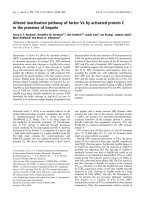
The structure of recombinant LOV2 domain of the
fern Adiantum capillus-veneris has been determined by
X-ray crystallography in the dark as well as the light
state [12,13] (Fig. 3). The protein is characterized by
five antiparallel b-sheets and four a-helices that form a
central pocket harboring the FMN chromophore. A
light-induced change in the position of the side chain
of cysteine 966 is well in line with the adduct forma-
tion [12–14]. In addition to the cysteine 966 residue,
11 amino acid residues were shown to contact the
FMN chromophore via hydrogen bonds or van der
Waals contacts. Notably, these residues are highly con-
served in all LOV domains, indicating a canonical
FMN binding motif (Fig. 2).
NMR studies with the LOV2 domain of Avena sati-
va showed that the photoadduct formation involves a
conformational change in the ribityl side chain of the
flavin cofactor as indicated by
31
P and
13
C NMR data
[10]. It was also shown that the adduct formation trig-
gers the unfolding of the helical domain Ja, which
AB
Fig. 1. Organization of phototropins used in the preent study. A, A. sativa NPH1-1 (accession no. O49003); B, A. capillus-veneris phy3
(accession no Q9ZWQ6).
Fig. 2. Amino acid sequence alignment of domains used in the present study. Amino acid residues derived from the vector are italicized.
Asterisks indicate amino acid residues of protein from A. capillus-veneris in direct contact with the FMN chromophore [13]. Identical amino
acid residues are shown in black, and similar amino acid residues are in grey shadow typeface. The formation of a cysteinyl-flavin-C(4a) cova-
lent adduct after irradiation of the LOV–FMN complex with blue light is shown and the cystein residue involved in adduct formation is
marked by an arrow.
W. Eisenreich et al.
13
C NMR of phototropin domains
FEBS Journal 274 (2007) 5876–5890 ª 2007 The Authors Journal compilation ª 2007 FEBS 5877
serves as a linker between the LOV2 domain and the
kinase domain in the LOV2 domain of A. sativa [11].
That unfolding is believed to modulate the activity of
the kinase domain, which is conducive to its autophos-
phorylation.
The exact role of the LOV1 domain in regulating
phototropin activity is not fully understood, but the
overall architecture of LOV1 from Chlamydomonas was
found to be almost identical with that of LOV2 [15].
Recent studies indicate that LOV2 acts as the principal
light sensing domain, which is coupled via the Ja helix
with the kinase activity [11], whereas LOV1 may play a
crucial role in receptor dimerization [16,17].
The present study was initiated in order to monitor
more closely the light-induced chemical shift modula-
tion of the FMN cofactors complexed to LOV
domains of different origins. For the unequivocal
assignment of the
13
C NMR signals of all carbon
atoms in the FMN chromophore, the apoproteins were
reconstituted with random and ordered
13
C isotopo-
logue libraries of FMN. The signal intensity modula-
tion reflecting the different isotopologue compositions
in samples with random isotopologue libraries of
FMN served as the basis for isotope abundance editing
of the
13
C NMR signals. The method can be adapted
for NMR signal assignment in a variety of other pro-
tein ⁄ ligand systems.
Results
We have reported earlier on the preparation of isoto-
pologue mixtures of 6,7-dimethyl-8-ribityllumazine (3;
Fig. 4) by in vivo biotransformation of
13
C-labeled glu-
cose using a recombinant Escherichia coli strain [4].
These isotopologue mixtures were used in the present
study as a starting material for the preparation of iso-
topologue mixtures of FMN by enzyme-assisted syn-
thesis.
The transformation of 3 into riboflavin catalyzed by
the enzyme riboflavin synthase proceeds as a dismuta-
tion whereby two equivalents of 3 are transformed into
one equivalent each of riboflavin (5; Fig. 4) and
5-amino-6-ribitylamino-2,4(1H,3H)-pyrimidinedione (4;
Fig. 4). In order to avoid the inherent loss of isotope-
labeled precursor, the second product 4 resulting from
the dismutation can be reconverted into 3 by treatment
with lumazine synthase using 3,4-dihydroxy-2-buta-
none 4-phosphate as cosubstrate. The cosubstrate can
be prepared in appropriately
13
C-labeled form by
enzymatic conversion of
13
C-labeled glucose. By that
approach, the yield of riboflavin based on isotope-
labeled 3 can be optimized (for details, see Experimen-
tal procedures).
The riboflavin arising by the in vitro biotransforma-
tion can be converted into FMN by treatment with
riboflavin kinase in situ; ATP required as kinase sub-
strate can be conveniently recycled using phosphoenol
pyruvate as phosphate donor. One-pot reaction mix-
tures catalyzing the formation of FMN from randomly
labeled 3 and specifically
13
C-labeled glucose comprise
nine enzyme catalysts and afford the product at a yield
of over 90% based on the
13
C-labeled 3 [3].
Synthetic genes specifying the LOV1 (Fig. 5) and
LOV2 domains of phototropin NPH1-1 from A. sativa
and the LOV2 domain of phototropin phy3 from the
fern A. capillus-veneris were constructed as described
A
B
Fig. 3. Adiantum phy3 LOV2 structures. (A) dark and (B) light state
(protein databank ID code 1G28, respectively, 1JNU) [12,13]. Atoms
in amino acid residues are colored by elements: carbon, white; oxy-
gen, red; nitrogen, blue; sulfur, yellow.
13
C NMR of phototropin domains W. Eisenreich et al.
5878 FEBS Journal 274 (2007) 5876–5890 ª 2007 The Authors Journal compilation ª 2007 FEBS
in the Experimental procedures. All genes were opti-
mized for hyperexpression in E. coli host strains. Gen-
erally, the assembled DNA fragments were cloned into
an expression vector specifying fusion proteins com-
prising hisactophilin from Dictyostelium discoideum
and a thrombin cleavage site. All sequences have been
deposited in GenBank. The cognate fusion proteins
were expressed efficiently in recombinant E. coli strains
and could be bound to nickel-chelating Sepharose due
to the large number of histidine residues present in the
hisactophilin domain. The column was washed with
buffer containing 8 m urea to release the protein-
bound FMN, and the resulting apoprotein was recon-
stituted on the column with isotope-labeled FMN. The
protein was then eluted with imidazole. The solution
was treated with thrombin and passed again through a
nickel-chelating column in order to remove the cleaved
hisactophilin domain that was bound, whereas the
LOV2 domains were not retained.
Figure 6 shows
13
C NMR signals of the recombi-
nant LOV2 domain from A. capillus-veneris (LOV2
fern
)
reconstituted with [U-
13
C
17
]FMN and with two isoto-
pologue mixtures of FMN obtained by biotransforma-
tion of [2-
13
C
1
]- or [3-
13
C
1
]glucose, respectively. The
left panel shows spectra that were acquired under dark
conditions. In the spectrum of protein reconstituted
with universally
13
C-labeled FMN (Fig. 6A), all signals
with the exception of C(2) appear as broadened multi-
plets due to
13
C
13
C coupling of directly adjacent
carbon atoms. In the samples reconstituted with the
isotopologue mixtures, the carbon signals of the bound
FMN appear as singlets, and their apparent intensities
vary over a wide range (Fig. 6B,C). This intensity vari-
ation is due to the presence of the singly
13
C-labeled
isotopologues at different abundances in the FMN iso-
topologue mixtures. The relative intensities of the indi-
vidual carbon signals observed in the protein sample
reflect the relative abundances of the different FMN
isotopologues (cf.
13
C enrichments of the FMN speci-
mens from the different
13
C-labeled glucoses indicated
by filled circles in Fig. 6) and constitute the basis for
unequivocal signal assignment.
Fig. 4. Synthesis of isotopologue libraries of FMN from [2-
13
C
1
]glucose. 1, Ribulose 5-phosphate; 2, 3,4-dihydroxy-2-butanone 4-phosphate;
3, 6,7-dimethyl-8-ribityllumazine; 4, 5-amino-6-ribitylamino-2,4(1H,3H)-pyrimdinedione; 5, riboflavin.
W. Eisenreich et al.
13
C NMR of phototropin domains
FEBS Journal 274 (2007) 5876–5890 ª 2007 The Authors Journal compilation ª 2007 FEBS 5879
For example, the position 8a methyl group, but not
the position 7a methyl group, is significantly labeled in
the sample of 3 obtained by biotransformation of
[2-
13
C
1
]glucose, and the signal detected at 23.2 p.p.m.
in the spectrum with the isotopologue library from
[2-
13
C
1
]glucose can be clearly assigned to C(8a)
(Fig. 6B). The C(7a) atom is not
13
C-enriched from
either [2-
13
C
1
]- or [3-
13
C
1
]glucose; therefore, no signal
Fig. 5. Construction of a synthetic gene for
A. sativa LOV1 domain. Alignment of the
wild-type DNA sequence (ASNPH1), and the
synthetic DNA sequence (ASLOV1-syn) with
5¢ and 3¢ overhangs including the synthetic
BglII and HindIII sites. Changed codons are
shaded in black. New single restriction sites
are shaded in grey. Oligonucleotides used
as forward primers are drawn above, and
reverse primers below, the aligned DNA
sequences.
Fig. 6.
13
C NMR signals of
13
C-labeled FMN complexed to LOV2 domain from A. capillus-veneris under dark conditions or under blue light
irradiation. A, [U-
13
C
17
]FMN; B, FMN obtained from [2-
13
C
1
]glucose; C, FMN obtained from [3-
13
C
1
]glucose. Asterisks indicate impurities.
13
C NMR of phototropin domains W. Eisenreich et al.
5880 FEBS Journal 274 (2007) 5876–5890 ª 2007 The Authors Journal compilation ª 2007 FEBS
can be detected in the
13
C NMR spectra of the corre-
sponding protein samples. On the other hand, a second
methyl signal (doublet with a coupling constant of
43 Hz) is observed at 21.9 p.p.m. in the spectrum with
[U-
13
C
17
]FMN as a cofactor. It is immediately obvious
that this signal has to be assigned to C(7a).
Due to the specific
13
C enrichments in the ribityl
moiety of the FMN samples, the signals for C(1¢),
C(2¢) and C(4¢) are observed in the isotopologue mix-
ture from [2-
13
C
1
]glucose, whereas only the signals for
C(2¢) and C(3¢) are detected in the spectrum of the iso-
topologue mixture from [3-
13
C
1
]glucose with higher
intensity of the C(2¢) signal. On this basis, all ribityl
signals can be unequivocally assigned (Table 1 and
Fig. 6).
Using the same isotopologue editing approach,
unequivocal signal assignments can be obtained for the
carbon atoms of the isoalloxazine ring. Thus, label
from [2-
13
C
1
]glucose is diverted to the ring carbon
atoms 4a, 5a, 6 and 8 with
13
C enrichments of
6 > 4a > 5a 8 (cf. filled circles in Fig. 6). The car-
bon atoms 4, 5a, 7, 8, 9a and 10a acquire
13
C label
from [3-
13
C
1
]glucose with enrichments in the order of
5a 8 > 10a > 4 > 9a 7. Indeed, in the signal
region for aromatic carbon atoms (115–165 p.p.m),
four signals were observed in the protein samples with
FMN from [2-
13
C
1
]glucose (Fig. 6B), and six signals
were detected with FMN from [3-
13
C
1
]glucose
(Fig. 6C). The signal intensities were found to vary in
the same pattern as determined for the free FMN iso-
topologue mixture, and thus provided the basis for the
assignments. Additional validation is provided by the
simultaneous detection of the signals for C(8), and
C(5a) in both samples because both molecular posi-
tions acquire
13
C enrichment from [2-
13
C
1
]glucose, as
well as from [3-
13
C
1
]glucose. Due to low
13
C enrich-
ments of C(9) in the used FMN libraries, no signal
should be detectable for that atom. However, a signal
for C(9) has to be present in the sample with
[U-
13
C
17
]FMN and was indeed observed at
118.9 p.p.m. (Fig. 6A). In summary, the observed sig-
nal intensities in the spectra with universally
13
C-
labeled FMN and two isotopologue libraries of FMN
(i.e. obtained from biotransformation of [2-
13
C
1
]- and
[3-
13
C
1
]glucose) allowed the assignments of all 17 car-
bon atoms of FMN. The results are summarized in
Table 2. The validity of the experimental approach
was confirmed by signal assignments using an ordered
library of
13
C-labeled FMN isotopologues. More spe-
cifically, we measured the
13
C NMR chemical shifts
of seven selectively
13
C-labeled FMN isotopologues
Table 1.
13
C abundance of FMN obtained from [2-
13
C
1
]glucose and
FMN obtained from [3-
13
C
1
]glucose bound to the LOV2 domain
from A. capillus-veneris under dark and light conditions. The corre-
sponding values with the isotopologue mixtures of free FMN are
given for comparison. On the basis of the low signal-to-noise ratios
of the NMR spectra, the errors can be estimated as ± 30% of a
given
13
C abundance value.
Carbon
position
13
C abundance (%)
[2-
13
C
1
]glucose [3-
13
C
1
]glucose
Free
FMN
LOV2-bound
FMN
Free FMN
LOV2-bound
FMN
Dark Light Dark Light
43131
c
+
b
4a 49 +
b
ND
a
5a 16 26 ND
a
87 70 +
b
687++
b
++
b
71626+
b
816+
b
+
b
87 ++
b
++
b
8a 87 ++
b
++
b
9a 16 +
b
+
b
10a 43 ++
b
+
b
1¢ 72 90 +
b
2¢ 30 27 ND
a
62 81 ++
b
3¢ 25 28 +
b
4¢ 34 34
c
+
b
a
Not determined due to signal overlapping.
b
Signal observed at high
(+) and very high intensity (+ +).
c
Reference value.
Table 2. NMR chemical shifts of TARF, free FMN and FMN bound
to LOV2 domain from A. capillus-veneris in dark and light condi-
tions.
FMN
atom
NMR chemical shifts (p.p.m.)
TARF
Free
FMN
LOV-2 bound FMN
Dark Light
2 154.4 159.8 159.4 159.2
4 159.2 63.7 161.3 165.9
4a 135.1 136.2 134.2 65.7
5a 134.5 136.4 136.2 130.1
6 132.5 131.8 133.1 119.1
7 137.2 140.4 138.9 136.0
7a 19.5 19.9 21.9 21.8
8 148.7 151.7 150.2 130.1
8a 21.6 22.2 23.2 22.2
9 115.8 118.3 118.9 119.6
9a 131.4 133.5 134.4 127.7
10a 150.8 152.1 150.8 156.7
1¢ 45.0 48.8 44.8 46.8
2¢ 70.2 70.7 68.1 66.7
3¢ 70.0 74.0 75.1 75.5
4¢ 69.5 73.1 72.9 73.0
5¢ 62.1 66.4 65.8 66.7
P 5.1 4.8 4.1
W. Eisenreich et al.
13
C NMR of phototropin domains
FEBS Journal 274 (2007) 5876–5890 ª 2007 The Authors Journal compilation ª 2007 FEBS 5881
bound to the LOV2 domain of A. capillus-veneris
(Fig. 7). The chemical shifts observed with these sam-
ples were in agreement with the signal assignments
made on the basis of the random isotopologue libraries
(Fig. 6).
The interpretation of the NMR chemical shifts of
protein-bound flavins is usually based on the compari-
son with the chemical shifts of free flavins [1]. There-
fore, the published assignments for free FMN [10]
were checked by the isotopologue abundance editing
method using labeled FMN obtained from [1-
13
C
1
]-
[2-
13
C
1
]- and [3-
13
C
1
]glucose. The previous assignments
[10] of the isoalloxazine ring carbons could be com-
pletely confirmed (Table 2 and Fig. 6). The previous,
tentative assignments of C(3¢) and C(4¢) [10] had to be
interchanged. All
13
C NMR assignments of tetra-
acetylriboflavin (TARF) were assigned by 2D
13
C inadequate experiments using [U-
13
C
17
]TARF. In
this case, the previous assignment for C(2¢ ) and C(4¢)
[18,19] had to be interchanged (Table 2).
The isotopologue editing method was then used to
assign the
13
C NMR signals of FMN bound to the
LOV domains under study under blue light irradiation
conditions. In order to keep photodamage of the pro-
tein as low as possible, the acquisition times were
somewhat reduced under blue light as compared to
dark conditions. As a consequence, the signal-to-noise
ratios of the NMR spectra of the irradiated samples
were lower than those of the corresponding spectra in
the dark (Fig. 6, right column). The signal assignments
obtained from the random isotopologue libraries
matched those from the selectively labeled FMN sam-
ples (Fig. 7 and supplementary Fig. S1). The results
are presented in Table 2 and confirm previous assign-
ments for LOV2 from A. sativa (LOV2
oat
) [10], except
that the chemical shifts due to C(7) and C(8), C(6) and
C(9), as well as those due to C(3¢) and C(4¢), have to
be interchanged.
In analogy to the above procedure, the chemical
shifts of the LOV1 domain from A. sativa (LOV1
oat
)
were also investigated. The results are given in supple-
mentary Tables S1 and S2 and supplementary Figs S4
and S5. In summary, the FMN signals of (LOV2
oat
)
and (LOV1
oat
) were detected at similar chemical shifts
(± 0.2 p.p.m), with the exception of the signals for
C(5¢) and C(7a), which were upfield shifted by
0.5 p.p.m and 0.7 p.p.m., respectively, and the signals
for C(2) and C(4), which were downfield shifted by
1.6 p.p.m and 0.7 p.p.m., respectively, in the LOV1
domain. A correlation diagram of the chemical shifts
for all proteins investigated in this study is shown in
Fig. 8.
Fig. 7.
13
C NMR signals of of
13
C-labeled
FMN with LOV2 domain from A. capillus-
veneris under dark conditions. A,
[U-
13
C
17
]FMN; B, [xylene-
13
C
8
]FMN; C,
[7a,9-
13
C
2
]FMN; D, [6,8a-
13
C
2
]FMN; E,
[4,10a-
13
C
2
]FMN; F, [7,9a-
13
C
2
]FMN; G,
[5a,8-
13
C
2
]FMN; H, [4a-
13
C
1
]FMN. Asterisks
indicate impurities.
13
C NMR of phototropin domains W. Eisenreich et al.
5882 FEBS Journal 274 (2007) 5876–5890 ª 2007 The Authors Journal compilation ª 2007 FEBS
Discussion
The approach described in the present study opens a
new way to unambiguously assign all carbon atoms in
the
13
C NMR spectra of protein-bound flavin cofac-
tors using no more than three FMN samples (i.e. a
uniformly labeled and two partially labeled flavin
samples that can be biosynthetically obtained by in vivo
biotransformation of [2-
13
C
1
]- and [3-
13
C
1
]glucose).
Since, in the latter two cases, the degree of
13
C enrich-
ment of a given carbon atom in the flavin samples dif-
fers, the
13
C NMR signal strength (amplitude) of a
given carbon atom provides an additional constraint in
the signal assignment procedure. By contrast to previ-
ous NMR work on flavoproteins [18], where various
iosotopologues, selectively enriched in the xylene
moiety of flavin, also were used [20], the isotopologues
obtained by the new biosynthetic approach allow
assignment of the carbon atoms of the ribityl side
chain in the NMR spectra of flavin. The
13
C chemical
shifts of these atoms can provide important informa-
tion about the binding interaction between the hydro-
xyl groups of the side chain of flavin and the
apoprotein, and could also report possible conforma-
tional changes of the side chain (e.g. due to reduction
of a flavoprotein).
In supplementary Table S2, the
13
C chemical shifts
of LOV1 domain of A. sativa in the two states are
listed. In the dark state, the chemical shifts of the iso-
alloxazine moiety of flavin are very similar to those
observed with LOV2 of the same species and of dif-
ferent organisms (Fig. 8). Most of the differences
(± 0.3 p.p.m) between the two sets are within the
accuracy limits of chemical shift determination, except
for C(8) of LOV2
fern
, which is upfield shifted by
0.6 p.p.m., and C(8a) of LOV2
fern
, which is downfield
shifted by 0.7 p.p.m., respectively, compared to
LOV1
oat
and LOV2
oat
. The chemical shifts of the side
chain carbon atoms 1¢ and 3¢ of LOV2
fern
show signifi-
cant differences, which may be ascribed to variation in
the strength of the hydrogen bond of the correspond-
ing hydroxyl groups with the proteins and ⁄ or to con-
formational changes in the side chain (Fig. 8). A
similar effect is shown by the proteins in the blue light
irradiated state. The greatest difference in chemical
shifts is observed for C(2) of LOV1
oat
, which is down-
field shifted by 1.6 p.p.m. compared to the LOV2
molecules. Similarly, the C(4) and C(7a) of LOV1
oat
are downfield shifted by 0.7 p.p.m and upfield shifted
by 0.5 p.p.m., respectively, compared to LOV2
oat
and
LOV2
fern
. A significant difference is also observed for
C(9) and C(1¢) of LOV2
fern,
which are upfield shifted
by 0.9 and 0.6 p.p.m., respectively, and C(2¢), which is
downfield shifted by 0.7 p.p.m., compared to LOV1
oat
and LOV2
oat
.
Based on extensive
13
C and
15
N NMR studies on
free flavins in aprotic and protic media [21], which
have shown that a direct correlation exists between the
p-electron density and the
13
C chemical shift of a
particular atom of the flavin molecule, the observed
Fig. 8.
13
C Chemical shifts of
13
C-labeled FMN in complex with
LOV domains: black lines, dark conditions; blue lines, irradiated
with blue light.
W. Eisenreich et al.
13
C NMR of phototropin domains
FEBS Journal 274 (2007) 5876–5890 ª 2007 The Authors Journal compilation ª 2007 FEBS 5883
chemical shifts can be interpreted in terms of the elec-
tronic structure of the protein-bound flavin and its
perturbation by binding interaction and chemical reac-
tions. Thus, the dark state interaction between FMN
and the LOV domains under study is characterized by
strong hydrogen bonding with the C(2)O group of
flavin. The strength of the hydrogen bond corresponds
approximately to that of free FMN in water, indi-
cating polarization of the flavin along the axis
C(8)-C(6)-C(9a)-N(5)-C(10a)-C(2). This is manifested
by the observed downfield shifts of the corresponding
C atoms (Table 2 and supplementary Table S2).
Although there exists a hydrogen bond between the
protein and the flavin at C(4)O group, its strength is
considerably weaker than that observed in free FMN
in water. These observations are in good agreement
with recent X-ray data showing a distance of 0.31 nm
between the N
d
group of N998 and the oxygen atom
of the C(2)O group (Table 3). To the C(4)O group,
two hydrogen bonds (N
e
group of Q1029, N
d
group of
N1008) have been suggested by X-ray data, yet the
distance between the bond-forming atoms is larger
than that observed at C(2)O, supporting our inter-
pretation. A strong hydrogen bond to C(4)O would
have influenced the chemical shift of the C(4a) atom
by a downfield shift compared to that of FMN [10].
The even slightly upfield shifted resonance of the
C(4a) atom in comparison to TARF indicates extra
p-electron density allocation to this position, released
from the N(10) atom, which is downfield shifted
compared to TARF, as shown previously [10]. The
resonance position of C(4a) is thus in full agreement
with the weak hydrogen bond observed at C(4)O. The
partial positive charge created on N(10) [21] by the
release of electron density onto C(4a) is distributed
mainly onto the C(5a) and the C(9) atom, and, to a
lesser extent, onto the C(7) atom, in agreement with
the fact that the latter atom experiences a smaller
downfield shift than the other two atoms. The data
demonstrate that, with regard to the isoalloxazine
moiety of flavin, there are only minor electronic differ-
ences of the prosthetic group of the different proteins
investigated in the present study. Taking also into
account the previously published
15
N NMR data on
LOV2 [10], it can be concluded that the chemical shift
of the N(5) atom of flavin indicates no hydrogen bond
formation with this atom and a rather hydrophobic
environment at this site. This interpretation of the
NMR data is fully supported by the X-ray data [22].
On the other hand, the chemical shift of the N(1) atom
indicates the presence of a strong hydrogen bond at
this site, although the X-ray data suggest the absence
of a hydrogen bond. The opposite holds for the N(3)
atom: the X-ray data propose a hydrogen bond with
the protein whereas the
15
N NMR data suggest the
absence of such a bond.
The chemical shifts of the C(3¢) and C(4¢) atoms
of the side chain of protein-bound flavin resemble
Table 3. Distances between FMN atoms (Ligand) and amino acid residues of LOV2 from Adiantum phy3 (chimeric fern photopreceptor)
[12,13]. For comparison,
13
C chemical shifts of bound FMN atoms are given.
Atom Dark state Light state
Ligand Protein
Distance
(A
˚
)
13
C NMR chemical
shifts (p.p.m.)
Distance
(A
˚
)
13
C NMR chemical
shifts (p.p.m.)
C(4a) S
c
[C(966)] 4.2 134.2 1.8 65.7
O
e
[Q(1029)] 4.7 4.0
O(4) N
e
[Q(1029)] 3.5 161.3 [C(4)] 3.1 165.9 [C(4)]
N
d
[N(1008)] 3.4 3.4
N(3) O
d
[N(998)] 2.8 3.0
O(2) N
d
[N(998)] 3.1 159.4 [C(2)] 3.3 159.2 [C(2)]
N(5) C
e
[F(1010)] 3.2 136.2 [C(5a)] 3.8 130.1 [C(5a)]
O
d
[N(965)] 2.7 2.9
O(2¢)N
d
[N(965)] 3.9 68.1 [C(2¢)] 3.9 66.7 [C(2¢)]
O(H
2
O-2) 3.4 3.4
O(H
2
O-1) 3.6 3.6
O(3¢)O(H
2
O-1) 3.1 75.1 [C(3¢)] 3.1 75.5 [C(3¢)]
O(4¢)O
e
(Q970) 3.3 72.9 [C(4¢)] 3.7 73.0 [C(4¢)]
N
e
(Q970) 3.2 3.0
O(1) (on P) N
e
[R(967)] 2.6 2.5
O(2) (on P) N
g
[R(967)] 2.6 4.8 (P) 2.7 4.1 (P)
O(3) (on P) N
g
[R(983)] 2.8 3.0
O(3) (on P) N
e
[R(983)] 2.6 2.7
13
C NMR of phototropin domains W. Eisenreich et al.
5884 FEBS Journal 274 (2007) 5876–5890 ª 2007 The Authors Journal compilation ª 2007 FEBS
those of FMN in water, indicating stronger hydrogen
bonding interactions with the hydroxyl group at C(3¢)
and a somewhat weaker one with that at C(4¢)
compared to those observed in FMN. This hydrogen
bonding pattern agrees with that observed by X-ray
crystallography [22]. The resonance position due to
C(1¢) reflects the increased sp
2
hybridization of N(10)
[21]. Both C(2¢) and C(5¢) are upfield shifted by more
than 1 p.p.m. compared to those of FMN. Whereas
the X-ray data indicate no hydrogen bond between the
C(5¢)O group and the protein, but a strong one at
C(2¢)O, the NMR data do not indicate hydrogen
bonding at these atoms. It is suggested that the appar-
ent absence of a hydrogen bond at C(2¢)O, as revealed
by
13
C NMR, may be masked by a counteracting
factor, most probably a conformational change.
Upon blue light irradiation of the proteins, rather
drastic changes are observed in the NMR spectra [10].
The most obvious one is the large upfield shift
()68.5 p.p.m) of the C(4a) resonance. This proves the
conversion of the C(4a) atom from sp
2
to sp
3
hybrid-
ization, in line with the formation of a covalent bond
between this atom of flavin, and the sulfur atom of
C966 in LOV2 of A. capillus-veneris (Fig. 2). The reso-
nance line of N(5) also undergoes a large upfield shift
()283 p.p.m) [10], indicating the change from an aro-
matic to an aliphatic nitrogen atom. The other carbon
atoms of flavin most affected by conversion of the pro-
tein by light are: C(8) ()19.9 p.p.m), C(6) ()14.0 pm),
C(9a) ()6.7 p.p.m) and C(10a) (+ 5.9 p.p.m). All these
atoms are involved in the possible mesomeric struc-
tures of oxidized flavin [21] that are disturbed by the
C(4a) substitution. The upfield shifts of the resonances
of these atoms, with the exception of C(10a), which
shows a downfield shifted signal, demonstrates the
allocation of the incoming electron density at these
positions. The downfield shift of the resonance line
due to C(10a) is caused by the higher electron density
withdrawal from this atom by the further polarization
of the C(2)O group compared to that of the molecule
under dark conditions. Since the sp
2
hybridization of
the N(10) atom increases considerably upon formation
of the C(4a) adduct [10], the upfield shifts of the reso-
nances of C(5a) ()6.1 p.p.m) and C(7) ()2.9 p.p.m)
atoms can be ascribed to electron density release onto
these atoms from N(10). Overall, with regard to the
chemical shifts of the carbon atoms of the isoalloxa-
zine ring, the electronic structure of the different pro-
teins investigated in the present study is very similar, if
not almost identical. Only the chemical shifts of the
C(2), C(4) and C(4a) atoms of the adduct of LOV1
and LOV2 proteins differ considerably from each
other. The chemical shifts of the former protein are
downfield from those due to LOV2, indicating stronger
hydrogen bonding in LOV1 than in LOV2 at these
positions. The hydrogen bond pattern as observed by
NMR of the oxidized proteins is also observed in the
adduct forms. The hydrogen bond at N(3), as observed
by X-ray, is now also evident in the NMR data.
Whereas the chemical shifts of the C(4a) signals in
LOV2
oat
and LOV1
oat
are very similar, the corre-
sponding signal in LOV2
fern
is upfield shifted with
respect to the former. This observation suggests some
structural difference(s) at the C(4a) position between
the proteins from oat and that from fern.
The hydrogen bonding pattern observed for the
C(3¢) and the C(4¢) atoms of the ribityl side chain in
the oxidized proteins is also observed in the corre-
sponding adducts, but the strength of hydrogen bond-
ing with the C(3¢)O group is considerably increased,
especially that in LOV2
fern
. With regard to C(5¢) atom,
the LOV2 proteins exhibit similar chemical shifts for
this atom, whereas that of LOV1 is upfield shifted by
0.5 p.p.m. The chemical shift for the C(2¢) atom
increases in the order: LOV2
oat
, LOV1
oat
and LOV2-
fern
, possibly reflecting variations in the strength of
hydrogen bonding interactions at this position.
The
13
C NMR data show that there exist some subtle
differences between the proteins investigated in the pres-
ent study, as far as the isoalloxazine moiety of the flavin
in the resting and photo-adduct state is concerned. The
largest difference among the three proteins is observed
for the resonance line of the C(2)O group (downfield
shift) of LOV1 in the photo-adduct state. However,
there are some resonances of ribityl side chain carbon
atoms, which differ to a greater extent among the three
proteins (Fig. 8), indicating variations in the interaction
with the proteins and ⁄ or conformational differences.
The LOV1 and LOV2 domains from A. sativa have
also been investigated by optical (light absorption, fluo-
rescence, CD) and chemical techniques (kinetics) [9,23].
The light absorption and
13
C NMR data indicate a
hydrophobic environment at the isoalloxazine moiety of
the protein-bound flavin, whereas the fluorescence quan-
tum yield of flavin bound to the two proteins reflect
probably the sequence difference between the two pro-
teins in the neighborhood of the flavin (residue 1010 is
phenylalanine in LOV2 of A. capillus-veneris and the
positionally equivalent residue in LOV1 of A. sativa is
leucine 117). From these data, it can be concluded that
the microenvironment of flavin in LOV2 is more hydro-
phobic than that in LOV1. This property of the proteins
appears to be correlated with the kinetic data, where it
has been determined that LOV2 is more reactive to form
the photo-adduct than LOV1 and its photo-adduct
is more stable than that of LOV1 [9]. Moreover, the
W. Eisenreich et al.
13
C NMR of phototropin domains
FEBS Journal 274 (2007) 5876–5890 ª 2007 The Authors Journal compilation ª 2007 FEBS 5885
light-driven formation of the adduct is dependent on
light intensity, whereas the dissociation reaction of the
adduct into its constituents is dependent on temperature
[9]. In addition, the active site of the proteins, especially
LOV2, is practically inaccessible to external molecules
(e.g. dithionite). From this, it is obvious that the active
site of these flavoproteins differs considerably from
those of other flavoproteins in that the former cannot be
reduced by external agents. These facts demonstrate the
specific tuning of these proteins for only one particular
reaction, the photochemically driven, reversible reaction
to form an intermediate adduct between the flavin mole-
cule and a specific component of the associated protein.
Experimental procedures
Materials
Restriction enzymes were obtained from New England Bio-
labs (Schwalbach, Germany). T4 DNA ligase was from
Gibco BRL (Karlsruhe, Germany). EXT DNA Polymerase
and Taq polymerase were from Finnzymes (Epsoo,
Finland). Oligonucleotides were synthesized by Thermo
Electron GmbH (Ulm, Germany). A Plasmid Miniprep Kit
from PEQLab (Erlangen, Germany) was used for plasmid
DNA isolation and purification. DNA fragments and PCR
amplificates were purified with a Gel Extraction Kit or Cycle
Pure Kit from PEQLab [7a,9-
13
C
2
]-, [4a-
13
C
1
]-, [5a,8-
13
C
2
]-,
[7,9a-
13
C
2
]-, [4,10a-
13
C
2
]-, [6,8a-
13
C
2
]- and [xylene-
13
C
8
]ribo-
flavin were synthesized as described previously [2,3].
[U-
13
C
17
]riboflavin was prepared by fermentation of
Bacillus subtilis strain RB50PRF6 with [U-
13
C
6
]glucose as
carbon source [24]. Random libraries of isotopologues of
6,7-dimethyl-8-ribityllumazine were prepared as described
previously [4]. Recombinant riboflavin kinase from Schizo-
saccharomyces pombe, 3,4-dihydroxy-2-butanone 4-phos-
phate synthase, riboflavin synthase from E. coli and
6,7-dimethyl-8-ribityllumazine synthase from B. subtilis were
prepared as described previously [25–28]. Thrombin from
bovine plasma, hexokinase from yeast, pyruvate kinase
from rabbit muscle, glucose 6-phosphate dehydrogenase
from Leuconostoc mesenteroides, glutamate dehydro-
genase from bovine liver, and 6-phosphogluconate dehydro-
genase from B. subtilis were purchased from Sigma-Aldrich
(Taufkirchen, Germany). The transformation of riboflavin
into TARF was performed in accordance with a previously
described procedure [29].
Preparation of random isotopologue libraries
of FMN
A solution (total volume, 20 mL) containing 100 mm Tris,
pH 8, 10 mm MgCl
2
, 3 mg of [2-
13
C
1
]- or [3-
13
C
1
]glucose,
3.5 mg of phosphoenol pyruvate, 0.5 mg of ATP, 6 mg of
2-oxoglutaric acid, 0.6 mg of NAD
+
, 2.5 mg of NH
4
Cl,
5mgofa
13
C-isotopologue mixture of 6,7-dimethyl-8-ribi-
tyllumazine obtained by biotransformation of [2-
13
C
1
]- or
[3-
13
C
1
]glucose, 5 units of hexokinase, 5 units of pyruvate
kinase, 5 units of glucose 6-phosphate dehydrogenase,
5 units of glutamate dehydrogenase, 5 units of 6-phospho-
gluconate dehydrogenase, 2 mg of 3,4-dihydroxy-2-buta-
none 4-phosphate synthase, 2 mg of lumazine synthase and
4 mg of riboflavin synthase was incubated at 37 ° C for 4 h.
Riboflavin kinase (0.5 mg), pyruvate kinase (5 units) and
phosphoenol pyruvate (3.5 mg) were added, and the reac-
tion mixture was incubated at 37 °C. The reaction was
monitored by TLC [Cellulose F, Merck AG, Darmstadt,
Germany; eluent, 3% (w ⁄ v) ammonium chloride]. Typi-
cally, reactions were completed after approximately 16 h.
Strains and plasmids
Bacterial strains and plasmids used in this study are
summarized in Table 4. Bacteria were grown at 37 °Cin
LB medium. Ampicillin (170 mgÆL
)1
) and kanamycin
(15 mgÆL
)1
) were added as required.
Preparation of a synthetic gene coding for
A. sativa phototropin NPH1-1 LOV1 domain
The partially complementary oligonucleotides ASLOV1-1
and ASLOV1-2 were annealed and treated with a mixture
of deoxyoligonucleotide triphosphates and DNA polymer-
ase. The resulting 112 bp segment was elongated by a series
of four PCR amplifications using pairwise combinations
of oligonucleotides according to Table 5 (oligonucleotides
ASLOV1-3 ⁄ ASLOV1-4, ASLOV1-5 ⁄ ASLOV1-6, ASLOV1-
7 ⁄ ASLOV1-8 and ASLOV1-9 ⁄ ASLOV1-8) (Fig. 5). The
resulting 376 bp DNA fragment was digested with BglII
and HindIII and ligated into the plasmid pNCO113-hisacto-
philin(C49S mutant), which had been treated with BamHI
and HindIII affording the expression plasmid pNCO-HIS-
ACT-ASLOV1-syn (Table 4).
Preparation of a synthetic gene coding for
A. sativa phototropin NPH1-1 LOV2 domain
The ORF coding for the LOV2 domain of A. sativa was con-
structed in analogy to the gene for LOV1 described above.
The synthesis was performed using pairwise combinations of
oligonucleotides given in Table 5 (ASLOV2-1 ⁄ ASLOV2-2,
ASLOV2-3 ⁄ ASLOV2-4, ASLOV2-5 ⁄ ASLOV2-6, ASLOV2-7 ⁄
ASLOV2-8, ASLOV2-9 ⁄ ASLOV2-10 and ASLOV2-9 ⁄
ASLOV2-12). The resulting 501 bp DNA fragment was
digested with BamHI and HindIII and ligated into the plas-
mid pNCO113-hisactophilin(C49S mutant), which had been
treated with the same restriction endonucleases affording the
expression plasmid pNCO-HISACT-ASLOV2-syn (Table 4).
13
C NMR of phototropin domains W. Eisenreich et al.
5886 FEBS Journal 274 (2007) 5876–5890 ª 2007 The Authors Journal compilation ª 2007 FEBS
Preparation of a synthetic gene coding for
A. capillus-veneris phototropin PHY3 LOV2
domain
The ORF coding for the LOV2 domain of A. capillus-vene-
ris was constructed in analogy to the gene for LOV1
described above. The synthesis was performed using pair-
wise combinations of oligonucleotides given in Table 5
(ACVLOV2-1 ⁄ ACVLOV2-2, ACVLOV2-3 ⁄ ACVLOV2-4,
ACVLOV2-5 ⁄ ACVLOV2-6 and ACVLOV2-7 ⁄ ACVLOV2-
8). The resulting 369 bp DNA fragment was digested
with BamHI and HindIII and ligated into the plasmid
pNCO113-hisactophilin [32], which had been treated with
the same restriction endonucleases affording the expression
plasmid pNCO-HISACT-ACVLOV2-syn (Table 4).
Transformation of bacterial cells
Ligation mixtures were transformed into E. coli XL1-Blue
cells. Transformants were selected on LB solid medium sup-
plemented with ampicillin (170 mgÆL
)1
). The plasmids were
reisolated and were analyzed by restriction analysis and by
DNA sequencing. Sequenced expression plasmids were
transformed into E. coli M15[pREP4] cells carrying the
pREP4 repressor plasmid for the overexpression of lac
repressor protein. Kanamycin (15 mgÆ L
)1
) and ampicillin
(170 mgÆL
)1
) were added to secure the maintenance of both
plasmids in the host strain.
Bacterial culture
Bacterial strains and plasmids used in this study are sum-
marized in Table 4. E. coli strains M15[pREP4]-pNCO-
HISACT-ASLOV1-syn, M15[pREP4]-pNCO-HISACT-
ASLOV2-syn and M15[pREP4]-pNCO-HISACT-ACV-
LOV2-syn were grown to an attenuance of 0.7 at D
600 nm
in LB medium supplemented with ampicillin (170 mgÆL
)1
)
and kanamycin (15 mgÆL
)1
). Isopropyl thio-b-d-galacto-
side was added to a final concentration of 1 mm and
incubation was continued with shaking at 30 °C over-
night. The cells were harvested by centrifugation and
stored at )20 °C.
Protein isolation
Frozen bacterial cell mass (10 g) was thawed in 20 mL of
50 mm sodium phosphate, pH 8, containing 300 mm NaCl,
10 mm imidazole and 0.02% sodium azide. The mixture
was ultrasonically treated and centrifuged. The supernatant
was passed through a column of nickel-chelating Sepharose
(1.5 · 7 cm), which had been equilibrated with 50 mm
sodium phosphate, pH 8, containing 300 mm NaCl, 10 mm
imidazole and 0.02% sodium azide (buffer A). The column
was washed with 75 mL of buffer A containing 8 m urea
and subsequently with 20 mL of buffer A containing
220 lm
13
C-labeled FMN. The reconstituted protein was
eluted with 50 mm sodium phosphate, pH 8, containing
300 mm NaCl and 1 m imidazole.
Thrombin cleavage
The combined fractions from the nickel-chelating Sepharose
column were desalted and transferred to a buffer containing
50 mm Tris hydrochloride, pH 7.5, 200 mm KCl and
2.5 mm CaCl
2
using a HiPrep 26 ⁄ 10 Desalting column
Table 4. Bacterial strains and plasmids used in the present study.
Strain ⁄ plasmid Relevant characteristics
Accession
numbers Reference
E. coli strains
XL1-Blue recA1, endA1, gyrA96, thi-1, hsdR17, supE44,
relA1, lac[F¢, proAB, lacI
q
ZDM15, Tn10(tet
r
)]
[30]
M15[pREP4] lac, ara, gal, mtl, recA
+
, uvr
+
, Str
R
(pREP4: Kan
R
, lacI) [31]
Expression plasmids
pNCO113-hisactophilin pNCO113 vector with the gene coding for hisactophilin
of D. discoideum with 3¢-BamHI and HindIII cloning sites
AF544403 [32]
pNCO113-hisactophilin
(C49S mutant)
pNCO113 vector with a gene coding for a cystein-free mutant
of hisactophilin of D. discoideum and a thrombin cleavage
site with 3¢-BamHI and HindIII cloning sites
EF469737 Present study
pNCO-HISACT-ASLOV1-syn pNCO-HISACT-(C49S)-BH vector with the LOV1 domain
(amino acids 130–244) of phototropin NPH1-1 of A. sativa
EF493212 Present study
pNCO-HISACT-ASLOV2-syn pNCO-HISACT-(C49S)-BH vector with the LOV2 domain
(amino acids 404–559) of phototropin NPH1-1 of A. sativa
EF493211 Present study
pNCO-HISACT-ACVLOV2-syn pNCO-HISACT-BH vector with the LOV2 domain
(amino acids 925–1032) of phototropin PHY3 of
A. capillus-veneris
EF493213 Present study
pRFN4 Vector containing the rib-operon of B. subtilis AY386222 [4]
W. Eisenreich et al.
13
C NMR of phototropin domains
FEBS Journal 274 (2007) 5876–5890 ª 2007 The Authors Journal compilation ª 2007 FEBS 5887
(2.6 · 10 cm; Amersham Biosciences, Little Chalfont, UK).
Thrombin (5 units per mg of protein) was added. The mix-
ture was incubated at room temperature for 16 h and was
then placed on a nickel-chelating Sepharose column
(1.5 · 7 cm), which was developed with buffer A. Yellow
fluorescent fractions were combined and concentrated by
ultrafiltration to a volume of 8 mL. The protein was trans-
ferred to 25 mm sodium ⁄ potassium phosphate, pH 7, using
a HiPrep 26 ⁄ 10 Desalting column (10 · 2.6 cm). The solu-
tion was concentrated to a volume of 0.5 mL by ultrafiltra-
tion. The protein yield was 5 mg (A. sativa LOV1 and
LOV2) or 2.5 mg (A. capillus-veneris LOV2) per 1 g of wet
cell pellet. Essentially all unlabeled FMN could be removed
from the protein using the described procedure. According
to the FMN concentration determined from absorption of
the LOV samples at 470 nm, the efficiency of protein recon-
stitution with labeled FMN was more than 90% (mol per
mol).
NMR spectroscopy
1
H-decoupled
13
C NMR spectra were recorded at 17 °C
with a 500 MHz spectrometer from Bruker Instruments
(Karlsruhe, Germany). The repetition time was 3.2 s and
the overall experimental time was 5–15 h for a single spec-
trum. Samples contained 25 mm sodium ⁄ potassium phos-
phate, pH 7, and 10 mg of recombinant LOV2 or LOV1
domain per mL. The samples were continuously irradiated
with blue light emitted by a photodiode (455 nm, 175 mW;
Luxeon Star ⁄ O Batwing, Lumileds Lighting, San Jose, CA,
USA) using an optical fibre where indicated. 2D inade-
quate experiments were performed according to standard
Bruker software (xwinnmr).
Determination of
13
C enrichments
Whenever possible,
13
C enrichments were determined by
quantitative NMR spectroscopy. For this purpose,
13
C NMR spectra of LOV proteins in complex with FMN
obtained by biotransformation of selectively
13
C-labeled
glucose and universally
13
C-labeled glucose, respectively,
were measured under the same experimental conditions.
The ratios of the signal integrals were then calculated for
each respective carbon atom. Relative
13
C abundances were
normalized to the known abundance for particular carbon
atoms of the free ligand. In poor quality spectra,
Table 5. Oligonucleotides used for the construction of the synthetic genes coding for A. sativa LOV1 and LOV2 domains, and A. capillus-
veneris LOV2 domain. Recognition sites used for cloning are underlined.
Designation Sequence (5¢ to 3¢)
ASLOV1-1 TTCCTCCAAGGTTCCGGCACGGATCCAGCTGAGATTGCCAAGATCCGTCAGGCTCTGGCAAATGGTTCGAAC
ASLOV1-2 GCGGTACCGTCTTTCTTGTAGTTGAGGACACGGCCGCAGTAGTTCGAACCATTTGCCAGAGCCTGACGGATC
ASLOV1-3 CAACATGACCGGTTACACATCCAAGGAAGTGGTAGGTCGTAACTGTCGTTTCCTCCAAGGTTCCGGCACGGATC
ASLOV1-4 CTTCATCCTTGATTGGTGCAATGGTCAGGAGATTCCAGAATGCGGTACCGTCTTTCTTGTAGTTG
ASLOV1-5 CCAGGTCACCCAATCATGTACGCAAGCGCTGGTTTCTTCAACATGACCGGTTACACATCCAAG
ASLOV1-6 ACTTACTTCCACTTGCATGCCGATGAACTTGAGGACACGACCTTCTTCATCCTTGATTGGTGCAATGG
ASLOV1-7 GCACTGTCCGCATTCCAACAGACCTTCGTAGTTTCGGACGCCAGCCGTCCAGGTCACCCAATCATGTACGCAAG
ASLOV1-8 ATTATTAT
AAGCTTATTCAGTGTATTTACTTACTTCCACTTGCATGCCGATGAA
ASLOV1-9 ATAATAATA
AGATCTGCACTGTCCGCATTCCAACAGACCTTC
ASLOV2-1 GCGCAAAATTCGTGATGCCATCGATAACCAAACAGAGGTCACTGTACAGCTGATTAATTATACAAAG
ASLOV2-2 AGGCTGCAAGTGAAAGAGGTTCCAGAACTTTTTACCACTCTTTGTATAATTAATCAGCTGTACAGTGAC
ASLOV2-3 AACTGCCGTTTTCTTCAAGGTCCTGAAACCGATCGCGCGACAGTGCGCAAAATTCGTGATGCCATCGATAAC
ASLOV2-4 GGACACCAATAAAGTACTGGACATCACCCTTCTGATCACGCATAGGCTGCAAGTGAAAGAGGTTCCAG
ASLOV2-5 AGTTTCTTGCAGTTGACAGAATATTCGCGAGAAGAAATTCTGGGTCGTAACTGCCGTTTTCTTCAAGGTCC
ASLOV2-6 CACGCTCGGCCGCATCACGGACATGTTCGGTACCATCCAACTGGACACCAATAAAGTACTGGACATC
ASLOV2-7 GTCATTACTGACCCACGTTTGCCAGATAATCCCATTATCTTCGCGTCCGATAGTTTCTTGCAGTTGACAGAATATTC
ASLOV2-8 CATCAATATTTTCTGCAGTTTTCTTAATCAGCATGACACCCTCACGCTCGGCCGCATCACGGACATG
ASLOV2-9 ATAATA
GGATCCGAATTTCTTGCTACTACACTTGAACGTATTGAGAAGAACTTTGTCATTACTGACCCACGTTTGCCAG
ASLOV2-10 CCTCTGGACGCAGATTAGCATCTGGAAGTTCTTTTGCCGCCTCATCAATATTTTCTGCAGTTTTCTTAATC
ASLOV2-12 TATTATTAT
AAGCTTAGTTAGCCCACAAATCCTCTGGACGCAGATTAGCATCTGG
ACVLOV2-1 GAGGAAGTCCTAGGTAACAACTGCCGTTTCCTGCAGGGCCGCGGTACTGATCGTAAAGCAGTGCAG
ACVLOV2-2 GACATCGCGCTGCTCCTTGACTGCATCACGGATCAGCTGCACTGCTTTACGATCAGTACCGCGGCC
ACVLOV2-3 CATCTTCGCGAGTGACCGGTTTCTGGAGCTCACGGAGTATACACGTGAGGAAGTCCTAGGTAACAACTGC
ACVLOV2-4 CCAAAAGGCGCGCCCACCTTTTGTATAGTTTAAAACCTGTACAGTGACATCGCGCTGCTCCTTGACTGC
ACVLOV2-5 AAGTCTTTCGTGATCACAGATCCTCGTTTACCAGACAACCCTATCATCTTCGCGAGTGACCGGTTTCTG
ACVLOV2-6 GGACGTCGCCATTTTCATCACGCATGACTTGAAGATGGAAGAGATTCCAAAAGGCGCGCCCACCTTTTG
ACVLOV2-7 ATAATA
GGATCCGGTCTGGTACCACGCGGTGAGCGTATCGGTAAGTCTTTCGTGATCACAGATCCTC
ACVLOV2-8 TATTATTAT
AAGCTTACATCTCCTGCTGAACTCCGATGAAATATTGGACGTCGCCATTTTCATCACGCATG
13
C NMR of phototropin domains W. Eisenreich et al.
5888 FEBS Journal 274 (2007) 5876–5890 ª 2007 The Authors Journal compilation ª 2007 FEBS
13
C enrichments were estimated from the signal heights
(Table 1 and supplementary Table S1).
Acknowledgements
This work was supported by grants from the Deutsche
Forschungsgemeinschaft (SFB533, TP A5), the Fonds
der Chemischen Industrie and the Hans-Fischer-Gesell-
schaft e.V.
References
1Mu
¨
ller F (1992) Nuclear magnetic resonance studies on
flavoproteins. Chemistry and Biochemistry of Flavopro-
teins (Mu
¨
ller F, ed.), pp. 557–595. CRC Press, Boca
Raton, FL.
2 van Schagen CG & Mu
¨
ller F (1981) A
13
C nuclear-mag-
netic-resonance study on free flavins and Megasphaera
elsdenii and Azotobacter vinelandii flavodoxin.
13
C-
enriched flavins as probes for the study of flavoprotein
active sites. Eur J Biochem 120, 33–39.
3Ro
¨
misch W, Eisenreich W, Richter G & Bacher A
(2002) Rapid one-pot synthesis of riboflavin isotopo-
mers. J Org Chem 67, 8890–8894.
4 Illarionov B, Fischer M, Lee CY, Bacher A & Eisenr-
eich W (2004) Rapid preparation of isotopolog libraries
by in vivo transformation of
13
C-glucose. Studies on
6,7-dimethyl-8-ribityllumazine, a biosynthetic precursor
of vitamin B
2
. J Org Chem 69, 5588–5594.
5 Otto MK, Jayaram M, Hamilton RM & Delbru
¨
ck M
(1981) Replacement of riboflavin by an analog in the
blue-light photoreceptor of phycomyces. Proc Natl Acad
Sci USA 78, 266–269.
6 Zhulin IB, Taylor BL & Dixon R (1997) PAS domain
S-boxes in Archaea, Bacteria and sensors for oxygen
and redox. Biochem Sci 22, 331–333.
7 Huala E, Oeller PW, Liscum E, Han IS, Larsen E &
Briggs WR (1997) Arabidopsis NPH1: a protein kinase
with a putative redox-sensing domain. Science 278,
2120–2123.
8 Taylor BL & Zhulin IB (1999) PAS domains: internal
sensors of oxygen, redox potential, and light. Microbiol
Mol Biol Rev 63, 479–506.
9 Salomon M, Christie JM, Knieb E, Lempert U & Briggs
WR (2000) Photochemical and mutational analysis of
the FMN-binding domains of the plant blue light recep-
tor, phototropin. Biochemistry 39, 9401–9410.
10 Salomon M, Eisenreich W, Du
¨
rr H, Schleicher E, Knieb
E, Massey V, Ru
¨
diger W, Mu
¨
ller F, Bacher A & Rich-
ter G (2001) An optomechanical transducer in the blue
light receptor phototropin from Avena sativa. Proc Natl
Acad Sci USA 98, 12357–12361.
11 Harper SM, Christie JM & Gardner KH (2004)
Disruption of the LOV-J alpha helix interaction
activates phototropin kinase activity. Biochemistry 43,
16184–16192.
12 Crosson S & Moffat K (2002) Photoexcited structure of
a plant photoreceptor domain reveals a light-driven
molecular switch. Plant Cell 14, 1067–1075.
13 Crosson S & Moffat K (2001) Structure of a flavin-
binding plant photoreceptor domain: insights into light-
mediated signal transduction. Proc Natl Acad Sci USA
98, 2995–3000.
14 Schleicher E, Kowalczyk RM, Kay CW, Hegemann P,
Bacher A, Fischer M, Bittl R, Richter G & Weber S
(2004) On the reaction mechanism of adduct formation
in LOV domains of the plant blue-light receptor photo-
tropin. J Am Chem Soc 126, 11067–11076.
15 Fedorov R, Schlichting I, Hartmann E, Domratcheva
T, Fuhrmann M & Hegemann P (2003) Crystal struc-
tures and molecular mechanism of a light-induced sig-
naling switch: the Phot-LOV1 domain from
Chlamydomonas reinhardtii. Biophys J 84, 2474–2482.
16 Briggs WR, Beck CF, Cashmore AR, Christie JM,
Hughes J, Jarillo JA, Kagawa T, Kanegae H, Liscum E,
Nagatani A et al. (2001) The phototropin family of
photoreceptors. Plant Cell 13, 993–997.
17 Salomon M, Lempert U & Ru
¨
diger W (2004) Dimer-
ization of the plant photoreceptor phototropin is
probably mediated by the LOV1 domain. FEBS Lett
572, 8–10.
18 Vervoort J, Mu
¨
ller F, Mayhew SG, van den Berg WA,
Moonen CT & Bacher A (1986) A comparative carbon-
13, nitrogen-15, and phosphorus-31 nuclear magnetic
resonance study on the flavodoxins from Clostridium
MP,
Megasphaera elsdenii, and Azotobacter vinelandii.
Biochemistry 25, 6789–6799.
19 Eisenreich W, Kemter K, Bacher A, Mulrooney SB,
Williams CH Jr & Mu
¨
ller F (2004)
13
C-,
15
N- and
31
P-NMR studies of oxidized and reduced low molecu-
lar mass thioredoxin reductase and some mutant pro-
teins. Eur J Biochem 271, 1437–1452.
20 Sedlmaier H, Mu
¨
ller F, Keller PJ & Bacher A (1987)
Enzymatic synthesis of riboflavin and FMN specifically
labeled with
13
C in the xylene ring. Z Naturforsch [C]
42, 425–429.
21 Moonen CT, Vervoort J & Muller F (1984) Reinvestiga-
tion of the structure of oxidized and reduced flavin:
carbon-13 and nitrogen-15 nuclear magnetic resonance
study. Biochemistry 23, 4859–4867.
22 Harper SM, Neil LC & Gardner KH (2003) Structural
basis of a phototropin light switch. Science 301, 1541–
1544.
23 Swartz TE, Corchnoy SB, Christie JM, Lewis JW, Szun-
di I, Briggs WR & Bogomolni RA (2001) The photo-
cycle of a flavin-binding domain of the blue light
photoreceptor phototropin. J Biol Chem 276, 36493–
36500.
W. Eisenreich et al.
13
C NMR of phototropin domains
FEBS Journal 274 (2007) 5876–5890 ª 2007 The Authors Journal compilation ª 2007 FEBS 5889
24 Dwyer TM, Mortl S, Kemter K, Bacher A, Fauq A &
Frerman FE (1999) The intraflavin hydrogen bond in
human electron transfer flavoprotein modulates redox
potentials and may participate in electron transfer.
Biochemistry 38, 9735–9745.
25 Richter G, Volk R, Krieger C, Lahm HW, Ro
¨
thlisber-
ger U & Bacher A (1992) Biosynthesis of riboflavin:
cloning, sequencing, and expression of the gene coding
for 3,4-dihydroxy-2-butanone 4-phosphate synthase of
Escherichia coli. J Bacteriol 174, 4050–4056.
26 Eberhardt S, Richter G, Gimbel W, Werner T & Bacher
A (1996) Cloning, sequencing, mapping and hyper-
expression of the ribC gene coding for riboflavin
synthase of Escherichia coli. Eur J Biochem 242,
712–719.
27 Bauer S, Kemter K, Bacher A, Huber R, Fischer M &
Steinbacher S (2003) Crystal structure of Schizosaccharo-
myces pombe riboflavin kinase reveals a novel ATP and
riboflavin-binding fold. J Mol Biol 326, 1463–1473.
28 Fischer M, Haase I, Kis K, Meining W, Ladenstein R,
Cushman M, Schramek N, Huber R & Bacher A (2003)
Enzyme catalysis via control of activation entropy: site-
directed mutagenesis of 6,7-dimethyl-8-ribityllumazine
synthase. J Mol Biol 326, 783–793.
29 Mu
¨
ller F (1971) Synthesis of 2-substituted riboflavin
analogs. Methods Enzymol 18B , 453–458.
30 Bullock WO, Fernandez JM & Short JM (1987) XL1-
Blue: a high efficiency plasmid transforming recA
Escherichia coli strain with beta-galactosidase selection.
Biotechniques 5, 376–378.
31 Zamenhof PJ & Villarejo M (1972) Construction and
properties of Escherichia coli strains exhibiting a -com-
plementation of b-galactosidase fragments in vivo.
J Bacteriol 110, 171–178.
32 Kay CW, Schleicher E, Kuppig A, Hofner H, Ru
¨
diger
W, Schleicher M, Fischer M, Bacher A, Weber S &
Richter G (2003) Blue light perception in plants.
Detection and characterization of a light-induced
neutral flavin radical in a C450A mutant of phototro-
pin. J Biol Chem 278, 10973–10982.
Supplementary material
The following supplementary material is available
online:
Fig. S1.
13
C NMR signals of
13
C-labeled FMN with
LOV2 domain from A. capillus-veneris irradiated with
blue light.
Fig. S2.
13
C NMR signals of
13
C-labeled FMN with
LOV2 domain from A. sativa under dark conditions.
Fig. S3.
13
C NMR signals of
13
C-lableled FMN with
LOV2 domain from A. sativa under blue light irradia-
tion.
Fig. S4.
13
C NMR signals of
13
C-labeled FMN with
LOV1 domain from A. sativa under dark conditions.
Fig. S5.
13
C NMR signals of
13
C-lableled FMN with
LOV1 domain from A. sativa under blue light irradia-
tion.
Table S1.
13
C abundance of FMN obtained from
[2-
13
C1]glucose and FMN obtained from [3-
13
C1]glu-
cose bound to the LOV1 domain from A. sativa under
dark and light conditions.
Table S2.
13
C NMR chemical shifts of TARF, free
FMN and FMN bound to LOV1 domain from A. sati-
va under dark and light conditions.
This material is available as part of the online article
from
Please note: Blackwell Publishing is not responsible
for the content or functionality of any supplementary
materials supplied by the authors. Any queries (other
than missing material) should be directed to the corre-
sponding author for the article.
13
C NMR of phototropin domains W. Eisenreich et al.
5890 FEBS Journal 274 (2007) 5876–5890 ª 2007 The Authors Journal compilation ª 2007 FEBS