Báo cáo khoa học: KCTD5, a putative substrate adaptor for cullin3 ubiquitin ligases docx
Bạn đang xem bản rút gọn của tài liệu. Xem và tải ngay bản đầy đủ của tài liệu tại đây (777.46 KB, 11 trang )
KCTD5, a putative substrate adaptor for cullin3
ubiquitin ligases
Yolanda Bayo
´
n
1
, Antonio G. Trinidad
1
, Marı
´a
L. de la Puerta
1
, Marı
´a
del Carmen Rodrı
´guez
1
,
Jori Bogetz
2
, Ana Rojas
3
, Jose
´
M. De Pereda
4
, Souad Rahmouni
5
, Scott Williams
2
,
Shu-ichi Matsuzawa
6
, John C. Reed
6
, Mariano Sa
´
nchez Crespo
1
, Tomas Mustelin
2
and
Andre
´
s Alonso
1
1 Instituto de Biologı
´
a y Gene
´
tica Molecular, CSIC-Universidad de Valladolid, Spain
2 Program of Inflammation, Inflammatory and Infectious Disease Center, and Program of Signal Transduction, Burnham Institute for Medical
Research, La Jolla, CA, USA
3 Structural Bioinformatics Group, Centro Nacional de Investigaciones Oncolo
´
gicas, Madrid, Spain
4 Centro de Investigacio
´
n del Ca
´
ncer, CSIC-Universidad de Salamanca, Spain
5 Department of Pathology B-35, University of Lie
`
ge, CHU of Lie
`
ge, Belgium
6 Program of Apoptosis and Cell Death, Burnham Institute for Medical Research, La Jolla, CA, USA
The BTB (bric-a-brac, tramtrak and broad com-
plex) ⁄ POZ (poxvirus zinc finger) domain is a protein–
protein interaction domain first described in several
proteins of Drosophila melanogaster and poxvirus [1,2].
BTB ⁄ POZ domain-containing proteins constitute a
diverse group of proteins involved in transcriptional
repression, cytoskeletal regulation, and ion channel
function [3]. More recently, some BTB proteins have
been characterized as substrate-specific adaptors for
cullin(CUL)3-based E3 ligases [4–7]. The BTB domain
of these substrate-specific adaptors binds to CUL3,
whereas additional domains in these polypeptides, such
as zinc fingers, meprin and traf homology (MATH)
domain, and Kelch repeats, work as substrate recogni-
tion domains. The first protein shown to be regulated
by a CUL3 ligase was MEI-1 in Caenorhaditis elegans.
This protein is part of the katanin-like microtubule
severing complex [5,6] and is recruited to CUL3 by the
Keywords
BTB; cullin; E3 ligases; KCTD; ubiquitin
Correspondence
A. Alonso, Instituto de Biologı
´
a y Gene
´
tica
Molecular, CSIC-Universidad de Valladolid,
c ⁄ Sanz y Fore
´
ss⁄ n, 47003 Valladolid, Spain
Fax: +34 983 184800
Tel: +34 983 184839
E-mail:
(Received 7 April 2008, revised 30 May
2008, accepted 3 June 2008)
doi:10.1111/j.1742-4658.2008.06537.x
Potassium channel tetramerization domain (KCTD) proteins contain a
bric-a-brac, tramtrak and broad complex (BTB) domain that is most simi-
lar to the tetramerization domain (T1) of voltage-gated potassium chan-
nels. Some BTB-domain-containing proteins have been shown recently to
participate as substrate-specific adaptors in multimeric cullin E3 ligase reac-
tions by recruiting proteins for ubiquitination and subsequent degradation
by the proteasome. Twenty-two KCTD proteins have been found in the
human genome, but their functions are largely unknown. In this study, we
have characterized KCTD5, a new KCTD protein found in the cytosol of
cultured cell lines. The expression of KCTD5 was upregulated post-trans-
criptionally in peripheral blood lymphocytes stimulated through the T-cell
receptor. KCTD5 interacted specifically with cullin3, bound ubiquitinated
proteins, and formed oligomers through its BTB domain. Analysis of the
interaction with cullin3 showed that, in addition to the BTB domain, some
amino acids in the N-terminus of KCTD5 are required for binding to
cullin3. These findings suggest that KCTD5 is a substrate-specific adaptor
for cullin3-based E3 ligases.
Abbreviations
AU, arbitrary unit; BTB, bric-a-brac, tramtrak and broad complex; CT, cycle threshold; CUL, cullin; GFP, green fluorescent protein; GST,
glutathione S-transferase; HA, hemagglutinin; IL-2, interleukin-2; KCTD, potassium channel tetramerization domain; MATH, meprin and traf
homology; PBL, peripheral blood lymphocyte; PHA, phytohemagglutinin; PMA, 4b-phorbol 12-myristate 13-acetate; POZ, poxvirus zinc finger;
Ub, ubiquitin.
3900 FEBS Journal 275 (2008) 3900–3910 ª 2008 The Authors Journal compilation ª 2008 FEBS
BTB protein MEL-26. In mammalian cells, a few other
BTB proteins, e.g. SPOP, a BTB-MATH protein, and
KEAP1, a BTB-KELCH protein, have been described
as adaptors of CUL3-based E3 ligases [8]. CUL3 is
one of the seven cullins found in the human genome
(CUL1, CUL2, CUL3, CUL4A, CUL4B, CUL5 and
CUL7), and most of them bind to adaptors through
their BTB domains, which, in turn, bind to additional
proteins that work as substrate-specific adaptors. Thus,
in SKP1–CUL1–F-box, the archetypical cullin E3
ligase, CUL1 binds on the N-terminus to the adaptor
Skp1 that associates with an F-box protein working as
substrate-specific adaptor, and on the C-terminus to
the RING domain-containing protein Roc1 ⁄ Rbx ⁄ Hrt
[9]. Cullin E3 ligases are multimeric RING E3 ligases
that participate in protein ubiquitination, a process
mediated by a three-step enzymatic cascade. Ubiquitin
(Ub) is initially activated by the Ub-activating enzyme
(E1) and then transferred to a Ub-conjugating enzyme
(E2), which associates with a third protein, the Ub
ligase (E3), involved in recruiting the substrates for
ubiquitination and, therefore, providing specificity to
this process [10]. Ubiquitination is involved in a wide
range of cellular functions, such as cell proliferation,
differentiation, and apoptosis, mainly by targeting pro-
teins for degradation by the 26S proteasome, but it is
also involved in protein transport and signaling
through additional mechanisms [10,11].
Although the human genome might include about
400 BTB proteins [8], only a few have been shown to
work as substrate-binding proteins for CUL3 E3 ligases.
In this connection, potassium channel tetramerization
domain (KCTD) proteins form a group of proteins
containing a BTB domain, the function of which is lar-
gely unknown. Herein, we report the characterization
of KCTD5, a new POZ ⁄ BTB protein that is a putative
new substrate-specific adaptor for CUL3-based E3
ligases.
Results and Discussion
KCTD5 was identified in a yeast two-hybrid screening
for the dual-specificity phosphatase VHR while look-
ing for adaptors that help us to understand how this
phosphatase targets its substrates, Erk and Jnk. The
clone obtained in this assay contained a cDNA
sequence present in public databases with the Genbank
accession number NM_018992. Next, we tested VHR
interaction with KCTD5 in mammalian cells, and
could not find evidence for this. Nevertheless, we con-
tinued the study of this new protein. First, we studied
the expression of this gene, finding that its mRNA was
expressed in all the tissues and cell lines tested
(Fig. 1A). On the contrary, protein expression was
only observed in transformed cells and was absent
from primary cells, such as peripheral blood leukocytes
(PBLs), mouse brain cells or human brain cells
(Fig. 1B, lanes 6, 9 and 10), thus suggesting that its
expression was upregulated post-transcriptionally.
Prompted by these results, especially by the differences
observed between the expression of mRNA and pro-
tein in PBLs, we hypothesized that KCTD5 might be
induced by mitogens such as as phytohemagglutinin
(PHA) and interleukin-2 (IL-2) in these cells. Using
these stimuli, we observed a 2.5-fold increase (Fig. 1D,
lane 5) in mRNA expression and an 84.7-fold increase
in protein expression at 48 h (Fig. 1C, lane 7) in PBLs
stimulated with PHA. To investigate whether other
stimuli known to induce T-cell proliferation increase
KCTD5 protein, 4b-phorbol 12-myristate 13-acetate
(PMA) plus ionomycin and a combination of antibod-
ies for the T-cell and CD28 receptors, which mimic
antigen stimulation, were used. As shown in Fig. 1E,
these stimuli increased KCTD5 protein to an extent
similar to that observed for PHA. As RT-PCR assays
lack enough sensitivity to detect changes in the amount
of mRNA, quantitative PCR assays were conducted in
order to detect subtle changes in KCTD5 mRNA. As
shown in Fig. 1F, there was only a slight decrease of
KCTD5 mRNA after PHA stimulation of PBLs. These
data suggested that these stimuli regulated KCTD5 at
a post-transcriptional level, by increasing either the
translation or the stability of KCTD5 protein. In the
latter case, this would imply that KCTD5 is an unsta-
ble protein in the absence of stimuli. In this regard,
treatment of resting PBLs with MG132, a proteasome
inhibitor, has no effect on KCTD5 protein (data not
shown), meaning that stimulus-dependent translation is
involved in increasing the quantity of KCTD5 protein
in PBLs. Altogether, these data suggest that KCTD5
expression is mainly regulated by a post-transcriptional
mechanism in PBLs, possibly at the translational level.
Several databases were searched to find homologs of
KCTD5, using as query its BTB domain. Although the
BTB domain is present in proteins from all eukaryotic
groups, when the query included the KCTD5 C-termi-
nal region in addition to the BTB domain, homologs
were only found among the metazoans. However, no
protein was found with a BTB domain followed by the
C-terminus of KCTD5 in plants and fungi. An align-
ment of KCTD5 orthologs in several species is shown
in Fig. 2A. Among BTB proteins, KCTD5 is grouped
with potassium channels. The similarity with potas-
sium channels is restricted to the T1 domain, which is
a BTB domain. Whereas cullins are present in all
eukaryotes, KCTD5-like proteins appeared later in
Y. Bayo
´
n et al. KCTD5, a new substrate-specific adaptor for Cul3
FEBS Journal 275 (2008) 3900–3910 ª 2008 The Authors Journal compilation ª 2008 FEBS 3901
evolution in multicellular organisms, most likely to ful-
fil a new function, which is at the present time
unknown. Searches for human paralogs, using as query
the BTB domain of KCTD5 to generate a phylogenetic
tree (Fig. 2B), gave 22 sequences. Some of these
human paralogs are found in highly similar groups
with conserved sequences out of the BTB domain used
for this analysis, e.g. the group constituted by KCTD5,
KCTD2, and KCTD17. Elements recently cloned have
been included in two groups: (a) the group formed by
polymerase d and proliferating cell nuclear antigen-
interacting proteins, tumor necrosis factor-a-induced
protein 1 [12], KCTD13 product polymerase delta-
interacting protein 1 [12], and KCTD10 [13]; and (b)
the group formed by the leftover-related proteins
KCTD8, Pfetin (predominantly fetal expressed T1
domain) (KCTD12), and KCTD16, which are involved
in development [14]. For the remaining sequences there
are clear paralogy relationships, which indicate close
relationships within the sequences, as in the case of
KCTD3 and Q8TBC3, a human homolog of mouse
seta-binding protein-1 [15], KCTD1 and KCTD15,
and KCTD21 and KCTD6. Most of these sequences
remain uncharacterized. This analysis of KCTD
sequences shows that they form a group clearly differ-
entiated from the voltage-gated potassium channels,
not only by the absence of transmembrane domains,
but also on the basis of the differences in BTB
sequences.
To determine the subcellular localization of KCTD5,
green fluorescent protein (GFP)–KCTD5 was trans-
fected and detected by confocal microscopy (Fig. 3A).
Whereas GFP alone is found in the nucleus as well as
in the cytosol, fusion of KCTD5 to GFP restricts the
expression of the fusion protein, GFP–KCTD5, to the
cytosol. Furthermore, HEK293 cells were transfected
with a plasmid expressing myc–KCTD5, and this
protein was detected by immunocytochemistry in the
cytosol (Fig. 3B). As it has been recently reported that
deletion of the C-terminus of KCTD5 [16] changes its
location to the nucleus, cells were transfected with
different deletion mutants of KCTD5. Immunocyto-
chemistry of these cells showed that these constructs
were again detected in the cytosol (Fig. 3B). Therefore,
in our hands, KCTD5 is detected only in the cytosol.
As we had a specific antibody for KCTD5, we tried
several times to reveal the endogenous protein with
this antibody, but we could not see any specific bind-
ing, so we consider that this antibody is not suitable
for immunocytochemistry.
Although it has been proposed that all the proteins
containing a BTB domain are substrate-specific
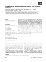
Fig. 1. KCTD5 expression. (A) RNA from different tissues and cell lines was analyzed by RT-PCR using specific primers for KCTD5. A plas-
mid encoding KCTD5 was used as a positive control (lane 1) for the RT-PCR. (B) Expression of KCTD5 in different cell types detected by
immunoblot with antibody to KCTD5 (upper panel). b-Actin was detected by immunoblot on the same membrane as an internal control of
protein loading (lower panel). (C) Time course of the expression of KCTD5 protein in PBLs stimulated with PHA. (D) Time course of the
expression of KCTD5 mRNA in PBLs stimulated with PHA, where numbers indicate hours of stimulation. (E) Expression of KCTD5 protein in
PBLs subjected to various stimuli. (F) Levels of KCTD5 mRNA assayed by quantitative PCR in PBLs stimulated with PHA. TCR+CD28 indi-
cates antibodies specific for T-cell receptor plus CD28.
KCTD5, a new substrate-specific adaptor for Cul3 Y. Bayo
´
n et al.
3902 FEBS Journal 275 (2008) 3900–3910 ª 2008 The Authors Journal compilation ª 2008 FEBS
adaptors for cullin ubiquitin ligases [5,6], in the case of
CUL3, most of the adaptors described so far belong to
the Kelch group. Thus, we investigated whether
KCTD5 could interact with CUL3. To test this inter-
action, HEK293 cells were transfected with plasmids
encoding CUL1, CUL2, CUL3, CUL4A and CUL4B
A
B
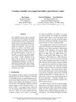
Fig. 2. Analysis of KCTD5 homologs. (A) Multiple protein sequence alignment of various KCTD5 orthologous sequences from different spe-
cies. (B) Phylogenetic tree built from human paralogs of KCTD5 using the BTB domain of 23 peptides. The BTB domain (T1 domain) of the
voltage-gated potassium channel KCNC1 protein is included in the analysis to root the tree.
Y. Bayo
´
n et al. KCTD5, a new substrate-specific adaptor for Cul3
FEBS Journal 275 (2008) 3900–3910 ª 2008 The Authors Journal compilation ª 2008 FEBS 3903
along with KCTD5. Total lysates were prepared from
these cells and used for immunoprecipitation assays. A
specific interaction of KCTD5 with CUL3 was
observed (Fig. 4A, lane 6), but not with the other cul-
lins (Fig. 4A, lane 5 for CUL1 and data not shown).
This interaction was confirmed in primary cells by car-
rying out immunoprecipitation assays in lysates from
PBLs stimulated with PHA for 2 days. Under these
conditions, CUL3 was detected by immunoblot in
KCTD5 precipitates (Fig. 4B, lane 2), but not when
the immunoprecipitation was carried out with an irrel-
evant antibody (Fig. 4B, lane 1). Then, the ability to
form a functional E3 ligase complex with CUL3 and
Rbx1 was assayed. Expression vectors for these pro-
teins were transfected into HEK293 cells, and cell
lysates were subjected to immunoprecipitation with
antibody to myc. As KCTD5 was precipitated when
CUL3 was present in the lysate (Fig. 4C, lane 5), this
result indicates that KCTD5 is part of a canonical cul-
lin-based E3 ligase complex. A faint band is also seen
in Fig. 4C (lane 2) that is probably due to the interac-
tion of Rbx1 with endogenous CUL3.
We also addressed whether KCTD5 could be ubiqui-
tinated, based on the fact that other BTB adaptor pro-
teins have been shown to be substrates of E3 ligases.
To do this, we transfected cells with expression vectors
for myc–Ub and hemagglutinin (HA)–KCTD5, and
cell lysates were immunoprecipitated with an antibody
specific for HA. The precipitates showed the presence
of ubiquitinated proteins (Fig. 4D) by immunoblotting.
To distinguish between covalent and noncovalent Ub
binding to KCTD5, we repeated this experiment, lys-
ing the cells with a highly denaturing buffer containing
8 m urea. Under these conditions, no smear was
detected in the KCTD5 immunoprecipitation and nor
was a KCTD5 ladder observed in Ub precipitates,
which is typical of ubiquitinated proteins (data not
shown). In addition, we could not detect KCTD5
ubiquitination in in vitro assays (data not shown).
Thus, unlike to what has been described for other
BTB proteins that work as substrate-specific adaptors,
KCTD5 is not ubiquitinated.
The interaction between BTB proteins and CUL3 is
considered to be mediated by the BTB domain and
the N-terminal region of CUL3 [8], mainly on the
basis of assays in which deletion of the BTB domain
and the N-terminal region of CUL3 is accompanied
by loss of binding. To analyze in detail KCTD5 bind-
ing to CUL3, pull-down and immunoprecipitation
assays with a series of deletion mutants of KCTD5
and CUL3 were carried out (Fig. 5A,C). These exper-
iments showed that the C-terminal region of KCTD5
was dispensable for CUL3 interaction, whereas the
B
A
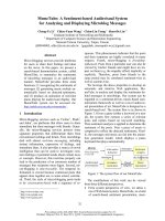
Fig. 3. Subcellular localization of KCTD5. (A) Left panels: fluores-
cence images of HEK293 cells transfected with either GFP or GFP–
KCTD5. Right panels: phase contrast images of the same cells. (B)
Immunofluorescence staining of HEK293 cells transfected with
plasmids encoding KCTD5 and several deletion mutants with mAb
to myc followed by a secondary antibody labeled with Alexa
Fluor 594.
KCTD5, a new substrate-specific adaptor for Cul3 Y. Bayo
´
n et al.
3904 FEBS Journal 275 (2008) 3900–3910 ª 2008 The Authors Journal compilation ª 2008 FEBS
BTB domain alone (45–145 amino acids), although
essential for this interaction, was not sufficient
(Fig. 5B). In fact, it required additional amino acids
(40–45) on the N-terminus, outside of the BTB fold,
as the 40–145 amino acid peptide is the smallest moi-
ety able to interact with CUL3. Studies with other
BTB proteins, e.g. SPOP [17]or the BTB protein
At1g21780 from Arabidopsis thaliana [18], have also
shown that other parts of their sequence, in addition
to the BTB domain, are involved in the association
with CUL3. On the other hand, the CUL3 region
involved in this interaction was the N-terminus, as
described for other BTB proteins, because a deletion
of 75 amino acids in the N-terminus of CUL3 com-
pletely abrogated the binding of KCTD5 to CUL3
(Fig. 5D, lane 8). Therefore, this detailed study on
the interaction of KCTD5 with CUL3 shows that the
sole BTB domain of KCTD5 does not support this
Fig. 4. KCTD5 interacts with CUL3 and ubiquitinated proteins. (A) HEK293 cells were transfected with plasmids encoding myc–KCTD5, HA–
CUL1, and HA–CUL3, as indicated. Cell lysates were subjected to immunoprecipitation (IP) with antibody to myc followed by immunoblotting
with antibodies to HA and myc. Expression of the tagged proteins is shown in the lower panels as WCL (whole cell lysate). (B) Lysates from
PBLs treated with PHA for 2 days (upper panel) were immunoprecipitated with either KCTD5 or an irrelevant IgG antibody and then blotted
with antibodies to CUL3 (upper panel) and KCTD5. The panels marked WCL show the expression levels of KCTD5 and CUL3 in the PBL
whole cell lysates. (C) HEK293 cells were transfected with plasmids encoding myc–Rbx1, HA–CUL3 and GST–KCTD5, lysates from these
cells were processed for pull-down with GST beads, and the presence of KCTD5 in the precipitates was checked by western blotting with
antibody to GST, followed by anti-HA and anti-myc blots to detect HA–CUL3 and myc–Rbx1. WCLs were immunoblotted with antibodies to
GST, HA and myc to assess the expression of the tagged proteins. (D) HEK293 cells were transfected with plasmids encoding for
HA–KCTD5 and myc–ubiquitin. Cell lysates were immunoprecipitated with antibody to HA, and ubiquitinated proteins that interact with
KCTD5 were detected with antibody to myc. WCLs were immunoblotted with the antibodies to HA and myc to assess the expression of
the tagged proteins.
Y. Bayo
´
n et al. KCTD5, a new substrate-specific adaptor for Cul3
FEBS Journal 275 (2008) 3900–3910 ª 2008 The Authors Journal compilation ª 2008 FEBS 3905
association and requires additional amino acids in the
N-terminus of this domain.
As the BTB domain is responsible for homo-oligo-
merization in BTB proteins [3], we addressed whether
KCTD5 might form homo-oligomers. For this purpose,
HEK293 cells were transfected with different constructs
of KCTD5 to show this association by either immuno-
precipitation or pull-down assays (Fig. 6A,B). We found
the BTB domain to be essential for KCTD5 oligomeri-
zation, as peptides expressing the KCTD5 N-terminal
region (N55) or the C-terminal sequence (POZCO,
amino acids 145–234) could not interact with themselves
(Fig. 6B). As the POZ ⁄ BTB domain of KCTD5 is dis-
tantly related to the T1 domain of voltage-gated potas-
sium channels, this fact was taken as a hint that KCTD5
could also tetramerize. To address this issue, gel exclu-
sion chromatography was run with recombinant
KCTD5 protein and KCTD5 was collected in fractions
consistent with the estimated molecular mass of an oct-
amer (Fig. 6C), which in turn can be explained by the
formation of two tetramers.
Taken together, our results show that the BTB
domain of KCTD5 is not able to bind alone to CUL3,
indicating that although it is critical for this associa-
tion, other sequences contribute to the binding of sub-
strate-specific adaptors to CUL3, namely, five amino
acids in the N-terminus of the BTB domain. In addi-
tion to the BTB fold, KCTD5 presents two other
regions: 40 amino acids in the N-terminal sequence,
which include a low-complexity region (12–33 amino
Fig. 5. Mapping the interaction of KCTD5 with CUL3. (A) Schematic diagram of the several KCTD5 deletion mutants used in this study. (B)
Plasmids for KCTD5 and different deletion mutants expressed as GST fusion proteins were transfected, along with HA–CUL3, in HEK293
cells. Lysates were subjected to pull-down assays with glutathione–Sepharose beads, and the presence of CUL-3 in the precipitates was
detected by immunoblot with antibody to HA, followed by antibody to GST. The expression of the proteins was checked in the whole cell
lysate (WCL) by western blot with antibodies to HA and CUL3. (C) Schematic diagram of the CUL3 deletion mutants used in this study. (D)
myc–KCTD5 was expressed in HEK293 cells along with different deletion mutants of HA–CUL3. The presence of the different CUL3 pep-
tides was checked in the myc immunoprecipitates by western blot with an antibody to HA. myc–KCTD5 was detected in the immunoprecipi-
tates by immunoblot with antibody to myc. The same antibodies were used to show the expression in the WCL (lower panels).
KCTD5, a new substrate-specific adaptor for Cul3 Y. Bayo
´
n et al.
3906 FEBS Journal 275 (2008) 3900–3910 ª 2008 The Authors Journal compilation ª 2008 FEBS
acids), and 88 amino acids in the C-terminus (PO-
ZCO). Taking into account that KCTD5 could be an
adaptor of CUL3 E3 ligases, we favor the hypothesis
that the POZCO region could participate in substrate
recognition, and that this could be a new protein inter-
action domain conserved through evolution, as seen in
orthologs. The fact that KCTD5 can form octamers
and the recent description of heterodimerization of
CUL3 [19] would indicate that complexes of higher
order could be formed among CUL3 and BTB
substrate adaptors, implying the recruitment of a great
number of substrates by these E3 ligases.
Although scarce, the information available about
KCTD proteins suggests that these proteins might be
involved in development and cellular differentiation.
For example, in zebra fish, three members of this group
– lov (leftover), ron (righton), and dex (dexter) – are
expressed asymmetrically in the left and right zebrafish
diencephalons [14]. Pfetin, a human ortholog of lov and
ron genes, encoded by human gene KCTD12, is detected
as mRNA preferentially expressed in fetal organs [20],
with the highest expression levels in the cochlea.
Another KCTD protein, KCTD11 ⁄ REN, is also regu-
lated developmentally in the nervous system [21], and it
has been implicated in the regulation of the Hedgehog
pathway [22]. The information presented in this article
would indicate that KCTD proteins might function by
recruiting specific substrates involved in development
and cellular differentiation for ubiquitination by CUL3
Ub ligases and degradation by the proteasome. As
regards KCTD5, there is another report that shows its
ability to interact with two viral regulatory proteins,
Rep68 and Rep78, of the adeno-associated virus type 2,
which are essential for viral DNA replication and gene
expression [16], although no relationship was established
with CUL3.
In summary, in this study we present evidence that
KCTD5 is a new substrate-specific adaptor for CUL3-
based Ub ligases. Our data indicate that a relevant
mechanism underlying the physiological role of KCTD
proteins includes recruitment of proteins to CUL3-
based E3 Ub ligases for degradation in the protea-
some. As identification of substrates recruited to the
proteasome would be very valuable for understanding
the function of these proteins, we are pursuing
the identification of KCTD5-interacting proteins,
especially those that are ubiquitinated.
Experimental procedures
Antibodies and reagents
Tissue culture reagents were from Cambrex (Verviers,
Belgium). The 12CA5 mAb against HA was from Roche
(Indianapolis, IN, USA), anti-HA clone HA.11 was from
Covance (Berkely, CA, USA), anti-glutathione S-transferase
(GST) and mAb against myc (9E10) were from Santa Cruz
Fig. 6. KCTD5 oligomerization. (A) HEK293 cells were transfected
with plasmids encoding for GST–KCTD5 and different deletion
mutants of KCTD5, and cell lysates were subjected to pull-down
with Glutathione–Sepharose beads and immunoblotted with a spe-
cific antibody to FLAG followed by antibody to GST. (B) HEK293
cells were transfected with HA–KCTD5 and several plasmids that
expressed different deletion mutants of KCTD5 as GST-fusion pro-
teins. Anti-HA immunoprecipitates of the cell lysates were analyzed
by immunoblot with antibody to GST followed by antibody to HA.
(C) Gel filtration chromatography of KCTD5 recombinant protein
produced in bacteria. The presence of KCTD5 in the fractions was
analyzed by immunoblot with antibody to KCTD5. Numbers under
the arrows indicate the chromatography fractions in which mole-
cular mass markers are eluted.
Y. Bayo
´
n et al. KCTD5, a new substrate-specific adaptor for Cul3
FEBS Journal 275 (2008) 3900–3910 ª 2008 The Authors Journal compilation ª 2008 FEBS 3907
Biotechnology Inc. (Santa Cruz, CA, USA), anti-cullin 3 was
from Abcam (Cambridge, UK), and mAbs against b-actin,
PHA, FLAG M2 mAb and PMA were from Sigma Chemical
Co. (St Louis, MO, USA). Antibodies against CD3
(UCHT1) and CD28 (clone CD28.2) were from BD Pharm-
ingen (Franklin Lakes, NJ, USA). MG-132 was from Calbio-
chem (Darmstadt, Germany). IL-2 was from PreprotechEC
(Rocky Hill, NJ, USA). Goat anti-(mouse IgG) conjugated
with Alexa FluorÒ 594 was from Molecular Probes (Eugene,
OR, USA). A mouse mAb was raised against recombinant
full-length KCTD5. Human MTC panel II was from
Clontech (Mountain View, CA, USA).
Plasmids and mutagenesis
Standard molecular biology techniques were used to gener-
ate the different constructs used in this study. All constructs
were verified by nucleotide sequencing. KCTD5 from a
Jurkat cDNA library obtained from Origene (Rockville,
MD, USA) was cloned in the pEF plasmid and served as a
template for the different KCTD5 plasmids used in this
study. HA–cullin1 and HA–cullin3 expression plasmids
were a kind gift of C. Geisen (Department of Medical
Oncology, Dana-Farber Cancer Institute, Boston, MA,
USA). Cullin4A and cullin4B were generously provided by
K. Tanaka (Department of Molecular Oncology, Tokyo
Metropolitan Institute of Medical Science, Japan) [23].
Cell culture and transfections
PBLs were isolated from buffy coats of healthy donors by
centrifugation at 700 g for 30 min on Ficoll–Hypaque (GE
Healthcare) cushions. Monocytes ⁄ macrophages were elimi-
nated by adherence to plastic for 1 h at 37 °C. Proliferation
was induced by PHA and IL-2, which was added after 48 h
with PHA, antibodies to CD3 plus antibodies to CD28, or
PMA plus ionomycin. Jurkat T-leukemia cells were kept at
logarithmic growth in RPMI-1640 medium supplemented
with 10% fetal bovine serum, 2 mml-glutamine, 1 mm
sodium pyruvate, nonessential amino acids, 100 UÆmL
)1
penicillin G, and 100 lgÆmL
)1
streptomycin. Transfection
of Jurkat T cells was performed as described previously
[24]. HEK293 cells were maintained at 37 °C in DMEM
supplemented with 10% fetal bovine serum, 2 mml-gluta-
mine, 100 UÆmL
)1
penicillin G, and 100 lgÆmL
)1
strepto-
mycin. For transient transfection, HEK293 cells were
transfected using the calcium phosphate precipitation
method [25].
Immunoprecipitation, GST pull-down,
SDS
⁄
PAGE, and immunoblotting
These procedures were performed done as reported previ-
ously [24]. Briefly, cells were lysed in 20 mm Tris ⁄ HCl,
pH 7.5, 150 mm NaCl, 5 mm EDTA containing 1% NP-40,
1mm Na
3
VO
4
,10lgÆmL
)1
aprotinin and leupeptin, and
1mm phenylmethanesulfonyl fluoride, and clarified by
centrifugation at 16 000 g for 10 min. The clarified lysates
were preabsorbed on protein G-Sepharose and then incu-
bated with antibody for 2 h; this was followed by overnight
incubation with protein G-Sepharose beads. Immune com-
plexes were washed three times in lysis buffer and resus-
pended in SDS sample buffer. Proteins resolved by
SDS ⁄ PAGE were transferred to a nitrocellulose membrane,
and immunoblotted with optimal dilutions of specific anti-
bodies followed by the appropriate anti-IgG–peroxidase
conjugate. Blots were developed by the enhanced chemilu-
minescence technique (ECL kit; GE Healthcare) according
to the manufacturer’s instructions. Pull-down of GST
fusion proteins was performed with glutathione–Sepharose
beads (GE Healthcare) incubated with the clarified lysates
for 2 h. The complexes were then washed and processed as
explained above for the immunoprecipitation. Some blots,
after being developed by chemiluminescence, were visual-
ized with a Bio-Rad VersaDoc chemiluminescence imager.
In this case, quantitation was carried out using quantity
one software from Bio-Rad.
RT-PCR
Total cellular RNA was extracted by the TRIzol method
(Life Technologies, Grand Island, NY, USA). The condi-
tions for cDNA first-strand synthesis and PCR reactions
were as described previously [26]. To address more exactly
the expression of KCTD5 mRNA, real-time RT-PCR was
carried out in RNA samples treated with DNase (Turbo-
DNA freeTM; Ambion, Austin, TX, USA). The resulting
cDNA was amplified in a PTC-200 apparatus equipped
with a Chromo4 detector (BioRad Laboratories), using
SYBR Green I mix containing HotStart polymerase
(ABgene, Epsom, UK). b-Actin was used as a housekeeping
gene to assess the relative abundance of KCTD5 mRNA,
using the comparative cycle threshold (CT) method for
relative expression. This method allows the relative
expression for a given cDNA using the formula: 2
)DCT
,
where DC
T
¼ DC
KCTD5
T
À DC
bÀactin
T
[27]. Therefore, one arbi-
trary unit (AU) corresponds to the expression of b-actin.
Indirect immunofluorescence and confocal
microscopy
HEK293 cells were cultured on coverslips and transiently
transfected with the indicated plasmids. Cells transfected
with GFP plasmids were fixed with 3.7% paraformaldehyde
and mounted on microscope slides, and GFP was then visu-
alized on an MRC-1024 confocal laser scanning microscope
(Bio-Rad). Phase contrast images were also taken. Immuno-
fluorescence staining of transfected KCTD5 was performed
KCTD5, a new substrate-specific adaptor for Cul3 Y. Bayo
´
n et al.
3908 FEBS Journal 275 (2008) 3900–3910 ª 2008 The Authors Journal compilation ª 2008 FEBS
as described previously [24]. HEK293 cells were washed in
NaCl ⁄ P
i
, fixed in 3.7% formaldehyde, permeabilized with
0.1% saponin in NaCl ⁄ P
i
, and blocked in the same medium
supplemented with 2.5% normal goat serum for 30 min at
room temperature. Primary and secondary antibodies were
diluted in the same buffer and incubated with the cells for 1 h
each at room temperature. After three washes with NaCl ⁄ P
i
,
the cells were mounted onto glass slides and viewed under a
confocal laser scanning microscope.
Gel filtration chromatography
For gel filtration chromatography, we used recombinant
KCTD5 produced in bacteria as His
6
-KCTD5 after
removal of the His-tag with thrombin. The protein solution
was fractionated through a Superdex 200 fast protein liquid
chromatography column (GE Healthcare), and collected in
fractions of 500 lL. Protein was precipitated with 10% tri-
chloroacetic acid and washed with acetone before addition
of SDS sample buffer and analysis by 10% SDS ⁄ PAGE.
Sequence analysis and alignments
For sequence retrieval, the BTB domain of human KCTD5
was used as query to retrieve the orthologs from the
UniPROT ( data-
base using the blast algorithm [28]. psi-blast [29] searches
retrieved 22 human paralogs. Multiple sequence alignments
of the BTB domain were conducted using muscle [30] and
probcons [31] in both the orthologs and the paralogs. To
generate reliable phylogenetic trees, Bayesian inference
using mrbayes v3.1.2 software was applied [32]. Multiple
alignments were done in two independent runs, with four
independent Markov chains in each run. One thousand five
hundred samples were used to estimate the posterior proba-
bility distribution. The amino acid model is a fixed rate
model using a mixture of fixed models. To compute a con-
sensus tree, we sampled 2502 from a total of 3002 trees in
two independent files (thus discarding 16% of the initial
samples prior to convergence). To root the tree, the
sequence of the BTB domain (T1) of the voltage potassium
channel KCNC1_HUM is included in the analysis.
Acknowledgements
We are grateful to Dr Keiji Tanaka for the CUL4A
and CULB cDNAs, to Dr Cristoff Geisen for the
CUL1 and CUL3 plasmids, and to Dr Joan Conaway
for the myc–Rbx1 plasmid. We thank the staff of
Centro de Hemoterapia y Hemodonacio
´
n de Castilla y
Leo
´
n for its help with the separation of leukocytes.
This work was supported by a grant from Programa
Nacional de Biologı
´
a Fundamental (Grant BFU2006-
01203 ⁄ BMC), Red Cardiovascular from Instituto de
Salud Carlos III. Y. Bayo
´
n is under contract within
the Ramo
´
n y Cajal Program of the Ministerio de Edu-
cacio
´
n y Ciencia of Spain, co-funded by the European
Social Fund through FEDER-FSE.
References
1 Bardwell VJ & Treisman R (1994) The POZ domain: a
conserved protein–protein interaction motif. Genes Dev
8, 1664–1677.
2 Zollman S, Godt D, Prive GG, Couderc JL & Laski
FA (1994) The BTB domain, found primarily in zinc
finger proteins, defines an evolutionarily conserved
family that includes several developmentally regulated
genes in Drosophila. Proc Natl Acad Sci USA 91,
10717–10721.
3 Stogios PJ, Downs GS, Jauhal JJ, Nandra SK & Prive
GG (2005) Sequence and structural analysis of BTB
domain proteins. Genome Biol 6, R82, doi: 10.1186/
gb-2005-6-10-r82.
4 Furukawa M, He YJ, Borchers C & Xiong Y (2003)
Targeting of protein ubiquitination by BTB–Cullin
3–Roc1 ubiquitin ligases. Nat Cell Biol 5, 1001–1007.
5 Pintard L, Willis JH, Willems A, Johnson JL, Srayko
M, Kurz T, Glaser S, Mains PE, Tyers M, Bowerman
B et al. (2003) The BTB protein MEL-26 is a substrate-
specific adaptor of the CUL-3 ubiquitin-ligase. Nature
425, 311–316.
6 Xu L, Wei Y, Reboul J, Vaglio P, Shin TH, Vidal M,
Elledge SJ & Harper JW (2003) BTB proteins are sub-
strate-specific adaptors in an SCF-like modular ubiqu-
itin ligase containing CUL-3. Nature 425, 316–321.
7 Geyer R, Wee S, Anderson S, Yates J & Wolf DA
(2003) BTB ⁄ POZ domain proteins are putative sub-
strate adaptors for cullin 3 ubiquitin ligases. Mol Cell
12, 783–790.
8 Petroski MD & Deshaies RJ (2005) Function and regu-
lation of cullin-RING ubiquitin ligases. Nat Rev Mol
Cell Biol 6, 9–20.
9 Pickart CM (2001) Mechanisms underlying ubiquitina-
tion. Annu Rev Biochem 70, 503–533.
10 Hochstrasser M (1996) Ubiquitin-dependent protein
degradation. Annu Rev Genet 30, 405–439.
11 Hershko A & Ciechanover A (1998) The ubiquitin
system. Annu Rev Biochem 67, 425–479.
12 Wolf FW, Marks RM, Sarma V, Byers MG, Katz RW,
Shows TB & Dixit VM (1992) Characterization of a
novel tumor necrosis factor-alpha-induced endothelial
primary response gene. J Biol Chem 267, 1317–1326.
13 He H, Tan CK, Downey KM & So AG (2001) A tumor
necrosis factor alpha- and interleukin 6-inducible pro-
tein that interacts with the small subunit of DNA poly-
merase delta and proliferating cell nuclear antigen. Proc
Natl Acad Sci USA 98, 11979–11984.
Y. Bayo
´
n et al. KCTD5, a new substrate-specific adaptor for Cul3
FEBS Journal 275 (2008) 3900–3910 ª 2008 The Authors Journal compilation ª 2008 FEBS 3909
14 Gamse JT, Kuan YS, Macurak M, Brosamle C, Thisse
B, Thisse C & Halpern ME (2005) Directional asymme-
try of the zebrafish epithalamus guides dorsoventral
innervation of the midbrain target. Development 132,
4869–4881.
15 Borinstein SC, Hyatt MA, Sykes VW, Straub RE, Lip-
kowitz S, Boulter J & Bogler O (2000) SETA is a multi-
functional adapter protein with three SH3 domains that
binds Grb2, Cbl, and the novel SB1 proteins. Cell Sig-
nal 12, 769–779.
16 Weger S, Hammer E, Gotz A & Heilbronn R (2007)
Identification of a cytoplasmic interaction partner of
the large regulatory proteins Rep78 ⁄ Rep68 of adeno-
associated virus type 2 (AAV-2). Virology 362, 192–206.
17 Kwon JE, La M, Oh KH, Oh YM, Kim GR, Seol JH,
Baek SH, Chiba T, Tanaka K, Bang OS et al. (2006)
BTB domain-containing speckle-type POZ protein
(SPOP) serves as an adaptor of Daxx for ubiquitination
by Cul3-based ubiquitin ligase. J Biol Chem 281,
12664–12672.
18 Figueroa P, Gusmaroli G, Serino G, Habashi J, Ma L,
Shen Y, Feng S, Bostick M, Callis J, Hellmann H et al.
(2005) Arabidopsis has two redundant Cullin3 proteins
that are essential for embryo development and that
interact with RBX1 and BTB proteins to form multi-
subunit E3 ubiquitin ligase complexes in vivo. Plant
Cell 17, 1180–1195.
19 Wimuttisuk W & Singer JD (2007) The Cullin3 ubiqu-
itin ligase functions as a Nedd8-bound heterodimer.
Mol Biol Cell 18, 899–909.
20 Resendes BL, Kuo SF, Robertson NG, Giersch AB,
Honrubia D, Ohara O, Adams JC & Morton CC (2004)
Isolation from cochlea of a novel human intronless gene
with predominant fetal expression. J Assoc Res Otolar-
yngol 5, 185–202.
21 Ferretti E, De Smaele E, Di Marcotullio L, Screpanti I
& Gulino A (2005) Hedgehog checkpoints in medullo-
blastoma: the chromosome 17p deletion paradigm.
Trends Mol Med 11, 537–545.
22 Di Marcotullio L, Ferretti E, De Smaele E, Argenti B,
Mincione C, Zazzeroni F, Gallo R, Masuelli L, Napo-
litano M, Maroder M et al. (2004) REN(KCTD11) is a
suppressor of Hedgehog signaling and is deleted in
human medulloblastoma. Proc Natl Acad Sci USA 101,
10833–10838.
23 Matsuda N, Azuma K, Saijo M, Iemura S, Hioki Y,
Natsume T, Chiba T, Tanaka K & Tanaka K (2005)
DDB2, the Xeroderma pigmentosum group E gene prod-
uct, is directly ubiquitylated by Cullin 4A-based ubiqu-
itin ligase complex. DNA Repair (Amst) 4, 537–545.
24 Alonso A, Rahmouni S, Williams S, van Stipdonk M,
Jaroszewski L, Godzik A, Abraham RT, Schoenberger
SP & Mustelin T (2003) Tyrosine phosphorylation
of VHR phosphatase by ZAP-70. Nat Immunol 4,
44–48.
25 Kingston RE, Chen CW, Okayama H & Rose JK
(2003) Transfection of DNA into eukaryotic cells. In
Current Protocols in Molecular Biology (Ausubel FM,
Brent R, Kingston RE, Moore DD, Seidman JG, Smith
JA & Struhl K, eds), pp. 9.1.4–9.1.9. John Wiley &
Sons, Boston, MA.
26 Fernandez N, Alonso S, Valera I, Vigo AG, Renedo M,
Barbolla L & Crespo MS (2005) Mannose-containing
molecular patterns are strong inducers of cyclooxygen-
ase-2 expression and prostaglandin E2 production in
human macrophages. J Immunol 174, 8154–8162.
27 Pfaffl MW (2001) A new mathematical model for rela-
tive quantification in real-time RT-PCR. Nucleic Acids
Res 29, 2002–2007.
28 Altschul SF, Gish W, Miller W, Myers EW & Lipman
DJ (1990) Basic local alignment search tool. J Mol Biol
215, 403–410.
29 Altschul SF, Madden TL, Schaffer AA, Zhang J,
Zhang Z, Miller W & Lipman DJ (1997) Gapped
BLAST and PSI-BLAST: a new generation of protein
database search programs. Nucleic Acids Res 25,
3389–3402.
30 Edgar RC (2004) MUSCLE: multiple sequence align-
ment with high accuracy and high throughput. Nucleic
Acids Res 32, 1792–1797.
31 Do CB, Mahabhashyam MS, Brudno M & Batzoglou S
(2005) ProbCons: probabilistic consistency-based multi-
ple sequence alignment. Genome Res 15, 330–340.
32 Ronquist F & Huelsenbeck JP (2003) MrBayes 3:
Bayesian phylogenetic inference under mixed models.
Bioinformatics 19, 1572–1574.
KCTD5, a new substrate-specific adaptor for Cul3 Y. Bayo
´
n et al.
3910 FEBS Journal 275 (2008) 3900–3910 ª 2008 The Authors Journal compilation ª 2008 FEBS