Báo cáo khoa học: Endothelial signalling events during leukocyte transmigration pptx
Bạn đang xem bản rút gọn của tài liệu. Xem và tải ngay bản đầy đủ của tài liệu tại đây (571.77 KB, 8 trang )
MINIREVIEW
Endothelial signalling events during leukocyte
transmigration
Peter L. Hordijk
Department of Molecular Cell Biology, Sanquin Research and Landsteiner Laboratory, Academic Medical Center, University of Amsterdam,
the Netherlands
Transendothelial migration (TEM) is an essential
aspect of the trafficking of leukocytes, as well as of
malignant cells. Much pathology is associated with
uncontrolled TEM, for instance in chronic inflamma-
tory disorders (asthma, rheumatoid arthritis, psoriasis)
and in metastasis. On the other hand, regulated leuko-
cyte trafficking is required for immune surveillance
and stem cell homing following transplantation proce-
dures. The multistep model for TEM [1] is well estab-
lished and probably applies to most transmigration
events, albeit that tissue specificity may result in addi-
tional complexity. For example, in the brain, the endo-
thelium constitutes the blood–brain barrier, which is
tighter than the endothelium in other tissues, resulting
in additional requirements for both leukocytes and the
endothelium to allow efficient transmigration.
Seminal work by the groups of Silverstein & Bender
[2,3] has triggered an ever-growing list of studies [4]
confirming the notion that endothelial cells (EC) not
only mediate leukocyte adhesion, but also actively par-
ticipate in the transmigration event. Most of these
studies have focussed on regulation of the paracellular
pathway, although, recently, analysis of transcellular
migration has also become fashionable. This review
will focus on events in the EC that have been implica-
ted in these different routes of TEM.
Keywords
adhesion; ICAM-1; migration;
transendothelial migration; VCAM-1
Correspondence
P. L. Hordijk, Department of Molecular Cell
Biology, Sanquin Research and Landsteiner
Laboratory, Academic Medical Center,
University of Amsterdam, Plesmanlaan 125,
1066 CX Amsterdam, the Netherlands
Fax: +31 20 5123474
Tel: +31 20 5123263
E-mail:
(Received 15 May 2006, accepted 17 July
2006)
doi:10.1111/j.1742-4658.2006.05440.x
The notion that it takes two to tango is certainly true for leukocyte trans-
endothelial migration. A growing pallet of leukocyte adhesion-induced
signaling events in endothelial cells have been identified, mediating both
short-term (i.e. permeability) as well as long-term (i.e. regulation of tran-
scription) effects. Efficient paracellular migration (i.e. through endothelial
cell–cell junctions) requires both intracellular calcium and the actin cyto-
skeleton, but also involves small GTPases, reactive oxygen species and pro-
tein kinases. The alternative route of transcellular migration appears to
depend on components such as caveolae and intermediate filaments. This
minireview discusses our current knowledge on the regulation of leukocyte
transmigration through endothelial signalling.
Abbreviations
CAM, cellular adhesion molecule; EC, endothelial cells; ERK, extracellular signal regulated kinase; ICAM, intercellular adhesion molecule;
JAM, junctional adhesion molecule; MAPK, mitogen-activated protein kinase; NOX, NADPH oxidase; PECAM-1, platelet ⁄ endothelial cell
adhesion molecule-1; ROS, reactive oxygen species; TEM, transendothelial migration; VCAM-1, vascular cell adhesion molecule-1;
VE-cadherin, vascular endothelial cadherin.
4408 FEBS Journal 273 (2006) 4408–4415 ª 2006 The Author Journal compilation ª 2006 FEBS
Endothelial docking structures
Rolling and adhesion of leukocytes over activated
endothelium (i.e. at sites of inflammation) is accompan-
ied by a complex response from the endothelial cells.
Initially, this comprises engagement and subsequent
clustering of endothelial adhesion molecules. These
include E-selectin and Ig-like cell adhesion molecules,
such as intercellular adhesion molecule (ICAM)-1,
ICAM-2, vascular cell adhesion molecule-1 (VCAM-1),
platelet ⁄ endothelial cell adhesion molecule-1
(PECAM-1) and members of the junctional adhesion
molecule (JAM) subfamily. Subsequent to leukocyte
adhesion, the EC show a pronounced morphological
response by forming ‘docking structures’ [5] or ‘trans-
migratory cups’ [6]. These are actin-rich membrane
extensions that form around the adherent leukocyte. In
these structures, not only are integrin ligands such as
ICAM-1 and VCAM-1 concentrated, but also adaptor
and linker molecules, such as ERM (ezrin, radixin,
moesin) proteins, vinculin, talin and a-actinin [5]. For-
mation of these structures requires calcium and, accord-
ing to some [5], but not to others [7], activation of the
Rho ⁄ p160ROCK pathway. As a result of the concen-
tration of adhesion and signaling molecules, docking
structures represent the main signaling ‘platforms’ from
which intracellular signaling into the EC is initiated.
There are indications that docking structures, and the
proteins therein, remain associated with the leukocytes
throughout the transmigration process [5,6]. This might
well be important for the sustained signaling that is
required for efficient crossing of the endothelial barrier.
Transcellular versus paracellular
migration
Leukocyte TEM has classically been considered to
occur at cell–cell junctions. It is now clear that, next to
this paracellular pathway, transcellular migration (i.e.
through the endothelial cell body) can also be
observed [8]. In vivo analysis showed that neutrophils
can cross the endothelial monolayer in a transcellular
manner [9]. Recently, a series of studies reported that
transcellular migration can also be observed in vitro
[6,10–13]. In particular, ICAM-1 has been associated
with transcellular migration [6,10,11,13]. It is obvious
that ICAM-1, being the main endothelial ligand for
b2-integrins, is crucial for TEM in general and for
polymorphonuclear cell transmigration in particular
[14]. Yet, Yang et al. [10] showed that prolonged
tumor necrosis factor-a treatment, or expression of an
ICAM-1–green fluorescent protein fusion on immortal-
ized EC, increases the relative contribution of transcel-
lular migration to polymorphonuclear cell diapedesis,
suggesting that ICAM-1 plays an active role in deter-
mining whether polymorphonuclear cells use the para-
cellular or the transcellular route. Whether VCAM-1
plays a similar role, for example, for monocytes, is not
known. Additional regulatory factors that might pro-
mote transcellular migration are the polygonal shape
of the EC or the levels of b2-integrin occupancy, shear
and the presence of chemokines on the EC [10,13].
The endothelial structures that mediate transcellular
migration were initially suggested to be vesiculo-vacuo-
lar organelles, which are abundant in EC and could
align to form a channel for macromolecules [15] and
perhaps even for migrating leukocytes. More recently,
transcellular migration was linked to caveolae, a sub-
class of membrane lipid rafts that may, by invagination,
detach from the membrane and mediate vesicular trans-
port. The protein caveolin, a key marker for caveolae,
was found to be enriched at the site of leukocyte–endo-
thelial cell contact [6]. Using a caveolin knockdown
approach, Millan et al. [11] showed that caveolin was
required for transcellular, but not for paracellular,
TEM. Another study, by Nieminen et al. [12], has
implicated the intermediate filament protein, vimentin,
in the process of lymphocyte transcellular migration.
However, its regulation and precise role in the transmi-
gration process remains to be determined.
Although these studies show that the paracellular
and transcellular pathways co-exist, considerable vari-
ation in the relative contribution of the transcellular
pathway to leukocyte TEM has been noted. This may
depend on the type of leukocyte, as Yang et al. repor-
ted efficient transcellular migration (up to 50% of the
total transmigration events) for neutrophils, whereas
T lymphocytes exclusively used the paracellular route
[10]. Contrasting findings have also been described
(i.e. that lymphocytes would preferentially use the
transcellular route) [12]. Similarly, the source of the
endothelium (microvascular versus macrovascular
[11]), and the state of activation of the endothelium
or the leukocytes [10,12,13], may also affect the relat-
ive importance of one route over the other. Yet, in
most of these studies, the contribution of the transcel-
lular pathway was only 10–30% to the total trans-
migration events. Intriguingly, down-regulation of
caveolin expression in human umbilical vein endothel-
ial cells blocked transcellular migration by T lympho-
blasts, but did not reduce the overall TEM, suggesting
that cells can switch from the transcellular to the
paracellular route without a significant reduction in
TEM efficiency [11]. The factors that determine the
choice of leukocytes for one or the other pathway
remain to be established.
P. L. Hordijk Endothelial signalling events
FEBS Journal 273 (2006) 4408–4415 ª 2006 The Author Journal compilation ª 2006 FEBS 4409
Signaling by endothelial adhesion
molecules
As mentioned above, leukocyte adhesion and the for-
mation of endothelial docking structures is associated
with the clustering of adhesion and signaling mole-
cules. It is very likely that this clustering is required
for efficient signal transduction into the EC which, at
least for some types of leukocyte, is important for effi-
cient TEM. Many cell surface (adhesion) proteins have
been implicated in leukocyte TEM, in particular Ig
family members. However, for only some of these has
the induction of intracellular signaling been causally
related to leukocyte transmigration.
E-selectin
Although in the classical multistep model for TEM,
selectins are usually depicted as mediating low-affinity
interactions to allow rolling, there is ample evidence for
the signaling capacity of E-selectin (CD62 E), both
towards the actin cytoskeleton [16] as well as to p42 mi-
togen-activated protein kinase (MAPK) ⁄ extracellular
signal regulated kinase (ERK) activation and the induc-
tion of c-fos [17]. Clustering of E-selectin, which is an
adhesion receptor for neutrophils and memory T cells,
results in its association with the actin cytoskeleton. In
addition, clustered E-selectin associates, through its
intracellular domain, with Ras, Raf and MAPK ⁄ ERK
kinase (MEK). These proteins trigger the downstream
signaling towards MAPK and c-fos [17,18]. Later stud-
ies showed that tyrosine phosphorylation of the E-se-
lectin intracellular tail is instrumental in these events
through the recruitment and activation of the SHP-2
phosphatase, which signals, via Shc and Grb2 adapter
proteins, to the Ras-MAPK pathway [19]. In addition,
E-selectin resides in caveolin-containing lipid rafts and
associates with phospholipase C gamma [20]. Raft dis-
ruption ablates the activation of phospholipase C
gamma, but not of MAPK, indicating that the activa-
tion of different signaling pathways can occur in dis-
tinct membrane subdomains [20].
PECAM-1
The Ig-like CAM, PECAM-1, mediates homotypic
interactions between leukocytes and EC and between
EC themselves at intercellular junctions. PECAM-1 has
been implicated in cell survival, angiogenesis, lung
development and experimental autoimmune encephalo-
myelitis [21,22]. Blocking antibodies to PECAM-1 inhi-
bit neutrophil and lymphocyte TEM in vitro [23,24]
(also see review by Petri & Bixel, this issue of FEBS). In
contrast to most other Ig-like CAMs, PECAM-1 has an
extended intracellular tail that encodes two immuno-
receptor tyrosine-based inhibition motifs and which is
subject to tyrosine phosphorylation by src-like kinases,
primarily in response to cell stimulation or PECAM-1
cross-linking [21]. These immunoreceptor tyrosine-based
inhibition motifs mediate, following phosphorylation,
association with the SHP-1 and SHP-2 tyrosine phos-
phatases, with the SH2 domain-containing inositol
5-phosphatase, SHIP, with adapter, proteins such as
Grb2, and with b- and c-catenin. PECAM-1 stimulates
integrin adhesion by activating Rap1 [25] and has been
associated with cell survival. Similarly to E-selectin,
PECAM-1 can activate MAPK via its association with
SHP2. Remarkably, although the signaling capacities of
PECAM-1 have been extensively studied, its relevance
as a signaling molecule in TEM is not clear. This may
also relate to the fact that PECAM-1-deficient mice
showed only limited problems in models of inflamma-
tion, although later studies reported that this result may
depend on the mice strain used [26]. Recently, however,
the modulation of cell–cell adhesion by PECAM-1 has
been proposed, based on studies in transfected epithelial
cells [27]. Whether these data can be readily translated
to EC remains to be seen.
JAMs
The family of JAM molecules concentrate in endothel-
ial tight junctions [28]. In addition, JAM proteins are
expressed by leukocytes. Several studies have clearly
shown that JAM family members are essential for leu-
kocyte TEM [8,29] (also see the review by Petri &
Bixel, this issue of FEBS). In addition, JAM proteins
have been implicated in cell signaling towards cell
polarity and the formation of cell–cell contact. JAMs
can associate, through C-terminal PDZ-binding motifs,
with a series of proteins, including ZO1, AF6, Par 3
and MUPP1 [28]. Despite their role in the regulation
of cell–cell adhesion and the fact that the JAMs clearly
have relevant signaling capacities, it is, as for
PECAM-1, not yet known whether they in fact trans-
mit signals into the EC that promote TEM.
ICAM-1
ICAM-1 is one of the main integrin ligands involved
in leukocyte TEM, in particular of lymphocytes and
neutrophils. ICAM-1 is expressed on resting endo-
thelium, but up-regulated upon activation by inflam-
matory stimuli. ICAM-1 has a short cytoplasmic tail
of 29 amino acids that associates to ERM (ezrin, ra-
dixin, moesin) proteins, as well as to a-actinin [30,31].
Endothelial signalling events P. L. Hordijk
4410 FEBS Journal 273 (2006) 4408–4415 ª 2006 The Author Journal compilation ª 2006 FEBS
ICAM-1 acts as an adhesion molecule and a signal
transducer in EC. ICAM-1 activates the p60src kinase,
which leads to phosphorylation of cortactin [32], trig-
gers release of intracellular calcium and activates the
Rho GTPase, which explains the effects of ICAM-1 on
the actin cytoskeleton and on contractility in EC [16].
These effects are mediated by the C-terminus of
ICAM-1 and are required for efficient TEM of
lymphocytes [31,33,34]. Moreover, ICAM-1 has been
shown to activate p60src via the activation of xanthine
oxidase, in a SHP2-dependent manner, leading to tyro-
sine phosphorylation of ezrin and p38 MAPK [35].
Finally, cell-permeable versions of the cytoplasmic tail
of ICAM-1 were found to block leukocyte TEM
[10,34,36]. In conclusion, ICAM-1 activates a series of
signaling events through its intracellular C-terminal tail
that are likely to increase endothelial permeability,
resulting in enhanced leukocyte TEM.
VCAM-1
The main b1-integrin ligand on the endothelium,
VCAM-1, is, in contrast to ICAM-1, absent from rest-
ing cells but greatly up-regulated by inflammatory
stimuli. Similarly to ICAM-1, VCAM-1 not only acts
as an adhesion receptor, but also as a signal transducer
upon binding of leukocytes. The cytoplasmic domain
of VCAM-1 is only 19 amino acids long and comprises
a type I PDZ-binding motif. However, whether specific
interactions are mediated by this motif is unknown; to
date, only ezrin and moesin have been shown to asso-
ciate with the cytoplasmic domain of VCAM-1 [5].
VCAM-1 clustering leads to the activation of Rac1,
production of reactive oxygen species (ROS), activa-
tion of p38 MAPK and changes in the actin cytoskele-
ton (i.e. stress fiber formation) (Fig. 1). These events
have all been associated with the increased endothelial
permeability (as measured by tracer molecules or trans-
endothelial resistance) that is induced by VCAM-1
cross-linking. VCAM-1-mediated leukocyte TEM is
also dependent on some of these signaling events,
including Rac1 and Rho activation [37]. Of particular
interest is the role of VCAM-1-induced production of
ROS. ROS are known to impair cell–cell adhesion in
EC and are important regulators of endothelial integ-
rity through their indirect stimulation of tyrosine
kinase activity. In addition, vascular ROS play an
important role in the development of cardiovascular
disease [38]. Conversely, scavenging ROS preserves
endothelial barrier function, prevents endothelial cell
migration and angiogenesis, and is atheroprotective.
The source of endothelial ROS has been suggested to
be the NADPH oxidase 2 (NOX2) which is, like its
relative NOX4, also expressed in EC [39,40]. NOX2 is
supposedly localized in the endothelial plasma mem-
brane, resulting in an extracellular release of ROS.
These have been proposed to activate metalloproteases
that would promote endothelial permeability by
proteolytic degradation of vascular endothelial cadher-
in (VE-cadherin) or of the extracellular matrix
(Fig. 2A) [39].
Our laboratory has shown that ROS production in
response to VCAM-1-mediated activation of Rac1 can
be observed intracellularly, and we have proposed a
role for the redox sensitive proline-rich tyrosine kinase
2 in the control of endothelial integrity through the
phosphorylation of b-catenin [41] (Fig. 2B). However,
our present knowledge of this pathway remains limited.
The molecular mechanism of VCAM-1-triggered acti-
vation of Rac1 is completely unknown, as is the poten-
tial role for the relatively abundant NOX4 protein in
VCAM-1 signalling. Also, the mechanism of VE-cadh-
erin inactivation through ROS (i.e. either by proteolytic
breakdown, or by reducing its homophilic adhesion
through reduction of its link to the actin cytoskeleton),
be it from the inside or the outside of the cells, requires
further analysis. In addition to ROS signaling, endo-
thelial integrity is also subject to regulation by the Rap1
GTPase, microtubule dynamics and by proteins that
control VE-cadherin internalization. To what extent
these events are also part of the process of leukocyte
TEM is presently unclear.
It is important to underscore that engaged, clustered
ICAM-1 and VCAM-1 may be in very close proximity
on the endothelial cell surface, in particular following
adhesion of leukocytes that use b1 and b2 integrins for
transmigration. This means that the signaling which is
induced by these molecules may also be intercon-
nected. The extent of cross-talk between ICAM-1 and
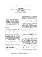
Fig. 1. Clustered vascular cell adhesion molecule-1 (VCAM-1) aligns
with actin stress fibers. Transient expression of the VCAM-1–green
fluorescent protein (GFP) fusion (green) shows its diffuse distribu-
tion over the endothelial cell surface (left panel). Cross-linking by a
VCAM-1 antibody induces clustering of the protein and alignment
of the VCAM-1–GFP clusters with actin stress fibers (right panel;
F-actin in blue).
P. L. Hordijk Endothelial signalling events
FEBS Journal 273 (2006) 4408–4415 ª 2006 The Author Journal compilation ª 2006 FEBS 4411
VCAM-1 induced signalling events is thus an import-
ant issue for future research.
Effects of shear
EC in various parts of the vasculature are exposed to
different levels of fluid shear stress. There is no doubt
that this shear force triggers and modulates endothel-
ial cell signaling and affects endothelial permeability,
proliferation, migration and gene expression [42].
Shear stress is strongly associated with the develop-
ment of atherosclerosis, which is an arterial disease
that occurs predominantly at sites of disturbed lami-
nar blood flow. VCAM-1 and E-selectin, in conjunc-
tion with the actin cytoskeleton, have been shown to
activate ERK2 in a shear-dependent manner [18].
Recently, the vascular endothelial growth factor
receptor, VE-cadherin and PECAM-1 were identified
as components of a shear detecting complex in EC
[43]. This complex mediates shear induced and ligand-
independent activation of src and of the phosphatidyl-
inositol-3-Kinase ⁄ Akt pathway and is required for the
activation of nuclear factor-jB at sites of disturbed
flow. Apart from affecting the EC, shear also pro-
motes chemokine-induced lymphocyte TEM, an effect
coined ‘chemorheotaxis’ [13]. Thus, shear force repre-
sents an additional level of regulation of both leuko-
cyte migration as well as endothelial signalling.
Targets of endothelial signaling
There appears to be at least two classes of endothelial
target downstream of the signaling that is initiated by
leukocyte binding. There are rapid effects on the actin
cytoskeleton and the VE–cadherin–catenin complex
and these appear to co-operate in mediating efficient
transendothelial migration. On the other hand, there
is evidence for activation of transcription factors, such
as nuclear factor-jB and c-fos [17]. The subsequent
up-regulation of cell adhesion molecules or metallo-
proteases may have important effects on the amplifi-
cation and ⁄ or duration of the inflammatory response.
Activation of ERK may well play a role in both
pathways, as ERK has been implicated in the regula-
tion of cell adhesion and migration [44]. In addition,
ERK is involved in the activation of c-fos and of
nuclear factor-jB. Thus, it appears that there is co-
operativity between the ERK and p38 MAPK path-
ways, as well as the ROS that are produced in the
EC, in altering the gene expression profile of activated
endothelium.
Concluding remarks
Along with the increased knowledge on the control of
endothelial integrity, the number of signaling compo-
nents that are implicated in the efficient transmigration
of leukocytes is also growing. The key players appear
to be small GTPases and the actin cytoskeleton, ROS,
MAPKs, cell-matrix adhesion molecules, transcription
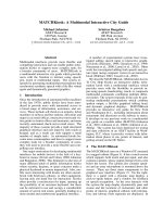
A
B
Fig. 2. Models for the action of vascular cell adhesion molecule-1
(VCAM-1)-derived reactive oxygen species (ROS) in controlling
transendothelial migration (TEM). According to one model (Fig. 2A),
VCAM-1 activates NADPH oxidase 2 (NOX2), which resides in the
plasma membrane, in a Rac1-dependent manner. Extracellularly
produced ROS activate metalloproteases, which degrade junctional
and ⁄ or matrix proteins. A second model (Fig. 2B) proposes ROS-
mediated activation of the proline-rich tyrosine kinase 2 and phos-
phorylation of b-catenin as instrumental in the transient loss of
vascular endothelial cadherin-mediated cell-to-cell contact, which
follows VCAM-1 engagment. Intercellular adhesion molecule-1-
mediated RhoA activation is required in both models, for providing
enhanced endothelial contractility. See the text for details.
Endothelial signalling events P. L. Hordijk
4412 FEBS Journal 273 (2006) 4408–4415 ª 2006 The Author Journal compilation ª 2006 FEBS
factors, and also enzymes such as calpain or activated
metalloproteases. A recurrent theme is that the type of
leukocyte, source of the EC, inflammatory stimulus
and the absence or presence of shear, will affect the
responses measured and thus the consequent implica-
tion of a particular event in TEM.
The complexity of this field is further boosted by
studies on the transcellular pathway. A major chal-
lenge will be to define whether this pathway is domin-
ant in specific tissues, as was proposed for the brain,
or perhaps associated with certain (pathological) con-
ditions. Also, it remains to be determined whether
transcellular migration is regulated by endothelial sign-
aling, or is primarily a result of the protrusive activity
of leukocytes. Thus, although we are not quite dancing
in the dark anymore, many issues remain that guaran-
tee a more complex, but no less interesting, future for
the research on leukocyte TEM.
Acknowledgements
Dr Jaap van Buul is gratefully acknowledged for crit-
ical reading. PLH is a fellow of the Landsteiner Foun-
dation for Blood Transfusion Research. I apologize to
all whose work could not be included because of space
constraints. VCAM-1-GFP was a kind gift from
Dr Sanchez-Madrid.
References
1 Springer TA (1994) Traffic signals for lymphocyte recir-
culation and leukocyte emigration: the multistep para-
digm. Cell 76, 301–314.
2 Huang AJ, Manning JE, Bandak TM, Ratau MC,
Hanser KR & Silverstein SC (1993) Endothelial cell
cytosolic free calcium regulates neutrophil migration
across monolayers of endothelial cells. J Cell Biol 120,
1371–1380.
3 Pfau S, Leitenberg D, Rinder H, Smith BR, Pardi R &
Bender JR (1995) Lymphocyte adhesion-dependent cal-
cium signaling in human endothelial cells. J Cell Biol
128, 969–978.
4 van Buul JD & Hordijk PL (2004) Signaling in leuko-
cyte transendothelial migration. Arterioscler Thromb
Vasc Biol 24, 824–833.
5 Barreiro O, Yanez-Mo M, Serrador JM, Montoya MC,
Vicente-Manzanares M, Tejedor R, Furthmayr H &
Sanchez-Madrid F (2002) Dynamic interaction of
VCAM-1 and ICAM-1 with moesin and ezrin in a novel
endothelial docking structure for adherent leukocytes.
J Cell Biol 157, 1233–1245.
6 Carman CV & Springer TA (2004) A transmigratory
cup in leukocyte diapedesis both through individual
vascular endothelial cells and between them. J Cell Biol
167, 377–388.
7 Carman CV, Jun CD, Salas A & Springer TA (2003)
Endothelial cells proactively form microvilli-like mem-
brane projections upon intercellular adhesion molecule 1
engagement of leukocyte LFA-1. J Immunol 171, 6135–
6144.
8 Engelhardt B & Wolburg H (2004) Mini-review: Trans-
endothelial migration of leukocytes: through the front
door or around the side of the house? Eur J Immunol
34, 2955–2963.
9 Feng D, Nagy JA, Pyne K, Dvorak HF & Dvorak AM
(1998) Neutrophils emigrate from venules by a transen-
dothelial cell pathway in response to FMLP. J Exp Med
187, 903–915.
10 Yang L, Froio RM, Sciuto TE, Dvorak AM, Alon R
& Luscinskas FW (2005) ICAM-1 regulates neutrophil
adhesion and transcellular migration of TNF-alpha-
activated vascular endothelium under flow. Blood 106,
584–592.
11 Millan J, Hewlett L, Glyn M, Toomre D, Clark P &
Ridley AJ (2006) Lymphocyte transcellular migration
occurs through recruitment of endothelial ICAM-1 to
caveola- and F-actin-rich domains. Nat Cell Biol 8,
113–123.
12 Nieminen M, Henttinen T, Merinen M, Marttila-Ichiha-
ra F, Eriksson JE & Jalkanen S (2006) Vimentin func-
tion in lymphocyte adhesion and transcellular
migration. Nat Cell Biol 8, 156–162.
13 Cinamon G, Shinder V, Shamri R & Alon R (2004)
Chemoattractant signals and beta 2 integrin occupancy
at apical endothelial contacts combine with shear stress
signals to promote transendothelial neutrophil migra-
tion. J Immunol 173, 7282–7291.
14 Kakkar AK & Lefer DJ (2004) Leukocyte and endothe-
lial adhesion molecule studies in knockout mice. Curr
Opin Pharmacol 4, 154–158.
15 Dvorak AM, Kohn S, Morgan ES, Fox P, Nagy JA &
Dvorak HF (1996) The vesiculo-vacuolar organelle
(VVO): a distinct endothelial cell structure that provides
a transcellular pathway for macromolecular extravasa-
tion. J Leukoc Biol 59, 100–115.
16 Lorenzon P, Vecile E, Nardon E, Ferrero E, Harlan
JM, Tedesco F & Dobrina A (1998) Endothelial cell
E- and P-selectin and vascular cell adhesion molecule-1
function as signaling receptors. J Cell Biol 142, 1381–
1391.
17 Hu Y, Kiely JM, Szente BE, Rosenzweig A & Gim-
brone MA Jr (2000) E-selectin-dependent signaling
via the mitogen-activated protein kinase pathway
in vascular endothelial cells. J Immunol 165, 2142–
2148.
18 Cuvelier SL, Paul S, Shariat N, Colarusso P & Patel
KD (2005) Eosinophil adhesion under flow conditions
P. L. Hordijk Endothelial signalling events
FEBS Journal 273 (2006) 4408–4415 ª 2006 The Author Journal compilation ª 2006 FEBS 4413
activates mechanosensitive signaling pathways in human
endothelial cells. J Exp Med 202 , 865–876.
19 Hu Y, Szente B, Kiely JM & Gimbrone MA Jr
(2001) Molecular events in transmembrane signaling
via E–selectin. SHP2 association, adaptor protein
complex formation and ERK1 ⁄ 2 activation. J Biol
Chem 276, 48549–48553.
20 Kiely JM, Hu Y, Garcia-Cardena G & Gimbrone
MA Jr (2003) Lipid raft localization of cell surface
E-selectin is required for ligation-induced activation
of phospholipase C gamma. J Immunol 171, 3216–
3224.
21 Newman PJ & Newman DK (2003) Signal transduction
pathways mediated by PECAM-1: new roles for an old
molecule in platelet and vascular cell biology. Arterios-
cler Thromb Vasc Biol 23, 953–964.
22 Graesser D, Solowiej A, Bruckner M, Osterweil E,
Juedes A, Davis S, Ruddle NH, Engelhardt B &
Madri JA (2002) Altered vascular permeability and
early onset of experimental autoimmune encephalo-
myelitis in PECAM-1-deficient mice. J Clin Invest 109,
383–392.
23 Bogen S, Pak J, Garifallou M, Deng X & Muller WA
(1994) Monoclonal antibody to murine PECAM-1
(CD31) blocks acute inflammation in vivo. J Exp Med
179, 1059–1064.
24 Christofidou-Solomidou M, Nakada MT, Williams J,
Muller WA & DeLisser HM (1997) Neutrophil platelet
endothelial cell adhesion molecule-1 participates in neu-
trophil recruitment at inflammatory sites and is down-
regulated after leukocyte extravasation. J Immunol 158,
4872–4878.
25 Reedquist KA, Ross E, Koop EA, Wolthuis RM,
Zwartkruis FJ, van Kooyk Y, Salmon M, Buckley CD
& Bos JL (2000) The small GTPase, Rap1, mediates
CD31-induced integrin adhesion. J Cell Biol 148, 1151–
1158.
26 Schenkel AR, Chew TW & Muller WA (2004) Platelet
endothelial cell adhesion molecule deficiency or
blockade significantly reduces leukocyte emigration in
a majority of mouse strains. J Immunol 173, 6403–
6408.
27 Wang Y & Sheibani N (2006) PECAM-1 isoform-speci-
fic activation of MAPK ⁄ Erks and small GTPases: impli-
cations in inflammation and angiogenesis. J Cell
Biochem 98, 451–468.
28 Ebnet K, Suzuki A, Ohno S & Vestweber D (2004)
Junctional adhesion molecules (JAMs): more molecules
with dual functions? J Cell Sci 117, 19–29.
29 Del Maschio A, De Luigi A, Martin-Padura I, Brock-
haus M, Bartfai T, Fruscella P, Adorini L, Martino G,
Furlan R, De Simoni MG et al. (1999) Leukocyte
recruitment in the cerebrospinal fluid of mice with
experimental meningitis is inhibited by an antibody to
junctional adhesion molecule (JAM). J Exp Med 190,
1351–1356.
30 Carpen O, Pallai P, Staunton DE & Springer TA (1992)
Association of intercellular adhesion molecule-1
(ICAM-1) with actin-containing cytoskeleton and alpha-
actinin. J Cell Biol 118, 1223–1234.
31 Greenwood J, Etienne-Manneville S, Adamson P &
Couraud PO (2002) Lymphocyte migration into the
central nervous system: implication of ICAM-1 signal-
ling at the blood–brain barrier. Vasc Pharmacol 38,
315–322.
32 Durieu-Trautmann O, Chaverot N, Cazaubon S, Stros-
berg AD & Couraud PO (1994) Intercellular adhesion
molecule 1 activation induces tyrosine phosphorylation
of the cytoskeleton-associated protein cortactin in brain
microvessel endothelial cells. J Biol Chem 269, 12536–
12540.
33 Adamson P, Etienne S, Couraud PO, Calder V &
Greenwood J (1999) Lymphocyte migration through
brain endothelial cell monolayers involves signaling
through endothelial ICAM-1 via a rho-dependent path-
way. J Immunol 162, 2964–2973.
34 Greenwood J, Amos CL, Walters CE, Couraud PO,
Lyck R, Engelhardt B & Adamson P (2003) Intracel-
lular domain of brain endothelial intercellular
adhesion molecule-1 is essential for T lymphocyte-
mediated signaling and migration. J Immunol 171,
2099–2108.
35 Wang Q, Pfeiffer GR & Gaarde WA (2003) Activation
of SRC tyrosine kinases in response to ICAM-1 ligation
in pulmonary microvascular endothelial cells. J Biol
Chem 278, 47731–47743.
36 Lyck R, Reiss Y, Gerwin N, Greenwood J, Adamson P
& Engelhardt B (2003) T-cell interaction with ICAM-
1 ⁄ ICAM-2 double-deficient brain endothelium in vitro:
the cytoplasmic tail of endothelial ICAM-1 is necessary
for transendothelial migration of T cells. Blood 102,
3675–3683.
37 van Wetering SBN, van Buul JD, Mul FP, Lommerse
I, Mous R, ten Klooster JP, Zwaginga JJ & Hordijk
PL (2003) VCAM-1-mediated Rac signaling controls
endothelial cell-cell contacts and leukocyte transmigra-
tion. Am J Physiol Cell Physiol 285, C343–C352.
38 Shah AM & Channon KM (2004) Free radicals and
redox signalling in cardiovascular disease. Heart 90,
486–487.
39 Deem TL & Cook-Mills JM (2004) Vascular cell adhe-
sion molecule 1 (VCAM-1) activation of endothelial cell
matrix metalloproteinases: role of reactive oxygen spe-
cies. Blood 104, 2385–2393.
40 van Buul JD, Fernandez-Borja M, Anthony EC & Hor-
dijk PL (2005) Expression and localization of NOX2
and NOX4 in primary human endothelial cells. Antioxid
Redox Signal 7, 308–317.
Endothelial signalling events P. L. Hordijk
4414 FEBS Journal 273 (2006) 4408–4415 ª 2006 The Author Journal compilation ª 2006 FEBS
41 van Buul JD, Anthony EC, Fernandez-Borja M,
Burridge K & Hordijk PL (2005) Proline-rich tyrosine
kinase 2 (Pyk2) mediates vascular endothelial-cadherin-
based cell-cell adhesion by regulating beta-catenin
tyrosine phosphorylation. J Biol Chem 280, 21129–
21136.
42 Tzima E (2006) Role of small GTPases in endothelial
cytoskeletal dynamics and the shear stress response.
Circ Res 98, 176–185.
43 Tzima E, Irani-Tehrani M, Kiosses WB, Dejana E, Sch-
ultz DA, Engelhardt B, Cao G, DeLisser H & Schwartz
MA (2005) A mechanosensory complex that mediates
the endothelial cell response to fluid shear stress. Nature
437, 426–431.
44 Howe AK, Aplin AE & Juliano RL (2002) Anchorage-
dependent ERK signaling – mechanisms and conse-
quences. Curr Opin Genet Dev 12, 30–35.
P. L. Hordijk Endothelial signalling events
FEBS Journal 273 (2006) 4408–4415 ª 2006 The Author Journal compilation ª 2006 FEBS 4415