Báo cáo khoa học: Factors affecting habituation of PC12 cells to ATP potx
Bạn đang xem bản rút gọn của tài liệu. Xem và tải ngay bản đầy đủ của tài liệu tại đây (382.37 KB, 8 trang )
Factors affecting habituation of PC12 cells to ATP
J. Russel Keath
1
and Edward W. Westhead
2
1
Department Neurobiology and Physiology, Northwestern University, Evanston, IL, USA;
2
Department of Biochemistry and
Molecular Biology, University of Massachusetts, Amherst, MA, USA
Extracellular ATP triggers catecholamine secretion from
PC12 cells by activating ionotropic purine receptors.
Repeated stimulation by ATP leads to habituation of the
secretory r esponse. In this paper, we use amperometric
detection to monitor the habituation of PC12 cells to mul-
tiple s timulations of ATP or its agonist. Cells habituate to
30 l
M
ATP slower than they do to 300 or 600 l
M
ATP.
Modifying external Mg
2+
affects the response of cells to
30 l
M
ATP, bu t does not affect habituation, suggesting that
habituation does not necessarily correspond to either sti-
mulus intensity or cellular r esponse. Mg
2+
affects the initial
response of PC12 cells to 2MeSATP in a manner similar
to ATP. Increasing external [Mg
2+
]to3.0m
M
, however,
eliminates habituation to 2M eSATP. This habituation can
be partially restored by costimulation with 100 l
M
UTP.
Background application of UTP increases habituation to
both ATP and 2MeSATP. T his suggests that ATP-sensitive
metabotropic (P
2
Y) receptors play a role in the habituation
process. Finally, although Ca
2+
influx through voltage-
operated calcium channels does not appear to contribute to
secretion during ATP stimulation, blocking these channels
with nicardipine increases habituation. This suggests a role
for voltage-operated calcium channels in the habituation
process.
Keywords: voltage-operated calcium channels; PC12 cells;
habituation; inactivation; P
2
X receptors.
While ATP is commonly known as an energy storage
molecule, i t a lso serves as a neurotransmitter. A TP activates
both ionotropic (P
2
X) receptors, triggering neurosecretion,
and metabotropic (P
2
Y) receptors, which induce the
production of inositol phosphates, diacylglycerol and cyclic
AMP, and inhibit
L
-type calcium channels [1].
PC12 cells are a convenient model f or ATP-induced
secretion. When stimulated, these cells release catechol-
amines, ATP, and a w ide v ariety of other n eurotransmitters
and neuromodulators [2,3]. Several ligands, including
purinergic and cholinergic ligands [3,4], trigger Ca
2+
influx,
which activates exocytotic catecholamine secretion. ATP,
for example, activates a ligand-gated cation channel
permeable to Na
+
and Ca
2+
, triggering exocytosis [3,5–
7]. Several factors modify the response of PC12 cells to
ATP, including stimulus intensity [8], exposure to neuro-
modulators [9] and previous stimulations that the cell m ay
have experienced [8,10].
One such modification is habituation, which is defined as
the progressive decrease in the response of a cell to
repetitively applied stimulations. Cheever and Koshland
[8,10] correlated habituation of the exocytotic response of
PC12 cells to ATP with a decrease in Ca
2+
influx during
ATP stimulation, elegantly demonstrating that habituation
to ATP is ultimately due to inactivation of the ionotropic
P
2
X receptors.
The results of some studies have suggested that the P
2
X
2
receptors found in PC12 cells do not readily inactivate
[11,12]. The studies cited, however, examined ion channels
expressed in HEK cells and oocytes. Cellular components
necessary for desensitization in the native environment of
the channels might not be present in the transfected cells.
Indeed, recent work by Ding and Sachs [13] shows
desensitization of P
2
X
2
channels in HEK cells under when
the cell membrane is punctured in the presence of external
Ca
2+
. We are therefore comfortable supporting the inter-
pretation of Cheever and Koshland.
Work by Chow and Wang [9] has suggested that
phosphorylation of receptor-channels is necessary for habi-
tuation. They transfected cells that do not normally express
P
2
X channels with P
2
X
2
receptor-channel cDNA from PC12
cells. B y m easuring ion influx triggered by ATP stimulation,
they demonstrated that the response of the cell to brief
stimulations with ATP did not desensitize unless t he cell was
treated with 8-Br-cAMP or the purified catalytic subunit of
PKA. Recent work by Chen and Bobbin [14] supports this
finding by showing that increasing protein kinase A phos-
phorylation of t he P
2
X receptor dow n-regulates P
2
X
activity. Other groups [15,16] have examined the structural
nature of P
2
X channels that allows habituation.
In this paper we show that habituation is not a necessary
consequence of stimulation, and suggest that habituation is
controlled by metabotropic receptors acted upon c oncom-
itantly w ith ATP activation of ionotropic receptors. We also
show that when ATP depolarizes cells, the subsequent
opening of
L
-type Ca
2+
channels does not enhance secretion
but does decrease habituation.
Correspondence to J. R. Keath, Northwestern University NBP 2145
Sheridan Road, Tech Institute Tech MG 90–92 Evanston, IL 60208,
USA. Fax: +1 847 4915211, Tel.: +1 847 4677785,
+1 847 4913789, E-mail:
Abbreviation: VOCC, volta ge-operated calcium channel.
(Received 27 May 2004, revised 6 August 2004,
accepted 23 August 2004)
Eur. J. Biochem. 271, 4034–4041 (2004) Ó FEBS 2004 doi:10.1111/j.1432-1033.2004.04341.x
Materials and methods
PC12 cell culture
PC12 cells were grown on cell culture dishes in Dulbecco’s
modified Eagle’s medium with 10% (v/v) horse serum
and 5% (v/v) fetal bovine serum, supplemented with
50 IUÆmL
)1
penicillin and 50 lgÆmL
)1
streptomycin. No
nerve growth factor was added t o solution. Cells were
nevertheless ob served to differentiate in culture, suggesting
the presence an e ndogenous growth factor. The culture
medium was replaced once every 3 days, and the cells were
passed to avoid confluence.
One day prior to an experiment, cells from culture dishes
were transferred to Petri dishes containing cytodex 3 beads.
Cell-coated beads were then loaded into an HPLC fitting
(total volume 62 lL) which served as a cell chamber. This
was t hen c onnected tothe flow-through apparatus (described
below) and placed in a wate r bath maintained at 30 °C.
Flow-through apparatus
Exocytosis of the PC12 cells was measured w ith an
amperometric detector mounted in a flow-through appar-
atus. Pressurized air was used to move the contents of the
buffer solution bottles through polyethylene lines to a six-
port injection valve. Stimulants were added to the back-
ground solution without affecting the pressure or flow rate
of the system. From the valve, solution traveled to the cell
chamber, flowed over the bead s, a nd passed over an
amperometric detector set at 0.45 V. Catecholamines that
passed over the electrode we re oxidiz ed, generating a
current proportional to their concentrations, which was
recorded on a chart recorder. Intensity of response was
measured as the maximum amplitude of current generated
during the secretory response to a given stimulation. Peak
amplitudes generally ranged from 1 to 50 n A. Current
across the electrode was monitored for the full d uration of
the experiment.
Cell stimulation in flow-through apparatus
Stimulation of the cells was a ccomplished using a s ix-port
injection valve. Solution containing either ATP or its
analogs was injected into the 100 lL loading loop of the
injector valve. When it was time to stimulate the cells,
the valve was switched so that the solution flowed through
the loading loop to the cell chamber. At a flow rate of
1mLÆmin
)1
, the cells were stimulated for 6s. Norepi-
nephrine standards were used to determine the response of
the d etector and the dispersion of ATP and its analogs
during stimulation. These t ests indicated that stimulants
loaded in the loading loop were diluted approximately
threefold by the time they reached the test chamber. All
stimulants were therefore injected into the loading loop at
three times the desired concentration.
In all experiments, the cells were given a single reference
stimulation in Locke’s solution (in m
M
: 154 NaCl, 5.6 KCl,
2.2 CaCl
2
,1.2MgCl
2
,10glucose,5HEPES,pH7.3)prior
to switching to test conditions (Fig. 1A). This was carried
out to ascertain if the test conditions affected the response of
the cell to the stimulant being used. During habituation the
cells were stimulated once every 5 min. If the background
solution of the cells was switched from the standard Locke’s
solution to a modified solution, e.g. a Locke’s solution with
100 l
M
UTP, the cells were allowed 10 min to adjust to the
change in conditions before the habituation stimulations
were begun.
This reference stimulation was also carried out to
normalize the results of each study. The distribution and
configuration of the cells on the beads was not generally
uniform. This not only makes it impossible to count the
cells, but also interferes with determining a ctive cell numbers
using other methods, such as total protein a ssay, which do
not reflect the degree to which cells have access to medium.
Data were therefore recorded as ratios (described in data
analysis). By doing this, we consider only the secretory sites
of the cells that are exposed to the medium.
In contrast to experiments in which plates of cells are
stimulated for minutes to measure h abituation, our experi-
ments are for much shorter times and the amount of
catecholamine release is under 1% of cell content. Direct
evidence that the habituation we observe is not depletion of
secretion-ready g ranules is shown by t he data of Fig. 2 (bars
6 and 10), 4, and 5. In 3.0 m
M
Mg
2+
, ATP and 2MeSATP
cause equivalent secretion but very different degrees of
habituation.
Data analysis
To determine t he effect of a test condition on the response of
PC12 cells to a stimulant, the first response of cells under
test conditions was divided by the response of t he cells to an
identical stimulation under control conditions given 10
minutes earlier (Fig. 1, B/A). To allow co mparisons of the
relative amplitude of cellular responses, each response was
scaled to a standard, in t his c ase 300 l
M
ATP un der control
conditions. This was accomplished by m ultiplying the effect
of each condition to a stimulus (B/A) by the ratio of the
cellular response of that stimulus to 300 l
M
ATP (F/G).
The term Ôscaled responseÕ will refer to the response of PC12
cells to a stimulus under a particular condition that has been
normalized to the response of PC12 cells to 300 l
M
ATP
under control conditions. The scaled response of PC12 cells
to ATP and 2MeSATP in the various conditions studied
areshowninFig.2.
Habituation of the cells to a stimulant under different
conditions (as shown in Figs 3–6) is reported a s relative
response, which is defined as the ratios of the amplitude of
each response (B,C,D,E) in the run to the amplitude of the
initial r esponse of that run (B). Habituation w ill be recorded
in text as a percentage of the fourth stimulation relative
to the first stimulation of the habituation test. That is
(E/B) · 100% ± SEM.
Habituation d ata was analyzed with two-way
ANOVA
s
with repeated measures followed by Bonferroni’s post-hoc
tests. One-way
ANOVA
s were used to determine significant
differences in secretory responses. Analysis was carried out
using
SPSS
9.0 for W indows (SPSS Inc.). Significant differ-
ences were assumed at P < 0.05. Constraints in growing
conditions, apparatus requirements, and resources often
made it impractical t o run a full complement of c ontrol r uns
per e xperiment. Only one or two control runs therefore
typically accompanied each set of experimental runs. The
Ó FEBS 2004 Factors affecting habituation (Eur. J. Biochem. 271) 4035
control group was run to make sure that the cells and
conditions of that day were performing in the same manner
that they had on previous occasions. T he experimental
groups were then compared with the accumulated total of
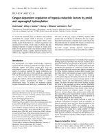
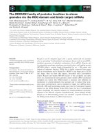
Fig. 1. Method fo r data analysis. Cells were stimulated once un der control conditions (A), switched to test co nditions, a llowed 10 min to adjust to
changes in conditions, and given four stimulations (B–E) s paced 5 min a part. Comparisons between stimulants (30 l
M
ATP and 300 l
M
ATP, for
example) were made by stimulating individual groups o f PC12 ce lls wit h both stimulants (F,G) under control conditions. The effect of test
conditions on cellular response to a stimulus was determined by dividing the peak current generated by the first st imulation under test conditions (B)
by the peak cu rrent gen erated u nder co ntrol co nditions ( A). The rat io o f F /G was then u sed to s cale the cellular r esponses to the various stimuli and
conditions to a single standard, 300 l
M
ATP under con trol cond itions (Fig. 2). Habituation w as recorded as the peak current of each stimulation in
test conditions (B,C,D,E) divided by the p eak current o f the fi rst stimulation in test conditions (B). The line in the recording h as been en hanced to
allow easier visualization.
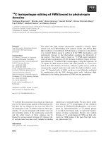
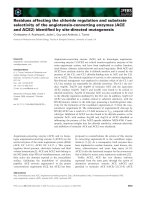
Fig. 2. Initial responses of PC12 cells to stimulation by A TP and
2MeSATP. Responses were normalized as described in the Materials
and methods a nd Fig. 1. ÔBCKÕ indicates t he presence of 100 l
M
UTP
in the background solu tion. ÔCo-StÕ indic ates t he use o f 1 00 l
M
UTP as
a costimulant. An asterisk indic ates a significant d ifference fro m 30 l
M
ATP unde r test conditions to 30 l
M
ATP under control conditions
(P < 0.05). Double asterisks in dicate a s ignifican t differenc e bet ween
theresponseofPC12cellsto60m
M
2MeSATP under test conditions
and 60 l
M
2MeSATP under control conditions ( P < 0.05). The triple
asterisks indicates a significant difference between the response of
PC12 cells to stimulation with 60 l
M
2MeSATP/100 l
M
UTP in 0 m
M
Mg
2+
and the response to an identical stimulation in 3.0 m
M
Mg
2+
(P <0.05).
Fig. 3. Effect of [ ATP] on habituation o f PC12 cells to ATP. Cells were
stimulated with 30 l
M
ATP (e, n ¼ 14), 300 l
M
ATP (h, n ¼ 16), or
600 l
M
ATP (n, n ¼ 3). Asterisk indicates a significant difference
from the habituation of cells to 300 l
M
ATP (P < 0.05). Error bars
denote one SEM.
4036 J. R. Keath and E. W. Westhead (Eur. J. Biochem. 271) Ó FEBS 2004
the control group runs. Analysis of variance within the
control runs did not reveal significant variation when the
runs were grouped according to day or month, indicating
that the degree of h abituation observed in response to
stimuli is reproducible.
Materials
ATP, BaCl
2
,CaCl
2
, Cytodex 3 beads, fetal bovine serum,
gramicidin, HEPES, KCl, 2MeSATP, MgCl
2
, nicardipine,
and UTP were obtained from S igma (St Louis, MO, USA).
Glucose and K
2
HPO
4
were purchased from Fisher Scientific
(Pittsburgh, PA, USA). Horse serum was purchased from
Intergen (Purchase, New York, NY, USA). Dulbecco’s
medium, penicillin, and streptomycin were purchased from
Life Technologies, Inc. (Grand Island, NY, USA). PC12
cells were a gift from G. Guroff (NICDH, NIH, Bethesda,
MD, USA).
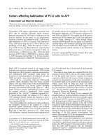
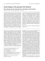
Fig. 5. Effect of prolonged UTP exposure on the habituation of PC12
cells to ATP (A, squares) or 2MeSATP (B, circles). Cells were stimu-
lated with 300 l
M
ATP or 60 l
M
2MeSATP in either a r eg u lar L o ck e’s
solution (open symbols, n ¼ 16 for ATP, 11 for 2MeSATP) or in a
background solu tion co ntaining 100 l
M
UTP (solid symbols, n ¼ 3for
ATP, 3 f or 2MeSATP). Asterisks in dic ate a significant d ifference from
the h abituation of cells in the Loc ke’s solution. Error b ars denote one
SEM.
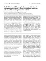
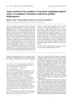
Fig. 4. Effect of Mg
2+
on the habituation o f PC12 cells to 30 l
M
ATP
(A), 60 l
M
2MeSATP (B) and 60 l
M
2MeSATP with 100 l
M
UTP
(C). All cells were stimulated once in Locke’s solution containing
1.2 m
M
Mg
2+
before switching to solutions in which the [Mg
2+
]was
adjusted to 0.0 m
M
Mg
2+
(solid symbols, solid lines, n ¼ 3forATP,3
for 2MeSATP, 3 for 2MeSATP with UTP), 1.2 m
M
Mg
2+
(open
symbols, solid line, n ¼ 14 for ATP, 11 for 2MeSATP, 3 for 2MeS-
ATP with UTP) or 3 .0 m
M
Mg
2+
(open symbols, d otted lines, n ¼ 3
for ATP, 3 for 2MeSATP, 3 for 2MeSATP with UTP). Asterisk
indicates a significant difference from the ha bituation of cells in 1.2 m
M
Mg
2+
(P < 0.05). Error bars denote one SEM.
Ó FEBS 2004 Factors affecting habituation (Eur. J. Biochem. 271) 4037
Results
To determine how the extent of habituation d epends on the
strength of stimulation, we first altered the strength of
stimulation by changing the concentration of the stimulant,
ATP. The cells were stimulated with three concentrations of
ATP: 30 l
M
, which produces a release of catech olamine
roughly half of the maximum r elease possible (Fig. 2, bar 4);
300 l
M
, commonly used concentration to cause maximum
secretory response (Fig. 2, bar 1); and 600 l
M
,whichgives
the same secretory response as 300 l
M
ATP (data not
shown) but might set in motion ATP-activated processes
with lower sensitivity to ATP than t hose involved in
exocytosis.
The degree of habituation observed when the cells were
stimulated with 30 l
M
ATP ( 81 ± 2%, n ¼ 14) was
significantly less than that seen with 300 l
M
ATP
(72 ± 1%, n ¼ 16) and 600 l
M
ATP (71 ¼ /– 2%, n ¼
3) (Fig. 3). There was no significant difference between the
habituation p roduced by 300 and 600 l
M
ATP. Thus, initial
results suggested that habituation is affected in parallel w ith
the secretory response.
The second way stimulation intensity was modified was
by changing the Mg
2+
concentration. Mg
2+
is known to
complex with ATP [17], altering the balance of free and
complexed ATP. ATP receptors differ in their relative
affinity for ATP and its Mg
2+
complex, thus Mg
2+
lowers
the ionotropic receptor’s a ffinity for ATP, but may not
similarly a ffect other ATP receptors [18,19]. Changing
[Mg
2+
]from0.0to1.2m
M
Mg
2+
halved the initial
secretory response of PC12 cells to 30 l
M
ATP, while an
increase to 3.0 m
M
Mg
2+
reduced the initial secretory
response to a quarter of that seen in 0.0 m
M
Mg
2+
(Fig. 2,
bars 3–5). This is in agreement with the findings of several
groups [18–23]. Mg
2+
concentration had no effect on the
response o f the cells to a saturating concentration of 300 l
M
ATP (data not shown). This is also in agreement with
other groups [19,22]. We therefore focused our attention
on 30 l
M
ATP.
We examined the effect of Mg
2+
on habituation of cells
to 30 l
M
ATP (Fig. 4A). Initial response to 30 l
M
ATP is
twice as great in the 0 m
M
Mg
2+
solution, as in the 1.2 m
M
Mg
2+
solution approximately s imilar to t he difference
between 300 l
M
ATP and 30 l
M
ATP in 1.2 m
M
Mg
2+
.
ANOVA
analysis does not indicate that differences in the
habituation curves o f the three [Mg
2+
] conditions are
statistically significant (0.0 m
M
Mg
2+
¼ 75 ± 2%, n ¼ 3,
1.2 m
M
Mg
2+
¼ 81 ± 2%, n ¼ 14, 3.0 m
M
Mg
2+
¼
83 ± 6%, n ¼ 3). This suggests t hat habituation does not
necessarily correlate with stimulus intensity, and suggests
that other factors may be involved.
ATP activates not only P
2
X receptors but also metabo-
tropic P
2
Y receptors on PC12 cells [24]. Work described in
the introduction suggests a number of possible ways in
which these P
2
Y triggered pathways could affect habitu-
ation. The ATP analog 2MeSATP is a good agonist of the
ionotropic receptor, but unlike ATP has little ability to
activate the phospholipase C pathway [25]. 2MeSATP c an
therefore test t he involvement of the phospholipase C
pathway in the habituation of P
2
X mediated exocytosis.
For these studies, we used 60 l
M
2MeSATP, which
produced a secretory response in 1.2 m
M
Mg
2+
solution
similar to that of 30 l
M
ATP at the same [Mg
2+
]. Figure 2
(bars 6–8) shows the effect of altering the [Mg
2+
]onthe
response o f PC12 cells to 60 l
M
2MeSATP. The response of
the cells in a 0.0-m
M
Mg
2+
solution was significantly higher
than the response in a 1.2-m
M
Mg
2+
solution that, in turn,
was significantly higher than the response in a 3.0 m
M
Mg
2+
solution. As with ATP, Mg
2+
interferes with
exocytosis elicited by 2MeSATP, presumably by interfering
with the binding of 2MeSATP to P
2
XandP
2
Y receptors.
PC12 cells in 0.0 m
M
and 1.2 m
M
Mg
2+
habituated to
60 l
M
2MeSATP (0.0 m
M
Mg
2+
¼ 72 ± 3%, n ¼ 3,
1.2 m
M
Mg
2+
¼ 76 ± 2%, n ¼ 11) to roughly the same
degree that they did to 3 0 l
M
ATP ( Fig. 4B) . Increa sing the
concentration of external Mg
2+
from 1.2 m
M
to 3.0 m
M
,
however, virtually eliminated habituation to 2MeSATP
(1.02 ± 4%, n ¼ 3). This clearly shows that habituation is
Fig. 6. Other factor s affecting habituation to ATP. (A) Effect o f the
L
-type VOCC b lock er n icardipine on the habituation o f PC12 cells to
300 l
M
ATP. Cells were stimulated with 300 l
M
ATP in normal
Locke’s solution (h, n ¼ 16) or a solution containing 10 l
M
nicardi-
pine (j, n ¼ 3). (B) Comparison of cells desens itized to 300 l
M
ATP
in back ground solutions containing either 2.2 m
M
Ca
2+
(h, n ¼ 16)
or 0.6 m
M
Ba
2+
(j, n ¼ 3). An asterisk indicates a significant differ-
ence from the h abituation of cells under control conditions. Error bars
denote one SEM.
4038 J. R. Keath and E. W. Westhead (Eur. J. Biochem. 271) Ó FEBS 2004
not a necessary consequence of stimulation. It takes more
than simple activation of P
2
X receptors to desensitize them.
The uncoupling of secretion and habituation shown in
Fig. 5 suggests that one or more metabotropic purinergic
receptors involved in habituation are more sensitive to
Mg
2+
than the P
2
X receptor.
An establishe d difference between ATP and 2MeSATP is
that the latter does not activate the phospholipase C
pathway in P C12 cells. UTP is a specific P
2
Y agonist that
activates this pat hway [ 26]. If this pathway promotes
habituation to ATP in 3.0 m
M
[Mg
2+
] where none is seen
to 2MeSATP, UTP might restore habituation by activating
that pathway.
When UTP was used as a costimulant, it caused no
significant change in initial secretory response at 0 m
M
Mg
2+
, but significantly decreased the effect of increasing
[Mg
2+
] on e xocytosis elicited from the cells (compare Fig. 2,
bars 6–8 with 10–12). UTP a lone did not produce a
significant amount of exocytosis in our PC12 cells, ruling
out direct stimulation of P
2
X receptors by UTP. A
background solution containing UTP d oes not affect
secretion in response to 2MeSATP (compare Fig. 2, bars
7 and 9), showing that UTP is not affecting secretion by
sequestering Mg
2+
, in agreement with published dissoci-
ation constants (not shown). It seems likely that the
synergistic increase in secretion is due to the Ca
2+
released
by UTP from internal stores. While insufficient to trigger
substantial secretion, it reduces the diffusion of Ca
2+
entering through the ion channels, thus increasing the
effective [Ca
2+
] at the secretory sites.
At 0.0 and 1.2 m
M
Mg
2+
, habituation to costimulations
with 2MeSATP a nd UTP w ere not significantly greater than
habituation to 2MeSATP alone (Fig. 4C) (0.0 m
M
Mg
2+
¼ 65 ± 1%, n ¼ 3, 1.2 m
M
Mg
2+
¼ 69 ± 1%,
n ¼ 3). While UTP did not completely restore habituation
to 2MeSATP a t 3.0 m
M
Mg
2+
to levels seen when ATP w as
the stimulant, it did significantly increase it (78 ± 3%, n ¼
3). Therefore the differenc e in the effect of high [Mg
2+
]on
the habituation of cells to ATP and 2MeSATP can be
attributed in part to metabotropi c activity stimulated via the
UTP-sensitive P
2
Y receptor.
Having examined the effect that costimulation with UTP
had on the response and habituation of PC12 cells to
2MeSATP and ATP, we then looked at the impact of
including UTP in the background solution. We hypothes-
ized that the second messenger activity required f or
habituation can be triggered b y UTP, so t hat activating
the UTP pathway continuously could e ither increase
habituation by priming the inactivating pathway or reduce
habituation by desensitizing the inactivatory pathway.
Figure 2 (bars 1, 2, 7, and 9) shows that a continuous
application of 100 l
M
UTP in the background solution had
no significant effect on the initial response of cells to either
300 l
M
ATP or 60 l
M
2MeSATP. In contrast, Fig. 5(A,B)
shows that a background of 100 l
M
UTP s ignificantly
increased the habituation of PC12 cells to both A TP
(51 ± 2%, n ¼ 3) and 2MeSATP (55 ± 1%, n ¼ 3)
stimulations. This is a very different outcome from that
observed when UTP was used as a costimulant. UTP
costimulation increased secretory response, but did not
affect habituation. We have suggested that UTP’s effect on
secretion w as due to Ca
2+
released from internal stores. It i s
reasonable to suggest that after 10 min of continuous UTP
stimulation, the released Ca
2+
has been sequestered and
removed from the internal milieu. This would explain why
UTP i n t he background did n ot increase secretion. The
impact of UTP on habituation will be addressed in the
discussion.
Studies by Fasolato et al. [21] and our labo ratory
(G. Balan, unpublished data) suggested that cation influx
through P
2
X receptor-channels during ATP stimulation is
sufficient to activate VOCCs, allowing Ca
2+
to enter the
cell. More recently studies have confirmed this pathway and
investigated it in detail [27]. However, several researchers
[3,28–31] have demonstrated that treatment with VOCC
blockers does not affect the total amount of Ca
2+
that
enters a cell during ATP stimulation.
We explored the possible role of the
L
-type VOCC in
habituation by looking at both the initial response and the
habituation of PC12 cells to ATP in the presence of the
VOCC blocker nicardipine (10 l
M
). As with other experi-
ments in which the background solution was altered, the
cells were exposed to nicardipine for 10 min before being
stimulated to ATP o r 2 MeSATP. This p rovided ample time
for nicardipine to block
L
-type VOCC activity.
Nicardipine d id not significantly affect the response of t he
cells in any case (data not shown), in agreement with
findings quoted above but in contrast to the result of Kim’s
laboratory [ 20]. In contrast to the lack of effect of
nicardipine on the initial response, Fig. 6A shows that
10 l
M
nicardipine increases habituation of PC12 cells to
300 l
M
ATP (48% ± 2%, n ¼ 10). Similar effects were
observed when 3 0 l
M
ATP and 60 l
M
2MeSATP were u sed
as stimulants (data not shown). Even though Ca
2+
influx
through the
L
-type VOCCs appears to have little role in
secretion during ATP stimulation, it does decrease habitu-
ation.
Nakazawa and collaborators [30,32] have demonstrated
that high levels of [Ca
2+
]
in
can prevent ion flow through
bothVOCCsandP
2
X receptor-channels in PC12 cells.
Others [33–35] have demonstrated that this inhibition of ion
flow through VOCCs is likely due to Ca
2+
directly binding
to a cytosolic region of the channels. To assess the effects
that this might have on habituation, the 2.2 m
M
Ca
2+
in the
external solution was replaced with 0.6 m
M
Ba
2+
,which
triggers exocytosis in a manner and magnitude similar to
Ca
2+
, but does not inactivate ion channels to as great a
degree [13].
Figure 6B shows that replacing 2.2 m
M
Ca
2+
with
0.6 m
M
Ba
2+
produced a dramatic increase in the degree
of habituation produced by 300 l
M
ATP (42% ± 2%,
n ¼ 3). T his supports the i dea that blockage o f ion channels
by high [Ca
2+
]
in
can decrease the habituation of PC12 cells
to ATP.
Discussion
Although this paper represents only a beginning in the study
of habituation to ATP, three important findings are clearly
demonstrated. T he first is that habituation does not
necessarily correspond with either stimulus intensity or
amount of secretion. Support for this comes from the study
employing 2 MeSATP in the presence of 3.0 m
M
Mg
2+
.
2MeSATP (60 l
M
) stimulation produces a secretory
Ó FEBS 2004 Factors affecting habituation (Eur. J. Biochem. 271) 4039
response approximating that of 30 l
M
ATP, and the
secretion produced by both stimuli are similarly reduced
by the increase in [Mg
2+
], yet in 3.0 m
M
Mg
2+
habituation
to ATP is unchanged while habituation to 2MeSATP is
essentially eliminated. The secretory responses are nearly
identical, but habituation patterns are dramatically differ-
ent. Support for this finding can also be provided by
comparing the effects of UTP as a costimulant and UTP in
the background solution. When UTP was used as a
costimulant, it increased 2MeSATP induced secretion, but
had n o effect on habituation. While UTP in the background
solution did not increase secretion, it produced a dramatic
increase in habitu ation. Our d ata t herefore shows t hat there
is no necessary correlation between habituation and stimu-
lus intensity or level of secretion.
The second significant finding is that there is a role for
multiple purinergic receptor types in the habituation
process. This is shown most clearly in the lack of habitu-
ation of cells to multiple stimulation with 2MeSATP in the
presence of 3.0 m
M
Mg
2+
, in contrast to the habituation to
ATP observed a t the same [Mg
2+
] and an equivalent level of
secretion. The fact that the combination of U TP and
2MeSATP causes habituation intermediate between ATP
alone and 2 MeSATP indicates that the UTP-sensitive P
2
Y
purinergic receptor likely plays a role but is not the only
metabotropic purinergic receptor i nvolved i n habituation. If
it were, we would expect complete recovery of habituation,
instead of partial recovery. The UTP-sensitive P
2
Y receptor
activates phospholipase C, leading to release of Ca
2+
from
subcellular stores and activation of protein kinase C. Other
purinergic m etabotropic r eceptors can activate other second
messenger pathways. Due to the complexity of purinergic
signaling pathways, it may be very difficult to determine the
exact pathway leading to habituation until more specific
antagonists become available.
The third important finding is that factors that modify
Ca
2+
influx affect the habituation process, as shown by
increased habituation when
L
-type VOCCs are blocked by
nicardipine. Ca
2+
regulation of the habituation process is
also demonstrated by increased habituation when Ba
2+
is
used in place of Ca
2+
to support secretion. These conclu-
sions are in accord with previous work showing inactivation
of VOCCs and ATP gated channels by Ca
2+
[30,32] and
with recent work showing a Ca
2+
effect on habituation of
P
2
X channels using patch clamp methods [13].
To explain how blocking
L
-type VOCCs could increase
habituation, we make four postulations. We first postu-
late that habituation is due to the desensitization of P
2
X
receptors. This is reasonable given previous findings [8–
10,14]. Second, we postulate that P
2
X channels must be
in the open, active, state for desensitization to occur. The
need is shown in the experiments where UTP was present
in the background solution prior to and during habitu-
ation. It is important to note that background UTP does
not affect the initial response to ATP, only the
subsequent ones, i.e. the habituation process. This clearly
shows that while the cell is primed for h abituation, the
process requires activation of the P
2
X receptor. Third, we
postulate that inactivation of P
2
X receptors due to direct
Ca
2+
binding, as described by Nakazawa and Hess [32],
is more rapidly reversible than the longer term desensi-
tization triggered by the P2Y pathway. Finally, we
postulate that the Ca
2+
block protects these receptor-
channels from the longer term desensitization.
During ATP stimulation, Ca
2+
will enter the c ell through
both t he P 2X receptors and any VOCCs on the cell
membrane. Internal [Ca
2+
] will rise rapidly, therefore Ca
2+
blockage and protection of the P
2
X channel will be rapid,
allowing little opportunity for P
2
Y-dependent desensitiza-
tion to occur. If the
L
-type channels are blocked, Ca
2+
will
enter the cell more slowly and take longer to reach c hannel-
inactivating concentrations. This will allow a greater
window of opportunity for t he desensitization of P
2
X
receptor. With or without
L
-type channels, Ca
2+
influx will
continue until [Ca
2+
]
in
reaches levels which block first the
VOCCs and then the P
2
X receptor-channels. B locking
VOCCs can therefore increase the likelihood of P
2
X
desensitization without affecting total Ca
2+
influx.
Our explanation allows us to accou nt for the increase in
habituation observed when Ca
2+
is replaced with Ba
2+
.A
higher internal concentration of Ba
2+
is required to
inactivate the P
2
X receptor-channels [13,30]. This will
extend the time t hat these channels are a ctive, and t herefore
vulnerable to the desensitization processes.
This interpretation also allows a potential explanation of
the activity of VOCC blockers o n the response of the cells to
ATP stimulation. Variation between strains of PC12 cells
will likely include differences in ion channel densities. In
strains where the density of P
2
X receptors is sufficient to
trigger maximum exocytosis, VOCCs will merely contribute
totherateofCa
2+
influx, not the fin al [Ca
2+
]. In strains
where P
2
X receptor density is smaller, VOCCs may have a
greater effect.
Finally, our explanation of the mechanics of ATP
habituation also allows us to explain a finding of Cheever
and Koshland [8] in which they found that desensitizing
PC12 cells to depolarization did not desensitize them to ATP,
but did increase the rate at which they desensitized to ATP.
When they desensitized their cells to depolarization, they
inactivated the voltage-operated c hannels. A ccording to o ur
explanation, this loss of VOCC activity would not decrease
the response to ATP, but it would increase the amoun t of
time that the P
2
X receptor-channels remained open during
stimulation. This longer time wou ld result in a greater
opportunity for the habituation process to take place, and
therefore a greater degree of observed habituation.
In summary, we have pr ovided evidence that habituation
of PC12 cells to ATP is a proc ess separate from the secretory
process and that it involves P
2
Y receptor pathways. We
have also produced a model that a llows for the contribution
of VOCCs to Ca
2+
influx and a role in habituation during
ATP stimulation without affecting the secretion that this
stimulation produces.
Acknowledgement
We are grateful to Dr. David G ross for h elpful discussions and
suggestions.
References
1. Burnstock, G. (1997) The past, present and future of purine
nucleotides as signaling molecules. Neuropharmacology 36, 1127–
1139.
4040 J. R. Keath and E. W. Westhead (Eur. J. Biochem. 271) Ó FEBS 2004
2. Greene, L.A. & Rein, G. (1977) Release of (
3
H)norepinephrine
from a clonal line of p heochromocytoma cells (PC12) by nicotinic
cholinergic stimulation. Brain Res. 138, 521–528.
3. Inoue, K., Nakazawa, K., Fujimori, K. & Takanaka, A. (1989)
Extracellular adenosine 5¢-triphosphate-evoked norepinephrine
secretion not relating to voltage-gated Ca channels in pheo-
chromocytom a P C12 ce lls. Neurosci. Lett. 106, 294–299.
4. Greene, L.A. & Rein, G. (1977) Release, storage and uptake of
catecholamines by a clonal cell lin e of nerv e growth fa ctor (NGF)
responsive pheo-chromoc ytoma cells. Brain Res. 129, 247–263.
5. Fred holm, B.B., Abbracchio, M.P., Burnstock, G., Daly, J.W.,
Harden, T.K., Jacobson, K.A., Leff, P. & Williams, M. (1994)
Nomenclature and classific ation of purinoceptors. Pharmacol.
Rev. 46, 143–156.
6. Nakazawa, K ., Fujimori, K., T akanaka, A. & Inoue, K. (1990) An
ATP-activated conductance in pheochromocytoma cells and its
suppression by extracellular calcium. J. Physiol. 428, 257–272.
7. Nakazawa, K., Fujimori, K., Takanaka, A. & Inoue, K. (1991)
Comparison of adenosine triphosphate- and nicotine-activated
inward currents in rat phaeochromocytom a cells. J. Ph ysiol. 434,
647–660.
8. Cheever, L. & Koshland, D.E. Jr (1994) Habituation of neuro-
secretory responses to extracellular ATP in P C12 cells. J. Neurosci.
14, 4831–4838.
9. Chow, Y.W. & Wang, H.L. (1998) Functional modulation of
P
2
X
2
receptors by cyclic AMP-dependent protein kinase. J. Neu-
rochem. 70, 2606–2612.
10. Cheever, L. & Koshland, D.E. J r (1992) Retention o f habituation
in PC12 cells. Proc. Natl Acad. Sci. USA 89, 10084–10088.
11. Ding, S. & Sachs, F. (1999) Single channel properties of P
2
X
2
purinoceptors. J. Ge n. Physiol. 113, 695–720.
12. North, R.A. (2002) Molecular physiology of P2X receptors.
Physiol. Rev. 82, 1013–1067.
13. Ding, S. & Sac hs, F. (2000) Inactivation of P
2
X
2
purinoceptors by
divalent cations. J. Physiol. 522, 199–214.
14. Chen, C. & Bobbin, R.P. (1998) P
2
X receptors in cochlear Deiters’
cells. Br.J.Pharmacol.124, 337–344.
15. Boue-Grabot, E., Archambault, V . & Seguela, P. (2000) A protein
kinase C site highly c onserved in P
2
X subunits controls the
desensitization kinetics of P
2
X(2) ATP-gated channels. J. Biol.
Chem. 275, 10190–10195.
16. Brandle , U., Spielmanns, P., Osteroth, R., Sim, J., Surprenant, A.,
Buell, G., Ru ppersberg, J.P., Plinkert, P.K., Zenne r, H.P. &
Glowatzki, E. (1984) Desensitization of the P
2
X(2) receptor con-
trolled by alternative splicing. FEBS Lett. 404, 294–298.
17. Pecoraro, V.L., Hermes, J.D. & Cleland, W.W. (1984) Stability
constants of Mg
2+
and Cd
2+
complexes of adenine nucleotides
and thionucleotides and rate constants for formation and disso-
ciation of MgATP and MgADP. Biochemistry 23, 5262–5271.
18. Reichsman, F., S antos, S. & Westhead, E.W. (1995) Two d istinct
ATP r eceptors activate calcium entry and internal calcium release
in bovine chromaffin cells. J. Neurochem. 65, 2080–2086.
19. Rhoads,A.R.,Parui,R.,Vu,N.D.,Cadogan,R.&Wagner,P.D.
(1993) ATP-induced secretion in PC12 cells and photoaffinity
labeling of receptors. J. Neur och em. 61 , 1657–1666.
20. Choi, S.Y. & Kim, K.T. (1996) Characterization of Na
+
influx
mediated by ATP(4-)-activated P2 purinoceptors in PC12 cells.
Br.J.Pharmacol.118, 935–940.
21. Fasolato, C., Pizzo, P. & Pozzan, T. (1990) Receptor-mediated
calcium influx in PC12 cells. ATP and bradykinin a ctivate two
independent pathways. J. Biol. Chem. 265, 20351–20355.
22. Kim, W.K. & Rabin, R.A. (1994) Characterization of the pur-
inergic P2 receptors in PC12 cells: evidence for a novel subtype.
J. Biol. Chem. 269, 6471–6477.
23. Trezise,D.J.,Bell,N.J.,Kennedy,I.&Humphrey,P.P.(1994)
Effects of divalent cations on the potency of ATP and related
agonists in the rat isolated vagus nerve: implications for P2 pur-
inoceptor classification. Br. J. Pharmacol. 113, 463–470.
24. Unterberger, U., Moskvina, E., Scholze, T., Freissmuth, M. &
Boehm, S. (2002) Inhibition of adenylyl cyclase by neuronal P
2
Y
receptors. B r. J. P ha rmac ol. 135, 673–684.
25. Nikodijevic, B., Sei, Y., Shin,Y.&Daly,J.W.(1994)Effectsof
ATP and UTP in pheochromocytoma PC12 cells: evidence for
thepresenceofthreeP2receptors, only one of which subserves
stimulation of norepinephrine release. Cell.Mol.Neurobiol.14,
27–47.
26. Koizumi, S., Nakazawa, K. & Inoue, K. (1995) Inhibition by
Zn
2+
of uridine 5¢-triphosphate-induced Ca
2+
-influx but not
Ca
2+
-mobilization in rat phaeochromocytoma cells. Br.J.Phar-
macol. 115, 1502–1508.
27. Hur, E.M., Park, T.J. & Kim, K.T. (2001) Coupling of 1-type
voltage-sensitive c alcium channels to P
2
X(2) p urino ceptors in P C-
12 cells. Am. J. Physiol. Cell. Physiol. 280, C1121–C1129.
28.Grohovaz,F.,Zacchetti,D.,Clementi,E.,Lorenzon,P.,
Meldolesi, J. & Fumagalli, G. (1991) [Ca
2+
]
i
imaging in PC12
cells: multiple response pattern s t o receptor ac tivatio n reveal
new aspects of transmembrane signaling. J. Cell Biol. 113 , 1341–
1350.
29. Michel, A.D., Grahames, C.B. & Humphrey, P.P. (1996) Func-
tional characterization of P2 purinoceptors in P C12 c ells by
measurement of radiolabelled calcium influx. Naunyn Schmiede-
bergs Arch. Pharmacol. 354, 562–571.
30. Nakazawa, K. & Inoue, K. (1992) Roles of Ca
2+
influx through
ATP-activated channe ls in catecholamine relea se from pheo-
chromocytom a P C12 ce lls. J. Neurophysiol. 68, 2026–2032.
31. Raha, S., de Souza, L.R. & Reed, J.K. (1993) Intracellular sig-
nalling by nucleotide receptors in PC12 pheochromocytoma cells.
J. Cell. Physiol. 154, 623–630.
32. Nakazawa, K. & Hess, P. (1993) Block by calcium of ATP-acti-
vated channels in pheochromocytoma cells. J. Gen. Physiol. 101,
377–392.
33. de Leon, M., Wang, Y., Jones, L., Perez-Reyes, E., Wei, X.,
Soong, T.W., Snutch, T.P. & Yue, D.T. (1995) Essential Ca
2+
-
binding motif for C a
2+
-sensitive inactivation of 1-type C a
2+
channels. Science 270, 1502–1506.
34. Haack, J.A. & Rosenberg, R.L. ( 1994) Calcium-dependent
inactivation of 1-type calcium channels in planar lipid bilayers.
Biophys. J. 66, 1051–1060.
35. Imredy, J.P. & Yue, D.T. (1994) Mechanism of Ca
2+
-sensitive
inactivation of 1-type Ca
2+
channels. Neuron 12, 1301–1318.
Ó FEBS 2004 Factors affecting habituation (Eur. J. Biochem. 271) 4041