Plant physiology - Chapter 8 Photosynthesis: Carbon Reactions pdf
Bạn đang xem bản rút gọn của tài liệu. Xem và tải ngay bản đầy đủ của tài liệu tại đây (526.6 KB, 26 trang )
Photosynthesis:
Carbon Reactions
8
Chapter
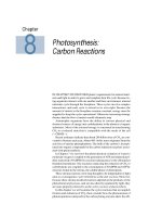
IN CHAPTER 5 WE DISCUSSED plants’ requirements for mineral nutri-
ents and light in order to grow and complete their life cycle. Because liv-
ing organisms interact with one another and their environment, mineral
nutrients cycle through the biosphere. These cycles involve complex
interactions, and each cycle is critical in its own right. Because the
amount of matter in the biosphere remains constant, energy must be
supplied to keep the cycles operational. Otherwise increasing entropy
dictates that the flow of matter would ultimately stop.
Autotrophic organisms have the ability to convert physical and
chemical sources of energy into carbohydrates in the absence of organic
substrates. Most of the external energy is consumed in transforming
CO
2
to a reduced state that is compatible with the needs of the cell
(—CHOH—).
Recent estimates indicate that about 200 billion tons of CO
2
are con-
verted to biomass each year. About 40% of this mass originates from the
activities of marine phytoplankton. The bulk of the carbon is incorpo-
rated into organic compounds by the carbon reduction reactions associ-
ated with photosynthesis.
In Chapter 7 we saw how the photochemical oxidation of water to
molecular oxygen is coupled to the generation of ATP and reduced pyri-
dine nucleotide (NADPH) by reactions taking place in the chloroplast
thylakoid membrane. The reactions catalyzing the reduction of CO
2
to
carbohydrate are coupled to the consumption of NADPH and ATP by
enzymes found in the stroma, the soluble phase of chloroplasts.
These stroma reactions were long thought to be independent of light
and, as a consequence, were referred to as the dark reactions. However,
because these stroma-localized reactions depend on the products of the
photochemical processes, and are also directly regulated by light, they
are more properly referred to as the carbon reactions of photosynthesis.
In this chapter we will examine the cyclic reactions that accomplish
fixation and reduction of CO
2
, then consider how the phenomenon of
photorespiration catalyzed by the carboxylating enzyme alters the effi-
ciency of photosynthesis. This chapter will also describe
biochemical mechanisms for concentrating carbon dioxide
that allow plants to mitigate the impact of photorespira-
tion: CO
2
pumps, C
4
metabolism, and crassulacean acid
metabolism (CAM). We will close the chapter with a con-
sideration of the synthesis of sucrose and starch.
THE CALVIN CYCLE
All photosynthetic eukaryotes, from the most primitive alga
to the most advanced angiosperm, reduce CO
2
to carbohy-
drate via the same basic mechanism: the photosynthetic car-
bon reduction cycle originally described for C
3
species (the
Calvin cycle, or reductive pentose phosphate [RPP] cycle).
Other metabolic pathways associated with the photosyn-
thetic fixation of CO
2
, such as the C
4
photosynthetic carbon
assimilation cycle and the photorespiratory carbon oxida-
tion cycle, are either auxiliary to or dependent on the basic
Calvin cycle.
In this section we will examine how CO
2
is fixed by the
Calvin cycle through the use of ATP and NADPH generated
by the light reactions (Figure 8.1), and how the Calvin cycle
is regulated.
The Calvin Cycle Has Three Stages:Carboxylation,
Reduction,and Regeneration
The Calvin cycle was elucidated as a result of a series of
elegant experiments by Melvin Calvin and his colleagues
in the 1950s, for which a Nobel Prize was awarded in 1961
(see
Web Topic 8.1). In the Calvin cycle, CO
2
and water
from the environment are enzymatically combined with a
five-carbon acceptor molecule to generate two molecules
of a three-carbon intermediate. This intermediate (3-phos-
phoglycerate) is reduced to carbohydrate by use of the ATP
and NADPH generated photochemically. The cycle is com-
pleted by regeneration of the five-carbon acceptor (ribu-
lose-1,5-bisphosphate, abbreviated RuBP).
The Calvin cycle proceeds in three stages (Figure 8.2):
1. Carboxylation of the CO
2
acceptor ribulose-1,5-bispho-
sphate, forming two molecules of 3-phosphoglycerate,
the first stable intermediate of the Calvin cycle
2. Reduction of 3-phosphoglycerate, forming gyceralde-
hyde-3-phosphate, a carbohydrate
3. Regeneration of the CO
2
acceptor ribulose-1,5-bisphos-
phate from glyceraldehyde-3-phosphate
The carbon in CO
2
is the most oxidized form found in
nature (+4). The carbon of the first stable intermediate, 3-
phosphoglycerate, is more reduced (+3), and it is further
reduced in the glyceraldehyde-3-phosphate product (+1).
Overall, the early reactions of the Calvin cycle complete the
reduction of atmospheric carbon and, in so doing, facilitate
its incorporation into organic compounds.
The Carboxylation of Ribulose Bisphosphate Is
Catalyzed by the Enzyme Rubisco
CO
2
enters the Calvin cycle by reacting with ribulose-1,5-
bisphosphate to yield two molecules of 3-phosphoglycerate
(Figure 8.3 and Table 8.1), a reaction catalyzed by the chloro-
plast enzyme ribulose bisphosphate carboxylase/oxy-
genase, referred to as rubisco (see
Web Topic 8.2). As indi-
146 Chapter 8
Light
Light reactions
Chlorophyll
Carbon reactions
Triose
phosphates
O
2
H
2
O
CO
2
+
H
2
O
(CH
2
O)
n
NADP
+
ADP
P
i
NADPH
ATP
+
+
FIGURE 8.1 The light and carbon reactions of photosynthe-
sis. Light is required for the generation of ATP and
NADPH. The ATP and NADPH are consumed by the car-
bon reactions, which reduce CO
2
to carbohydrate (triose
phosphates).
ADP
NADPH
ATP
ATP
+
NADP
+
ADP
P
i
+
CO
2
+
H
2
O
Start of cycle
3-phosphoglycerate
Ribulose-1,5-
bisphosphate
Glyceraldehyde-3-
phosphate
Sucrose, starch
Regeneration
Carboxylation
Reduction
FIGURE 8.2 The Calvin cycle proceeds in three stages: (1)
carboxylation, during which CO
2
is covalently linked to a
carbon skeleton; (2) reduction, during which carbohydrate
is formed at the expense of the photochemically derived
ATP and reducing equivalents in the form of NADPH; and
(3) regeneration, during which the CO
2
acceptor ribulose-
1,5-bisphosphate re-forms.
HC
C
CH
2
OP
OH
O
HOH
C
CH
2
OPO
3
2–
COO
–
C
HOH
CH
2
OP
C
HOH
CH
2
OP
CH
2
OPO
3
2–
CH
2
OP
O
C
HO
CO
H
OH
OH
H
H
C
C
C
CH
2
OH
C
O
3 CO
2
3 H
2
O
6 H
+
Ribulose
1,5-bisphosphate
1,3-bisphosphoglycerate
3-phosphoglycerate
Rubisco
Phosphoglycerate
kinase
Glyceraldehyde
3-phosphate
dehydrogenase
Glyceraldehyde
3-phosphate
NADPH
NADP
+
ADP
6 ATP
3 ADP
3 ATP
P
i
P
i
6
OP
CH
2
OP
C
HOH
CH
2
OP
O
C
H
C
HOH
C
HOH
CH
2
OP
O
C
H
6
+
6 H
+
+
6
6
Triose
phosphate
G3P DHAP
Dihydroxy-
acetone
phosphate
Dihydroxy-
acetone
phosphate
CH
2
OH
C
O
CH
2
OP
Triose
phosphate
isomerase
CH
2
OPO
3
2–
CH
2
OP
HO
CO
H
OH
OH
H
H
C
C
C
OH
H
C
Fructose
1,6-bisphosphate
Fructose
1,6-bisphosphatase
CH
2
OH
CH
2
OP
HO
CO
H
OH
OH
H
H
C
C
C
Fructose
6-phosphate
CH
2
OH
CH
2
OP
HO
CO
H
OH
H
C
C
Xylulose
5-phosphate
CH
2
OH
CH
2
OP
HO
CO
H
OH
H
C
C
Xylulose
5-phosphate
CH
2
OH
CH
2
OP
H
CO
OH
OH
H
C
C
Ribulose
5-phosphate
CH
2
OH
CH
2
OP
H
CO
OH
OH
H
C
C
Ribulose
5-phosphate
O
C
H
CH
2
OP
HOH
OH
H
C
HOH
C
C
Ribose
5-phosphate
Aldolase
H
2
O
P
i
H
2
O
Transketolase
Transketolase
Aldolase
Erythrose
4-phosphate
Ribulose
5-phosphate
3-epimerase
Phosphoribulokinase
Sedoheptulose
1,7-bisphosphate
Sedoheptulose
1,7-bisphosphatase
CH
2
OH
CH
2
OP
HO
CO
H
OH
OH
H
H
C
C
C
OH
H
C
Sedoheptulose
7-phosphate
CH
2
OH
CH
2
OP
H
CO
OH
OH
H
C
C
Ribulose
5-phosphate
Ribulose
5-phosphate
isomerase
Ribulose
5-phosphate
3-epimerase
FIGURE 8.3 The Calvin cycle. The carboxylation of three molecules of ribulose-1,5-
bisphosphate leads to the net synthesis of one molecule of glyceraldehyde-3-phos-
phate and the regeneration of the three molecules of starting material. This process
starts and ends with three molecules of ribulose-1,5-bisphosphate, reflecting the
cyclic nature of the pathway.
cated by the full name, the enzyme also has an oxygenase
activity in which O
2
competes with CO
2
for the common
substrate ribulose-1,5-bisphosphate (Lorimer 1983). As we
will discuss later, this property limits net CO
2
fixation.
As shown in Figure 8.4, CO
2
is added to carbon 2 of ribu-
lose-1,5-bisphosphate, yielding an unstable, enzyme-bound
intermediate, which is hydrolyzed to yield two molecules of
the stable product 3-phosphoglycerate (see Table 8.1, reac-
tion 1). The two molecules of 3-phosphoglycerate—labeled
“upper” and “lower” on the figure—are distinguished by
the fact that the upper molecule contains the newly incor-
porated carbon dioxide, designated here as *CO
2
.
Two properties of the carboxylase reaction are especially
important:
1. The negative change in free energy (see Chapter 2 on
the web site for a discussion of free energy) associated
with the carboxylation of ribulose-1,5-bisphosphate is
large; thus the forward reaction is strongly favored.
2.The affinity of rubisco for CO
2
is sufficiently high to
ensure rapid carboxylation at the low concentrations
of CO
2
found in photosynthetic cells.
Rubisco is very abundant, representing up to 40% of the
total soluble protein of most leaves. The concentration of
rubisco active sites within the chloroplast stroma is calcu-
lated to be about 4 mM, or about 500 times greater than the
concentration of its CO
2
substrate (see Web Topic 8.3).
Triose Phosphates Are Formed in the Reduction
Step of the Calvin Cycle
Next in the Calvin cycle (Figure 8.3 and Table 8.1), the 3-
phosphoglycerate formed in the carboxylation stage under-
goes two modifications:
1. It is first phosphorylated via 3-phosphoglycerate
kinase to 1,3-bisphosphoglycerate through use of the
ATP generated in the light reactions (Table 8.1, reac-
tion 2).
2. Then it is reduced to glyceraldehyde-3-phosphate
through use of the NADPH generated by the light
reactions (Table 8.1, reaction 3). The chloroplast
enzyme NADP:glyceraldehyde-3-phosphate dehy-
drogenase catalyzes this step. Note that the enzyme
is similar to that of glycolysis (which will be dis-
148 Chapter 8
TABLE 8.1
Reactions of the Calvin cycle
Enzyme Reaction
1. Ribulose-1,5-bisphosphate carboxylase/oxygenase 6 Ribulose-1,5-bisphosphate + 6 CO
2
+ 6 H
2
O →
12 (3-phosphoglycerate) + 12 H
+
2. 3-Phosphoglycerate kinase 12 (3-Phosphoglycerate) + 12 ATP →
12 (1,3-bisphosphoglycerate) + 12 ADP
3. NADP:glyceraldehyde-3-phosphate dehydrogenase 12 (1,3-Bisphosphoglycerate) + 12 NADPH + 12 H
+
→
12 glyceraldehye-3-phosphate + 12 NADP
+
+ 12 P
i
4. Triose phosphate isomerase 5 Glyceraldehyde-3-phosphate →
5 dihydroxyacetone-3-phosphate
5. Aldolase 3 Glyceraldehyde-3-phosphate + 3 dihydroxyacetone-
3-phosphate → 3 fructose-1,6-bisphosphate
6. Fructose-1,6-bisphosphatase 3 Fructose-1,6-bisphosphate + 3 H
2
O → 3 fructose-
6-phosphate + 3 P
i
7. Transketolase 2 Fructose-6-phosphate + 2 glyceraldehyde-3-phosphate →
2 erythrose-4-phosphate + 2 xylulose-5-phosphate
8. Aldolase 2 Erythrose-4-phosphate + 2 dihydroxyacetone-3-phosphate →
2 sedoheptulose-1,7-bisphosphate
9. Sedoheptulose-1,7,bisphosphatase 2 Sedoheptulose-1,7-bisphosphate + 2 H
2
O → 2 sedoheptulose-
7-phosphate + 2 P
i
10. Transketolase 2 Sedoheptulose-7-phosphate + 2 glyceraldehyde-3-phosphate →
2 ribose-5-phosphate + 2 xylulose-5-phosphate
11a. Ribulose-5-phosphate epimerase 4 Xylulose-5-phosphate → 4 ribulose-5-phosphate
11b. Ribose-5-phosphate isomerase 2 Ribose-5-phosphate → 2 ribulose-5-phosphate
12. Ribulose-5-phosphate kinase 6 Ribulose-5-phosphate + 6 ATP → 6 ribulose-1,5-bisphosphate +
6 ADP + 6 H
+
Net: 6 CO
2
+ 11 H
2
O + 12 NADPH + 18 ATP → Fructose-6-phosphate + 12 NADP
+
+ 6 H
+
+ 18 ADP + 17 P
i
Note:P
i
stands for inorganic phosphate.
cussed in Chapter 11), except that NADP rather than
NAD is the coenzyme. An NADP-linked form of the
enzyme is synthesized during chloroplast develop-
ment (greening), and this form is preferentially used
in biosynthetic reactions.
Operation of the Calvin Cycle Requires the
Regeneration of Ribulose-1,5-Bisphosphate
The continued uptake of CO
2
requires that the CO
2
accep-
tor, ribulose-1,5-bisphosphate, be constantly regenerated.
To prevent depletion of Calvin cycle intermediates, three
molecules of ribulose-1,5-bisphosphate (15 carbons total)
are formed by reactions that reshuffle the carbons from the
five molecules of triose phosphate (5 × 3 = 15 carbons). This
reshuffling consists of reactions 4 through 12 in Table 8.1
(see also Figure 8.3):
1. One molecule of glyceraldehyde-3-phosphate is con-
verted via triose phosphate isomerase to dihydroxy-
acetone-3-phosphate in an isomerization reaction
(reaction 4).
2. Dihydroxyacetone-3-phosphate then undergoes aldol
condensation with a second molecule of glyceralde-
hyde-3-phosphate, a reaction catalyzed by aldolase to
give fructose-1,6-bisphosphate (reaction 5).
3. Fructose-1,6-bisphosphate occupies a key position in
the cycle and is hydrolyzed to fructose-6-phosphate
(reaction 6), which then reacts with the enzyme trans-
ketolase.
4. A two-carbon unit (C-1 and C-2 of fructose-6-phos-
phate) is transferred via transketolase to a third mol-
ecule of glyceraldehyde-3-phosphate to give ery-
throse-4-phosphate (from C-3 to C-6 of the fructose)
and xylulose-5-phosphate (from C-2 of the fructose
and the glyceraldehyde-3-phosphate) (reaction 7).
5. Erythrose-4-phosphate then combines via aldolase
with a fourth molecule of triose phosphate (dihy-
droxyacetone-3-phosphate) to yield the seven-carbon
sugar sedoheptulose-1,7-bisphosphate (reaction 8).
6. This seven-carbon bisphosphate is then hydrolyzed
by way of a specific phosphatase to give sedoheptu-
lose-7-phosphate (reaction 9).
7. Sedoheptulose-7-phosphate donates a two-carbon
unit to the fifth (and last) molecule of glyceralde-
hyde-3-phosphate via transketolase and produces
ribose-5-phosphate (from C-3 to C-7 of sedoheptu-
lose) and xylulose-5-phosphate (from C-2 of the sedo-
heptulose and the glyceraldehyde-3-phosphate)
(reaction 10).
8. The two molecules of xylulose-5-phosphate are con-
verted to two molecules of ribulose-5-phosphate sug-
ars by a ribulose-5-phosphate epimerase (reaction
11a). The third molecule of ribulose-5-phosphate is
formed from ribose-5-phosphate by ribose-5-phos-
phate isomerase (reaction 11b).
9. Finally, ribulose-5-phosphate kinase catalyzes the phos-
phorylation of ribulose-5-phosphate with ATP, thus
regenerating the three needed molecules of the initial
CO
2
acceptor, ribulose-1,5-bisphosphate (reaction 12).
The Calvin Cycle Regenerates Its
Own Biochemical Components
The Calvin cycle reactions regenerate the biochemical inter-
mediates that are necessary to maintain the operation of the
cycle. But more importantly, the rate of operation of the
Calvin cycle can be enhanced by increases in the concentra-
tion of its intermediates; that is, the cycle is autocatalytic. As
a consequence, the Calvin cycle has the metabolically desir-
able feature of producing more substrate than is consumed,
as long as triose phosphate is not being diverted elsewhere:
5 RuBP
4–
+ 5 CO
2
+ 9 H
2
O + 16 ATP
4–
+ 10 NADPH →
6 RuBP
4–
+ 14 P
i
+ 6 H
+
+ 16 ADP
3–
+ 10 NADP
+
The importance of this autocatalytic property is shown
by experiments in which previously darkened leaves or
isolated chloroplasts are illuminated. In such experiments,
CO
2
fixation starts only after a lag, called the induction
period, and the rate of photosynthesis increases with time
in the first few minutes after the onset of illumination. The
Photosynthesis: Carbon Reactions 149
1
CH
2
OPO
3
2–
*CO
2
*CO
2
–
5
CH
2
OPO
3
2–
2
CO
3
C OHH
4
C OHH
Ribulose-1,5-bisphosphate 3-Phosphoglycerate
1
CH
2
OPO
3
2–
5
CH
2
OPO
3
2–
2
C
3
C O
HO
*CO
2
–
1
CH
2
OPO
3
2–
2
C
OH
H
OH
3
CO
2
–
4
C
5
CH
2
OPO
3
2–
H
4
C OHH
2-Carboxy-3-ketoarabinitol-
1,5-bisphosphate
(a transient, unstable,
enzyme-bound intermediate)
Carboxylation
H
2
O
Hydrolysis
+
“Upper”
“Lower”
FIGURE 8.4 The carboxyla-
tion of ribulose-1,5-bisphos-
phate by rubisco.
increase in the rate of photosynthesis during the induction
period is due in part to the activation of enzymes by light
(discussed later), and in part to an increase in the concen-
tration of intermediates of the Calvin cycle.
Calvin Cycle Stoichiometry Shows That Only
One-Sixth of the Triose Phosphate Is Used
for Sucrose or Starch
The synthesis of carbohydrates (starch, sucrose) provides
a sink ensuring an adequate flow of carbon atoms through
the Calvin cycle under conditions of continuous CO
2
uptake. An important feature of the cycle is its overall sto-
ichiometry. At the onset of illumination, most of the triose
phosphates are drawn back into the cycle to facilitate the
buildup of an adequate concentration of metabolites. When
photosynthesis reaches a steady state, however, five-sixths
of the triose phosphate contributes to regeneration of the
ribulose-1,5-bisphosphate, and one-sixth is exported to the
cytosol for the synthesis of sucrose or other metabolites that
are converted to starch in the chloroplast.
An input of energy, provided by ATP and NADPH, is
required in order to keep the cycle functioning in the fixa-
tion of CO
2
. The calculation at the end of Table 8.1 shows
that in order to synthesize the equivalent of 1 molecule of
hexose, 6 molecules of CO
2
are fixed at the expense of 18
ATP and 12 NADPH. In other words, the Calvin cycle con-
sumes two molecules of NADPH and three molecules of
ATP for every molecule of CO
2
fixed into carbohydrate.
We can compute the maximal overall thermodynamic
efficiency of photosynthesis if we know the energy content
of the light, the minimum quantum requirement (moles of
quanta absorbed per mole of CO
2
fixed; see Chapter 7), and
the energy stored in a mole of carbohydrate (hexose).
Red light at 680 nm contains 175 kJ (42 kcal) per quan-
tum mole of photons. The minimum quantum requirement
is usually calculated to be 8 photons per molecule of CO
2
fixed, although the number obtained experimentally is 9 to
10 (see Chapter 7). Therefore, the minimum light energy
needed to reduce 6 moles of CO
2
to a mole of hexose is
approximately 6 × 8 × 175 kJ = 8400 kJ (2016 kcal). How-
ever, a mole of a hexose such as fructose yields only 2804
kJ (673 kcal) when totally oxidized.
Comparing 8400 and 2804 kJ, we see that the maximum
overall thermodynamic efficiency of photosynthesis is
about 33%. However, most of the unused light energy is
lost in the generation of ATP and NADPH by the light reac-
tions (see Chapter 7) rather than during operation of the
Calvin cycle.
We can calculate the efficiency of the Calvin cycle more
directly by computing the changes in free energy associated
with the hydrolysis of ATP and the oxidation of NADPH,
which are 29 and 217 kJ (7 and 52 kcal) per mole, respec-
tively. We saw in the list summarizing the Calvin cycle reac-
tions that the synthesis of 1 molecule of fructose-6-phos-
phate from 6 molecules of CO
2
uses 12 NADPH and 18 ATP
molecules. Therefore the Calvin cycle consumes (12 × 217)
+ (18 × 29) = 3126 kJ (750 kcal) in the form of NADPH and
ATP, resulting in a thermodynamic efficiency close to 90%.
An examination of these calculations shows that the
bulk of the energy required for the conversion of CO
2
to
carbohydrate comes from NADPH. That is, 2 mol NADPH
× 52 kcal mol
–1
= 104 kcal, but 3 mol ATP × 7 kcal mol
–1
=
21 kcal. Thus, 83% (104 of 125 kcal) of the energy stored
comes from the reductant NADPH.
The Calvin cycle does not occur in all autotrophic cells.
Some anaerobic bacteria use other pathways for auto-
trophic growth:
• The ferredoxin-mediated synthesis of organic acids
from acetyl– and succinyl– CoAderivatives via a
reversal of the citric acid cycle (the reductive car-
boxylic acid cycle of green sulfur bacteria)
• The glyoxylate-producing cycle (the hydroxypropi-
onate pathway of green nonsulfur bacteria)
• The linear route (acetyl-CoApathway) of acetogenic,
methanogenic bacteria
Thus although the Calvin cycle is quantitatively the most
important pathway of autotrophic CO
2
fixation, others
have been described.
REGULATION OF THE CALVIN CYCLE
The high energy efficiency of the Calvin cycle indicates that
some form of regulation ensures that all intermediates in
the cycle are present at adequate concentrations and that
the cycle is turned off when it is not needed in the dark. In
general, variation in the concentration or in the specific
activity of enzymes modulates catalytic rates, thereby
adjusting the level of metabolites in the cycle.
Changes in gene expression and protein biosynthesis
regulate enzyme concentration. Posttranslational modifi-
cation of proteins contributes to the regulation of enzyme
activity. At the genetic level the amount of each enzyme
present in the chloroplast stroma is regulated by mecha-
nisms that control expression of the nuclear and chloroplast
genomes (Maier et al. 1995; Purton 1995).
Short-term regulation of the Calvin cycle is achieved by
several mechanisms that optimize the concentration of
intermediates. These mechanisms minimize reactions oper-
ating in opposing directions, which would waste resources
(Wolosiuk et al. 1993). Two general mechanisms can change
the kinetic properties of enzymes:
1. The transformation of covalent bonds such as the
reduction of disulfides and the carbamylation of
amino groups, which generate a chemically modified
enzyme.
2. The modification of noncovalent interactions, such as
the binding of metabolites or changes in the composi-
150 Chapter 8
tion of the cellular milieu (e.g., pH). In addition, the
binding of the enzymes to the thylakoid membranes
enhances the efficiency of the Calvin cycle, thereby
achieving a higher level of organization that favors
the channeling and protection of substrates.
Light-Dependent Enzyme Activation Regulates
the Calvin Cycle
Five light-regulated enzymes operate in the Calvin cycle:
1. Rubisco
2. NADP:glyceraldehyde-3-phosphate dehydrogenase
3. Fructose-1,6-bisphosphatase
4. Sedoheptulose-1,7-bisphosphatase
5. Ribulose-5-phosphate kinase
The last four enzymes contain one or more disulfide
(—S—S—) groups. Light controls the activity of these four
enzymes via the ferredoxin–thioredoxin system, a cova-
lent thiol-based oxidation–reduction mechanism identified
by Bob Buchanan and colleagues (Buchanan 1980; Wolo-
siuk et al. 1993; Besse and Buchanan 1997; Schürmann and
Jacquot 2000). In the dark these residues exist in the oxi-
dized state (—S—S—), which renders the enzyme inactive
or subactive. In the light the —S—S— group is reduced to
the sulfhydryl state (—SH HS—). This redox change leads
to activation of the enzyme (Figure 8.5). The resolution of
the crystal structure of each member of the ferredoxin–
thioredoxin system and of the target enzymes fructose-1,6-
bisphosphatase and NADP:malate dehydrogenase (Dai et
al. 2000) have provided valuable information about the
mechanisms involved.
This sulfhydryl (also called dithiol) signal of the regula-
tory protein thioredoxin is transmitted to specific target
enzymes, resulting in their activation (see
Web Topic 8.4).
In some cases (such as fructose-1,6-bisphosphatase), the
thioredoxin-linked activation is enhanced by an effector
(e.g., fructose-1,6-bisphosphate substrate).
Inactivation of the target enzymes observed upon
darkening appears to take place by a reversal of the reduc-
tion (activation) pathway. That is, oxygen converts the
thioredoxin and target enzyme from the reduced state
(—SH HS—) to the oxidized state (—S—S—) and, in so
doing, leads to inactivation of the enzyme (see Figure 8.5;
see also
Web Topic 8.4). The last four of the enzymes listed
here are regulated directly by thioredoxin; the first, rubisco,
is regulated indirectly by a thioredoxin accessory enzyme,
rubisco activase (see the next section).
Rubisco Activity Increases in the Light
The activity of rubisco is also regulated by light, but the
enzyme itself does not respond to thioredoxin. George
Lorimer and colleagues found that rubisco is activated
when activator CO
2
(a different molecule from the sub-
strate CO
2
that becomes fixed) reacts slowly with an
uncharged ε-NH
2
group of lysine within the active site of
the enzyme. The resulting carbamate derivative (a new
anionic site) then rapidly binds Mg
2+
to yield the activated
complex (Figure 8.6).
Two protons are released during the formation of the
ternary complex rubisco–CO
2
–Mg
2+
, so activation is pro-
moted by an increase in both pH and Mg
2+
concentration.
Thus, light-dependent stromal changes in pH and Mg
2+
(see the next section) appear to facilitate the observed acti-
vation of rubisco by light.
In the active state, rubisco binds another molecule
of CO
2
, which reacts with the 2,3-enediol form of ribulose-
1,5-bisphosphate (P—O—CH
2
—COH
—
—
COH—CHOH—
CH
2
O—P) yielding 2-carboxy-3-ketoribitol 1,5-bisphos-
Photosynthesis: Carbon Reactions 151
Light
Photosystem I
Ferredoxin Ferredoxin
H
+
(oxidized) (reduced)
Inactive Active
(oxidized) (reduced)
(oxidized)(reduced)
Ferredoxin:
thioredoxin
reductase
Thioredoxin Thioredoxin
SH HS
SH HS
SS
SS
Target enzyme Target enzyme
FIGURE 8.5 The ferredoxin–thioredoxin system reduces
specific enzymes in the light. Upon reduction, biosynthetic
enzymes are converted from an inactive to an active state.
The activation process starts in the light by a reduction of
ferredoxin by photosystem I (see Chapter 7). The reduced
ferredoxin plus two protons are used to reduce a catalyti-
cally active disulfide (—S—S—) group of the iron–sulfur
enzyme ferredoxin:thioredoxin reductase, which in turn
reduces the highly specific disulfide (—S—S—) bond of the
small regulatory protein thioredoxin (see Web Topic 8.4 for
details). The reduced form (—SH HS—) of thioredoxin then
reduces the critical disulfide bond (converts —S—S— to
—SH HS—) of a target enzyme and thereby leads to activa-
tion of that enzyme. The light signal is thus converted to a
sulfhydryl, or —SH, signal via ferredoxin and the enzyme
ferredoxin:thioredoxin reductase.
phate. The extreme instability of the latter intermediate
leads to the cleavage of the bond that links carbons 2 and 3
of ribulose-1,5-bisphosphate, and as a consequence, rubisco
releases two molecules of 3-phosphoglycerate.
The binding of sugar phosphates, such as ribulose-1,5-
bisphosphate, to rubisco prevents carbamylation. The
sugar phosphates can be removed by the enzyme rubisco
activase, in a reaction that requires ATP. The primary role
of rubisco activase is to accelerate the release of bound
sugar phosphates, thus preparing rubisco for carbamyla-
tion (Salvucci and Ogren 1996, see also
Web Topic 8.5).
Rubisco is also regulated by a natural sugar phosphate,
carboxyarabinitol-1-phosphate, that closely resembles the
six-carbon transition intermediate of the carboxylation
reaction. This inhibitor is present at low concentrations in
leaves of many species and at high concentrations in leaves
of legumes such as soybean and bean. Carboxyarabinitol-
1-phosphate binds to rubisco at night, and it is removed by
the action of rubisco activase in the morning, when photon
flux density increases.
Recent work has shown that in some plants rubisco acti-
vase is regulated by the ferredoxin–thioredoxin system
(Zhang and Portis 1999). In addition to connecting thiore-
doxin to all five regulatory enzymes of the Calvin cycle,
this finding provides a new mechanism for linking light to
the regulation of enzyme activity.
Light-Dependent Ion Movements
Regulate Calvin Cycle Enzymes
Light causes reversible ion changes in the stroma that influ-
ence the activity of rubisco and other chloroplast enzymes.
Upon illumination, protons are pumped from the stroma
into the lumen of the thylakoids. The proton efflux is cou-
pled to Mg
2+
uptake into the stroma. These ion fluxes
decrease the stromal concentration of H
+
(pH 7 → 8) and
increase that of Mg
2+
. These changes in the ionic composi-
tion of the chloroplast stroma
are reversed upon darkening.
Several Calvin cycle en-
zymes (rubisco, fructose-1,6-
bisphosphatase, sedoheptu-
lose-1,7-bisphosphatase, and
ribulose-5-phosphate kinase)
are more active at pH 8 than
at pH 7 and require Mg
2+
as a
cofactor for catalysis. Hence
these light-dependent ion
fluxes enhance the activity of
key enzymes of the Calvin
cycle (Heldt 1979).
Light-Dependent Membrane
Transport Regulates the
Calvin Cycle
The rate at which carbon is ex-
ported from the chloroplast plays
a role in regulation of the Calvin cycle. Carbon is exported
as triose phosphates in exchange for orthophosphate via
the phosphate translocator in the inner membrane of the
chloroplast envelope (Flügge and Heldt 1991). To ensure
continued operation of the Calvin cycle, at least five-sixths
of the triose phosphate must be recycled (see Table 8.1 and
Figure 8.3). Thus, at most one-sixth can be exported for
sucrose synthesis in the cytosol or diverted to starch syn-
thesis within the chloroplast. The regulation of this aspect
of photosynthetic carbon metabolism will be discussed fur-
ther when the syntheses of sucrose and starch are consid-
ered in detail later in this chapter.
THE C
2
OXIDATIVE PHOTOSYNTHETIC
CARBON CYCLE
An important property of rubisco is its ability to catalyze
both the carboxylation and the oxygenation of RuBP. Oxy-
genation is the primary reaction in a process known as
photorespiration. Because photosynthesis and photores-
piration work in diametrically opposite directions, pho-
torespiration results in loss of CO
2
from cells that are simul-
taneously fixing CO
2
by the Calvin cycle (Ogren 1984;
Leegood et al. 1995).
In this section we will describe the C
2
oxidative photo-
synthetic carbon cycle—the reactions that result in the par-
tial recovery of carbon lost through oxidation.
Photosynthetic CO
2
Fixation and Photorespiratory
Oxygenation Are Competing Reactions
The incorporation of one molecule of O
2
into the 2,3-ene-
diol isomer of ribulose-1,5-bisphosphate generates an
unstable intermediate that rapidly splits into 2-phospho-
glycolate and 3-phosphoglycerate (Figure 8.7 and Table 8.2,
reaction 1). The ability to catalyze the oxygenation of ribu-
lose-1,5-bisphosphate is a property of all rubiscos, regard-
152 Chapter 8
Rubisco Rubisco Rubisco Rubisco
Lys
NH
3
+
Lys
NH
2
Lys
NH
CO
2
H
+
H
+
COO
–
Lys
NH
COO
–
Mg
2+
Mg
2+
Mg
2+
H
+
H
+
Carbamylation
Inactive Active
FIGURE 8.6 One way in which rubisco is activated involves the formation of a car-
bamate–Mg
2+
complex on the ε-amino group of a lysine within the active site of the
enzyme. Two protons are released. Activation is enhanced by the increase in Mg
2+
concentration and higher pH (low H
+
concentration) that result from illumination.
The CO
2
involved in the carbamate–Mg
2+
reaction is not the same as the CO
2
involved in the carboxylation of ribulose-1,5-bisphosphate.
2 POCH
2
— (CHOH)
3
— H
2
COP
Ribulose-1,5-bisphosphate
2 POCH
2
— CHOH — CO
2
–
3-phosphoglycerate
POCH
2
— CHOH — CO
2
–
3-phosphoglycerate
HOCH
2
— HOCH — CO
2
–
Glycerate
HOCH
2
— CO — CO
2
–
Hydroxypyruvate
Serine
HOCH
2
— H
2
NCH — CO
2
–
Serine
2 POCH
2
— CO
2
–
2-phosphoglycolate
2 HOCH
2
— CO
2
–
Glycolate
2 Glycolate
2 H
2
N CH
2
— CO
2
–
Glycine
2 Glycine
HO
2
C
— (CH
2
)
2
—
CH N H
2
—
CO
2
Gluta mate
HO
2
C
— (CH
2
)
2
—
CO
—
CO
2
a-ketoglutarate
Glutamate
Glutamate
HO
2
C
— (CH
2
)
2
—
CO
—
CO
2
a-ketoglutarate
a-ketoglutarate
Calvin cycle
2 O
2
2 H
2
O
2 OCH
— CO
2
–
Glyoxylate
NADH
NAD
+
ATP
ADP
P
i
2
2 O
2
2 H
2
O
2
2 H
2
O
H
2
OCO
2
O
2
O
2
NADHNAD
+
PEROXISOME
MITOCHONDRION
CHLOROPLAST
(2.1)
(2.2)
(2.10)
(2.3)(2.4)
(2.5)
(2.9)
(2.8)
(2.6, 2.7)
+
NH
4
+
Glycerate
FIGURE 8.7 The main reactions of the photorespiratory
cycle. Operation of the C
2
oxidative photosynthetic cycle
involves the cooperative interaction among three
organelles: chloroplasts, mitochondria, and peroxisomes.
Two molecules of glycolate (four carbons) transported from
the chloroplast into the peroxisome are converted to
glycine, which in turn is exported to the mitochondrion
and transformed to serine (three carbons) with the concur-
rent release of carbon dioxide (one carbon). Serine is trans-
ported to the peroxisome and transformed to glycerate. The
latter flows to the chloroplast where it is phosphorylated to
3-phosphoglycerate and incorporated into the Calvin cycle.
Inorganic nitrogen (ammonia) released by the mitochon-
drion is captured by the chloroplast for the incorporation
into amino acids by using appropiate skeletons (α-ketoglu-
tarate). The heavy arrow in red marks the assimilation of
ammonia into glutamate catalyzed by glutamine syn-
thetase. In addition, the uptake of oxygen in the peroxi-
some supports a short oxygen cycle coupled to oxidative
reactions. The flow of carbon, nitrogen and oxygen are indi-
cated in black, red and blue, respectively. See Table 8.2 for a
description of each numbered reaction.
less of taxonomic origin. Even the rubisco from anaerobic,
autotrophic bacteria catalyzes the oxygenase reaction when
exposed to oxygen.
As alternative substrates for rubisco, CO
2
and O
2
com-
pete for reaction with ribulose-1,5-bisphosphate because
carboxylation and oxygenation occur within the same
active site of the enzyme. Offered equal concentrations of
CO
2
and O
2
in a test tube, angiosperm rubiscos fix CO
2
about 80 times faster than they oxygenate. However, an
aqueous solution in equilibrium with air at 25°C has a
CO
2
:O
2
ratio of 0.0416 (see Web Topics 8.2 and 8.3). At
these concentrations, carboxylation in air outruns oxy-
genation by a scant three to one.
The C
2
oxidative photosynthetic carbon cycle acts as a
scavenger operation to recover fixed carbon lost during
photorespiration by the oxygenase reaction of rubisco (
Web
Topic 8.6). The 2-phosphoglycolate formed in the chloro-
plast by oxygenation of ribulose-1,5-bisphosphate is
rapidly hydrolyzed to glycolate by a specific chloroplast
phosphatase (Figure 8.7 and Table 8.2, reaction 2). Subse-
quent metabolism of the glycolate involves the cooperation
of two other organelles: peroxisomes and mitochondria
(see Chapter 1) (Tolbert 1981).
Glycolate leaves the chloroplast via a specific trans-
porter protein in the envelope membrane and diffuses to
the peroxisome. There it is oxidized to glyoxylate and
hydrogen peroxide (H
2
O
2
) by a flavin mononucleotide-
dependent oxidase: glycolate oxidase (Figure 8.7 and Table
8.2, reaction 3). The vast amounts of hydrogen peroxide
released in the peroxisome are destroyed by the action of
catalase (Table 8.2, reaction 4) while the glyoxylate under-
goes transamination (reaction 5). The amino donor for this
transamination is probably glutamate, and the product is
the amino acid glycine.
Glycine leaves the peroxisome and enters the mito-
chondrion (see Figure 8.7). There the glycine decarboxylase
multienzyme complex catalyzes the conversion of two mol-
ecules of glycine and one of NAD
+
to one molecule each of
serine, NADH, NH
4
+
and CO
2
(Table 8.2, reactions 6 and
7). This multienzyme complex, present in large concentra-
tions in the matrix of plant mitochondria, comprises four
proteins, named H-protein (a lipoamide-containing
polypeptide), P-protein (a 200 kDa, homodimer, pyridoxal
phosphate-containing protein), T-protein (a folate-de-
pendent protein), and L-protein (a flavin adenine
nucleotide–containing protein).
The ammonia formed in the oxidation of glycine dif-
fuses rapidly from the matrix of mitochondria to chloro-
plasts, where glutamine synthetase combines it with car-
bon skeletons to form amino acids. The newly formed
serine leaves the mitochondria and enters the peroxisome,
where it is converted first by transamination to hydrox-
ypyruvate (Table 8.2, reaction 8) and then by an NADH-
dependent reduction to glycerate (reaction 9).
154 Chapter 8
TABLE 8.2
Reactions of the C
2
oxidative photosynthetic carbon cycle
Enzyme Reaction
1. Ribulose-1,5-bisphosphate carboxylase/oxygenase 2 Ribulose-1,5-bisphosphate + 2 O
2
→ 2 phosphoglycolate +
(chloroplast) 2 3-phosphoglycerate + 4 H
+
2. Phosphoglycolate phosphatase (chloroplast) 2 Phosphoglycolate + 2 H
2
O → 2 glycolate + 2 P
i
3. Glycolate oxidase (peroxisome) 2 Glycolate + 2 O
2
→ 2 glyoxylate + 2 H
2
O
2
4. Catalase (peroxisome) 2 H
2
O
2
→ 2 H
2
O + O
2
5. Glyoxylate:glutamate aminotransferase (peroxisome) 2 Glyoxylate + 2 glutamate → 2 glycine + 2 α-ketoglutarate
6. Glycine decarboxylase (mitochondrion) Glycine + NAD
+
+ H
+
+ H
4
-folate → NADH + CO
2
+ NH
4
+
+
methylene-H
4
-folate
7. Serine hydroxymethyltransferase (mitochondrion) Methylene-H
4
-folate + H
2
O + glycine → serine + H
4
-folate
8. Serine aminotransferase (peroxisome) Serine + α-ketoglutarate → hydroxypyruvate + glutamate
9. Hydroxypyruvate reductase (peroxisome) Hydroxypyruvate + NADH + H
+
→ glycerate + NAD
+
10. Glycerate kinase (chloroplast) Glycerate + ATP → 3-phosphoglycerate + ADP + H
+
Note: Upon the release of glycolate from the chloroplast (reactions 2 → 3),the interplay of this organelle with the peroxisome and the mitochon-
drion drives the following overall reaction:
2 Glycolate + glutamate + O
2
→ glycerate + α-ketoglutarate + NH
4
+
+ CO
2
+ H
2
O
The 3-phosphoglycerate formed in the chloroplast (reaction 10) is converted to ribulose-1,5-bisphosphate via the reductive and regenerative
reactions of the Calvin cycle.The ammonia and α-ketoglutarate are converted to glutamate in the chloroplast by ferrodoxin-linked glutamate
synthase (GOGAT).
P
i
stands for inorganic phosphate.
Amalate-oxaloacetate shuttle transfers NADH from the
cytoplasm into the peroxisome, thus maintaining an ade-
quate concentration of NADH for this reaction. Finally,
glycerate reenters the chloroplast, where it is phosphory-
lated to yield 3-phosphoglycerate (Table 8.2, reaction 10).
In photorespiration, various compounds are circulated
in concert through two cycles. In one of the cycles, carbon
exits the chloroplast in two molecules of glycolate and
returns in one molecule of glycerate. In the other cycle,
nitrogen exits the chloroplast in one molecule of glutamate
and returns in one molecule of ammonia (together with
one molecule of α-ketoglutarate) (see Figure 8.7).
Thus overall, two molecules of phosphoglycolate (four
carbon atoms), lost from the Calvin cycle by the oxygenation
of RuBP, are converted into one molecule of 3-phospho-
glycerate (three carbon atoms) and one CO
2
. In other words,
75% of the carbon lost by the oxygenation of ribulose-1,5-bis-
phosphate is recovered by the C
2
oxidative photosynthetic
carbon cycle and returned to the Calvin cycle (Lorimer 1981).
On the other hand, the total organic nitrogen remains
unchanged because the formation of inorganic nitrogen
(NH
4
+
) in the mitochondrion is balanced by the synthesis
of glutamine in the chloroplast. Similarly, the use of NADH
in the peroxisome (by hydroxypyruvate reductase) is bal-
anced by the reduction of NAD
+
in the mitochondrion (by
glycine decarboxylase).
Competition between Carboxylation and
Oxygenation Decreases the Efficiency of
Photosynthesis
Because photorespiration is concurrent with photosyn-
thesis, it is difficult to measure the rate of pho-
torespiration in intact cells. Two molecules of 2-
phosphoglycolate (four carbon atoms) are
needed to make one molecule of 3-phospho-
glycerate, with the release of one molecule of
CO
2
; so theoretically one-fourth of the carbon
entering the C
2
oxidative photosynthetic carbon
cycle is released as CO
2
.
Measurements of CO
2
release by sunflower
leaves support this calculated value. This result
indicates that the actual rate of photosynthesis is
approximately 120 to 125% of the measured rate.
The ratio of carboxylation to oxygenation in air
at 25°C is computed to be between 2.5 and 3.
Further calculations indicate that photorespira-
tion lowers the efficiency of photosynthetic car-
bon fixation from 90% to approximately 50%.
This decreased efficiency can be measured as
an increase in the quantum requirement for CO
2
fixation under photorespiratory conditions (air
with high O
2
and low CO
2
) as opposed to non-
photorespiratory conditions (low O
2
and high
CO
2
).
Carboxylation and Oxygenation Are Closely
Interlocked in the Intact Leaf
Photosynthetic carbon metabolism in the intact leaf reflects
the integrated balance between two mutually opposing
and interlocking cycles (Figure 8.8). The Calvin cycle can
operate independently, but the C
2
oxidative photosynthetic
carbon cycle depends on the Calvin cycle for a supply of
ribulose-1,5-bisphosphate. The balance between the two
cycles is determined by three factors: the kinetic properties
of rubisco, the concentrations of the substrates CO
2
and O
2
,
and temperature.
As the temperature increases, the concentration of CO
2
in a solution in equilibrium with air decreases more than
the concentration of O
2
does (see Web Topic 8.3). Conse-
quently, the concentration ratio of CO
2
to O
2
decreases as
the temperature rises. As a result of this property, pho-
torespiration (oxygenation) increases relative to photosyn-
thesis (carboxylation) as the temperature rises. This effect
is enhanced by the kinetic properties of rubisco, which also
result in a relative increase in oxygenation at higher tem-
peratures (Ku and Edwards 1978). Overall, then, increas-
ing temperatures progressively tilt the balance away from
the Calvin cycle and toward the oxidative photosynthetic
carbon cycle (see Chapter 9).
The Biological Function of Photorespiration
Is Unknown
Although the C
2
oxidative photosynthetic carbon cycle
recovers 75% of the carbon originally lost from the Calvin
cycle as 2-phosphoglycolate, why does 2-phosphoglycolate
form at all? One possible explanation is that the formation
Photosynthesis: Carbon Reactions 155
Electron transport
and the Calvin cycle
C
2
oxidative photosynthetic
carbon cycle
Ribulose
1,5-bisphosphate
3-Phosphoglycerate
2-Phosphoglycolate
CO
2
CO
2
O
2
O
2
(Net
carbon
gain)
(Net
carbon
loss)
FIGURE 8.8 The flow of carbon in the leaf is determined by the balance
between two mutually opposing cycles. Whereas the Calvin cycle is
capable of independent operation in the presence of adequate sub-
strates generated by photosynthetic electron transport, the C
2
oxidative
photosynthetic carbon cycle requires continued operation of the Calvin
cycle to regenerate its starting material, ribulose-1,5-bisphosphate.
of 2-phosphoglycolate is a consequence of the chemistry of
the carboxylation reaction, which requires an intermediate
that can react with both CO
2
and O
2
.
Such a reaction would have had little consequence in
early evolutionary times if the ratio of CO
2
to O
2
in air were
higher than it is today. However, the low CO
2
:O
2
ratios
prevalent in modern times are conducive to photorespira-
tion, with no other function than the recovery of some of
the carbon present in 2-phosphoglycolate.
Another possible explanation is that photorespiration is
important, especially under conditions of high light inten-
sity and low intercellular CO
2
concentration (e.g., when
stomata are closed because of water stress), to dissipate
excess ATP and reducing power from the light reactions,
thus preventing damage to the photosynthetic apparatus.
Arabidopsis mutants that are unable to photorespire grow
normally under 2% CO
2
, but they die rapidly if transferred
to normal air. There is evidence from work with transgenic
plants that photorespiration protects C
3
plants from pho-
tooxidation and photoinhibition (Kozaki and Takeba 1996).
Further work is needed to improve our understanding of
the function of photorespiration.
CO
2
-CONCENTRATING MECHANISMS I:
ALGAL AND CYANOBACTERIAL PUMPS
Many plants either do not photorespire at all, or they do so
to only a limited extent. These plants have normal rubis-
cos, and their lack of photorespiration is a consequence of
mechanisms that concentrate CO
2
in the rubisco environ-
ment and thereby suppress the oxygenation reaction.
In this and the two following sections we will discuss
three mechanisms for concentrating CO
2
at the site of car-
boxylation:
1. C
4
photosynthetic carbon fixation (C
4
)
2. Crassulacean acid metabolism (CAM)
3. CO
2
pumps at the plasma membrane
The first two of these CO
2
-concentrating mechanisms are
found in some angiosperms and involve “add-ons” to the
Calvin cycle. Plants with C
4
metabolism are often found in
hot environments; CAM plants are typical of desert envi-
ronments. We will examine each of these two systems after
we consider the third mechanism: a CO
2
pump found in
aquatic plants that has been studied extensively in unicel-
lular cyanobacteria and algae.
When algal and cyanobacterial cells are grown in air
enriched with 5% CO
2
and then transferred to a low-CO
2
medium, they display symptoms typical of photorespira-
tion (O
2
inhibition of photosynthesis at low concentration
of CO
2
). But if the cells are grown in air containing 0.03%
CO
2
, they rapidly develop the ability to concentrate inor-
ganic carbon (CO
2
plus HCO
3
–
) internally. Under these
low-CO
2
conditions, the cells no longer photorespire.
At the concentrations of CO
2
found in aquatic environ-
ments, rubisco operates far below its maximal specific
activity. Marine and freshwater organisms overcome this
drawback by accumulating inorganic carbon by the use of
CO
2
and HCO
3
–
pumps at the plasma membrane. ATP
derived from the light reactions provides the energy nec-
essary for the active uptake of CO
2
and HCO
3
–
. Total inor-
ganic carbon inside some cyanobacterial cells can reach
concentrations of 50 mM (Ogawa and Kaplan 1987). Recent
work indicates that a single gene encoding a transcription
factor can regulate the expression of genes that encode the
components of the CO
2
-concentrating mechanism in algae
(Xiang et al. 2001).
The proteins that function as CO
2
–HCO
3
–
pumps are not
present in cells grown in high concentrations of CO
2
but are
induced upon exposure to low concentrations of CO
2
. The
accumulated HCO
3
–
is converted to CO
2
by the enzyme car-
bonic anhydrase, and the CO
2
enters the Calvin cycle.
The metabolic consequence of this CO
2
enrichment is
suppression of the oxygenation of ribulose bisphosphate
and hence also suppression of photorespiration. The ener-
getic cost of this adaptation is the additional ATP needed
for concentrating the CO
2
.
CO
2
-CONCENTRATING MECHANISMS II:
THE C
4
CARBON CYCLE
There are differences in leaf anatomy between plants that
have a C
4
carbon cycle (called C
4
plants) and those that pho-
tosynthesize solely via the Calvin photosynthetic cycle (C
3
plants). Across section of a typical C
3
leaf reveals one major
cell type that has chloroplasts, the mesophyll. In contrast,
a typical C
4
leaf has two distinct chloroplast-containing cell
types: mesophyll and bundle sheath (or Kranz, German for
“wreath”) cells (Figure 8.9).
There is considerable anatomic variation in the arrange-
ment of the bundle sheath cells with respect to the meso-
phyll and vascular tissue. In all cases, however, operation
of the C
4
cycle requires the cooperative effort of both cell
types. No mesophyll cell of a C
4
plant is more than two or
three cells away from the nearest bundle sheath cell (see
Figure 8.9A). In addition, an extensive network of plas-
modesmata (see Figure 1.27) connects mesophyll and bun-
dle sheath cells, thus providing a pathway for the flow of
metabolites between the cell types.
Malate and Aspartate Are Carboxylation Products
of the C
4
Cycle
Early labeling of C
4
acids was first observed in
14
CO
2
label-
ing studies of sugarcane by H. P. Kortschack and colleagues
and of maize by Y. Karpilov and coworkers. When leaves
were exposed for a few seconds to
14
CO
2
in the light, 70 to
80% of the label was found in the C
4
acids malate and
aspartate—a pattern very different from the one observed
in leaves that photosynthesize solely via the Calvin cycle.
156 Chapter 8
Photosynthesis: Carbon Reactions 157
Bundle sheath cells
Mesophyll cells
(D)
FIGURE 8.9 Cross-sections of leaves, showing the anatomic
differences between C
3
and C
4
plants. (A) A C
4
monocot,
saccharum officinarum (sugarcane). (135×) (B) A C
3
monocot,
Poa sp. (a grass). (240×) (C) A C
4
dicot, Flaveria australasica
(Asteraceae). (740×) The bundle sheath cells are large in C
4
leaves (A and C), and no mesophyll cell is more than two
or three cells away from the nearest bundle sheath cell.
These anatomic features are absent in the C
3
leaf (B). (D)
Three-dimensional model of a C
4
leaf. (A and B © David
Webb; C courtesy of Athena McKown; D after Lüttge and
Higinbotham; E from Craig and Goodchild 1977.)
(B)(A)
(C)
(E)
Plasmodesmata
In pursuing these initial observations, M. D. Hatch and
C. R. Slack elucidated what is now known as the C
4
pho-
tosynthetic carbon cycle (C
4
cycle) (Figure 8.10). They
established that the C
4
acids malate and aspartate are the
first stable, detectable intermediates of photosynthesis in
leaves of sugarcane and that carbon atom 4 of malate sub-
sequently becomes carbon atom 1 of 3-phosphoglycerate
(Hatch and Slack 1966). The primary carboxylation in
these leaves is catalyzed not by rubisco, but by PEP (phos-
phoenylpyruvate) carboxylase (Chollet et al. 1996).
The manner in which carbon is transferred from car-
bon atom 4 of malate to carbon atom 1 of 3-phospho-
glycerate became clear when the involvement of meso-
phyll and bundle sheath cells was elucidated. The
participating enzymes occur in one of the two cell types:
PEP carboxylase and pyruvate–orthophosphate dikinase
are restricted to mesophyll cells; the decarboxylases and
the enzymes of the complete Calvin cycle are confined to
the bundle sheath cells. With this knowledge, Hatch and
Slack were able to formulate the basic model of the cycle
(Figure 8.11 and Table 8.3).
The C
4
Cycle Concentrates CO
2
in
Bundle Sheath Cells
The basic C
4
cycle consists of four stages:
1. Fixation of CO
2
by the carboxylation of
phosphoenolpyruvate in the mesophyll
cells to form a C
4
acid (malate and/or
aspartate)
2. Transport of the C
4
acids to the bundle
sheath cells
3. Decarboxylation of the C
4
acids within
the bundle sheath cells and generation
of CO
2
, which is then reduced to carbo-
hydrate via the Calvin cycle
158 Chapter 8
Carboxylation
Decarboxylation
Regeneration
HCO
3
–
Phosphoenol-
pyruvate
C
4
acid
C
4
acid
C
3
acid
C
3
acid
CO
2
Calvin cycle
Atmospheric CO
2
Mesophyll
cell
Bundle
sheath
cell
Plasma
membrane
Cell wall
FIGURE 8.10 The basic C
4
photosynthetic carbon cycle involves four
stages in two different cell types: (1) Fixation of CO
2
into a four-carbon
acid in a mesophyll cell; (2) Transport of the four-carbon acid from the
mesophyll cell to a bundle sheath cell; (3) Decarboxylation of the four-car-
bon acid, and the generation of a high CO
2
concentration in the bundle
sheath cell. The CO
2
released is fixed by rubisco and converted to carbo-
hydrate by the Calvin cycle.(4) Transport of the residual three-carbon acid
back to the mesophyll cell, where the original CO
2
acceptor, phospho-
enolpyruvate, is regenerated.
TABLE 8.3
Reactions of the C
4
photosynthetic carbon cycle
Enzyme Reaction
1. Phosphoenolpyruvate (PEP) carboxylase Phosphoenolpyruvate + HCO
3
–
→ oxaloacetate + P
i
2. NADP:malate dehydrogenase Oxaloacetate + NADPH + H
+
→ malate + NADP
+
3. Aspartate aminotransferase Oxaloacetate + glutamate → aspartate + α-ketoglutarate
4. NAD(P) malic enzyme Malate + NAD(P)
+
→ pyruvate + CO
2
+ NAD(P)H + H
+
5. Phosphoenolpyruvate carboxykinase Oxaloacetate + ATP → phosphoenolpyruvate + CO
2
+ ADP
6. Alanine aminotransferase Pyruvate + glutamate ↔ alanine + α-ketoglutarate
7. Adenylate kinase AMP + ATP → 2 ADP
8. Pyruvate–orthophosphate dikinase Pyruvate + P
i
+ ATP → phosphoenolpyruvate + AMP + PP
i
9. Pyrophosphatase PP
i
+ H
2
O → 2 P
i
Note:P
i
and PP
i
stand for inorganic phosphate and pyrophosphate,respectively.
4. Transport of the C
3
acid (pyruvate or alanine) that is
formed by the decarboxylation step back to the meso-
phyll cell and regeneration of the CO
2
acceptor phos-
phoenolpyruvate
One interesting feature of the cycle is that regeneration
of the primary acceptor—phosphoenolpyruvate—con-
sumes two “high-energy” phosphate bonds: one in the
reaction catalyzed by pyruvate–orthophosphate dikinase
(Table 8.3, reaction 8) and another in the conversion of PP
i
to 2P
i
catalyzed by pyrophosphatase (reaction 9; see also
Figure 8.11).
Shuttling of metabolites between mesophyll and bundle
sheath cells is driven by diffusion gradients along numer-
ous plasmodesmata, and transport within the cells is reg-
ulated by concentration gradients and the operation of spe-
cialized translocators at the chloroplast envelope. The cycle
thus effectively shuttles CO
2
from the atmosphere into the
bundle sheath cells. This transport process generates a
much higher concentration of CO
2
in the bundle sheath cells
than would occur in equilibrium with the external atmos-
phere. This elevated concentration of CO
2
at the carboxyla-
tion site of rubisco results in suppression of the oxygenation
of ribulose-1,5-bisphosphate and hence of photorespiration.
Discovered in the tropical grasses, sugarcane, and
maize, the C
4
cycle is now known to occur in 16 families of
both monocotyledons and dicotyledons, and it is particu-
larly prominent in Gramineae (corn, millet, sorghum,
sugarcane), Chenopodiaceae (Atriplex), and Cyperaceae
(sedges). About 1% of all known species have C
4
metabo-
lism (Edwards and Walker 1983).
There are three variations of the basic C
4
pathway that
occur in different species (see
Web Topic 8.7). The varia-
tions differ principally in the C
4
acid (malate or aspartate)
transported into the bundle sheath cells and in the manner
of decarboxylation.
The Concentration of CO
2
in Bundle Sheath Cells
Has an Energy Cost
The net effect of the C
4
cycle is to convert a dilute solution
of CO
2
in the mesophyll cells into a concentrated CO
2
solu-
tion in cells of the bundle sheath. Studies of a PEP car-
boxylase–deficient mutant of Amaranthus edulis clearly
showed that the lack of an effective mechanism for con-
centrating CO
2
in the bundle sheath markedly enhances
photorespiration in a C
4
plant (Dever et al. 1996).
Thermodynamics tells us that work must be done to
establish and maintain the CO
2
concentration gradient in
the bundle sheath (for a detailed discussion of theomody-
namics, see Chapter 2 on the web site). This principle also
applies to the operation of the C
4
cycle. From a summation
COO¯
OPO
3
2–
HCO
3
–
Atmospheric CO
2
C
CH
2
NADPH
NADP
+
ATP
P
i
P
i
P
i
+
PP
i
+
2
Pyruvate-
phosphate
dikinase
PEP carboxylaseMalate
dehydrogenase
Malic enzyme
Phosphoenol-
pyruvate (PEP)
COO¯
O
C
CH
3
Pyruvate
COO¯
O
C
CH
2
CO
2
–
CO
2
Oxaloacetate
COO¯
CHOH
CH
2
CO
2
–
Malate
NADPH
NADP
+
Carbonic
anhydrase
Mesophyll cell
Bundle sheath cell
Calvin cycle
AMP
+
ATP
2 ADP
Adenylate
kinase
FIGURE 8.11 The C
4
photosynthetic pathway. The hydrolysis of two ATP drives the
cycle in the direction of the arrows, thus pumping CO
2
from the atmosphere to the
Calvin cycle of the chloroplasts from bundle sheath cells.
of the reactions involved, we can calculate the energy cost
to the plant (Table 8.4). The calculation shows that the CO
2
-
concentrating process consumes two ATP equivalents (2
“high-energy” bonds) per CO
2
molecule transported. Thus
the total energy requirement for fixing CO
2
by the com-
bined C
4
and Calvin cycles (calculated in Tables 8.4 and 8.1,
respectively) is five ATP plus two NADPH per CO
2
fixed.
Because of this higher energy demand, C
4
plants pho-
tosynthesizing under nonphotorespiratory conditions (high
CO
2
and low O
2
) require more quanta of light per CO
2
than
C
3
leaves do. In normal air, the quantum requirement of C
3
plants changes with factors that affect the balance between
photosynthesis and photorespiration, such as temperature.
By contrast, owing to the mechanisms built in to avoid
photorespiration, the quantum requirement of C
4
plants
remains relatively constant under different environmental
conditions (see Figure 9.23).
Light Regulates the Activity of Key C
4
Enzymes
Light is essential for the operation of the C
4
cycle because
it regulates several specific enzymes. For example, the
activities of PEP carboxylase, NADP:malate dehydroge-
nase, and pyruvate–orthophosphate dikinase (see Table 8.3)
are regulated in response to variations in photon flux den-
sity by two different processes: reduction–oxidation of thiol
groups and phosphorylation–dephosphorylation.
NADP:malate dehydrogenase is regulated via the thiore-
doxin system of the chloroplast (see Figure 8.5). The enzyme
is reduced (activated) upon illumination of leaves and is
oxidized (inactivated) upon darkening. PEP carboxylase is
activated by a light-dependent phosphorylation–dephos-
phorylation mechanism yet to be characterized.
The third regulatory member of the C
4
pathway, pyru-
vate–orthophosphate dikinase, is rapidly inactivated by an
unusual ADP-dependent phosphorylation of the enzyme
when the photon flux density drops (Burnell and Hatch
1985). Activation is accomplished by phosphorolytic cleav-
age of this phosphate group. Both reactions, phosphory-
lation and dephosphorylation, appear to be catalyzed by a
single regulatory protein.
In Hot,Dry Climates,the C
4
Cycle Reduces
Photorespiration and Water Loss
Two features of the C
4
cycle in C
4
plants overcome the dele-
terious effects of higher temperature on photosynthesis that
were noted earlier. First, the affinity of PEP carboxylase for
its substrate, HCO
3
–
, is sufficiently high that the enzyme is
saturated by HCO
3
–
in equlibrium with air levels of CO
2
.
Furthermore, because the substrate is HCO
3
–
, oxygen is not
a competitor in the reaction. This high activity of PEP car-
boxylase enables C
4
plants to reduce the stomatal aperture
and thereby conserve water while fixing CO
2
at rates equal
to or greater than those of C
3
plants. The second beneficial
feature is the suppression of photorespiration resulting
from the concentration of CO
2
in bundle sheath cells
(Marocco et al. 1998).
These features enable C
4
plants to photosynthesize more
efficiently at high temperatures than C
3
plants, and they are
probably the reason for the relative abundance of C
4
plants
in drier, hotter climates. Depending on their natural envi-
ronment, some plants show properties intermediate
between strictly C
3
and C
4
species.
CO
2
-CONCENTRATING MECHANISMS III:
CRASSULACEAN ACID METABOLISM
A third mechanism for concentrating CO
2
at the site of
rubisco is found in crassulacean acid metabolism (CAM).
Despite its name, CAM is not restricted to the family Cras-
sulaceae (Crassula, Kalanchoe, Sedum); it is found in numer-
ous angiosperm families. Cacti and euphorbias are CAM
plants, as well as pineapple, vanilla, and agave.
The CAM mechanism enables plants to improve water
use efficiency. Typically, a CAM plant loses 50 to 100 g of
water for every gram of CO
2
gained, compared with val-
ues of 250 to 300 g and 400 to 500 g for C
4
and C
3
plants,
160 Chapter 8
TABLE 8.4
Energetics of the C
4
photosynthetic carbon cycle
Phosphoenolpyruvate + H
2
O + NADPH + CO
2
(mesophyll) → malate + NADP
+
+ P
i
(mesophyll)
Malate + NADP
+
→ pyruvate + NADPH + CO
2
(bundle sheath)
Pyruvate + P
i
+ ATP → phosphoenolpyruvate + AMP + PP
i
(mesophyll)
PP
i
+ H
2
O → 2 P
i
(mesophyll)
AMP + ATP → 2ADP
Net: CO
2
(mesophyll) + ATP + 2 H
2
O → CO
2
(bundle sheath) + 2ADP + 2 P
i
Cost of concentrating CO
2
within the bundle sheath cell = 2 ATP per CO
2
Note:As shown in reaction 1 of Table 8.3,the H
2
O and CO
2
shown in the first line of this table actually react with phospho-
enolpyruvate as HCO
3
–
.
P
i
and PP
i
stand for inorganic phosphate and pyrophosphate,respectively.
respectively (see Chapter 4). Thus, CAM plants have a
competitive advantage in dry environments.
The CAM mechanism is similar in many respects to the
C
4
cycle. In C
4
plants, formation of the C
4
acids in the mes-
ophyll is spatially separated from decarboxylation of the
C
4
acids and from refixation of the resulting CO
2
by the
Calvin cycle in the bundle sheath. In CAM plants, forma-
tion of the C
4
acids is both temporally and spatially sepa-
rated. At night, CO
2
is captured by PEP carboxylase in the
cytosol, and the malate that forms from the oxaloacetate
product is stored in the vacuole (Figure 8.12). During the
day, the stored malate is transported to the chloroplast and
decarboxylated by NADP-malic enzyme, the released CO
2
is fixed by the Calvin cycle, and the NADPH is used for
converting the decarboxylated triose phosphate product to
starch.
The Stomata of CAM Plants Open at Night and
Close during the Day
CAM plants such as cacti achieve their high water use effi-
ciency by opening their stomata during the cool, desert
nights and closing them during the hot, dry days. Closing
the stomata during the day minimizes water loss, but
because H
2
O and CO
2
share the same diffusion pathway,
CO
2
must then be taken up at night.
CO
2
is incorporated via carboxylation of phospho-
enolpyruvate to oxaloacetate, which is then reduced to
malate. The malate accumulates and is stored in the large
vacuoles that are a typical, but not obligatory, anatomic fea-
ture of the leaf cells of CAM plants (see Figure 8.12). The
accumulation of substantial amounts of malic acid, equiv-
alent to the amount of CO
2
assimilated at night, has long
been recognized as a nocturnal acidification of the leaf
(Bonner and Bonner 1948).
With the onset of day, the stomata close, preventing loss
of water and further uptake of CO
2
. The leaf cells deacid-
ify as the reserves of vacuolar malic acid are consumed.
Decarboxylation is usually achieved by the action of
NADP-malic enzyme on malate (Drincovich et al. 2001).
Because the stomata are closed, the internally released CO
2
cannot escape from the leaf and instead is fixed and con-
verted to carbohydrate by the Calvin cycle.
Photosynthesis: Carbon Reactions 161
CO
2
Dark: Stomata opened Light: Stomata closed
CO
2
uptake and
fixation: leaf
acidification
Open stoma permits
entry of CO
2
and
loss of H
2
O
Atmospheric Decarboxylation of stored
malate and refixation of internal
CO
2
: deacidification
Closed stoma
prevents H
2
O loss
and CO
2
uptake
HCO
3
–
Phosphoenol-
pyruvate
PEP carboxylase
Oxaloacetate
Malate
Malic acid
Triose
phosphate
Starch
P
i
NADH
NAD
+
NAD
+
malic
dehydrogenase
Chloroplast Vacuole
Chloroplast
Vacuole
CO
2
Malic acid
Malate
Starch
Pyruvate
Calvin
cycle
NADP
+
malic
enzyme
FIGURE 8.12 Crassulacean acid metabolism (CAM). Temporal separation of CO
2
uptake
from photosynthetic reactions: CO
2
uptake and fixation take place at night, and decar-
boxylation and refixation of the internally released CO
2
occur during the day. The adap-
tive advantage of CAM is the reduction of water loss by transpiration, achieved by the
stomatal opening during the night.
The elevated internal concentration of CO
2
effectively
suppresses the photorespiratory oxygenation of ribulose
bisphosphate and favors carboxylation. The C
3
acid result-
ing from the decarboxylation is thought to be converted
first to triose phosphate and then to starch or sucrose, thus
regenerating the source of the original carbon acceptor.
Phosphorylation Regulates the Activity of PEP
Carboxylase in C
4
and CAM Plants
The CAM mechanism that we have outlined in this discus-
sion requires separation of the initial carboxylation from the
subsequent decarboxylation, to avoid a futile cycle. In addi-
tion to the spatial and temporal separation exhibited by C
4
and CAM plants, respectively, a futile cycle is avoided by
the regulation of PEPcarboxylase (Figure 8.13). In C
4
plants
the carboxylase is “switched on,” or active, during the day
and in CAM plants during the night. In both C
4
and CAM
plants, PEP carboxylase is inhibited by malate and activated
by glucose-6-phosphate (see
Web Essay 8.1 for a detailed
discussion of the regulation of PEP carboxylase).
Phosphorylation of a single serine residue of the CAM
enzyme diminishes the malate inhibition and enhances the
action of glucose-6-phosphate so that the enzyme becomes
catalytically more active (Chollet et al. 1996; Vidal and
Chollet 1997) (see Figure 8.13). The phosphorylation is cat-
alyzed by a PEP carboxylase-kinase. The synthesis of this
kinase is stimulated by the efflux of Ca
2+
from the vacuole
to the cytosol and the resulting activation of a
Ca
2+
/calmodulin protein kinase (Giglioli-Guivarc’h et al.
1996; Coursol et al. 2000; Nimmo 2000; Bakrim et al. 2001).
Some Plants Adjust Their Pattern of CO
2
Uptake to
Environmental Conditions
Plants have many mechanisms that maximize water and
CO
2
supply during development and reproduction. C
3
plants regulate the stomatal aperture of their leaves during
the day, and stomata close during the night. C
4
and CAM
plants utilize PEP carboxylase to fix CO
2
, and they separate
that enzyme from rubisco either spatially (C
4
plants) or
temporally (CAM plants).
Some CAM plants show longer-term regulation and are
able to adjust their pattern of CO
2
uptake to environmental
conditions. Facultative CAM plants such as the ice plant
(Mesembryanthemum crystallinum) carry on C
3
metabolism
under unstressed conditions, and they shift to CAM in
response to heat, water, or salt stress. This form of regulation
requires the expression of numerous CAM genes in response
to stress signals (Adams et al. 1998; Cushman 2001).
In aquatic environments, cyanobacteria and green algae
have abundant water but find low CO
2
concentrations in
their surroundings and actively concentrate inorganic CO
2
intracellularly. In diatoms, which abound in the phyto-
plankton, a CO
2
-concentrating mechanism operates simul-
taneously with a C
4
pathway (Reinfelder et al. 2000).
Diatoms are a fine example of photosynthetic organisms
that have the capacity to use different CO
2
-concentrating
mechanisms in response to environmental fluctuations.
SYNTHESIS OF STARCH AND SUCROSE
In most species, sucrose is the principal form of carbohydrate
translocated throughout the plant by the phloem. Starch is an
insoluble stable carbohydrate reserve that is present in almost
all plants. Both starch and sucrose are synthesized from the
triose phosphate that is generated by the Calvin cycle (see
Table 8.1) (Beck and Ziegler 1989). The pathways for the syn-
thesis of starch and sucrose are shown in Figure 8.14.
Starch Is Synthesized in the Chloroplast
Electron micrographs showing prominent starch deposits,
as well as enzyme localization studies, leave no doubt that
the chloroplast is the site of starch synthesis in leaves (Fig-
ure 8.15). Starch is synthesized from triose phosphate via
fructose-1,6-bisphosphate (Table 8.5 and Figure 8.14). The
glucose-1-phosphate intermediate is converted to ADP-glu-
cose via ADP-glucose pyrophosphorylase (Figure 8.14 and
Table 8.5, reaction 5) in a reaction that requires ATP and
generates pyrophosphate (PP
i
, or H
2
P
2
O
7
2–
).
As in many biosynthetic reactions, the pyrophosphate
is hydrolyzed via a specific inorganic pyrophosphatase to
two orthophosphate (P
i
) molecules (Table 8.5, reaction 6),
thereby driving reaction 5 toward ADP-glucose synthesis.
Finally, the glucose moiety of ADP-glucose is transferred
to the nonreducing end (carbon 4) of the terminal glucose
of a growing starch chain (Table 8.5, reaction 7), thus com-
pleting the reaction sequence.
Sucrose Is Synthesized in the Cytosol
The site of sucrose synthesis has been studied by cell frac-
tionation, in which the organelles are isolated and sepa-
rated from one another. Enzyme analyses have shown that
sucrose is synthesized in the cytosol from triose phosphates
162 Chapter 8
PEP carboxylase
Inactive day form
PEP carboxylase
Active night form
Kinase
Phosphatase
H
2
O
Inhibited
by malate
Insensitive
to malate
OH
Ser
O P
Ser
P
i
ATP
ADP
FIGURE 8.13 Diurnal regulation of CAM phosphoenolpyru-
vate (PEP) carboxylase. Phosphorylation of the serine
residue (Ser-OP) yields a form of the enzyme which is
active during the night and relatively insensitive to malate.
During the day, dephosphorylation of the serine (Ser-OH)
gives a form of the enzyme which is inhibited by malate.
by a pathway similar to that of starch—that is, by way of
fructose-1,6-bisphosphate and glucose-1-phosphate (Fig-
ure 8.14 and Table 8.6, reactions 2–6).
In sucrose synthesis, the glucose-1-phosphate is con-
verted to UDP-glucose via a specific UDP-glucose
pyrophosphorylase (Table 8.6, reaction 7) that is analogous
to the ADP-glucose pyrophosphorylase of chloroplasts. At
this stage, two consecutive reactions complete the synthe-
sis of sucrose (Huber and Huber 1996). First, sucrose-6-
phosphate synthase catalyzes the reaction of UDP-glucose
with fructose-6-phosphate to yield sucrose-6-phosphate
and UDP (Table 8.6, reaction 9). Second, the sucrose-6-
phosphate phosphatase (phosphohydrolase) cleaves the
phosphate from sucrose-6-phosphate, yielding sucrose
(Table 8.6, reaction 10). The latter reaction, which is essen-
tially irreversible, pulls the former in the direction of
sucrose synthesis.
As in starch synthesis, the pyrophosphate formed in the
reaction catalyzed by UDP-glucose pyrophosphorylase
(Table 8.6, reaction 7) is hydrolyzed, but not immediately
as in the chloroplasts. Because of the absence of an inor-
ganic pyrophosphatase, the pyrophosphate can be used by
other enzymes, in transphosphorylation reactions. One
example is fructose-6-phosphate phosphotransferase, an
enzyme that catalyzes a reaction like the one catalyzed by
phosphofructokinase (Table 8.6, reaction 4a) except that
pyrophosphate replaces ATP as the phosphoryl donor.
Acomparison of the reactions in Tables 8.5 and 8.6 (as
illustrated in Figure 8.14) reveals that the conversion of
triose phosphates to glucose-1-phosphate in the pathways
Triose phosphates
Sucrose
phosphate
UDP-glucose
Sucrose
P
i
translocator
Triose phosphates
UTP
ADP-glucose
CHLOROPLAST
CYTOSOL
Glucose-1-
phosphate
Glucose-6-
phosphate
Glucose-1-
phosphate
Glucose-6-
phosphate
Fructose-6-phosphate
Fructose-6-phosphate
Fructose-1,6-bisphosphate
Fructose-1,6-bisphosphate
Starch
H
2
O
ATP
P
i
P
i
P
i
P
i
P
i
P
i
PP
i
PP
i
Calvin cycle
Glucose-6-
phosphate
Starch
synthase
(5-7)
Hexose
phosphate
isomerase
(5-3)
Phospho-
glucomutase
(5-4)
Fructose-1, 6-
biphosphatase
(5-2)
Sucrose phosphate
phosphatase
(6-10)
Sucrose phosphate
synthase
(6-9)
UDP-glucose
pyrophosphorylase
(6-7)
Phospho-
glucomutase
(6-6)
Hexose phosphate
isomerase
(6-5)
Fructose-1, 6-
bisphosphatase
(6-4a)
Aldolase
(6-3)
ADP glucose
pyro-
phosphorylase
(5-5)
Pyrophosphatase
(5-6)
Aldolase
(5-1)
(6-1)
FIGURE 8.14 The syntheses of starch and sucrose are compet-
ing processes that occur in the chloroplast and the cytosol,
respectively. When the cytosolic P
i
concentration is high,
chloroplast triose phosphate is exported to the cytosol via the
P
i
in exchange for P
i
, and sucrose is synthesized. When the
cytosolic P
i
concentration is low, triose phosphate is retained
within the chloroplast, and starch is synthesized. The num-
bers facing the arrows are keyed to Tables 8.5 and 8.6.
leading to the synthesis of starch and sucrose have several
steps in common. However, these pathways utilize
isozymes (different forms of enzymes catalyzing the same
reaction) that are unique to the chloroplast or cytosol.
The isozymes show markedly different properties. For
example, the chloroplastic fructose-1,6-bisphosphatase is
regulated by the thioredoxin system but not by fructose-
2,6-bisphosphate and AMP. Conversely, the cytosolic form
of the enzyme is regulated by fructose-2,6-bisphosphate
(see the next section), is sensitive to AMP especially in the
presence of fructose-2,6-bisphosphate, and is unaffected by
thioredoxin.
Aside from the cytosolic fructose-1,6-bisphosphatase,
sucrose synthesis is regulated at the level of sucrose phos-
phate synthase, an allosteric enzyme that is activated by
glucose-6-phosphate and inhibited by orthophosphate. The
enzyme is inactivated in the dark by phosphorylation of
a specific serine residue via a protein kinase and activated
in the light by dephosphorylation via a protein phos-
phatase. Glucose-6-phosphate inhibits the kinase, and P
i
inhibits the phosphatase.
The recent purification and cloning of sucrose-6-phos-
phate phosphatase from rice leaves (Lund et al. 2000) is
providing new information on the molecular and func-
tional properties of this enzyme. These studies indicate that
sucrose-6-phosphate synthase and sucrose-6-phosphatase
exist as a supramolecular complex showing an enzymatic
activity that is higher than that of the isolated constituent
enzymes (Salerno et al. 1996). This noncovalent interaction
of the two enzymes involved in the last two steps of
sucrose synthesis points to a novel regulatory feature of
carbohydrate metabolism in plants.
The Syntheses of Sucrose and
Starch Are Competing
Reactions
The relative concentrations of ortho-
phosphate and triose phosphate are
major factors that control whether
photosynthetically fixed carbon is
partitioned as starch in the chloro-
plast or as sucrose in the cytosol.
The two compartments communi-
cate with one another via the phos-
phate/triose phosphate translocator,
also called the phosphate transloca-
tor (see Table 8.6, reaction 1), a strict
stoichiometric antiporter.
The phosphate translocator cat-
alyzes the movement of orthophos-
phate and triose phosphate in oppo-
site directions between chloroplast
and cytosol. Alow concentration of
orthophosphate in the cytosol limits
the export of triose phosphate from
the chloroplast through the translo-
cator, thereby promoting the synthesis of starch. Con-
versely, an abundance of orthophosphate in the cytosol
inhibits starch synthesis within the chloroplast and pro-
motes the export of triose phosphate into the cytosol,
where it is converted to sucrose.
Orthophosphate and triose phosphate control the activ-
ity of several regulatory enzymes in the sucrose and starch
biosynthetic pathways. The chloroplast enzyme ADP-glu-
cose pyrophosphorylase (see Table 8.5, reaction 5) is the key
enzyme that regulates the synthesis of starch from glucose-
1-phosphate. This enzyme is stimulated by 3-phospho-
glycerate and inhibited by orthophosphate. A high con-
centration ratio of 3-phosphoglycerate to orthophosphate
is typically found in illuminated chloroplasts that are
actively synthesizing starch. Reciprocal conditions prevail
in the dark.
Fructose-2,6-bisphosphate is a key control molecule that
allows increased synthesis of sucrose in the light and
decreased synthesis in the dark. It is found in the cytosol in
minute concentrations, and it exerts a regulatory effect on
the cytosolic interconversion of fructose-1,6-bisphosphate
and fructose-6-phosphate (Huber 1986; Stitt 1990):
CH
2
OH
–2
O
3
POCH
2
OH
HO
OPO
3
2–
H
H
H
O
Fructose-2,6-bisphosphate
(a regulatory metabolite)
OH
CH
2
OPO
3
2–
–2
O
3
POCH
2
HO
OH
H
H
H
O
Fructose-1,6-bisphosphate
(an intermediary metabolite)
164 Chapter 8
Thylakoid
Starch grain
FIGURE 8.15 Electron micrograph of a bundle sheath cell from maize, showing the
starch grains in the chloroplasts. (15,800×) (Photo by S. E. Frederick, courtesy of E.
H. Newcomb.)
Photosynthesis: Carbon Reactions 165
TABLE 8.5
Reactions of starch synthesis from triose phosphate in chloroplasts
1. Fructose-1,6,bisphosphate aldolase
Dihydroxyacetone-3-phosphate + glyceraldehyde-3-phosphate→ fructose-1,6-bisphosphate
2. Fructose-1,6-bisphosphatase
Fructose-1,6-bisphosphate + H
2
O→ fructose-6-phosphate + P
i
3. Hexose phosphate isomerase
Fructose-6-phosphate → glucose-6-phosphate
4. Phosphoglucomutase
Glucose-6-phosphate → glucose-1-phosphate
5. ADP-glucose pyrophosphorylase
Glucose-1-phosphate + ATP → ADP-glucose + PP
i
6. Pyrophosphatase
PP
i
+ H
2
O → 2 P
i
+ 2H
+
7. Starch synthase
ADP-glucose + (1,4-α-
D-glucosyl)
n
→ ADP + (1,4-α-D-glucosyl)
n+1
Note: Reaction 6 is irreversible and “pulls”the preceding reaction to the right.
P
i
and PP
i
stand for inorganic phosphate and pyrophosphate,respectively.
CO
CH
2
OPO
3
2–
CH
2
OH
C
C
HO
OH
H
CH
2
OPO
3
2–
CH
2
OPO
3
2–
2–
O
3
POH
2
C
HO
HO
OH
H
H
H
O
CH
2
OPO
3
2–
2–
O
3
POH
2
C
HO
HO
OH
H
H
H
O
CH
2
OH
OH
2–
O
3
POH
2
C
HO
HO
OH
H
H
H
O
CH
2
OH
2–
O
3
POH
2
C
HO
HO
OH
H
H
H
O
CH
2
OPO
3
2–
OH
OH
HO
H
H
H
H
H
O
OH
CH
2
OPO
3
2–
OH
OH
HO
H
H
H
H
H
O
OPO
3
2–
CH
2
OH
HO
OH
HO
H
H
H
H
H
O
OPO
3
2–
CH
2
OH
HO
OH
HO
H
H
H
H
H
O
O
CH
2
OH
HO
OH
HO
H
H
H
H
O
P
O
O
O
–
O
O
–
P O Adenosine
O
CH
2
OH
HO
OH
HO
H
H
H
H
O
P
O
O
O
–
O
O
–
P O Adenosine
O
CH
2
OH
OH
OH
OH
H
H
H
H
H
O
O
CH
2
OH
OH
OH
O
H
H
H
H
H
O
O
CH
2
OH
OH
OH
H
H
H
H
O
Nonreducing end of a
starch chain with
n residues
Elongated starch with
n + 1 residues
166 Chapter 8
CO
CH
2
OPO
3
2–
CH
2
OH
C
C
HO
OH
H
CH
2
OPO
3
2–
CO
CH
2
OPO
3
2–
CH
2
OH
C
C
HO
OH
H
CH
2
OPO
3
2–
CH
2
OPO
3
2–
2–
O
3
POH
2
C
HO
HO
OH
H
H
H
O
CH
2
OPO
3
2–
2–
O
3
POH
2
C
HO
HO
OH
H
H
H
O
CH
2
OH
2–
O
3
POH
2
C
HO
HO
OH
H
H
H
O
CH
2
OH
2–
O
3
POH
2
C
HO
HO
OH
H
H
H
O
CH
2
OPO
3
2–
2–
O
3
POH
2
C
HO
HO
OH
H
H
H
O
CH
2
OH
2–
O
3
POH
2
C
HO
HO
OH
H
H
H
O
CH
2
OPO
3
2–
OH
OH
OH
HO
H
H
H
H
H
O
CH
2
OPO
3
2–
OH
OH
OH
HO
H
H
H
H
H
O
CH
2
OH
HO
OPO
3
2–
OH
HO
H
H
H
H
H
O
CH
2
OH
HO
OPO
3
2–
OH
HO
H
H
H
H
H
O
O
P
O
O
–
O
–
O
P
O
–
O
O
–
O
P O Uridine
CH
2
OH
OH
OH
HO
H
H
H
H
H
O
P
O
O
O
–
O
O
–
PO O Uridine
TABLE 8.6
Reactions of sucrose synthesis from triose phosphate in the cytosol
1. Phosphate/triose phosphate translocator
Triose phosphate (chloroplast) + P
i
(cytosol) → triose phosphate (cytosol) + P
i
(chloroplast)
2. Triose phosphate isomerase
Dihydroxyacetone-3-phosphate → glyceraldehyde-3-phosphate
3. Fructose-1,6-bisphosphate aldolase
Dihydroxyacetone-3-phosphate + glyceraldehyde-3-phosphate→ fructose-1,6-bisphosphate
4a. Fructose-1,6-phosphatase
Fructose-1,6-bisphosphate + H
2
O → fructose-6-phosphate + P
i
4b. PP
i
-linked phosphofructokinase
Fructose-6-phosphate + PP
i
→ fructose-1,6-bisphosphate + P
i
5. Hexose phosphate isomerase
Fructose-6-phosphate → glucose-6-phosphate
6. Phosphoglucomutase
Glucose-6-phosphate → glucose-1-phosphate
7. UDP-glucose pyrophosphorylase
Glucose-1-phosphate + UTP → UDP-glucose + PP
i
Increased cytosolic fructose-2,6-bisphosphate is associated
with decreased rates of sucrose synthesis because fructose-
2,6-bisphosphate is a powerful inhibitor of cytosolic fructose-
1,6-bisphosphatase (see Table 8.6, reaction 4a) and an activa-
tor of the pryophosphate-dependent (PP
i
-linked) phospho-
fructokinase (reaction 4b). But what, in turn, controls the
cytosolic concentration of fructose-2,6-bisphosphate?
Fructose-2,6-bisphosphate is synthesized from fructose-
6-phosphate by a special fructose-6-phosphate 2-kinase
(not to be confused with the fructose-6-phosphate 1-kinase
of glycolysis) and is degraded specifically by fructose-2,6-
bisphosphatase (not to be confused with fructose-1,6-bis-
phosphatase of the Calvin cycle). Recent evidence suggests
that, as in animal cells, both plant activities reside on a sin-
gle polypeptide chain.
The kinase and phosphatase activities are controlled by
orthophosphate and triose phosphate. Orthophosphate
stimulates fructose-6-phosphate 2-kinase and inhibits fruc-
tose-2,6-bisphosphatase; triose phosphate inhibits the 2-
kinase (Figure 8.16). Consequently, a low cytosolic ratio of
triose phosphate to orthophosphate promotes the forma-
tion of fructose-2,6-bisphosphate, which in turn inhibits
the hydrolysis of cytosolic fructose-1,6-bisphosphate and
slows the rate of sucrose synthesis. Ahigh cytosolic ratio
of triose phosphate to orthophosphate has the opposite
effect.
Light regulates the concentration of these activators and
inhibitors through the reactions associated with photo-
synthesis and thereby controls the concentration of fruc-
tose-2,6-bisphosphate in the cytosol. The glycolytic
enzyme phosphofructokinase also functions in the con-
version of fructose-6-phosphate to fructose-1,6-bisphos-
phate, but in plants it is not appreciably affected by fruc-
tose-2,6-bisphosphate.
The activity of phosphofructokinase in plants appears
to be regulated by the relative concentrations of ATP, ADP,
and AMP. The remarkable plasticity of plants was once
again illustrated by recent gene deletion experiments with
transformed tobacco plants. This experiment shows that
the transformed plants can grow without a functional
pyrophosphate-dependent fructose-6-phosphate kinase
enzyme. In this case the conversion of fructose-6-phosphate
to fructose-1,6-bisphosphate is apparently catalyzed exclu-
sively by phosphofructokinase (Paul et al. 1995).
Photosynthesis: Carbon Reactions 167
TABLE 8.6 (continued)
Reactions of sucrose synthesis from triose phosphate in the cytosol
8. Pyrophosphatase
PP
i
+H
2
O → 2 P
i
+ 2 H
+
9. Sucrose phosphate synthase
UDP-glucose + fructose-6-phosphate → UDP + sucrose-6-phosphate
10. Sucrose phosphate phosphatase
Sucrose-6-phosphate + H
2
O → sucrose+ P
i
Note:Reaction 1 takes place on the chloroplast inner envelope membrane.Reactions 2 through 10 take place in the cytosol.Reaction 8 is irre-
versible and “pulls”the preceding reaction to the right.
P
i
and PP
i
stand for inorganic phosphate and pyrophosphate,respectively .
CH
2
OH
OH
OH
HO
H
H
H
H
H
O
O
P
O
O
O
–
O
O
–
P O Uridine
CH
2
OH
2–
O
3
PO CH
2
HO
HO
OH
H
H
H
O
CH
2
OH
HO
HO
O
H
H
H
O
CH
2
OH
OH
OH
HO
H
H
H
H
H
2–
O
3
PO CH
2
O
CH
2
OH
HO
HO
O
H
H
H
O
CH
2
OH
OH
OH
HO
H
H
H
H
H
O
2–
O
3
PO CH
2
CH
2
OH
HO
HO
O
H
H
H
O
CH
2
OH
OH
OH
HO
H
H
H
H
H
O
HOH
2
C
SUMMARY
The reduction of CO
2
to carbohydrate via the carbon-linked
reactions of photosynthesis is coupled to the consumption of
NADPH and ATP synthesized by the light reactions of thy-
lakoid membranes. Photosynthetic eukaryotes reduce CO
2
via the Calvin cycle that takes place in the stroma, or soluble
phase, of chloroplasts. Here, CO
2
and water are combined
with ribulose-1,5-bisphosphate to form two molecules of 3-
phosphoglycerate, which are reduced and converted to car-
bohydrate. The continued operation of the cycle is ensured
by the regeneration of ribulose-1,5-bisphosphate. The Calvin
cycle consumes two molecules of NADPH and three mole-
cules of ATP for every CO
2
fixed and, provided these sub-
strates, has a thermodynamic efficiency close to 90%.
Several light-dependent systems act jointly to regulate
the Calvin cycle: changes in ions (Mg
2+
and H
+
), effector
metabolites (enzyme substrates), and protein-mediated sys-
tems (rubisco activase, ferredoxin–thioredoxin system).
The ferredoxin–thioredoxin control system plays a ver-
satile role by linking light to the regulation of other chloro-
plast processes, such as carbohydrate breakdown, pho-
tophosphorylation, fatty acid biosynthesis, and mRNA
translation. Control of these reactions by light separates
opposing biosynthetic from degradative processes and
thereby minimizes the waste of resources that would occur
if the processes operated concurrently.
Rubisco, the enzyme that catalyzes the carboxylation of
ribulose-1,5-bisphosphate, also acts as an oxygenase. In
both cases the enzyme must be carbamylated to be fully
active. The carboxylation and oxygenation reactions take
place at the active site of rubisco. When reacting with oxy-
gen, rubisco produces 2-phosphoglycolate and 3-phos-
phoglycerate from ribulose-1,5-bisphosphate rather than
two 3-phosphoglycerates as with CO
2
, thereby decreasing
the efficiency of photosynthesis.
The C
2
oxidative photosynthetic carbon cycle rescues
the carbon lost as 2-phosphoglycolate by rubisco oxyge-
nase activity. The dissipative effects of photorespiration are
avoided in some plants by mechanisms that concentrate
CO
2
at the carboxylation sites in the chloroplast. These
mechanisms include a C
4
photosynthetic carbon cycle,
CAM metabolism, and “CO
2
pumps” of algae and
cyanobacteria.
The carbohydrates synthesized by the Calvin cycle are
converted into storage forms of energy and carbon: sucrose
and starch. Sucrose, the transportable form of carbon and
energy in most plants, is synthesized in the cytosol, and its
synthesis is regulated by phosphorylation of sucrose phos-
phate synthase. Starch is synthesized in the chloroplast.
The balance between the biosynthetic pathways for sucrose
and starch is determined by the relative concentrations of
metabolite effectors (orthophosphate, fructose-6-phosphate,
3-phosphoglycerate, and dihydroxyacetone phosphate).
These metabolite effectors function in the cytosol by way
of the enzymes synthesizing and degrading fructose-2,6-bis-
phosphate, the regulatory metabolite that plays a primary
role in controlling the partitioning of photosynthetically
fixed carbon between sucrose and starch. Two of these effec-
tors, 3-phosphoglycerate and orthophosphate, also act on
168 Chapter 8
P
i
P
i
P
i
ATP
ADP
Sucrose synthesis
Fructose-1,6-bisphosphate
Fructose-6-phosphate
Glycolysis
PP-Fructose-
6-phosphate
kinase
Fructose-1,6-
bisphosphatase
Fructose-6-
phosphate
Fructose-2, 6-
bisphosphate
PP
Activates
Inhibits
Activated by:
Orthophosphate (P
i
)
Fructose-6-phosphate
Inhibited by:
Dihydroxyacetone
phosphate
3-phosphoglycerate
Inhibited by:
Orthophosphate (P
i
)
Fructose-6-phosphate
Fructose-2,6-
bisphosphatase
Fructose-6-
phosphate 2-
kinase
(A) (B)
FIGURE 8.16 Regulation of the cytosolic interconversion of fructose-6-phosphate and
fructose-1,6-bisphosphate. (A) The key metabolites in the allocation between glycolysis
and sucrose synthesis. The regulatory metabolite fructose 2,6-bisphosphate regulates
the interconversion by inhibiting the phosphatase and activating the kinase, as shown.
(B) The synthesis of fructose-2,6-bisphosphate itself is under strict regulation by the
activators and inhibitors shown in the figure.
starch synthesis in the chloroplast by allosterically regulat-
ing the activity of ADP-glucose pyrophosphorylase. In this
way the synthesis of starch from triose phosphates during
the day can be separated from its breakdown, which is
required to provide energy to the plant at night.
Web Material
Web Topics
8.1 How the Calvin Cycle Was Elucidated
Experiments carried out in the 1950s led to the
discovery of the path of CO
2
fixation.
8.2 Rubisco:A Model Enzyme for Studying Struc-
ture and Function
As the most abundant enzyme on Earth,rubisco
was obtained in quantities sufficient for elucidat-
ing its structure and catalytic properties.
8.3 Carbon Dioxide:Some Important Physico-
chemical Properties
Plants have adapted to the properties of CO
2
by
altering the reactions catalyzing its fixation.
8.4 Thioredoxins
First found to regulate chloroplast enzymes,
thioredoxins are now known to play a regulatory
role in all types of cells.
8.5 Rubisco Activase
Rubisco is unique among Calvin cycle enzymes
in its regulation by a specific protein, rubisco
activase.
8.6 Operation of the C
2
Oxidative Photosynthetic
Carbon Cycle
The enzymes of the C
2
oxidative photosynthetic
carbon cycle are localized in three different
organelles.
8.7 Three Variations of C
4
Metabolism
Certain reactions of the C
4
photosynthetic path-
way differ among plant species.
Web Essay
8.1 Modulation of Phosphoenolpyruvate Car-
boxylase in C
4
and CAM Plants
The CO
2
-fixing enzyme, phosphoenolpyruvate
carboxylase is regulated differently in C
4
and
CAM species.
Chapter References
Adams, P., Nelson, D. E., Yamada, S., Chmara, W., Jensen, R. G.,
Bohnert, H. J., and Griffiths, H. (1998) Tansley Review No. 97;
Growth and development of Mesembryanthemum crystallinum.
New Phytol. 138:171–190.
Bakrim, N., Brulfert, J., Vidal, J., and Chollet, R. (2001) Phospho-
enolpyruvate carboxylase kinase is controlled by a similar sig-
naling cascade in CAM and C
4
plants. Biochem. Biophys. Res. Com-
mun. 286: 1158–1162.
Beck, E., and Ziegler, P. (1989) Biosynthesis and degradation of starch
in higher plants. Annu. Rev. Plant Physiol. Plant Mol. Biol. 40:
95–118.
Besse, I., and Buchanan, B. B. (1997) Thioredoxin-linked plant and ani-
mal processes: The new generation. Bot. Bull. Acad. Sinica 38: 1–11.
Bonner, W., and Bonner, J. (1948) The role of carbon dioxide in acid
formation by succulent plants. Am. J. Bot. 35: 113–117.
Buchanan, B. B. (1980) Role of light in the regulation of chloroplast
enzymes. Annu. Rev. Plant Phsyiol. 31: 341–394.
Burnell, J. N., and Hatch, M. D. (1985) Light–dark modulation of leaf
pyruvate, P
i
dikinase. Trends Biochem. Sci. 10: 288–291.
Chollet, R., Vidal, J., and O’Leary, M. H. (1996) Phosphoenolpyru-
vate carboxylase: A ubiquitous, highly regulated enzyme in
plants. Annu. Rev. Plant Physiol. Plant Mol. Biol. 47: 273–298.
Coursol, S., Giglioli-Guivarc’h, N., Vidal, J., and Pierre J N. (2000)
An increase in the phosphoinositide-specific phospholipase C
activity precedes induction of C
4
phosphoenolpyruvate car-
boxylase phosphorylation in illuminated and NH
4
Cl-treated pro-
toplasts from Digitaria sanguinalis. Plant J. 23: 497–506.
Craig, S., and Goodchild, D. J. (1977) Leaf ultrastructure of Triodia
irritans: A C
4
grass possessing an unusual arrangement of pho-
tosynthetic tissues. Aust. J. Bot. 25: 277–290.
Cushman, J. C. (2001) Crassulacean acid metabolism: A plastic
photosynthetic adaptation to arid environments. Plant Physiol.
127: 1439–1448.
Dai, S., Schwendtmayer, C., Schürmann, P., Ramaswamy, S., and
Eklund, H. (2000) Redox signaling in chloroplasts: Cleavage of
disulfides by an iron-sulfur cluster. Science 287: 655–658.
Dever, L. V., Bailey, K. J., Lacuesta, M., Leegood, R. C., and Lea P. J.
(1996) The isolation and characterization of mutants of the C
4
plant Amaranthus edulis. Comp. Rend. Acad. Sci., III. 919–959.
Drincovich, M. F., Casati, P., and Andreo, C. S. (2001) NADP-malic
enzyme from plants: Aubiquitous enzyme involved in different
metabolic pathways. FEBS Lett. 490: 1–6.
Edwards, G. E., and Walker, D. (1983) C
3
, C
4
: Mechanisms and Cellu-
lar and Environmental Regulation of Photosynthesis. University of
California Press, Berkeley.
Flügge, U. I., and Heldt,. H. W. (1991) Metabolite translocators of the
chloroplast envelope. Annu. Rev. Plant Physiol. Plant Mol. Biol. 42:
129–144.
Frederick, S. E., and Newcomb, E. H. (1969) Cytochemical localiza-
tion of catalase in leaf microbodies (peroxisomes). J. Cell Biol. 43:
343–353.
Giglioli-Guivarc’h, N., Pierre, J N., Brown, S., Chollet, R., Vidal, J.,
and Gadal, P. (1996) The light-dependent transduction pathway
controlling the regulatory phosphorylation of C
4
phospho-
enolpyruvate carboxylase in protoplasts from Digitaria san-
guinalis. Plant Cell 8: 573–586.
Hatch, M. D., and Slack, C. R. (1966) Photosynthesis by sugarcane
leaves. Anew carboxylation reaction and the pathway of sugar
formation. Biochem. J. 101: 103–111.
Heldt, H. W. (1979) Light-dependent changes of stromal H
+
and
Mg
2+
concentrations controlling CO
2
fixation. In Photosynthesis II
(Encyclopedia of Plant Physiology, New Series, vol. 6) M. Gibbs and
E. Latzko, eds. Springer, Berlin, pp. 202–207.
Huber, S. C. (1986) Fructose-2,6-bisphosphate as a regulatory
metabolite in plants. Annu. Rev. Plant Physiol. 37: 233–246.
Huber, S. C., and Huber, J. L. (1996) Role and regulation of sucrose-
phosphate synthase in higher plants. Annu. Rev. Plant Physiol.
Plant Mol. Biol. 47: 431–444.
Kozaki, A., and Takeba, G. (1996) Photorespiration protects C
3
plants
from photooxidation. Nature 384: 557–560.
Photosynthesis: Carbon Reactions 169