Plant physiology - Chapter 13 Secondary Metabolites and Plant Defense docx
Bạn đang xem bản rút gọn của tài liệu. Xem và tải ngay bản đầy đủ của tài liệu tại đây (711.19 KB, 26 trang )
Secondary Metabolites and
Plant Defense
13
Chapter
IN NATURAL HABITATS, plants are surrounded by an enormous num-
ber of potential enemies. Nearly all ecosystems contain a wide variety
of bacteria, viruses, fungi, nematodes, mites, insects, mammals, and
other herbivorous animals. By their nature, plants cannot avoid these
herbivores and pathogens simply by moving away; they must protect
themselves in other ways.
The cuticle (a waxy outer layer) and the periderm (secondary pro-
tective tissue), besides retarding water loss, provide barriers to bacterial
and fungal entry. In addition, a group of plant compounds known as
secondary metabolites defend plants against a variety of herbivores and
pathogenic microbes. Secondary compounds may serve other important
functions as well, such as structural support, as in the case of lignin, or
pigments, as in the case of the anthocyanins.
In this chapter we will discuss some of the mechanisms by which
plants protect themselves against both herbivory and pathogenic organ-
isms. We will begin with a discussion of the three classes of compounds
that provide surface protection to the plant: cutin, suberin, and waxes.
Next we will describe the structures and biosynthetic pathways for the
three major classes of secondary metabolites: terpenes, phenolics, and
nitrogen-containing compounds. Finally, we will examine specific plant
responses to pathogen attack, the genetic control of host–pathogen inter-
actions, and cell signaling processes associated with infection.
CUTIN, WAXES, AND SUBERIN
All plant parts exposed to the atmosphere are coated with layers of lipid
material that reduce water loss and help block the entry of pathogenic
fungi and bacteria. The principal types of coatings are cutin, suberin, and
waxes. Cutin is found on most aboveground parts; suberin is present on
underground parts, woody stems, and healed wounds. Waxes are asso-
ciated with both cutin and suberin.
Cutin,Waxes, and Suberin Are Made Up of
Hydrophobic Compounds
Cutin is a macromolecule, a polymer consisting of many
long-chain fatty acids that are attached to each other by
ester linkages, creating a rigid three-dimensional net-
work. Cutin is formed from 16:0 and 18:1 fatty acids
1
with hydroxyl or epoxide groups situated either in the
middle of the chain or at the end opposite the carboxylic
acid function (Figure 13.1A).
Cutin is a principal constituent of the
cuticle, a mul-
tilayered secreted structure that coats the outer cell walls
of the epidermis on the aerial parts of all herba-
ceous plants (Figure 13.2). The cuticle is com-
posed of a top coating of wax, a thick middle
layer containing cutin embedded in wax (the
cuticle proper), and a lower layer formed of
cutin and wax blended with the cell wall sub-
stances pectin, cellulose, and other carbohydrates (the
cuticular layer). Recent research suggests that, in addi-
tion to cutin, the cuticle may contain a second lipid poly-
mer, made up of long-chain hydrocarbons, that has been
named
cutan (Jeffree 1996).
Waxes are not macromolecules, but complex mixtures of
long-chain acyl lipids that are extremely hydrophobic. The
most common components of wax are straight-chain alka-
nes and alcohols of 25 to 35 carbon atoms (see Figure 13.1B).
Long-chain aldehydes, ketones, esters, and free fatty acids
are also found. The waxes of the cuticle are synthesized by
epidermal cells. They leave the epidermal cells as droplets
that pass through pores in the cell wall by an unknown
mechanism. The top coating of cuticle wax often crystallizes
in an intricate pattern of rods, tubes, or plates (Figure 13.3).
Suberin is a polymer whose structure is very poorly
understood. Like cutin, suberin is formed from hydroxy or
epoxy fatty acids joined by ester linkages. However, suberin
differs from cutin in that it has dicarboxylic acids (see Fig-
ure 13.1C), more long-chain components, and a significant
proportion of phenolic compounds as part of its structure.
284 Chapter 13
(A) Hydroxy fatty acids that polymerize to make cutin:
HOCH
2
(CH
2
)
14
COOH
CH
3
(CH
2
)
8
CH(CH
2
)
5
COOH
(B) Common wax components:
Straight-chain alkanes CH
3
(CH
2
)
27
CH
3
CH
3
(CH
2
)
29
CH
3
Fatty acid ester CH
3
(CH
2
)
22
C — O(CH
2
)
25
CH
3
Long-chain fatty acid CH
3
(CH
2
)
22
COOH
Long-chain alcohol CH
3
(CH
2
)
24
CH
2
OH
(C) Hydroxy fatty acids that polymerize along with other
constituents to make suberin:
HOCH
2
(CH
2
)
14
COOH
HOOC(CH
2
)
14
COOH (a dicarboxylic acid)
O
OH
FIGURE 13.1 Constituents of (A) cutin, (B) waxes, and
(C) suberin.
1
Recall from Chapter 11 that the nomenclature for fatty
acids is X:Y, where X is the number of carbon atoms and Y
is the number of
cis double bonds.
Surface wax
Cuticle proper
(cutin embedded
in wax)
Cuticular layer
(cutin, wax, and
carbohydrates)
Cell wall
Plasma membrane
Epidermal
cell
Tonoplast
Middle lamella
Vacuole
(B)
Cuticle
Cuticular
layer
Primary
cell wall
Plasma
membrane
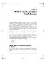
FIGURE 13.2 (A) Schematic drawing of the structure of the
plant cuticle, the protective covering on the epidermis of
leaves and young stems at the stage of full leaf expansion.
(B) Electron micrograph of the cuticle of a glandular cell
from a young leaf (
Lamium sp.), showing the presence of
the cuticle layers indicated in A, except for surface waxes,
which are not visible. (51,000
×) (A, after Jeffree 1996; B,
from Gunning and Steer 1996.)
(A)
Suberin is a cell wall constituent found in many loca-
tions throughout the plant. We have already noted its pres-
ence in the Casparian strip of the root endodermis, which
forms a barrier between the apoplast of the cortex and the
stele (see Chapter 4). Suberin is a principal component of
the outer cell walls of all underground organs and is asso-
ciated with the cork cells of the
periderm, the tissue that
forms the outer bark of stems and roots during secondary
growth of woody plants. Suberin also forms at sites of leaf
abscission and in areas damaged by disease or wounding.
Cutin,Waxes, and Suberin Help Reduce
Transpiration and Pathogen Invasion
Cutin, suberin, and their associated waxes form barriers
between the plant and its environment that function to keep
water in and pathogens out. The cuticle is very effective at
limiting water loss from aerial parts of the plant but does not
block transpiration completely because even with the stom-
ata closed, some water is lost. The thickness of the cuticle
varies with environmental conditions. Plant species native
to arid areas typically have thicker cuticles than plants from
moist habitats have, but plants from moist habitats often
develop thick cuticles when grown under dry conditions.
The cuticle and suberized tissue are both important in
excluding fungi and bacteria, although they do not appear
to be as important in pathogen resistance as some of the
other defenses we will discuss in this chapter. Many fungi
penetrate directly through the plant surface by mechanical
means. Others produce cutinase, an enzyme that hydrolyzes
cutin and thus facilitates entry into the plant.
SECONDARY METABOLITES
Plants produce a large, diverse array of organic compounds
that appear to have no direct function in growth and devel-
opment. These substances are known as
secondary
metabolites
, secondary products, or natural products. Sec-
ondary metabolites have no generally recognized, direct
roles in the processes of photosynthesis, respiration, solute
transport, translocation, protein synthesis, nutrient assim-
ilation, differentiation, or the formation of carbohydrates,
proteins, and lipids discussed elsewhere in this book.
Secondary metabolites also differ from primary metabo-
lites (amino acids, nucleotides, sugars, acyl lipids) in hav-
ing a restricted distribution in the plant kingdom. That is,
particular secondary metabolites are often found in only
one plant species or related group of species, whereas pri-
mary metabolites are found throughout the plant kingdom.
Secondary Metabolites Defend Plants against
Herbivores and Pathogens
For many years the adaptive significance of most plant sec-
ondary metabolites was unknown. These compounds were
thought to be simply functionless end products of metab-
olism, or metabolic wastes. Study of these substances was
pioneered by organic chemists of the nineteenth and early
twentieth centuries who were interested in these sub-
stances because of their importance as medicinal drugs,
poisons, flavors, and industrial materials.
More recently, many secondary metabolites have been
suggested to have important ecological functions in plants:
Secondary Metabolites and Plant Defense 285
10 mm
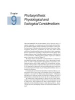
FIGURE 13.3 Surface wax
deposits, which form the top
layer of the cuticle, adopt dif-
ferent forms. These scanning
electron micrographs show the
leaf surfaces of two different
lines of
Brassica oleracea, which
differ in wax crystal structure.
(From Eigenbrode et al. 1991,
courtesy of S. D. Eigenbrode,
with permission from the
Entomological Society of
America.)
• They protect plants against being eaten by herbivores
(herbivory) and against being infected by microbial
pathogens.
• They serve as attractants for pollinators and seed-
dispersing animals and as agents of plant–plant
competition.
In the remainder of this chapter we will discuss the major
types of plant secondary metabolites, their biosynthesis,
and what is known about their functions in the plant, par-
ticularly their roles in defense.
Plant Defenses Are a Product of Evolution
We can begin by asking how plants came to have defenses.
According to evolutionary biologists, plant defenses must
have arisen through heritable mutations, natural selection,
and evolutionary change. Random mutations in basic
metabolic pathways led to the appearance of new com-
pounds that happened to be toxic or deterrent to herbi-
vores and pathogenic microbes.
As long as these compounds were not unduly toxic to
the plants themselves and the metabolic cost of producing
them was not excessive, they gave the plants that pos-
sessed them greater reproductive fitness than undefended
plants had. Thus the defended plants left more descen-
dants than undefended plants, and they passed their defen-
sive traits on to the next generation.
Interestingly, the very defense compounds that increase
the reproductive fitness of plants by warding off fungi, bac-
teria, and herbivores may also make them undesirable as
food for humans. Many important crop plants have been
artificially selected for producing relatively low levels of
these compounds, which of course can make them more
susceptible to insects and disease.
Secondary Metabolites Are Divided into
Three Major Groups
Plant secondary metabolites can be divided into three
chemically distinct groups: terpenes, phenolics, and nitro-
gen-containing compounds. Figure 13.4 shows in simpli-
286 Chapter 13
Erythrose-4-phosphate 3-Phosphoglycerate
(3-PGA)
Phosphoenolpyruvate Pyruvate
Acetyl CoA
Tricarboxylic
acid cycle
Aliphatic
amino acids
Aromatic
amino acids
Shikimic acid
pathway
Terpenes
Nitrogen-containing
secondary products
Phenolic
compounds
Malonic
acid pathway
MEP pathway
Mevalonic
acid pathway
SECONDARY CARBON METABOLISM
CO
2
Photosynthesis
PRIMARY CARBON METABOLISM
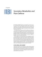
FIGURE 13.4 A simplified view of the major pathways of secondary-metabolite
biosynthesis and their interrelationships with primary metabolism.
fied form the pathways involved in the biosynthesis of sec-
ondary metabolites and their interconnections with pri-
mary metabolism.
TERPENES
The terpenes, or terpenoids, constitute the largest class of
secondary products. The diverse substances of this class are
generally insoluble in water. They are biosynthesized from
acetyl-CoA or glycolytic intermediates. After discussing the
biosynthesis of terpenes, we’ll examine how they act to
repel herbivores and how some herbivores circumvent the
toxic effects of terpenes.
Terpenes Are Formed by the Fusion of Five-
Carbon Isoprene Units
All terpenes are derived from the union of five-carbon ele-
ments that have the branched carbon skeleton of isopentane:
The basic structural elements of terpenes are sometimes
called
isoprene units because terpenes can decompose at
high temperatures to give isoprene:
Thus all terpenes are occasionally referred to as
isoprenoids.
Terpenes are classified by the number of five-carbon
units they contain, although extensive metabolic modifi-
cations can sometimes make it difficult to pick out the orig-
inal five-carbon residues. Ten-carbon terpenes, which con-
tain two C
5
units, are called monoterpenes; 15-carbon
terpenes (three C
5
units) are sesquiterpenes; and 20-carbon
terpenes (four C
5
units) are diterpenes. Larger terpenes
include
triterpenes (30 carbons), tetraterpenes (40 carbons),
and
polyterpenoids ([C
5
]
n
carbons, where n > 8).
There Are Two Pathways for Terpene Biosynthesis
Terpenes are biosynthesized from primary metabolites in
at least two different ways. In the well-studied
mevalonic
acid pathway
, three molecules of acetyl-CoA are joined
together stepwise to form mevalonic acid (Figure 13.5).
This key six-carbon intermediate is then pyrophosphory-
lated, decarboxylated, and dehydrated to yield
isopentenyl
diphosphate
(IPP
2
).
IPP is the activated five-carbon building block of ter-
penes. Recently, it was discovered that IPP also can be
formed from intermediates of glycolysis or the photosyn-
thetic carbon reduction cycle via a separate set of reactions
called the
methylerythritol phosphate (MEP) pathway
that operates in chloroplasts and other plastids (Lichten-
thaler 1999). Although all the details have not yet been elu-
cidated,
glyceraldehyde-3-phosphate and two carbon atoms
derived from
pyruvate appear to combine to generate an
intermediate that is eventually converted to IPP.
Isopentenyl Diphosphate and Its Isomer Combine
to Form Larger Terpenes
Isopentenyl diphosphate and its isomer, dimethylallyl
diphosphate (DPP), are the activated five-carbon building
blocks of terpene biosynthesis that join together to form
larger molecules. First IPP and DPP react to give geranyl
diphosphate (GPP), the 10-carbon precursor of nearly all
the monoterpenes (see Figure 13.5). GPP can then link to
another molecule of IPP to give the 15-carbon compound
farnesyl diphosphate (FPP), the precursor of nearly all the
sesquiterpenes. Addition of yet another molecule of IPP
gives the 20-carbon compound geranylgeranyl diphos-
phate (GGPP), the precursor of the diterpenes. Finally, FPP
and GGPP can dimerize to give the triterpenes (C
30
) and
the tetraterpenes (C
40
), respectively.
Some Terpenes Have Roles in Growth and
Development
Certain terpenes have a well-characterized function in
plant growth or development and so can be considered pri-
mary rather than secondary metabolites. For example, the
gibberellins, an important group of plant hormones, are
diterpenes. Sterols are triterpene derivatives that are essen-
tial components of cell membranes, which they stabilize by
interacting with phospholipids (see Chapter 11). The red,
orange, and yellow carotenoids are tetraterpenes that func-
tion as accessory pigments in photosynthesis and protect
photosynthetic tissues from photooxidation (see Chapter
7). The hormone abscisic acid (see Chapter 23) is a C
15
ter-
pene produced by degradation of a carotenoid precursor.
Long-chain polyterpene alcohols known as
dolichols
function as carriers of sugars in cell wall and glycoprotein
synthesis (see Chapter 15). Terpene-derived side chains,
such as the phytol side chain of chlorophyll (see Chapter
7), help anchor certain molecules in membranes. Thus var-
ious terpenes have important primary roles in plants. How-
ever, the vast majority of the different terpene structures
produced by plants are secondary metabolites that are pre-
sumed to be involved in defense.
Terpenes Defend against Herbivores in Many
Plants
Terpenes are toxins and feeding deterrents to many plant-
feeding insects and mammals; thus they appear to play
important defensive roles in the plant kingdom (Gershen-
zon and Croteau 1992). For example, the monoterpene
esters called
pyrethroids that occur in the leaves and flow-
H
3
C
H
2
C
CH — CH CH
2
H
3
C
H
3
C
CH — CH
2
— CH
3
Secondary Metabolites and Plant Defense 287
2
IPP is the abbreviation for isopentenyl pyrophosphate, an
earlier name for this compound. The other pyrophosphory-
lated intermediates in the pathway are also now referred to
as
diphosphates.
ers of Chrysanthemum species show very striking insecti-
cidal activity. Both natural and synthetic pyrethroids are
popular ingredients in commercial insecticides because of
their low persistence in the environment and their negligi-
ble toxicity to mammals.
In conifers such as pine and fir, monoterpenes accumu-
late in resin ducts found in the needles, twigs, and trunk.
These compounds are toxic to numerous insects, including
bark beetles, which are serious pests of conifer species
throughout the world. Many conifers respond to bark bee-
tle infestation by producing additional quantities of
monoterpenes (Trapp and Croteau 2001).
Many plants contain mixtures of volatile monoterpenes
and sesquiterpenes, called
essential oils, that lend a char-
288 Chapter 13
C
HOH
CH
2
OP
O
C
H
CH
3
O
O
OH
CC
CH
3
C
O
S CoA
HO
CH
3
C
COOH
CH
2
CH
2
CH
2
OH
CH
2
O
P P
CH
2
O
P P
CH
2
O
P P
CH
2
O
P P
CH
2
O
P P
CH
2
O
P P
OHH
3
C
CH
2
CH
O
CCH
2
OH OH
P
2×
2×
Glyceraldehyde
3-phosphate (C
3
)
Pyruvate (C
3
)
3× Acetyl-CoA (C
2
)
Mevalonic acid
Isopentenyl diphosphate (IPP, C
5
) Dimethyallyl diphosphate
(DMAPP, C
5
)
Geranyl diphosphate (GPP, C
10
)
Farnesyl diphosphate (FPP, C
15
)
Geranylgeranyl diphosphate (GGPP, C
20
)
Methylerythritol
phosphate (MEP)
Methylerythritol
phosphate
pathway
Mevalonate
pathway
Isoprene (C
5
)
Sesquiterpenes (C
15
)
Triterpenes (C
30
)
Polyterpenoids
Monoterpenes (C
10
)
Diterpenes (C
20
)
Tetraterpenes (C
40
)
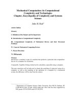
FIGURE 13.5 Outline of terpene biosynthesis. The basic 5-carbon units of terpenes
are synthesized by two different pathways. The phosphorylated intermediates, IPP
and DMAPP, are combined to make 10-carbon, 15-carbon and larger terpenes.
acteristic odor to their foliage. Peppermint, lemon, basil,
and sage are examples of plants that contain essential oils.
The chief monoterpene constituent of peppermint oil is
menthol; that of lemon oil is limonene (Figure 13.6).
Essential oils have well-known insect repellent proper-
ties. They are frequently found in glandular hairs that pro-
ject outward from the epidermis and serve to “advertise”
the toxicity of the plant, repelling potential herbivores even
before they take a trial bite. In the glandular hairs, the ter-
penes are stored in a modified extracellular space in the cell
wall (Figure 13.7). Essential oils can be extracted from
plants by steam distillation and are important commer-
cially in flavoring foods and making perfumes.
Recent research has revealed an interesting twist on the
role of volatile terpenes in plant protection. In corn, cotton,
wild tobacco, and other species, certain monoterpenes and
sesquiterpenes are produced and emitted only after insect
feeding has already begun. These substances repel
ovipositing herbivores and attract natural enemies, includ-
ing predatory and parasitic insects, that kill plant-feeding
insects and so help minimize further damage (Turlings et
al. 1995; Kessler and Baldwin 2001). Thus, volatile terpenes
are not only defenses in their own right, but also provide a
way for plants to call for defensive help from other organ-
isms. The ability of plants to attract natural enemies of
plant-feeding insects shows promise as a new, ecologically
sound means of pest control (see
Web Essay 13.1).
Among the nonvolatile terpene antiherbivore com-
pounds are the
limonoids, a group of triterpenes (C
30
) well
known as bitter substances in citrus fruit. Perhaps the most
powerful deterrent to insect feeding known is
azadirachtin
(Figure 13.8A), a complex limonoid from the neem tree
(
Azadirachta indica) of Africa and Asia. Azadirachtin is a
feeding deterrent to some insects at doses as low as 50 parts
per billion, and it exerts a variety of toxic effects (Aerts and
Mordue 1997). It has considerable potential as a commer-
cial insect control agent because of its low toxicity to mam-
mals, and several preparations containing azadirachtin are
now being marketed in North America and India.
The
phytoecdysones, first isolated from the common
fern,
Polypodium vulgare, are a group of plant steroids that
have the same basic structure as insect molting hormones
(Figure 13.8B). Ingestion of phytoecdysones by insects dis-
rupts molting and other developmental processes, often
with lethal consequences.
Triterpenes that are active against vertebrate herbivores
include cardenolides and saponins.
Cardenolides are gly-
cosides (compounds containing an attached sugar or sug-
ars) that taste bitter and are extremely toxic to higher ani-
mals. In humans, they have dramatic effects on the heart
muscle through their influence on Na
+
/K
+
-activated ATPases.
In carefully regulated doses, they slow and strengthen the
heartbeat. Cardenolides extracted from species of foxglove
Secondary Metabolites and Plant Defense 289
H
3
CCH
2
CH
3
Limonene
H
3
CCH
3
CH
3
OH
Menthol
(A)
(B)
FIGURE 13.6 Structures of limonene (A) and menthol (B).
These two well-known monoterpenes serve as defenses
against insects and other organisms that feed on these
plants. (A, photo © Calvin Larsen/Photo Researchers, Inc.;
B, photo © David Sieren/Visuals Unlimited.)
FIGURE 13.7 Monoterpenes and sesquiterpenes are commonly found in
glandular hairs on the plant surface. This scanning electron micrograph
shows a glandular hair on a young leaf of spring sunflower (
Balsamorhiza
sagittata
). Terpenes are thought to be synthesized in the cells of the hair
and are stored in the rounded cap at the top. This “cap” is an extracellular
space that forms when the cuticle and a portion of the cell wall pull away
from the remainder of the cell. (1105
×) (© J. N. A. Lott/Biological Photo
Service.)
(Digitalis) are prescribed to millions of patients for the treat-
ment of heart disease (see
Web Topic 13.1).
Saponins are steroid and triterpene glycosides, so
named because of their soaplike properties. The presence
of both lipid-soluble (the steroid or triterpene) and water-
soluble (the sugar) elements in one molecule gives
saponins detergent properties, and they form a soapy
lather when shaken with water. The toxicity of saponins is
thought to be a result of their ability to form complexes
with sterols. Saponins may interfere with sterol uptake
from the digestive system or disrupt cell membranes after
being absorbed into the bloodstream.
PHENOLIC COMPOUNDS
Plants produce a large variety of secondary products that
contain a phenol group—a hydroxyl functional group on
an aromatic ring:
These substances are classified as phenolic compounds.
Plant
phenolics are a chemically heterogeneous group of
nearly 10,000 individual compounds: Some are soluble only
in organic solvents, some are water-soluble carboxylic acids
and glycosides, and others are large, insoluble polymers.
In keeping with their chemical diversity, phenolics play
a variety of roles in the plant. After giving a brief account
of phenolic biosynthesis, we will discuss several principal
groups of phenolic compounds and what is known about
their roles in the plant. Many serve as defense compounds
against herbivores and pathogens. Others function in
mechanical support, in attracting pollinators and fruit dis-
persers, in absorbing harmful ultraviolet radiation, or in
reducing the growth of nearby competing plants.
Phenylalanine Is an Intermediate in the
Biosynthesis of Most Plant Phenolics
Plant phenolics are biosynthesized by several different
routes and thus constitute a heterogeneous group from a
metabolic point of view. Two basic pathways are involved:
the shikimic acid pathway and the malonic acid pathway
(Figure 13.9). The shikimic acid pathway participates in the
biosynthesis of most plant phenolics. The malonic acid
pathway, although an important source of phenolic sec-
ondary products in fungi and bacteria, is of less signifi-
cance in higher plants.
The
shikimic acid pathway converts simple carbohydrate
precursors derived from glycolysis and the pentose phos-
phate pathway to the aromatic amino acids (see
Web Topic
13.2) (Herrmann and Weaver 1999). One of the pathway
intermediates is shikimic acid, which has given its name to
this whole sequence of reactions. The well-known, broad-
spectrum herbicide glyphosate (available commercially as
Roundup) kills plants by blocking a step in this pathway (see
Chapter 2 on the web site). The shikimic acid pathway is pre-
sent in plants, fungi, and bacteria but is not found in animals.
Animals have no way to synthesize the three aromatic amino
acids—phenylalanine, tyrosine, and tryptophan—which are
therefore essential nutrients in animal diets.
The most abundant classes of secondary phenolic com-
pounds in plants are derived from phenylalanine via the
OH
290 Chapter 13
CH
3
CO
CH
3
CH
3
CH
3
H
3
C
O
O
O
O
OH
O
OH
HO
O
O
O
OC
CH
3
OC
CH
3
OC
O
(A) Azadirachtin, a limonoid
HO
O
OH
OH
HO
CH
3
CH
3
CH
3
OH
CH
3
H
3
C
(B) a-Ecdysone, an insect molting hormone
FIGURE 13.8 Structure of two
triterpenes, azadirachtin (A), and
α-ecdysone (B), which serve as
powerful feeding deterrents to
insects. (A, photo © Inga
Spence/Visuals Unlimited; B,
photo ©Wally Eberhart/Visuals
Unlimited.)
elimination of an ammonia molecule to form cinnamic acid
(Figure 13.10). This reaction is catalyzed by
phenylalanine
ammonia lyase
(PAL), perhaps the most studied enzyme
in plant secondary metabolism. PAL is situated at a branch
point between primary and secondary metabolism, so the
reaction that it catalyzes is an important regulatory step in
the formation of many phenolic compounds.
The activity of PAL is increased by environmental fac-
tors, such as low nutrient levels, light (through its effect on
phytochrome), and fungal infection. The point of control
appears to be the initiation of transcription. Fungal inva-
sion, for example, triggers the transcription of messenger
RNA that codes for PAL, thus increasing the amount of
PAL in the plant, which then stimulates the synthesis of
phenolic compounds.
The regulation of PAL activity in plants is made more
complex by the existence in many species of multiple PAL-
encoding genes, some of which are expressed only in spe-
cific tissues or only under certain environmental conditions
(Logemann et al. 1995).
Reactions subsequent to that catalyzed by PAL lead to
the addition of more hydroxyl groups and other sub-
stituents.
Trans-cinnamic acid, p-coumaric acid, and their
derivatives are simple phenolic compounds called
phenyl-
propanoids
because they contain a benzene ring:
and a three-carbon side chain. Phenylpropanoids are
important building blocks of the more complex phenolic
compounds discussed later in this chapter.
Now that the biosynthetic pathways leading to most
widespread phenolic compounds have been determined,
researchers have turned their attention to studying how these
pathways are regulated. In some cases, specific enzymes,
such as PAL, are important in controlling flux through the
pathway. Several transcription factors have been shown to
regulate phenolic metabolism by binding to the promoter
regions of certain biosynthetic genes and activating tran-
scription. Some of these factors activate the transcription of
large groups of genes (Jin and Martin 1999).
Some Simple Phenolics Are Activated by
Ultraviolet Light
Simple phenolic compounds are widespread in vascular
plants and appear to function in different capacities. Their
structures include the following:
• Simple phenylpropanoids, such as
trans-cinnamic
acid,
p-coumaric acid, and their derivatives, such as
caffeic acid, which have a basic phenylpropanoid car-
bon skeleton (Figure 13.11A):
• Phenylpropanoid lactones (cyclic esters) called
coumarins, also with a phenylpropanoid skeleton (see
Figure 13.11B)
• Benzoic acid derivatives, which have a
skeleton: which is formed from phenylpropanoids by
cleavage of a two-carbon fragment from the side
chain (see Figure 13.11C) (see also Figure 13.10)
As with many other secondary products, plants can elabo-
rate on the basic carbon skeleton of simple phenolic com-
pounds to make more complex products.
Many simple phenolic compounds have important roles
in plants as defenses against insect herbivores and fungi.
Of special interest is the phototoxicity of certain coumarins
called
furanocoumarins, which have an attached furan
ring (see Figure 13.11B).
C
1
C
6
C
6
C
3
C
6
Secondary Metabolites and Plant Defense 291
Shikimic acid
pathway
Erythrose-4
phosphate
(from pentose
phosphate pathway)
Phosphoenolpyruvic
acid (from glycolysis)
Acetyl-CoA
Miscellaneous
phenolics
Malonic acid
pathway
Phenylalanine
Cinnamic acid
Simple phenolics Flavonoids
Lignin
Hydrolyzable
tannins
Gallic
acid
C
3
C
6
[]
C
3
C
6
[]
n
C
3
C
6
[]
C
3
C
6
[]
C
1
C
6
[]
C
3
C
6
C
6
[]
Condensed tannins
n
C
3
C
6
C
6
[]
FIGURE 13.9 Plant phenolics are
biosynthesized in several differ-
ent ways. In higher plants, most
phenolics are derived at least in
part from phenylalanine, a prod-
uct of the shikimic acid pathway.
Formulas in brackets indicate the
basic arrangement of carbon
skeletons:
indicates a benzene ring, and
C3 is a three-carbon chain.
More detail on the pathway
from phenylalanine onward is
given in Figure 13.10.
C
6
These compounds are not toxic until they
are activated by light. Sunlight in the ultra-
violet A (UV-A) region (320–400 nm) causes
some furanocoumarins to become activated
to a high-energy electron state. Activated
furanocoumarins can insert themselves into
the double helix of DNA and bind to the
pyrimidine bases cytosine and thymine,
thus blocking transcription and repair and
leading eventually to cell death.
Phototoxic furanocoumarins are espe-
cially abundant in members of the Umbel-
liferae family, including celery, parsnip, and
parsley. In celery, the level of these com-
pounds can increase about 100-fold if the
plant is stressed or diseased. Celery pickers,
and even some grocery shoppers, have been
known to develop skin rashes from han-
dling stressed or diseased celery. Some
insects have adapted to survive on plants
that contain furanocoumarins and other
phototoxic compounds by living in silken
webs or rolled-up leaves, which screen out
the activating wavelengths (Sandberg and
Berenbaum 1989).
The Release of Phenolics into the Soil
May Limit the Growth of Other Plants
From leaves, roots, and decaying litter, plants
release a variety of primary and secondary
metabolites into the environment. Investiga-
tion of the effects of these compounds on
neighboring plants is the study of
allelopa-
thy
. If a plant can reduce the growth of
nearby plants by releasing chemicals into the
soil, it may increase its access to light, water,
and nutrients and thus its evolutionary fit-
ness. Generally speaking, the term
allelopathy
has come to be applied to the harmful effects
of plants on their neighbors, although a pre-
cise definition also includes beneficial effects.
Simple phenylpropanoids and benzoic
acid derivatives are frequently cited as hav-
ing allelopathic activity. Compounds such
as caffeic acid and ferulic acid (see Figure
13.11A) occur in soil in appreciable amounts
and have been shown in laboratory experi-
ments to inhibit the germination and growth
of many plants (Inderjit et al. 1995).
292 Chapter 13
NH
2
COOH
COOH
COSCoA
COOH
HO
OH O
OH
HO OH
O
HO
OH
O
OH
OH O
HO
O
OH
HO
OH O
HO
OH
O
OH O
HO
OH
OH O
HO
OH
O
OH
O
Phenylalanine
trans-Cinnamic acid
p-Coumaric acid
Phenylalanine ammonia lyase (PAL)
3 Malonyl-CoA molecules
Chalcone synthase
Benzoic acid
derivatives (Figure 13.11C)
Anthocyanins (Figure 13.13B)
Condensed tannins (Figure 13.15A)
Lignin precursors
(Web Topic 13.3)
NH
3
p-Coumaroyl-CoA
Chalcones
Flavanones
OH
Flavones
Isoflavones (isoflavonoids)
Flavonols
Dihydroflavonols
Caffeic acid
and other simple
phenylpropanoids
(Figure 13.11A)
Coumarins (Figure 13.11B)
CoA-SH
FIGURE 13.10 Outline of phenolic biosynthesis from phenylalanine. The formation
of many plant phenolics, including simple phenylpropanoids, coumarins, benzoic
acid derivatives, lignin, anthocyanins, isoflavones, condensed tannins, and other
flavonoids, begins with phenylalanine.
In spite of results such as these, the importance of
allelopathy in natural ecosystems is still controversial.
Many scientists doubt that allelopathy is a significant fac-
tor in plant–plant interactions because good evidence for
this phenomenon has been hard to obtain. It is easy to
show that extracts or purified compounds from one plant
can inhibit the growth of other plants in laboratory exper-
iments, but it has been very difficult to demonstrate that
these compounds are present in the soil in sufficient con-
centration to inhibit growth. Furthermore, organic sub-
stances in the soil are often bound to soil particles and may
be rapidly degraded by microbes.
In spite of the lack of supporting evidence, allelopathy
is currently of great interest because of its potential agri-
cultural applications. Reductions in crop yields caused by
weeds or residues from the previous crop may in some
cases be a result of allelopathy. An exciting future prospect
is the development of crop plants genetically engineered to
be allelopathic to weeds.
Lignin Is a Highly Complex Phenolic
Macromolecule
After cellulose, the most abundant organic substance in
plants is
lignin, a highly branched polymer of phenyl-
propanoid groups
that plays both primary and secondary roles. The precise
structure of lignin is not known because it is difficult to
extract lignin from plants, where it is covalently bound to
cellulose and other polysaccharides of the cell wall.
Lignin is generally formed from three different phenyl-
propanoid alcohols: coniferyl, coumaryl, and sinapyl, alco-
hols which are synthesized from phenylalanine via various
cinnamic acid derivatives. The phenylpropanoid alcohols are
joined into a polymer through the action of enzymes that
generate free-radical intermediates. The proportions of the
three monomeric units in lignin vary among species, plant
organs, and even layers of a single cell wall. In the polymer,
there are often multiple C—C and C—O—C bonds in each
phenylpropanoid alcohol unit, resulting in a complex struc-
ture that branches in three dimensions. Unlike polymers
such as starch, rubber, or cellulose, the units of lignin do not
appear to be linked in a simple, repeating way. However,
recent research suggests that a guiding protein may bind the
individual phenylpropanoid units during lignin biosynthe-
sis, giving rise to a scaffold that then directs the formation of
a large, repeating unit (Davin and Lewis 2000; Hatfield and
Vermerris 2001). (See
Web Topic 13.3 for the partial structure
of a hypothetical lignin molecule.)
Lignin is found in the cell walls of various types of sup-
porting and conducting tissue, notably the tracheids and
vessel elements of the xylem. It is deposited chiefly in the
thickened secondary wall but can also occur in the primary
wall and middle lamella in close contact with the celluloses
and hemicelluloses already present. The mechanical rigid-
ity of lignin strengthens stems and vascular tissue, allow-
ing upward growth and permitting water and minerals to
be conducted through the xylem under negative pressure
without collapse of the tissue. Because lignin is such a key
component of water transport tissue, the ability to make
lignin must have been one of the most important adapta-
tions permitting primitive plants to colonize dry land.
Besides providing mechanical support, lignin has signif-
icant protective functions in plants. Its physical toughness
deters feeding by animals, and its chemical durability makes
it relatively indigestible to herbivores. By bonding to cellu-
lose and protein, lignin also reduces the digestibility of these
substances. Lignification blocks the growth of pathogens
and is a frequent response to infection or wounding.
C
6
C
3
Secondary Metabolites and Plant Defense 293
H
OH
HO
CC
COOH
H
OCH
3
HO
CC
COOH
H
H
HO O O O O
O
OCH
3
CH
O
HO
OH
COOH
Caffeic acid
C
3
C
6
[]
Ferulic acid
Furan ring
Umbelliferone,
a simple coumarin
C
3
C
6
[]
Vanillin Salicylic acid
C
1
C
6
[]
Psoralen,
a furanocoumarin
(A)
(B)
(C)
Simple phenylpropanoids
Coumarins
Benzoic acid derivatives
FIGURE 13.11 Simple phenolic compounds play a great
diversity of roles in plants. (A) Caffeic acid and ferulic acid
may be released into the soil and inhibit the growth of
neighboring plants. (B) Psoralen is a furanocoumarin that
exhibits phototoxicity to insect herbivores. (C) Salicylic acid
is a plant growth regulator that is involved in systemic
resistance to plant pathogens.
There Are Four Major Groups of Flavonoids
The flavonoids are one of the largest classes of plant phe-
nolics. The basic carbon skeleton of a flavonoid contains 15
carbons arranged in two aromatic rings connected by a
three-carbon bridge:
This structure results from two separate biosynthetic path-
ways: the shikimic acid pathway and the malonic acid
pathway (Figure 13.12).
Flavonoids are classified into different groups, primar-
ily on the basis of the degree of oxidation of the three-car-
bon bridge. We will discuss four of the groups shown in
Figure 13.10: the anthocyanins, the flavones, the flavonols,
and the isoflavones.
The basic flavonoid carbon skeleton may have numer-
ous substituents. Hydroxyl groups are usually present at
positions 4, 5, and 7, but they may also be found at other
positions. Sugars are very common as well; in fact, the
majority of flavonoids exist naturally as glycosides.
Whereas both hydroxyl groups and sugars increase the
water solubility of flavonoids, other substituents, such as
methyl ethers or modified isopentyl units, make flavonoids
lipophilic (hydrophobic). Different types of flavonoids per-
form very different functions in the plant, including pig-
mentation and defense.
Anthocyanins Are Colored Flavonoids That
Attract Animals
In addition to predator–prey interactions, there are mutual-
istic associations among plants and animals. In return for the
reward of ingesting nectar or fruit pulp, animals perform
extremely important services for plants as carriers of pollen
and seeds. Secondary metabolites are involved in these
plant–animal interactions, helping to attract animals to flow-
ers and fruit by providing visual and olfactory signals.
The colored pigments of plants are of two principal
types: carotenoids and flavonoids.
Carotenoids, as we have
already seen, are yellow, orange, and red terpenoid com-
pounds that also serve as accessory pigments in photo-
synthesis (see Chapter 7).
Flavonoids are phenolic com-
pounds that include a wide range of colored substances.
The most widespread group of pigmented flavonoids is
the
anthocyanins, which are responsible for most of the red,
pink, purple, and blue colors observed in plant parts. By col-
oring flowers and fruits, the anthocyanins are vitally impor-
tant in attracting animals for pollination and seed dispersal.
Anthocyanins are glycosides that have sugars at position
3 (Figure 13.13B) and sometimes elsewhere. Without their
sugars, anthocyanins are known as
anthocyanidins (Figure
13.13A). Anthocyanin color is influenced by many factors,
including the number of hydroxyl and methoxyl groups in
ring B of the anthocyanidin (see Figure 13.13A), the presence
of aromatic acids esterified to the main skeleton, and the pH
of the cell vacuole in which these compounds are stored.
Anthocyanins may also exist in supramolecular complexes
along with chelated metal ions and flavone copigments. The
blue pigment of dayflower (
Commelina communis) was found
C
3
C
6
C
6
294 Chapter 13
A
8
54
63
2
7
C
3′
6′
1′
2′
5′
4′
B
O
1
Basic flavonoid skeleton
From shikimic acid
pathway via phenylalanine
From malonic
acid pathway
The three-carbon bridge
C
3
C
6
[]
C
6
[]
FIGURE 13.12 Basic flavonoid carbon skeleton. Flavonoids
are biosynthesized from products of the shikimic acid and
malonic acid pathways. Positions on the flavonoid ring sys-
tem are numbered as shown.
FIGURE 13.13 The structures of anthocyanidins (A) and
anthocyanin (B). The colors of anthocyanidins depend in
part on the substituents attached to ring B (see Table 13.1).
An increase in the number of hydroxyl groups shifts
absorption to a longer wavelength and gives a bluer color.
Replacement of a hydroxyl group with a methoxyl group
(OCH
3
) shifts absorption to a slightly shorter wavelength,
resulting in a redder color.
+
OH
HO
OH
AC
3′
2′
6′
1′
5′
4′
B
AC
B
OH
OH
HO
O
O
+
O
Anthocyanidin
Anthocyanin
Sugar
(A)
(B)
to consist of a large complex of six anthocyanin molecules,
six flavones, and two associated magnesium ions (Kondo et
al. 1992). The most common anthocyanidins and their colors
are shown in Figure 13.13 and Table 13.1.
Considering the variety of factors affecting anthocyanin
coloration and the possible presence of carotenoids as well,
it is not surprising that so many different shades of flower
and fruit color are found in nature. The evolution of flower
color may have been governed by selection pressures for
different sorts of pollinators, which often have different
color preferences.
Color, of course, is just one type of signal used to attract
pollinators to flowers. Volatile chemicals, particularly
monoterpenes, frequently provide attractive scents.
Flavonoids May Protect against Damage by
Ultraviolet Light
Two other major groups of flavonoids found in flowers are
flavones and flavonols (see Figure 13.10). These flavonoids
generally absorb light at shorter wavelengths
than anthocyanins do, so they are not visible to
the human eye. However, insects such as bees,
which see farther into the ultraviolet range of the
spectrum than humans do, may respond to
flavones and flavonols as attractant cues (Figure
13.14). Flavonols in a flower often form sym-
metric patterns of stripes, spots, or concentric
circles called
nectar guides (Lunau 1992). These
patterns may be conspicuous to insects and are
thought to help indicate the location of pollen and nectar.
Flavones and flavonols are not restricted to flowers; they
are also present in the leaves of all green plants. These two
classes of flavonoids function to protect cells from exces-
sive UV-B radiation (280–320 nm) because they accumulate
in the epidermal layers of leaves and stems and absorb
light strongly in the UV-B region while allowing the visible
(photosynthetically active) wavelengths to pass through
uninterrupted. In addition, exposure of plants to increased
UV-B light has been demonstrated to increase the synthe-
sis of flavones and flavonols.
Arabidopsis thaliana mutants that lack the enzyme chal-
cone synthase produce no flavonoids. Lacking flavonoids,
these plants are much more sensitive to UV-B radiation
than wild-type individuals are, and they grow very poorly
under normal conditions. When shielded from UV light,
however, they grow normally (Li et al. 1993). A group of
simple phenylpropanoid esters are also important in UV
protection in
Arabidopsis.
Secondary Metabolites and Plant Defense 295
TABLE 13.1
Effects of ring substituents on anthocyanidin color
Anthocyanidin Substituents Color
Pelargonidin 4′— OH Orange red
Cyanidin 3
′— OH, 4′— OH Purplish red
Delphinidin 3
′— OH,4′— OH,5′— OH Bluish purple
Peonidin 3
′— OCH
3
,4′— OH Rosy red
Petunidin 3′— OCH
3
,4′— OH, 5′— OCH
3
Purple
FIGURE 13.14 Black-eyed Susan (Rudbeckia sp.) as seen by
humans (A) and as it might appear to honeybees (B). (A)
To humans, the golden-eye has yellow rays and a brown
central disc. (B) To bees, the tips of the rays appear “light
yellow,” the inner portion of the rays “dark yellow,” and
the central disc “black.” Ultraviolet-absorbing flavonols are
found in the inner parts of the rays but not in the tips. The
distribution of flavonols in the rays and the sensitivity of
insects to part of the UV spectrum contribute to the
“bull’s-eye” pattern seen by honeybees, which presumably
helps them locate pollen and nectar. Special lighting was
used to simulate the spectral sensitivity of the honeybee
visual system. (Courtesy of Thomas Eisner.)
(B)
(A)
Other functions of flavonoids have recently been dis-
covered. For example, flavones and flavonols secreted into
the soil by legume roots mediate the interaction of legumes
and nitrogen-fixing symbionts, a phenomenon described in
Chapter 12. As will be discussed in Chapter 19, recent work
suggests that flavonoids also play a regulatory role in plant
development as modulators of polar auxin transport.
Isoflavonoids Have Antimicrobial Activity
The isoflavonoids (isoflavones) are a group of flavonoids in
which the position of one aromatic ring (ring B) is shifted
(see Figure 13.10). Isoflavonoids are found mostly in
legumes and have several different bio-
logical activities. Some, such as the
rotenoids, have strong insecticidal
actions; others have anti-estrogenic
effects. For example, sheep grazing on
clover rich in isoflavonoids often suffer
from infertility. The isoflavonoid ring sys-
tem has a three-dimensional structure
similar to that of steroids (see Figure
13.8B), allowing these substances to bind
to estrogen receptors. Isoflavonoids may
also be responsible for the anticancer
benefits of food prepared from soybeans.
In the past few years, isoflavonoids
have become best known for their role as
phytoalexins, antimicrobial compounds
synthesized in response to bacterial or
fungal infection that help limit the spread
of the invading pathogen. Phytoalexins
are discussed in more detail later in this
chapter.
Tannins Deter Feeding by
Herbivores
A second category of plant phenolic
polymers with defensive properties,
besides lignins, is the
tannins. The term
tannin was first used to describe com-
pounds that could convert raw animal
hides into leather in the process known
as tanning. Tannins bind the collagen
proteins of animal hides, increasing their
resistance to heat, water, and microbes.
There are two categories of tannins:
condensed and hydrolyzable.
Con-
densed tannins
are compounds formed
by the polymerization of flavonoid units
(Figure 13.15A). They are frequent con-
stituents of woody plants. Because con-
densed tannins can often be hydrolyzed
to anthocyanidins by treatment with
strong acids, they are sometimes called
pro-anthocyanidins.
Hydrolyzable tannins are heterogeneous polymers con-
taining phenolic acids, especially gallic acid, and simple
sugars (see Figure 13.15B). They are smaller than con-
densed tannins and may be hydrolyzed more easily; only
dilute acid is needed. Most tannins have molecular masses
between 600 and 3000.
Tannins are general toxins that significantly reduce the
growth and survivorship of many herbivores when added
to their diets. In addition, tannins act as feeding repellents
to a great diversity of animals. Mammals such as cattle,
deer, and apes characteristically avoid plants or parts of
plants with high tannin contents. Unripe fruits, for
296 Chapter 13
OH
HO
OH
OH
OH
AC
B
O
OH
HO
OH
OH
OH
O
OH
HO
OH
OH
OH
O
n
O
OH
OH
C
O
OH
C
O
OH
OH
OHHO
OH
OH
O
CCH
2
O
O
OH
OH
OH
HO
O
CO
HO
C
O
O
H
O
OH
H
O
CO
HO
HO
HO
OH
OHHO
CO
C
O
O
H
O
H
(A) Condensed tannin
(B) Hydrolyzable tannin
Gallic acid
FIGURE 13.15 Structure of some tannins formed from phenolic acids or
flavonoid units. (A) The general structure of a condensed tannin, where
n is
usually 1 to 10. There may also be a third —OH group on ring B. (B) The
hydrolyzable tannin from sumac (
Rhus semialata) consists of glucose and eight
molecules of gallic acid.
instance, frequently have very high tannin levels, which
may be concentrated in the outer cell layers.
Interestingly, humans often prefer a certain level of
astringency in tannin-containing foods, such as apples,
blackberries, tea, and red wine. Recently, polyphenols (tan-
nins) in red wine were shown to block the formation of
endothelin-1, a signaling molecule that makes blood ves-
sels constrict (Corder et al. 2001). This effect of wine tan-
nins may account for the often-touted health benefits of red
wine, especially the reduction in the risk of heart disease
associated with moderate red wine consumption.
Although moderate amounts of specific polyphenolics
may have health benefits for humans, the defensive prop-
erties of most tannins are due to their toxicity, which is gen-
erally attributed to their ability to bind proteins nonspecif-
ically. It has long been thought that plant tannins complex
proteins in the guts of herbivores by forming hydrogen
bonds between their hydroxyl groups and electronegative
sites on the protein (Figure 13.16A).
More recent evidence indicates that tannins and other
phenolics can also bind to dietary protein in a covalent fash-
ion (see Figure 13.16B). The foliage of many plants contains
enzymes that oxidize phenolics to their corresponding
quinone forms in the guts of herbivores (Felton et al. 1989).
Quinones are highly reactive electrophilic molecules that
readily react with the nucleophilic —NH
2
and —SH groups
of proteins (see Figure 13.16B). By whatever mechanism
protein–tannin binding occurs, this process has a negative
impact on herbivore nutrition. Tannins can inactivate her-
bivore digestive enzymes and create complex aggregates of
tannins and plant proteins that are difficult to digest.
Herbivores that habitually feed on tannin-rich plant
material appear to possess some interesting adaptations to
remove tannins from their digestive systems. For example,
some mammals, such as rodents and rabbits, produce sali-
vary proteins with a very high proline content (25–45%) that
have a high affinity for tannins. Secretion of these proteins
is induced by ingestion of food with a high tannin content
and greatly diminishes the toxic effects of tannins (Butler
1989). The large number of proline residues gives these pro-
teins a very flexible, open conformation and a high degree
of hydrophobicity that facilitates binding to tannins.
Plant tannins also serve as defenses against microor-
ganisms. For example, the nonliving heartwood of many
trees contains high concentrations of tannins that help pre-
vent fungal and bacterial decay.
NITROGEN-CONTAINING COMPOUNDS
A large variety of plant secondary metabolites have nitro-
gen in their structure. Included in this category are such
well-known antiherbivore defenses as alkaloids and
cyanogenic glycosides, which are of considerable interest
because of their toxicity to humans and their medicinal
properties. Most nitrogenous secondary metabolites are
biosynthesized from common amino acids.
In this section we will examine the structure and biolog-
ical properties of various nitrogen-containing secondary
metabolites, including alkaloids, cyanogenic glycosides, glu-
cosinolates, and nonprotein amino acids. In addition, we will
discuss the ability of
systemin, a protein released from dam-
aged cells, to serve as a wound signal to the rest of the plant.
Alkaloids Have Dramatic Physiological Effects on
Animals
The alkaloids are a large family of more than 15,000 nitro-
gen-containing secondary metabolites found in approxi-
mately 20% of the species of vascular plants. The nitrogen
atom in these substances is usually part of a
heterocyclic
ring
, a ring that contains both nitrogen and carbon atoms.
As a group, alkaloids are best known for their striking
pharmacological effects on vertebrate animals.
As their name would suggest, most alkaloids are alka-
line. At pH values commonly found in the cytosol (pH 7.2)
Secondary Metabolites and Plant Defense 297
OH N
H
2
OH
OH
HN
H
2
N
O
(A) Hydrogen bonding between tannins and protein
(B) Covalent bonding to protein after oxidation
Polyphenol oxidase
Tannin in phenol form
Tannin in quinone form
Tannin linked to protein
Tannin
Protein
Protein
Protein
Covalent bond
d
+
d
−
FIGURE 13.16 Proposed mechanisms for the interaction of
tannins with proteins. (A) Hydrogen bonds may form
between the phenolic hydroxyl groups of tannins and elec-
tronegative sites on the protein. (B) Phenolic hydroxyl
groups may bind covalently to proteins following activa-
tion by oxidative enzymes, such as polyphenol oxidase.
or the vacuole (pH 5 to 6), the nitrogen atom is protonated;
hence, alkaloids are positively charged and are generally
water soluble.
Alkaloids are usually synthesized from one of a few
common amino acids—in particular, lysine, tyrosine, and
tryptophan. However, the carbon skeleton of some alka-
loids contains a component derived from the terpene
pathway. Table 13.2 lists the major alkaloid types and their
amino acid precursors. Several different types, including
nicotine and its relatives (Figure 13.17), are derived from
ornithine, an intermediate in arginine biosynthesis. The B
vitamin nicotinic acid (niacin) is a precursor of the pyridine
(six-membered) ring of this alkaloid; the pyrrolidine (five-
membered) ring of nicotine arises from ornithine (Figure
13.18). Nicotinic acid is also a constituent of NAD
+
and
NADP
+
, which serve as electron carriers in metabolism.
The role of alkaloids in plants has been a subject of spec-
ulation for at least 100 years. Alkaloids were once thought
to be nitrogenous wastes (analogous to urea and uric acid
in animals), nitrogen storage compounds, or growth regu-
lators, but there is little evidence to support any of these
functions. Most alkaloids are now believed to function as
defenses against predators, especially mammals, because
298 Chapter 13
TABLE 13.2
Major types of alkaloids, their amino acid precursors, and well-known examples of each type
Biosynthetic
Alkaloid class Structure precursor Examples Human uses
Pyrrolidine Ornithine (aspartate) Nicotine Stimulant, depressant, tranquilizer
Tropane Ornithine Atropine Prevention of intestinal spasms, antidote to other
poisons, dilation of pupils for examination
Cocaine Stimulant of the central nervous system, local
anesthetic
Piperidine Lysine (or acetate) Coniine Poison (paralyzes motor neurons)
Pyrrolizidine Ornithine Retrorsine None
Quinolizidine Lysine Lupinine Restoration of heart rhythm
Isoquinoline Tyrosine Codeine Analgesic (pain relief ), treatment of coughs
Morphine Analgesic
Indole Tryptophan Psilocybin Halucinogen
Reserpine Treatment of hypertension, treatment of psychoses
Strychnine Rat poison, treatment of eye disorders
N
N
N
N
N
N
N
C
N
N
NH
3
C
N
N
N
O
O
O
O
CH
3
CH
3
CH
3
CH
3
OCH
3
NOC
CH
3
HO
N
HO
O
Cocaine
Morphine
Representative alkaloids
Caffeine
Nicotine
FIGURE 13.17 Examples of alkaloids, a diverse group of
secondary metabolites that contain nitrogen, usually as part
of a heterocyclic ring. Caffeine is a purine-type alkaloid
similar to the nucleic acid bases adenine and guanine. The
pyrrolidine (five-membered) ring of nicotine arises from
ornithine; the pyridine (six-membered) ring is derived from
nicotinic acid.
of their general toxicity and deter-
rence capability (Hartmann 1992).
Large numbers of livestock
deaths are caused by the ingestion
of alkaloid-containing plants. In
the United States, a significant per-
centage of all grazing livestock
animals are poisoned each year by
consumption of large quantities of
alkaloid-containing plants such as
lupines (
Lupinus), larkspur (Del-
phinium
), and groundsel (Senecio).
This phenomenon may be due to
the fact that domestic animals,
unlike wild animals, have not
been subjected to natural selection
for the avoidance of toxic plants.
Indeed, some livestock actually seem to prefer alkaloid-
containing plants to less harmful forage.
Nearly all alkaloids are also toxic to humans when taken
in sufficient quantity. For example, strychnine, atropine, and
coniine (from poison hemlock) are classic alkaloid poison-
ing agents. At lower doses, however, many are useful phar-
macologically. Morphine, codeine, and scopolamine are just
a few of the plant alkaloids currently used in medicine.
Other alkaloids, including cocaine, nicotine, and caffeine (see
Figure 13.17), enjoy widespread nonmedical use as stimu-
lants or sedatives.
On a cellular level, the mode of action of alkaloids in
animals is quite variable. Many alkaloids interfere with
components of the nervous system, especially the chemi-
cal transmitters; others affect membrane transport, protein
synthesis, or miscellaneous enzyme activities.
One group of alkaloids, the pyrrolizidine alkaloids, illus-
trates how herbivores can become adapted to tolerate plant
defensive substances and even use them in their own
defense (Hartmann 1999). Within plants, pyrrolizidine alka-
loids occur naturally as nontoxic N-oxides. In herbivore
digestive tracts, however, they are quickly reduced to
uncharged, hydrophobic tertiary alkaloids (Figure 13.19),
which easily pass through membranes and are toxic. Nev-
ertheless, some herbivores, such as cinnabar moth (
Tyria
jacobeae
), have developed the ability to reconvert tertiary
pyrrolizidine alkaloids to the nontoxic N-oxide form imme-
diately after its absorption from the digestive tract. These
herbivores may then store the N-oxides in their bodies as
defenses against their own predators.
Not all of the alkaloids that appear in plants are pro-
duced by the plant itself. Many grasses harbor endogenous
fungal symbionts that grow in the apoplast and synthesize
a variety of different types of alkaloids. Grasses with fun-
gal symbionts often grow faster and are better defended
Secondary Metabolites and Plant Defense 299
CH
2
NH
2
CH
NH
2
COOH
N
CH
3
+
N
N
CH
3
P
OH
2
C
H
OH
O
H
HO
N
COOH
+
N
COOH
H
2
C
H
2
C
Nicotinic acid mononucleotide (NADP
+
)
Nicotinic acid
Ornithine
N-Methyl pyrrolinium
Nicotine
FIGURE 13.18 Nicotine biosynthesis begins with the biosyn-
thesis of the nicotinic acid (niacin) from aspartate and glyc-
eraldehyde-3-phosphate. Nicotinic acid is also a component
of NAD
+
and NADP
+
, important participants in biological
oxidation–reduction reactions. The five-membered ring of
nicotine is derived from ornithine, an intermediate in argi-
nine biosynthesis.
H
3
C
OO
N
+
O
–
CH
3
O
O
HO CH
3
H
3
C
OO
N
CH
3
O
O
HO CH
3
N-oxide
(nontoxic form,
stored in plants)
Tertiary alkaloid
(toxic form)
Reduced in digestive
tracts of most herbivores
to toxic form
Oxidized to nontoxic
form by certain adapted
herbivores
FIGURE 13.19 Two forms of pyrrolizidine alkaloids occur in nature: the N-oxide
form and the tertiary alkaloid. The nontoxic N-oxide found in plants is reduced to
the toxic tertiary form in the digestive tracts of most herbivores. However, some
adapted herbivores can convert the toxic tertiary alkaloid back to the nontoxic N-
oxide. These forms are illustrated here for the alkaloid senecionine, found in species
of ragwort (
Senecio).
against insect and mammalian herbivores than those with-
out symbionts. Unfortunately, certain grasses with sym-
bionts, such as tall fescue, are important pasture grasses
that may become toxic to livestock when their alkaloid con-
tent is too high. Efforts are under way to breed tall fescue
with alkaloid levels that are not poisonous to livestock but
still provide protection against insects (see
Web Essay 13.2).
Like monoterpenes in conifer resin and many other anti-
herbivore defense compounds, alkaloids increase in
response to initial herbivore damage, fortifying the plant
against subsequent attack (Karban and Baldwin 1997). For
example,
Nicotiana attenuata, a wild tobacco that grows in
the deserts of the Great Basin, produces higher levels of
nicotine following herbivory. When it is attacked by nico-
tine-tolerant caterpillars, however, there is no increase in
nicotine. Instead, volatile terpenes are released that attract
enemies of the caterpillars. Clearly, wild tobacco and other
plants must have ways of determining what type of herbi-
vore is damaging their foliage. Herbivores might signal
their presence by the type of damage they inflict or the dis-
tinctive chemical compounds they release. Recently, the oral
secretions of caterpillars feeding on corn leaves were shown
to contain a fatty acid–amino acid conjugate that induced
the plant to produce defensive terpenes when applied to cut
leaves.
Cyanogenic Glycosides Release the Poison
Hydrogen Cyanide
Various nitrogenous protective compounds other than
alkaloids are found in plants. Two groups of these sub-
stances—cyanogenic glycosides and glucosinolates—are
not in themselves toxic but are readily broken down to give
off volatile poisons when the plant is crushed. Cyanogenic
glycosides release the well-known poisonous gas hydrogen
cyanide (HCN).
The breakdown of cyanogenic glycosides in plants is a
two-step enzymatic process. Species that make cyanogenic
glycosides also make the enzymes necessary to hydrolyze
the sugar and liberate HCN:
1. In the first step the sugar is cleaved by a glycosidase,
an enzyme that separates sugars from other mole-
cules to which they are linked (Figure 13.20).
2. In the second step the resulting hydrolysis product,
called an
α-hydroxynitrile or cyanohydrin, can
decompose spontaneously at a low rate to liberate
HCN. This second step can be accelerated by the
enzyme hydroxynitrile lyase.
Cyanogenic glycosides are not normally broken down
in the intact plant because the glycoside and the degrada-
tive enzymes are spatially separated, in different cellular
compartments or in different tissues. In sorghum, for exam-
ple, the cyanogenic glycoside dhurrin is present in the vac-
uoles of epidermal cells, while the hydrolytic and lytic
enzymes are found in the mesophyll (Poulton 1990).
Under ordinary conditions this compartmentation pre-
vents decomposition of the glycoside. When the leaf is
damaged, however, as during herbivore feeding, the cell
contents of different tissues mix and HCN forms.
Cyanogenic glycosides are widely distributed in the plant
kingdom and are frequently encountered in legumes,
grasses, and species of the rose family.
Considerable evidence indicates that cyanogenic glyco-
sides have a protective function in certain plants. HCN is a
fast-acting toxin that inhibits metalloproteins, such as the
iron-containing cytochrome oxidase, a key enzyme of mito-
chondrial respiration. The presence of cyanogenic glycosides
deters feeding by insects and other herbivores, such as snails
and slugs. As with other classes of secondary metabolites,
however, some herbivores have adapted to feed on
cyanogenic plants and can tolerate large doses of HCN.
The tubers of cassava (
Manihot esculenta), a high-carbo-
hydrate, staple food in many tropical countries, contain
high levels of cyanogenic glycosides. Traditional process-
ing methods, such as grating, grinding, soaking, and dry-
ing, lead to the removal or degradation of a large fraction
of the cyanogenic glycosides present in cassava tubers.
However, chronic cyanide poisoning leading to partial
paralysis of the limbs is still widespread in regions where
cassava is a major food source because the traditional
detoxification methods employed to remove cyanogenic
glycosides from cassava are not completely effective. In
addition, many populations that consume cassava have
poor nutrition, which aggravates the effects of the
cyanogenic glycosides.
300 Chapter 13
C
O—
CR′
R
N
C
OH
CR′
R
N
NC O + HC
R′
R
Glycosidase
Sugar
Sugar
Cyanogenic
glycoside
Hydrogen
cyanide
Cyanohydrin
Hydroxynitrile
lyase
or
spontaneous
Ketone
FIGURE 13.20 Enzyme-catalyzed hydrolysis of cyanogenic glycosides to release hydro-
gen cyanide. R and R
′ represent various alkyl or aryl substituents. For example, if R is
phenyl, R
′ is hydrogen, and the sugar is the disaccharide β-gentiobiose, the compound
is amygdalin (the common cyanogenic glycoside found in the seeds of almonds, apri-
cots, cherries, and peaches).
Efforts are currently under way to reduce the cyanogenic
glycoside content of cassava through both conventional
breeding and genetic engineering approaches. However, the
complete elimination of cyanogenic glycosides may not be
desirable because these substances are probably responsi-
ble for the fact that cassava can be stored for very long peri-
ods of time without being attacked by pests.
Glucosinolates Release Volatile Toxins
A second class of plant glycosides, called the glucosino-
lates
, or mustard oil glycosides, break down to release
volatile defensive substances. Found principally in the
Brassicaceae and related plant families, glucosinolates give
off the compounds responsible for the smell and taste of
vegetables such as cabbage, broccoli, and radishes.
The release of these mustard-smelling volatiles from
glucosinolates is catalyzed by a hydrolytic enzyme, called
a thioglucosidase or myrosinase, that cleaves glucose from
its bond with the sulfur atom (Figure 13.21). The resulting
aglycone, the nonsugar portion of the molecule, rearranges
with loss of the sulfate to give pungent and chemically
reactive products, including isothiocyanates and nitriles,
depending on the conditions of hydrolysis. These products
function in defense as herbivore toxins and feeding repel-
lents. Like cyanogenic glycosides, glucosinolates are stored
in the intact plant separately from the enzymes that
hydrolyze them, and they are brought into contact with
these enzymes only when the plant is crushed.
As with other secondary metabolites, certain animals are
adapted to feed on glucosinolate-containing plants without ill
effects. For adapted herbivores, such as the cabbage butterfly,
glucosinolates often serve as stimulants for feeding and egg
laying, and the isothiocyanates produced after glucosinolate
hydrolysis act as volatile attractants (Renwick et al. 1992).
Most of the recent research on glucosinolates in plant
defense has concentrated on rape, or canola (
Brassica
napus
), a major oil crop in both North America and Europe.
Plant breeders have tried to lower the glucosinolate levels
of rapeseed so that the high-protein seed meal remaining
after oil extraction can be used as animal food. The first
low-glucosinolate varieties tested in the field were unable
to survive because of severe pest problems. However, more
recently developed varieties with low glucosinolate levels
in seeds but high glucosinolate levels in leaves are able to
hold their own against pests and still provide a protein-rich
seed residue for animal feeding.
Nonprotein Amino Acids Defend against
Herbivores
Plants and animals incorporate the same 20 amino acids
into their proteins. However, many plants also contain
unusual amino acids, called
nonprotein amino acids, that
are not incorporated into proteins but are present instead
in the free form and act as protective substances. Nonpro-
tein amino acids are often very similar to common protein
amino acids. Canavanine, for example, is a close analog of
arginine, and azetidine-2-carboxylic acid has a structure
very much like that of proline (Figure 13.22).
Nonprotein amino acids exert their toxicity in various
ways. Some block the synthesis or uptake of protein amino
Secondary Metabolites and Plant Defense 301
Glucose
C
S
N
R
OSO
3
–
Glucose
C
SH
N
R
OSO
3
–
SO
4
2–
RNCS
RCN
Glucosinolate Aglycone Nitrile
Thioglucosidase Spontaneous
Isothiocyanate
FIGURE 13.21 Hydrolysis of glucosinolates to mustard-smelling volatiles. R repre-
sents various alkyl or aryl substituents. For example, if R is CH
2
—
—
CH—CH
2
–
, the
compound is sinigrin, a major glucosinolate of black mustard seeds and horserad-
ish roots.
CH CH
2
HOOC
COOH
CH
2
ONH
NH
CH
2
CH
2
CH
NH
2
CH
NH
NH
2
CH CH
2
HOOC
COOH
CH
2
CH
2
CH
2
CHCH
2
NH
CH
2
NH
2
CH
NH
NH
2
NH
Canavanine
Nonprotein amino acid Protein amino acid analog
Arginine
ProlineAzetidine-2-carboxylic acid
FIGURE 13.22 Nonprotein
amino acids and their pro-
tein amino acid analogs.
The nonprotein amino
acids are not incorporated
into proteins but are defen-
sive compounds found in
free form in plant cells.
acids; others, such as canavanine, can be mistakenly incor-
porated into proteins. After ingestion, canavanine is recog-
nized by the herbivore enzyme that normally binds arginine
to the arginine transfer RNA molecule, so it becomes incor-
porated into proteins in place of arginine. The usual result
is a nonfunctional protein because either its tertiary struc-
ture or its catalytic site is disrupted. Canavanine is less basic
than arginine and may alter the ability of an enzyme to bind
substrates or catalyze chemical reactions (Rosenthal 1991).
Plants that synthesize nonprotein amino acids are not
susceptible to the toxicity of these compounds. The jack
bean (
Canavalia ensiformis), which synthesizes large amounts
of canavanine in its seeds, has protein-synthesizing machin-
ery that can discriminate between canavanine and arginine,
and it does not incorporate canavanine into its own pro-
teins. Some insects that specialize on plants containing non-
protein amino acids have similar biochemical adaptations.
Some Plant Proteins Inhibit Herbivore Digestion
Among the diverse components of plant defense arsenals
are proteins that interfere with herbivore digestion. For
example, some legumes synthesize
α-amylase inhibitors
that block the action of the starch-digesting enzyme
α-amy-
lase. Other plant species produce
lectins, defensive proteins
that bind to carbohydrates or carbohydrate-containing pro-
teins. After being ingested by an herbivore, lectins bind to
the epithelial cells lining the digestive tract and interfere
with nutrient absorption (Peumans and Van Damme 1995).
The best-known antidigestive proteins in
plants are the proteinase inhibitors. Found in
legumes, tomatoes, and other plants, these sub-
stances block the action of herbivore proteolytic
enzymes. After entering the herbivore’s diges-
tive tract, they hinder protein digestion by bind-
ing tightly and specifically to the active site of
protein-hydrolyzing enzymes such as trypsin
and chymotrypsin. Insects that feed on plants
containing proteinase inhibitors suffer reduced
rates of growth and development that can be
offset by supplemental amino acids in their diet.
The defensive role of proteinase inhibitors has
been confirmed by experiments with transgenic
tobacco. Plants that had been transformed to
accumulate increased levels of proteinase
inhibitors suffered less damage from insect her-
bivores than did untransformed control plants
(Johnson et al. 1989).
Herbivore Damage Triggers a Complex
Signaling Pathway
Proteinase inhibitors and certain other defenses
are not continuously present in plants, but are
synthesized only after initial herbivore or
pathogen attack. In tomatoes, insect feeding
leads to the rapid accumulation of proteinase
inhibitors throughout the plant, even in undamaged areas
far from the initial feeding site. The systemic production of
proteinase inhibitors in young tomato plants is triggered
by a complex sequence of events:
1. Wounded tomato leaves synthesize
prosystemin, a
large (200 amino acid) precursor protein.
2. Prosystemin is proteolytically processed to produce
the short (18 amino acid) polypeptide called
sys-
temin
, the first (and so far only) polypeptide hor-
mone discovered in plants (Pearce et al. 1991) (Figure
13.23).
3. Systemin is released from damaged cells into the
apoplast.
4. Systemin is then transported out of the wounded leaf
via the phloem.
5. In target cells, systemin is believed to bind to a site
on the plasma membrane and initiate the biosynthe-
sis of
jasmonic acid, a plant growth regulator that
has wide-ranging effects (Creelman and Mullet 1997).
6. Jasmonic acid eventually activates the expression of
genes that encode proteinase inhibitors (see Figure
13.23). Other signals, such as ABA (abscisic acid), sal-
icylic acid, and pectin fragments from damaged plant
cell walls also appear to participate in this wound-
signaling cascade, but their specific roles are still
unclear.
302 Chapter 13
O
COOH
Herbivory
Systemin
(polypeptide
hormone)
Transported through phloem
to target cells in other organs
Receptor
Lipase
Membrane lipids
Free linolenic acid
Jasmonic acid biosynthesis
(see Figure 13.24)
Jasmonic acid
Activation of proteinase
inhibitor genes
Plasma
membrane
OUTSIDE
OF CELL
CYTOPLASM
Wounded leaf releases hormone
Signaling
pathway
FIGURE 13.23 Proposed signaling pathway for the rapid induction of
proteinase inhibitor biosynthesis in wounded tomato plants.
Jasmonic Acid Is a Plant Stress Hormone That
Activates Many Defense Responses
Jasmonic acid levels rise steeply in response to damage
caused by a variety of different herbivores and trigger the
formation of many different kinds of plant defenses besides
proteinase inhibitors, including terpenes and alkaloids. The
structure and biosynthesis of jasmonic acid have intrigued
plant biologists because of the parallels to some eicosanoids
that are central to inflammatory responses and other phys-
iological processes in mammals (see Chapter 14 on the web
site). In plants, jasmonic acid is synthesized from linolenic
acid (18:3), which is released from membrane lipids and
then converted to jasmonic acid as outlined in Figure 13.24.
Jasmonic acid is known to induce the transcription of a
host of genes involved in plant defense metabolism. The
mechanisms for this gene activation are slowly becoming
clear. For example, recent research on the Madagascar peri-
winkle (
Catharanthus roseus), which makes some valuable
anticancer alkaloids, identified a transcription factor that
responds to jasmonic acid by activating the expression of
several genes encoding alkaloid biosynthetic genes (van der
Fits and Memelink 2000). Interestingly, this transcription
factor also activates the genes of certain primary metabolic
pathways that provide precursors for alkaloid formation, so
it appears to be a master regulator of metabolism in Mada-
gascar periwinkle.
Direct demonstration of the role of jasmonic acid in
insect resistance has come from research with mutant lines
of
Arabidopsis that produce only low levels of jasmonic acid
(McConn et al. 1997). Such mutants are easily killed by
insect pests, such as fungus gnats, that normally do not
damage
Arabidopsis. However, application of exogenous
jasmonic acid can restore resistance nearly to the levels of
the wild-type plant.
PLANT DEFENSE AGAINST PATHOGENS
Even though they lack an immune system, plants are sur-
prisingly resistant to diseases caused by the fungi, bacteria,
viruses, and nematodes that are ever present in the envi-
ronment. In this section we will examine the diverse array
of mechanisms that plants have evolved to resist infection,
including the production of antimicrobial agents and a type
of programmed cell death (see Chapter 16) called the
hyper-
sensitive response
. Finally, we will discuss a special type of
plant immunity called
systemic acquired resistance.
Some Antimicrobial Compounds Are Synthesized
before Pathogen Attack
Several classes of secondary metabolites that we have
already discussed have strong antimicrobial activity when
tested in vitro; thus they have been proposed to function as
defenses against pathogens in the intact plant. Among
these are saponins, a group of triterpenes thought to dis-
rupt fungal membranes by binding to sterols.
Experiments performed in the laboratory of Anne
Osbourn at the John Innes Centre (Norwich, England) uti-
lized genetic approaches to demonstrate the role of saponins
in defense against pathogens of oat (Papadopoulou et al.
1999). Mutant oat lines with reduced saponin levels had
much less resistance to fungal pathogens than wild-type
oats. Interestingly, one fungal strain that normally grows on
oats was able to detoxify one of the principal saponins in the
plant. However, mutants of this strain that could no longer
detoxify saponins failed to infect oats, but could grow suc-
cessfully on wheat that did not contain any saponins.
Infection Induces Additional Antipathogen
Defenses
Some defenses are induced by herbivore attack or micro-
bial infection. Defenses that are produced only after initial
herbivore damage theoretically require a smaller invest-
ment of plant resources than defenses that are always pre-
sent, but they must be activated quickly to be effective. Like
proteinase inhibitors, other induced defenses appear to be
triggered by complex signal transduction networks, which
often involve jasmonic acid.
After being infected by a pathogen, plants deploy a
broad spectrum of defenses against invading microbes. A
common defense is the
hypersensitive response, in which
cells immediately surrounding the infection site die rapidly,
Secondary Metabolites and Plant Defense 303
COOH
O
COOH
O
COOH
Linolenic acid
12-Oxophytodienoic acid
Jasmonic acid
FIGURE 13.24 Steps in the pathway for conversion of
linolenic acid (18:3) to jasmonic acid.
depriving the pathogen of nutrients and preventing its
spread. After a successful hypersensitive response, a small
region of dead tissue is left at the site of the attempted inva-
sion, but the rest of the plant is unaffected.
The hypersensitive response is often preceded by the pro-
duction of
reactive oxygen species. Cells in the vicinity of the
infection synthesize a burst of toxic compounds formed by
the reduction of molecular oxygen, including the superoxide
anion (O
2
•
–
), hydrogen peroxide (H
2
O
2
) and the hydroxyl
radical (
•OH). An NADPH-dependent oxidase located on the
plasma membrane (Figure 13.25) is thought to produce O
2
•
–
,
which in turn is converted to
•OH and H
2
O
2
.
The hydroxyl radical is the strongest oxidant of these
active oxygen species and can initiate radical chain reac-
tions with a range of organic molecules, leading to lipid
peroxidation, enzyme inactivation, and nucleic acid degra-
dation (Lamb and Dixon 1997). Active oxygen species may
contribute to cell death as part of the hypersensitive
response or act to kill the pathogen directly.
Many species react to fungal or bacterial invasion by
synthesizing lignin or callose (see Chapter 10). These poly-
mers are thought to serve as barriers, walling off the
pathogen from the rest of the plant and physically block-
ing its spread. A related response is the modification of cell
wall proteins. Certain proline-rich proteins of the wall
become oxidatively cross-linked after pathogen attack in
an H
2
O
2
-mediated reaction (see Figure 13.25) (Bradley et
al. 1992). This process strengthens the walls of the cells in
the vicinity of the infection site, increasing their resistance
to microbial digestion.
Another defensive response to infection is the formation
of hydrolytic enzymes that attack the cell wall of the
pathogen. An assortment of glucanases, chitinases, and
other hydrolases are induced by fungal invasion. Chitin, a
polymer of
N-acetylglucosamine residues, is a principal
component of fungal cell walls. These hydrolytic enzymes
belong to a group of proteins that are closely associated
with pathogen infection and so are known as
pathogene-
sis-related
(PR) proteins.
Phytoalexins. Perhaps the best-studied response of plants
to bacterial or fungal invasion is the synthesis of
phy-
toalexins
. Phytoalexins are a chemically diverse group of
secondary metabolites with strong antimicrobial activity
that accumulate around the site of infection.
Phytoalexin production appears to be a common mech-
anism of resistance to pathogenic microbes in a wide range
of plants. However, different plant families employ differ-
ent types of secondary products as phytoalexins. For exam-
ple, isoflavonoids are common phytoalexins in the legume
family, whereas in plants of the potato family (Solanaceae),
such as potato, tobacco, and tomato, various sesquiterpenes
are produced as phytoalexins (Figure 13.26).
Phytoalexins are generally undetectable in the plant
before infection, but they are synthesized very rapidly after
microbial attack because of the activation of new biosyn-
thetic pathways. The point of control is usually the initia-
tion of gene transcription. Thus, plants do not appear to
store any of the enzymatic machinery required for phy-
toalexin synthesis. Instead, soon after microbial invasion
304 Chapter 13
Receptor
(R gene
product)
Ion fluxes,
change in
membrane
potential
Activation of genes for:
Plasma
membrane
Cell wall
OUTSIDE OF CELL
CYTOPLASM
Pathogen
Elicitor (product of an avr gene)
NADPH
oxidase
O
2
Reactive oxygen
species
Cell wall
cross-linking
Systemic
acquired
resistance
?
?
Hypersensitive response
Phytoalexin biosynthesis
Lignin biosynthesis
Salicylic acid biosynthesis
Biosynthesis of hydrolytic
enzymes
FIGURE 13.25 Many modes of antipathogen defense are induced by infection.
Fragments of pathogen molecules called elicitors initiate a complex signaling path-
way leading to the activation of defensive responses. Some bacterial protein elici-
tors are injected directly into the cell, where they interact with
R gene products.
they begin transcribing and translating the appropriate
mRNAs and synthesizing the enzymes de novo.
Although phytoalexins accumulate in concentrations that
have been shown to be toxic to pathogens in bioassays, the
defensive significance of these compounds in the intact plant
is not fully known. Recent experiments on genetically mod-
ified plants and pathogens have provided the first direct
proof of phytoalexin function in vivo. For example, when
tobacco was transformed with a gene catalyzing the biosyn-
thesis of the phenylpropanoid phytoalexin resveratrol, it
became much more resistant to a fungal pathogen than non-
transformed control plants were (Hain et al. 1993). In con-
trast,
Arabidopsis mutants deficient in the tryptophan-derived
phytoalexin camalexin were more susceptible than the wild-
type to a fungal pathogen. In other experiments, pathogens
that had been transformed with genes encoding phytoalexin-
degrading enzymes were then able to infect plants that were
normally resistant to them (Kombrink and Somssich 1995).
Some Plants Recognize Specific Substances
Released from Pathogens
Within a species, individual plants often differ greatly in
their resistance to microbial pathogens. These differences
often lie in the speed and intensity of a plant’s reactions.
Resistant plants respond more rapidly and more vigor-
ously to pathogens than susceptible plants. Hence it is
important to learn how plants sense the presence of
pathogens and initiate defense.
In the last few years, researchers have isolated over 20
different plant resistance genes, known as
R genes, that
function in defense against fungi, bacteria,
and nematodes. Most of the
R genes are
thought to encode protein receptors that rec-
ognize and bind specific molecules originat-
ing from pathogens. This binding alerts the
plant to the pathogen’s presence (see Figure
13.25). The specific pathogen molecules rec-
ognized are referred to as
elicitors, and they
include proteins, peptides, sterols, and poly-
saccharide fragments arising from the
pathogen cell wall, outer membrane, or a
secretion process (Boller 1995).
The
R gene products themselves are nearly
all proteins with a leucine-rich domain that is
repeated inexactly several times in the amino
acid sequence (see Chapter 14 on the web-
site). Such domains may be involved in elic-
itor binding and pathogen recognition. In
addition, the
R gene product is equipped to
initiate signaling pathways that activate the various modes
of antipathogen defense. Some
R genes encode a
nucleotide-binding site that binds ATP or GTP; others
encode a protein kinase domain (Young 2000).
R gene products are distributed in more than one place
in the cell. Some appear to be situated on the outside of the
plasma membrane, where they could rapidly detect elici-
tors; others are cytoplasmic to detect either pathogen mol-
ecules that are injected into the cell or other metabolic
changes indicating pathogen infection.
R genes constitute
one of the largest gene families in plants and are often clus-
tered together in the genome. The structures of
R gene clus-
ters may help generate
R gene diversity by promoting
exchange between chromosomes.
Studies of plant disease have revealed complex patterns
of host relationships between plants and pathogen strains.
Plant species are generally susceptible to the attack of certain
pathogen strains, but resistant to others. This specificity is
thought to be determined by interaction between the prod-
ucts of host
R genes and pathogen avr (avirulence) genes
believed to encode specific elicitors. According to current
thinking, successful resistance requires the elicitor, a product
of the pathogen
avr gene, to be rapidly recognized by a host
plant receptor, the product of an
R gene. Despite their name,
avr genes appear to encode factors that promote infection.
Exposure to Elicitors Induces a Signal
Transduction Cascade
Within a few minutes after pathogen elicitors have been
recognized by an R gene, complex signaling pathways are
Secondary Metabolites and Plant Defense 305
O
HO
O
OCH
3
OH
O
CH
3
H
3
C
OO
OH
HO
HO
CH
3
CH
3
CH
2
OH
HO
CH
3
CH
3
CH
3
CH
2
Medicarpin (from alfalfa)
Isoflavonoids from the Leguminosae (the pea family)
Glyceollin I (from soybean)
Rishitin (from potato and tomato)
Sesquiterpenes from the Solanaceae (the potato family)
Capsidiol (from pepper and tobacco)
Additional ring formed from a C
5
unit from the terpene pathway
FIGURE 13.26 Structure of some phytoalex-
ins—secondary metabolites with antimicrobial
properties that are rapidly synthesized after
microbial infection.
set in motion that lead eventually to defense responses (see
Figure 13.25). A common early element of these cascades is
a transient change in the ion permeability of the plasma
membrane.
R gene activation stimulates an influx of Ca
2+
and
H
+
ions into the cell and an efflux of K
+
and Cl
–
ions (Nürn-
berger and Scheel 2001). The influx of Ca
2+
activates the
oxidative burst that may act directly in defense (as already
described), as well as signaling other defense reactions. Other
components of pathogen-stimulated signal transduction
pathways include nitric oxide, mitogen-activated protein
(MAP) kinases, calcium-dependent protein kinases, jasmonic
acid, and salicylic acid (see the next section).
A Single Encounter with a Pathogen May Increase
Resistance to Future Attacks
When a plant survives the infection of a pathogen at one
site, it often develops increased resistance to subsequent
attacks at sites throughout the plant and enjoys protection
against a wide range of pathogen species. This phenome-
non, called
systemic acquired resistance (SAR), develops
over a period of several days following initial infection
(Ryals et al. 1996). Systemic acquired resistance appears to
result from increased levels of certain defense compounds
that we have already mentioned, including chitinases and
other hydrolytic enzymes.
Although the mechanism of SAR induction is still
unknown, one of the endogenous signals is likely to be
sal-
icylic acid
. The level of this benzoic acid derivative, a
compound rises dramatically in the zone of
infection after initial attack, and it is thought to establish
SAR in other parts of the plant, although salicylic acid itself
is not the mobile signal (Figure 13.27).
In addition to salicylic acid, recent studies suggest that
its methyl ester, methyl salicylate, acts as a volatile SAR-
inducing signal transmitted to distant parts of the plant
and even to neighboring plants (Shulaev et al. 1997). Thus,
even though plants lack immune systems like those present
in many animals, they have developed elaborate mecha-
nisms to protect themselves from disease-causing microbes.
SUMMARY
Plants produce an enormous diversity of substances that
have no apparent roles in growth and development
processes and so are classified under the heading of sec-
ondary metabolites. Scientists have long speculated that
these compounds protect plants from predators and
pathogens on the basis of their toxicity and repellency to
herbivores and microbes when tested in vitro. Recent
experiments on plants whose secondary-metabolite expres-
sion has been altered by modern molecular methods have
begun to confirm these defensive roles.
There are three major groups of secondary metabolites:
terpenes, phenolics, and nitrogen-containing compounds.
Terpenes, composed of five-carbon isoprene units, are tox-
ins and feeding deterrents to many herbivores.
Phenolics, which are synthesized primarily from prod-
ucts of the shikimic acid pathway, have several important
roles in plants. Lignin mechanically strengthens cell walls.
Flavonoid pigments function as shields against harmful
ultraviolet radiation and as attractants for pollinators and
fruit dispersers. Finally, lignin, flavonoids, and other phe-
nolic compounds serve as defenses against herbivores and
pathogens.
Members of the third major group, nitrogen-containing
secondary metabolites, are synthesized principally from
common amino acids. Compounds such as alkaloids,
cyanogenic glycosides, glucosinolates, nonprotein amino
acids, and proteinase inhibitors protect plants from a vari-
ety of herbivorous animals.
Plants have evolved multiple defense mechanisms
against microbial pathogens. Besides antimicrobial sec-
ondary metabolites, some of which are preformed and
some of which are induced by infection, other modes of
defense include the construction of polymeric barriers to
pathogen penetration and the synthesis of enzymes that
degrade pathogen cell walls. In addition, plants employ
specific recognition and signaling systems enabling the
rapid detection of pathogen invasion and initiation of a
vigorous defensive response. Once infected, some plants
also develop an immunity to subsequent microbial attacks.
C
1
C
6
306 Chapter 13
COOH
OH
COOCH
3
OH
Accumulation
of salicylic acid
Infection of
one leaf
Synthesis and
release of volatile
methyl salicylate
Airborne
transmission
of signal to
other parts of
plant (and
neighboring
plants)
Transmission of
signal to other
parts of plant via
vascular system,
resulting in
increased systemic
resistance
to pathogens
FIGURE 13.27 Initial pathogen infection may increase resis-
tance to future pathogen attack through development of
systemic acquired resistance.
For millions of years, plants have produced defenses
against herbivory and microbial attack. Well-defended
plants have tended to leave more survivors than poorly
defended plants, so the capacity to produce effective defen-
sive products has become widely established in the plant
kingdom. In response, many species of herbivores and
microbes have evolved the ability to feed on or infect plants
containing secondary products without being adversely
affected, and this herbivore and pathogen pressure has in
turn selected for new defensive products in plants.
The study of plant secondary metabolites has many
practical applications. By virtue of their biological activi-
ties against herbivorous animals and microbes, many of
these substances are employed commercially as insecti-
cides, fungicides, and pharmaceuticals, while others find
uses as fragrances, flavorings, medicinal drugs, and indus-
trial materials. The breeding of increased levels of sec-
ondary metabolites into crop plants has made it possible to
reduce the need for certain costly and potentially harmful
pesticides. In some cases, however, it has been necessary to
reduce the levels of naturally occurring secondary metabo-
lites to minimize toxicity to humans and domestic animals.
Web Material
Web Topics
13.1 Structure of Various Triterpenes
The structures of several triterpenes are given.
13.2 The Shikimic Acid Pathway
The biochemical pathway for the synthesis of
aromatic amino acids, the precursors of pheno-
lic compounds, is presented.
13.3 Detailed Chemical Structure of a Portion of a
Lignin Molecule
The partial structure of a hypothetical lignin
molecule from European beech (
Fagus sylvat-
ica)
is described.
Web Essays
13.1 Unraveling the Function of Secondary
Metabolites
Wild tobacco plants use alkaloids and terpenes
to defend themselves against herbivores.
13.2 Alkaloid-Making Fungal Symbionts
Fungal endophytes can enhance plant growth,
increase resistance to various stresses, and act
as “defensive mutualists” against herbivores.
Chapter References
Aerts, R. J., and Mordue, A. J. (1997) Feeding deterrence and toxoc-
ity of neem triterpenoids.
J. Chem. Ecol. 23: 2117–2132.
Boller, T. (1995) Chemoperception of microbial signals in plant cells.
Annu. Rev. Plant Physiol. Plant Mol. Biol. 46: 189–214.
Bradley, D. J., Kjellbom, P., and Lamb, C. J. (1992) Elicitor- and
wound-induced oxidative cross-linking of a proline-rich plant
cell wall protein: Anovel, rapid defense response.
Cell 70: 21–30.
Butler, L. G. (1989) Effects of condensed tannin on animal nutrition.
In
Chemistry and Significance of Condensed Tannins, R. W. Hem-
ingway and J. J. Karchesy, eds., Plenum, New York, pp. 391–402.
Corder, R., Douthwaite, J. A., Lees, D. M., Khan, N. Q., Viseu dos
Santos, A. C., Wood, E. G., and Carrier, M. J. (2001) Endothelin-1
synthesis reduced by red wine.
Nature 414: 863–864.
Creelman, R. A., and Mullet, J. E. (1997) Biosynthesis and action of
jasmonates in plants.
Annu. Rev. Plant Physiol. Plant Mol. Biol. 48:
355–381.
Davin, L. B., and Lewis, N. G. (2000) Dirigent proteins and dirigent
sites explain the mystery of specificity of radical precursor
coupling in lignan and lignin biosynthesis.
Plant Physiol. 123:
453–461.
Eigenbrode, S. D., Stoner, K. A., Shelton, A. M., and Kain, W. C.
(1991) Characteristics of glossy leaf waxes associated with resis-
tance to diamondback moth (Lepidoptera: Plutellidae) in
Bras-
sica oleracea
. J. Econ. Entomol. 83: 1609–1618.
Felton, G. W., Donato, K., Del Vecchio, R. J., and Duffey, S. S. (1989)
Activation of plant foliar oxidases by insect feeding reduces
nutritive quality of foliage for noctuid herbivores.
J. Chem. Ecol.
15: 2667–2694.
Gershenzon, J., and Croteau, R. (1992) Terpenoids. In
Herbivores:
Their Interactions with Secondary Plant Metabolites
, Vol. 1: The Chem-
ical Participants
, 2nd ed., G. A. Rosenthal and M. R. Berenbaum,
eds., Academic Press, San Diego, CA, pp. 165–219.
Gunning, B. E. S., and Steer, M. W. (1996)
Plant Cell Biology: Structure
and Function of Plant Cells.
Jones and Bartlett, Boston.
Hain, R., Reif, H J., Krause, E., Langebartels, R., Kindl, H., Vornam,
B., Wiese, W., Schmelzer, E., Schreier, P. H., Stoecker, R. H., and
Stenzel, K. (1993) Disease resistance results from foreign phy-
toalexin expression in a novel plant.
Nature 361: 153–156.
Hartmann, T. (1992) Alkaloids. In
Herbivores: Their Interactions with
Secondary Plant Metabolites
, Vol. 1: The Chemical Participants, 2nd
ed., G. A. Rosenthal and M. R. Berenbaum, eds., Academic Press,
San Diego, CA, pp. 79–121.
Hartmann, T. (1999) Chemical ecology of pyrrolizidine alkaloids.
Planta 207: 483–495.
Hatfield, R., and Vermerris, W. (2001) Lignin formation in plants. The
dilemma of linkage specificity.
Plant Physiol. 126: 1351–1357.
Herrmann, K. M., and Weaver, L. M. (1999) The shikimate pathway.
Annu. Rev. Plant Physiol. Plant Mol. Biol. 50: 473–503.
Inderjit, Dakshini, K. M. M., and Einhellig, F. A., eds. (1995)
Allelopa-
thy: Organisms, Processes, and Applications
. ACS Symposium series
American Chemical Society, Washington, DC.
Jeffree, C. E. (1996) Structure and ontogeny of plant cuticles. In
Plant
Cuticles: An Integrated Functional Approach
, G. Kerstiens, ed., BIOS
Scientific, Oxford, pp. 33–85.
Jin, H., and Martin, C. (1999) Multifunctionality and diversity within
the plant
MYB-gene family. Plant Mol. Biol. 41: 577–585.
Johnson, R., Narvaez, J., An, G., and Ryan, C. (1989) Expression of
proteinase inhibitors I and II in transgenic tobacco plants: Effects
on natural defense against
Manduca sexta larvae. Proc. Natl. Acad.
Sci. USA
86: 9871–9875.
Karban, R., and Baldwin, I. T. (1997)
Induced Responses to Herbivory.
University of Chicago Press, Chicago.
Secondary Metabolites and Plant Defense 307