Plant physiology - Chapter 16 Growth and Development pot
Bạn đang xem bản rút gọn của tài liệu. Xem và tải ngay bản đầy đủ của tài liệu tại đây (1.58 MB, 36 trang )
Growth and Development
16
Chapter
THE VEGETATIVE PHASE OF DEVELOPMENT begins with embryo-
genesis, but development continues throughout the life of a plant. Plant
developmental biologists are concerned with questions such as, How
does a zygote give rise to an embryo, an embryo to a seedling? How do
new plant structures arise from preexisting structures? Organs are gen-
erated by cell division and expansion, but they are also composed of tis-
sues in which groups of cells have acquired specialized functions, and
these tissues are arranged in specific patterns. How do these tissues form
in a particular pattern, and how do cells differentiate? What are the basic
principles that govern the size increase (growth) that occurs throughout
plant development?
Understanding how growth, cell differentiation, and pattern forma-
tion are regulated at the cellular, biochemical, and molecular levels is the
ultimate goal of developmental biologists. Such an understanding also
must include the genetic basis of development. Ultimately, development
is the unfolding of genetically encoded programs. Which genes are
involved, what is their hierarchical order, and how do they bring about
developmental change?
In this chapter we will explore what is known about these questions,
beginning with embryogenesis. Embryogenesis initiates plant develop-
ment, but unlike animal development, plant development is an ongoing
process. Embryogenesis establishes the basic plant body plan and forms
the meristems that generate additional organs in the adult.
After discussing the formation of the embryo, we will examine root
and shoot meristems. Most plant development is postembryonic, and it
occurs from meristems. Meristems can be considered to be cell factories
in which the ongoing processes of cell division, expansion, and differ-
entiation generate the plant body. Cells derived from meristems become
the tissues and organs that determine the overall size, shape, and struc-
ture of the plant.
Vegetative meristems are highly repetitive—they produce the same
or similar structures over and over again—and their activity can con-
tinue indefinitely, a phenomenon known as indeterminate
growth
. Some long-lived trees, such as bristlecone pines and
the California redwoods, continue to grow for thousands
of years. Others, particularly annual plants, may cease veg-
etative development with the initiation of flowering after
only a few weeks or months of growth. Eventually the
adult plant undergoes a transition from vegetative to repro-
ductive development, culminating in the production of a
zygote, and the process begins again. Reproductive devel-
opment will be discussed in Chapter 24.
Cells derived from the apical meristems exhibit specific
patterns of cell expansion, and these expansion patterns
determine the overall shape and size of the plant. We will ex-
amine how plant growth is analyzed after discussing meris-
tems, with an emphasis on growth patterns in space (rela-
tionship of plant structures) and time (when events occur).
Finally, despite their indeterminate growth habit, plants,
like all other multicellular organisms, senesce and die. At
the end of the chapter we will consider death as a devel-
opmental phenomenon, at both the cellular and organismal
levels. Foe an historical overviw of the study of plant
development, see
Web Essay 16.1.
EMBRYOGENESIS
The developmental process known as embryogenesis ini-
tiates plant development. Although embryogenesis usually
begins with the union of a sperm with an egg, forming a
single-celled
zygote, somatic cells also may undergo
embryogenesis under special circumstances. Fertilization
also initiates three other developmental programs: endo-
sperm, seed, and fruit development. Here we will focus on
embryogenesis because it provides the key to understand-
ing plant development.
Embryogenesis transforms a single-celled zygote into a
multicellular, microscopic, embryonic plant. A completed
embryo has the basic body plan of the mature plant and
many of the tissue types of the adult, although these are
present in a rudimentary form.
Double fertilization is unique to the flowering plants
(see
Web Topics 1.1 and 1.2). In plants, as in all other
eukaryotes, the union of one sperm with the egg forms a
single-celled zygote. In angiosperms, however, this event
is accompanied by a second fertilization event, in which
another sperm unites with two polar nuclei to form the
triploid endosperm nucleus, from which the
endosperm
(the tissue that supplies food for the growing embryo) will
develop.
Embryogenesis occurs within the
embryo sac of the
ovule while the ovule and associated structures develop
into the
seed. Embryogenesis and endosperm development
typically occur in parallel with seed development, and the
embryo is part of the seed. Endosperm may also be part of
the mature seed, but in some species the endosperm dis-
appears before seed development is completed. Embryo-
genesis and seed development are highly ordered, inte-
grated processes, both of which are initiated by double fer-
tilization. When completed, both the seed and the embryo
within it become dormant and are able to survive long
periods unfavorable for growth. The ability to form seeds
is one of the keys to the evolutionary success of
angiosperms as well as gymnosperms.
The fact that a zygote gives rise to an organized embryo
with a predictable and species-specific structure tells us
that the zygote is genetically programmed to develop in a
particular way, and that cell division, cell expansion, and
cell differentiation are tightly controlled during embryo-
genesis. If these processes were to occur at random in the
embryo, the result would be a clump of disorganized cells
with no definable form or function.
In this section we will examine these changes in greater
detail. We will focus on molecular genetic studies that have
been conducted with the model plant
Arabidopsis that have
provided insights into plant development
. It is most likely
that most angiosperms probably use similar developmen-
tal mechanisms that appeared early in the evolution of the
flowering plants and that the diversity of plant form is
brought about by relatively subtle changes in the time and
place where the molecular regulators of development are
expressed, rather than by different mechanisms altogether
(Doebley and Lukens 1998).
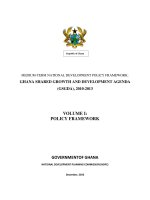
Arabidopsis thaliana is a member of the Brassicaceae, or
mustard family (Figure 16.1). It is a small plant, well suited
for laboratory culture and experimentation. It has been
called the
Drosophila of plant biology because of its wide-
spread use in the study of plant genetics and molecular
genetic mechanisms, particularly in an effort to understand
plant developmental change. It was the first higher plant
to have its genome completely sequenced. Furthermore,
there is a concerted international effort to understand the
function of every gene in the
Arabidopsis genome by the
year 2010. As a result, we are much closer to an under-
standing of the molecular mechanisms governing
Ara-
bidopsis
embryogenesis than of those for any other plant
species.
Embryogenesis Establishes the Essential Features
of the Mature Plant
Plants differ from most animals in that embryogenesis does
not directly generate the tissues and organs of the adult.
For example, angiosperm embryogenesis forms a rudi-
mentary plant body, typically consisting of an embryonic
axis and two cotyledons (if it is a dicot). Nevertheless,
embryogenesis establishes the two basic developmental
patterns that persist and can easily be seen in the adult
plant:
1. The apical–basal axial developmental pattern.
2. The radial pattern of tissues found in stems and
roots.
340 Chapter 16
Embryogenesis also establishes the primary meristems.
Most of the structures that make up the adult plant are gen-
erated after embryogenesis through the activity of meris-
stems. Although these primary meristems are established
during embryogenesis, only upon germination will they
become active and begin to generate the organs and tissues
of the adult.
Axial patterning. Almost all plants exhibit an axial polar-
ity
in which the tissues and organs are arrayed in a precise
order along a linear, or polarized, axis. The shoot apical
meristem is at one end of the axis, the root apical meristem
at the other. In the embryo and seedling, one or two cotyle-
dons are attached just below the shoot apical meristem.
Next in this linear array is the hypocotyl, followed by the
root, the root apical meristem, and the root cap. This axial
pattern is established during embryogenesis.
What may not be so obvious is the fact that any individ-
ual segment of either the root or the shoot also has apical and
basal ends with different, distinct physiological and structural
properties. For example, whereas adventitious roots develop
from the basal ends of stem cuttings, buds develop from the
apical ends, even if they are inverted (see Figure 19.12).
Radial patterning. Different tissues are organized in a pre-
cise pattern within plant organs. In stems and roots the tis-
sues are arranged in a radial pattern extending from the
outside of a stem or a root into its center. If we examine a
root in cross section, for example, we see three concentric
rings of tissues arrayed along a radial axis: An outermost
Silique (fruit)
Cauline (stem) leaf
(A) (B)
Rosette leaf
Roots
Internode
Petal
Sepal
Stamen Carpel
(C)
(D)
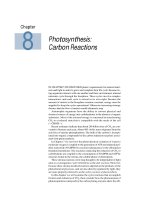
FIGURE 16.1 Arabidopsis thaliana. (A) Drawing of a mature
Arabidopsis plant showing the various organs. (B) Drawing of
a flower showing the floral organs. (C) An immature vegeta-
tive plant consisting of basal rosette leaves and a root system
(not shown). (D) A mature plant after most of the flowers
have matured and the siliques have developed. (A and B
after Clark 2001; C and D courtesy of Caren Chang.)
Growth and Development 341
layer of epidermal cells (the epidermis) covers a cylinder
of cortical tissue (the cortex), which in turn overlies the
vascular cylinder (the endodermis, pericycle, phloem, and
xylem) (Figure 16.2) (see Chapter 1).
The
protoderm is the meristem that gives rise to the epi-
dermis, the
ground meristem produces the future cortex and
endodermis, and the
procambium is the meristem that gives
rise to the primary vascular tissue and vascular cambium.
Arabidopsis Embryos Pass through Four Distinct
Stages of Development
The Arabidopsis pattern of embryogenesis has been studied
extensively and is the one we will present here, but keep
in mind that angiosperms exhibit many different patterns
of embryonic development, and this is only one type.
The most important stages of embryogenesis in
Ara-
bidopsis
, and many other angiosperms, are these:
1.
The globular stage embryo. After the first zygotic divi-
sion, the apical cell undergoes a series of highly
ordered divisions, generating an eight-cell (
octant)
globular embryo by 30 hours after fertilization
(Figure 16.3C). Additional precise cell divisions
Protoxylem
Pericycle
Endodermis
Cortex
Epidermis
Casparian strip
1 mm
FIGURE 16.2 The radial pattern of tissues found in plant
organs can be observed in a crosssection of the root. This
crosssection of an
Arabidopsis root was taken approximately
1 mm back from the root tip, a region in which the different
tissues have formed.
Apical cells
Basal cells
Cotyledon
Axis
Protoderm
Cotyledon
Axis
Root apex
Shoot apex
(A)
(B)
(D)
(E)
(F)
(G)
(H)
(C)
FIGURE 16.3 Arabidopsis embryogenesis is characterized by
a precise pattern of cell division. Successive stages of
embryogenesis are depicted here. (A) One-cell embryo after
the first division of the zygote, which forms the apical and
basal cells; (B) two-cell embryo; (C) eight-cell embryo; (D)
early globular stage, which has developed a distinct proto-
derm (surface layer); (E) early heart stage; (F) late heart
stage; (G) torpedo stage; (H) mature embryo. (From West
and Harada 1993 photographs taken by K. Matsudaira Yee;
courtesy of John Harada, © American Society of Plant
Biologists, reprinted with permission.)
50 µm
25 µm 25 µm 25 µm
25 µm
50 µm 50 µm 50 µm
342 Chapter 16
increase the number of cells in the sphere (Figure
16.3D).
2.
The heart stage embryo. This stage forms through
rapid cell divisions in two regions on either side of
the future shoot apex. These two regions produce
outgrowths that later will give rise to the cotyledons
and give the embryo bilateral symmetry (Figure
16.3E and F).
3.
The torpedo stage embryo. This stage forms as a result
of cell elongation throughout the embryo axis and
further development of the cotyledons (Figure
16.3G).
4.
The maturation stage embryo. Toward the end of
embryogenesis, the embryo and seed lose water and
become metabolically quiescent as they enter dor-
mancy (Figure 16.3H).
Cotyledons are food storage organs for many species,
and during the cotyledon growth phase, proteins, starch,
and lipids are synthesized and deposited in the cotyledons
to be utilized by the seedling during the heterotrophic
(nonphotosynthetic) growth that occurs after germination.
Although food reserves are stored in the
Arabidopsis cotyle-
dons, the growth of the cotyledons is not as extensive in
this species as it is in many other dicots. In monocots, the
food reserves are stored mainly in the endosperm. In
Ara-
bidopsis
and many other dicots, the endosperm develops
rapidly early in embryogenesis but then is reabsorbed, and
the mature seed lacks endosperm tissue.
The Axial Pattern of the Embryo Is Established
during the First Cell Division of the Zygote
Axial polarity is established very early in embryogenesis
(see
Web Topic 16.1). In fact, the zygote itself becomes
polarized and elongates approximately threefold before its
first division. The apical end of the zygote is densely cyto-
plasmic, but the basal half of the cell contains a large cen-
tral vacuole (Figure 16.4).
The first division of the zygote is asymmetric and occurs
at right angles to its long axis. This division creates two
cells—an apical and a basal cell—that have very different
fates (see Figure 16.3A). The smaller, apical daughter cell
receives more cytoplasm than the larger, basal cell, which
inherits the large zygotic vacuole. Almost all of the struc-
tures of the embryo, and ultimately the mature plant, are
derived from the smaller apical cell. Two vertical divisions
and one horizontal division of the apical cell generate the
eight-celled (octant) globular embryo (see Figure 16.3C).
The basal cell also divides, but all of its divisions are hor-
izontal, at right angles to the long axis. The result is a fila-
ment of six to nine cells known as the
suspensor that
attaches the embryo to the vascular system of the plant. Only
one of the basal cell derivatives contributes to the embryo.
The basal cell derivative nearest the embryo is known as the
hypophysis (plural hypophyses), and it forms the columella,
or central part of the root cap, and an essential part of the
root apical meristem known as the
quiescent center, which
will be discussed later in the chapter (Figure 16.5).
Even though the embryo is spherical throughout the
globular stage of embryogenesis (see Figure 16.3A–D), the
cells within the apical and basal halves of the sphere have
different identities and functions. As the embryo continues
to grow and reaches the heart stage, its axial polarity
becomes more distinct (see Figure 16.5), and three axial
regions can readily be recognized:
1. The
apical region gives rise to the cotyledons and shoot
apical meristem.
2. The
middle region gives rise to the hypocotyl, root,
and most of the root meristem.
3. The
hypophysis gives rise to the rest of the root meri-
stem (see Figure 16.5).
The cells of the upper and lower tiers of the early globular
stage embryo differ, and the embryo is divided into apical
and basal halves, reflecting the axial pattern imposed on
the embryo in the zygote.
The Radial Pattern of Tissue Differentiation Is First
Visible at the Globular Stage
The radial pattern of tissue differentiation is first observed
in the octant embryo (Figure 16.6). As cell division contin-
ues in the globular embryo, transverse divisions divide the
Zygote nucleus
Endosperm
nucleus
Embryo sac
Nucellus
Zygote
Ovule
integuments
Vacuole
FIGURE 16.4 Arabidopsis ovule containing the embryo sac at
about 4 hours after double fertilization. The zygote exhibits
a marked polarization. The terminal half of the zygote has
dense cytoplasm and a single large nucleus, while a large
central vacuole occupies the basal half of the cell. At this
stage, the embryo sac surrounding the zygote also contains
4 endosperm nuclei.
Growth and Development 343
Early seedling
Heart stage
Octant stage
Two-cell stage
Hypophysis
Suspensor
Basal cell
of suspensor
Central cells
Apical cells
Basal cell
Terminal cell
Shoot apical
meristem
Shoot apical
meristem
Cotyledons
Hypocotyl
Embryonic root
Root meristem
Quiescent center
Columella root cap
FIGURE 16.5 The apical–basal organization of
plant tissues and organs is established very
early in embryogenesis. This diagram illustrates
how the organs of the early
Arabidopsis seedling
originate from specific regions of the embryo.
(From Willemsen et al. 1998.)
Seedling
Cotyledons
Shoot apical
meristem
Root
Torpedo stage
Heart stage
Protoderm
Early globular stage
Hypophysis
Hypocotyl
Epidermis
Ground
meristem/
cortex and
epidermis
Vascular
cambium/
stele
Columella
of root cap
Quiescent
center
Root cap
FIGURE 16.6 The radial tissue patterns are also established during embryogene-
sis. This drawing illustrates the origin of the different tissues and organs from
embryonic regions in
Arabidopsis embryogenesis. The gray lines between the tor-
pedo and seedling stages indicate the regions of the embryo that give rise to
various regions of the seedling. The expanded regions represent boundaries
where developmental fate is somewhat flexible. (After Van Den Berg et al. 1995.)
344 Chapter 16
lower tier of cells radially into three regions.
These regions will become the radially arranged
tissues of the root and stem axes. The outermost
cells form a one-cell-thick surface layer, known as
the
protoderm. The protoderm covers both halves
of the embryo and will generate the epidermis.
Cells that will become the ground meristem
underlie the protoderm. The ground meristem
gives rise to the
cortex and, in the root and
hypocotyl, it will also produce the
endodermis.
The procambium is the inner core of elongated
cells that will generate the
vascular tissues and,
in the root, the
pericycle (see Figure 16.2).
Embryogenesis Requires Specific Gene
Expression
Analysis of Arabidopsis mutants that either fail to
establish axial polarity or develop abnormally
during embryogenesis has led to the identifica-
tion of genes whose expression participates in tis-
sue patterning during embryogenesis.
The GNOM gene: Axial patterning. Seedlings
homozygous for mutations in the
GNOM gene
lack both roots and cotyledons (Figure 16.7A)
(Mayer et al. 1993). Defects in
gnom embryos first
appear during the initial division of the zygote,
and they persist throughout embryogenesis. In
the most extreme mutants,
gnom embryos are
spherical and lack axial polarity entirely. We can conclude
that
GNOM gene expression is required for the establish-
ment of axial polarity.
1
The MONOPTEROS gene: Primary root and vascular
tissue. Mutations in the MONOPTEROS (MP) gene result
in seedlings that lack both a hypocotyl and a root, although
they do produce an apical region. The apical structures in
the
mp mutant embryos are not structurally normal, how-
ever, and the tissues of the cotyledons are disorganized
(Figure 16.7B) (Berleth and Jürgens 1993). Embryos of
mp
mutants first show abnormalities at the octant stage, and
they do not form a procambium in the lower part of the
globular embryo, the part that should give rise to the
hypocotyl and root. Later some vascular tissue does form
in the cotyledons, but the strands are improperly connected.
Although the
mp mutant embryos lack a primary root
when they germinate, they will form adventitious roots as
the seedlings grow into adult plants. The vascular tissues
in all organs of these mutant plants are poorly developed,
with frequent discontinuities. Thus the
MP gene is required
for the formation of the embryonic primary root, but not
for root formation in the adult plant. The
MP gene is
important for the formation of vascular tissue in postem-
bryonic development (Przemeck et al. 1996).
The SHORT ROOT and SCARECROW genes: Ground
tissue development. Genes have been identified that func-
tion in the establishment of the radial tissue pattern in the
root and hypocotyl during embryogenesis. These genes
also are required for maintenance of the radial pattern dur-
ing postembryonic development (Scheres et al. 1995; Di
Laurenzio et al. 1996). To identify these genes, investigators
isolated
Arabidopsis mutants that caused roots to grow
slowly (Figure 16.8B). Analysis of these mutants identified
several that have defects in the radial tissue pattern. Two
of the affected genes,
SHORT ROOT (SHR) and SCARE-
CROW
(SCR), are necessary for tissue differentiation and
cell differentiation not only in the embryo, but also in both
primary and secondary roots and in the hypocotyl.
Mutants of
SHR and SCR both produce roots with a sin-
gle-celled layer of ground tissue (Figure 16.8D). Cells mak-
ing up the single-celled layer of ground tissue have a
mixed identity and show characteristics of both endoder-
mal and cortical cells in plants with the
scr mutation. These
scr mutants also lack the cell layer called the starch sheath,
a structure that is involved in the growth response to gravity
(see Chapter 19). Roots of plants with the
shr mutation also
1
In discussions of plant and yeast genetics, wild-type (nor-
mal) genes are capitalized and italicized (in this case
GNOM),
and mutations are set in lowercase letters (here
gnom).
FIGURE 16.7 Genes whose functions are essential for Arabidopsis
embryogenesis have been identified by the selection of mutants in
which a stage of embryogenesis is blocked, such as
gnom and
monopteros. The development of mutant seedlings is contrasted here
with that of the wild type at the same stage of development. (A) The
GNOM gene helps establish apical–basal polarity. A plant homozy-
gous for
gnom is shown on the right. (B) The MONOPTEROS gene is
necessary for basal patterning and formation of the primary root.
Plants homozygous for the
monopteros mutation have a hypocotyl, a
normal shoot apical meristem, and cotyledons, but they lack the pri-
mary root. (A from Willemsen et al. 1998; B from Berleth and Jürgens
1993.)
MONOPTEROS genes control formation
of the primary rootGNOM genes control apical–
basal polarity
(B) Wild type
monopteros mutant
(A) Wild type gnom mutant
Growth and Development 345
have a single layer of ground tissue, but it has only cortical
cell characteristics and lacks endodermal characteristics.
The HOBBIT gene: The root meristem. The primary root
and shoot meristems are established during embryogene-
sis. Because in most cases they do not become active at this
time, the term
promeristem may be more appropriate to
describe these structures. A
promeristem may be defined
as an embryonic structure that will become a meristem
upon germination.
A molecular marker for the root promeristem has not
yet been identified, but it appears to be determined early
in embryogenesis. Root cap stem cells (the cells that divide
to produce the root cap) are formed from the hypophysis
at the heart stage of embryogenesis, indicating that the root
promeristem is established at least by this stage of embryo-
genesis (Figure 16.9). The expression of the
HOBBIT gene
may be an early marker of root meristem identity (Willem-
sen et al. 1998).
Stem cell Stem cell
Anticlinal
cell divisions
(A)
Daughter
cell
Periclinal
cell divisions
This step is
blocked in
scr mutants
Endodermal cell
Cortical cell
FIGURE 16.8 Mutations in the Arabidopsis gene SCARECROW (SCR)
alter the pattern of tissues in the root. (A) The cell divisions forming
the endodermis and cortex. The endodermal cells and cortical cells
are derived from the same initial cells as a result of two asymmetric
cell divisions. The cortical–endodermal stem cell (uncommitted cell)
expands and then divides anticlinally, reproducing itself and a
daughter cell. The daughter cell then divides periclinally to produce a
small cell that develops endodermal characteristics and a larger cell
that becomes a cortical cell. The second asymmetric division does not
occur in
scr mutants, and the daughter cell formed as a result of the
anticlinal division of the initial has characteristics of both cortical and
endodermal cells. (B) The growth of a 12-day-old wild-type seedling
(left) is compared with that of two 12-day-old seedlings homozygous
for a mutation in the
SCARECROW (SCR) gene (middle and right).
(C) Cross section of the primary root of a wild-type seedling. (D)
Cross section of the primary root of a seedling homozygous for the
scr mutant. (From Di Laurenzio et al. 1996; photos © Cell Press, cour-
tesy of P. Benfey.)
(B)
(D)
(C)
Epidermis
Cortex
Pericycle
Epidermis
Pericycle
Mutant
layer
cell
Endodermis
50 µm
50 µm
Wild type scr1 scr2
346 Chapter 16
Mutants of the HOBBIT (HBT) gene are defective in the
formation of a functional embryonic root, as are plants with
mp mutants. However, these two mutations act in very dif-
ferent ways. The
hbt mutants begin to show abnormalities
at the two- or four-cell stage, before the formation of the
globular embryo. The primary defect in
hbt mutants is in
the hypophyseal precursor, which divides vertically
instead of horizontally. As a result, the hypophysis does
not form, and the root meristem that subsequently forms
lacks a quiescent center and the columella (see Figure
16.9F). Embryos of
hbt mutants appear to have a root
meristem, but it does not function when the seedlings ger-
minate. Furthermore, plants grown from
hbt mutant
embryos are unable to form lateral roots.
The SHOOTMERISTEMLESS gene: The shoot promeri-
stem. The shoot promeristem can be recognized morpho-
logically by the torpedo stage of embryogenesis in
Ara-
bidopsis
. Oriented cell divisions of some of the cells
between the cotyledons result in a layered appearance of
this region that is characteristic of the shoot apical meri-
stem (as described later in the chapter). However, the pro-
genitors of these cells probably acquired the molecular
identity of the shoot apical meristem cells much earlier,
during the globular stage.
The
SHOOTMERISTEMLESS (STM) gene is expressed
specifically in the cells that will become the shoot apical
meristem, and its expression in these cells is required for
the formation of the shoot promeristem.
Arabidopsis plants
homozygous for a mutated, loss-of-function
STM gene do
not form a shoot apical meristem, and instead all the cells
in this region differentiate (Lincoln et al. 1994). The prod-
uct of the wild-type
STM gene appears to suppress cell dif-
ferentiation, ensuring that the meristem cells remain undif-
ferentiated.
STM mRNA can first be detected in one or two cells at
the apical end of the midglobular embryo. By the heart
stage,
STM expression is confined to a few cells between
the cotyledons (Long et al. 1996). Because
STM acts as a
marker for these cells, the shoot apical meristem must be
specified long before it can be recognized morphologically.
The
STM gene is necessary not only for the formation of
the embryonic shoot apical meristem, but also for the
maintenance of shoot apical meristem identity in the adult
plant. The role of the nucleus in controlling development
was first demonstrated in the giant algal unicell,
acetabu-
laria
(see Web Essay 16.2).
LRC
QC
COL
QC
(A) Wild type (B) hobbit mutant
(C) (D)
25 mm
25 mm
(E) (F)
FIGURE 16.9 The HOBBIT (HBT) gene is important for the
development of a functional root apical meristem. (A) Wild-
type
Arabidopsis seedling; (B) hobbit mutant seedling; (C)
root tip of wild type showing quiescent center (QC), col-
umella (COL) and lateral root cap (LRC); (D) root tip of
hob-
bit
mutant; (E) quiescent center and columella of wild-type;
(F) absence of quiescent center and columella in
hobbit. The
seedlings in A and B are both shown 7 days after germina-
tion (4
× magnification). Staining with iodine reveals starch
grains in the columella cells of the root cap in the wild type
(E). No starch grains are present in the
hbt mutant root tip
(F). (From Willemsen et al. 1998.)
Growth and Development 347
Embryo Maturation Requires Specific
Gene Expression
The Arabidopsis embryo enters dormancy after it has gen-
erated about 20,000 cells. Dormancy is brought about by
the loss of water and a general shutting down of gene tran-
scription and protein synthesis, not only in the embryo, but
also throughout the seed. To adapt the cell to the special
conditions of dormancy, specific gene expression is
required. For example, the
ABSCISIC ACID INSENSITIVE3
(ABI3) and FUSCA3 genes are necessary for the initiation
of dormancy and are sensitive to the hormone abscisic acid,
which is the signaling molecule that initiates seed and
embryo dormancy.
ABI3 also controls the expression of
genes encoding the storage proteins that are deposited in
the cotyledons during the maturation phase of embryogen-
esis (see Chapter 23).
The
LEAFY COTYLEDON1 (LEC1) gene also is active in
late embryogenesis. Because
lec1 mutants cannot survive
desiccation and do not enter dormancy, the embryos die
unless they are rescued through isolation before desicca-
tion occurs. The rescued embryos will germinate in culture
and produce fertile plants, which are like wild-type plants
except that they lack the 7S storage protein and they have
leaflike cotyledons with trichomes on their upper surface.
The normal appearance and development of the mature
lec1 mutants indicates that the LEC1 gene is required only
during embryogenesis. Although the most obvious defects
of the
lec1 mutants are seen only in the maturation phase
embryo, mRNA from
LEC1 gene expression can be
detected throughout embryogenesis. It has been proposed
that
LEC1 is a general repressor of vegetative development
and its expression is necessary throughout embryogenesis
(Lotan et al. 1998).
THE ROLE OF CYTOKINESIS IN
PATTERN FORMATION
One of the most striking features of tissue organization in
many plants, illustrated by
Arabidopsis, is the remarkably
precise pattern of oriented, often called
stereotypic, cell divi-
sions. This pattern of divisions generates files of cells
extending from the meristem toward the base of the plant.
Although the division pattern is not as precise in all other
species, the basic pattern of tissue formation is similar.
How important is the plane of cell division for the estab-
lishment of the tissue patterns found in plant organs?
The Stereotypic Cell Division Pattern Is Not
Required for the Axial and Radial Patterns of
Tissue Differentiation
Two Arabidopsis mutants, fass and ton, have dramatic effects
on the patterns of cell division in all stages of development
Wild-type Arabidopsis
(A) (B)
(D) (E)
(C)
(F)
Homozygous ton mutant
50 µm
FIGURE 16.10 Arabidopsis plants with
mutations in the
TON gene are
unable to form a preprophase band
of microtubules in cells at any stage
of division. Plants carrying this
mutation are highly irregular in their
cell division and expansion planes,
and as a result they are severely
deformed. However, they continue
to produce recognizable tissues and
organs in their correct positions.
Although the organs and tissues pro-
duced by these mutant plants are
highly abnormal, the radial tissue
pattern is not disturbed. (A–C) Wild-
type
Arabidopsis: (A) early globular
stage embryo; (B) seedling seen from
the top; (C) cross section of a root.
(D–F) Comparable stages of
Arabidopsis homozygous for the ton
mutation: (D) early embryogenesis;
(E) mutant seedling seen from the
top; (F) cross section of the mutant
root showing the random orientation
of the cells, but a near wild-type tis-
sue order; an outer epidermal layer
covers a multicellular cortex, which
in turn surrounds the vascular cylin-
der. (From Traas et al. 1995.)
60 µm
348 Chapter 16
and eliminate the stereotypic divisions seen in the wild
type (Torres-Ruiz and Jürgens 1994; Traas et al. 1995). These
mutations probably are in the same gene, and cells in
plants homozygous for the
ton (fass) mutation lack a cyto-
plasmic structure known as the
preprophase band of micro-
tubules. The preprophase band appears to be essential for
the orientation of the phragmoplast during cytokinesis, and
thus is required for oriented cell divisions (see Chapter 1
and
Web Topic 16.2).
The effects of the
ton (fass) mutation are seen from the
earliest stages of embryogenesis and persist throughout
development. The plants are tiny, never reaching more than
2 to 3 cm in height. They have misshapen leaves, roots, and
stems, and they are sterile (Figure 16.10D–F). Nevertheless,
the mutant plants not only establish an axial pattern, but
they have all the cell types and organs of the wild-type
plant, and these occur in their correct positions. The precise
numbers of cells found in each tissue layer are radically dif-
ferent in the mutants, but each tissue is present and in the
proper order.
The fact that these mutations do not prevent the estab-
lishment of the radial tissue pattern is strong evidence that
the stereotypic cell division pattern found in the
Arabidop-
sis
embryo and in the root is not essential for the radial pat-
tern of tissue differentiation.
An Arabidopsis Mutant with Defective Cytokinesis
Cannot Establish the Radial Tissue Pattern
The Arabidopsis mutant knolle is defective in cytokinesis, the
step at the end of mitosis in which a new wall is formed
partitioning the daughter nuclei into separate cells. The
KNOLLE gene encodes a syntaxin-like protein that is
important for vesicle fusion.
Syntaxins are proteins that
integrate into membranes, permitting the membranes to
fuse. Vesicle fusion is essential for cytokinesis (Figure
16.11).
FIGURE 16.11 Encoded by the KNOLLE gene, syntaxin pro-
teins play a critical role in the fusion of Golgi-derived mem-
branes, which is required for normal cytokinesis in most
organisms, including
Arabidopsis. (A) Electron micrograph of a
region of an
Arabidopsis embryo with the knolle mutation. The
box outlined is 5 mm wide. (B) Higher-magnification pho-
tomicrograph showing an incomplete and abnormal cross-
wall attached to the parent cell wall. (C) A model for the
fusion of vesicles during cell plate formation. A complex of
soluble proteins mediates the interaction of synaptobrevin
protein with the syntaxin protein (encoded by the
KNOLLE
gene) on the target membrane. (A and B from Lukowitz et al.
1996, courtesy of G. Jürgens; C after Assaad et al. 1996.)
(C)
Syntaxin protein
(in Arabidopsis coded
by KNOLLE gene)
Synaptobrevin
(a vesicle
membrane
protein)
C
C
N
N
Target membrane
Several soluble
proteins mediate
interactions of
membrane proteins
Vesicle membrane
(A)
(B)
e
o
n
n
Abnormal cross wall
Growth and Development 349
Although cell division is not blocked by the knolle muta-
tion, cell plate formation is irregular and often incomplete.
As a result, many cells are binucleate, while other cells are
only partly separated or are connected by large cytoplas-
mic bridges. The division planes also are irregular. These
irregularities have severe effects on development.
Plants homozygous for the
knolle mutation go through
embryogenesis, but the radial tissue pattern is severely dis-
rupted and an epidermal layer does not form in early
embryogenesis. The
knolle mutation does not prevent for-
mation of the apical–basal axis, and embryogenesis is com-
pleted, although the seedlings are very short-lived and die
soon after germination. The plants also lack functional
meristems.
The conclusion drawn from studies of the
knolle muta-
tion appears to contradict what we learned from the
ton
(fass) mutations. Both the knolle and the ton mutations dis-
rupt the normal pattern of cell division in embryonic and
postembryonic development. But whereas the
knolle muta-
tions block the establishment of the radial tissue pattern, in
the
ton mutants the pattern is established.
One difference between the
ton and the knolle mutations
is that the latter usually prevents the effective separation of
daughter cells during cytokinesis because the cell plate is
incomplete. Since cell–cell communication is important for
pattern formation, it may be necessary for cells to be iso-
lated effectively so that the information exchange can be
regulated. Even though the cytosol is continuous between
adjacent plant cells through plasmodesmata, complete cel-
lularization is required for normal development. Thus the
ton mutants are able to perceive positional information cor-
rectly, while the
knolle mutants cannot. For a review of the
mechanisms determining the plane of cell division in plant
cells, see
Web Essay 16.3.
MERISTEMS IN PLANT DEVELOPMENT
Meristems are populations of small, isodiametric (having
equal dimensions on all sides) cells with embryonic char-
acteristics. Vegetative meristems are self-perpetuating. Not
only do they produce the tissues that will form the body of
the root or stem, but they also continuously regenerate
themselves. A meristem can retain its embryonic character
indefinitely, possibly even for thousands of years in the
case of trees. The reason for this ability is that some meri-
stematic cells do not become committed to a differentiation
pathway, and they retain the capacity for cell division, as
long as the meristem remains vegetative.
Undifferentiated cells that retain the capacity for cell
division indefinitely are said to be
stem cells. Although his-
torically called
initial cells in plants, in function they are
very similar, if not identical, to animal stem cells (Weigel
and Jürgens 2002). When stem cells divide, on average one
of the daughter cells retains the identity of the stem cell,
while the other is committed to a particular developmen-
tal pathway (Figure 16.12).
Stem cells usually divide slowly. Their committed
daughters, however, may enter a period of rapid cell divi-
sion before they stop dividing and can be recognized as spe-
cific cell types. Stem cells represent the ultimate source of
all the cells in the meristem and the entire rest of the plant—
both roots, leaves, and other organs, as well as stems.
The Shoot Apical Meristem Is a Highly
Dynamic Structure
The vegetative shoot apical meristem generates the stem,
as well as the lateral organs attached to the stem (leaves
and lateral buds). The shoot apical meristem typically con-
tains a few hundred to a thousand cells, although the
Ara-
bidopsis
shoot apical meristem has only about 60 cells.
The shoot apical meristem is located at the extreme tip
of the shoot, but it is surrounded and covered by immature
leaves. These are the youngest leaves produced by the
activity of the meristem. It is useful to distinguish the shoot
apex from the meristem proper. The
shoot apex (plural
apices) consists of the apical meristem plus the most
recently formed leaf primordia. The
shoot apical meristem
is the undifferentiated cell population only and does not
include any of the derivative organs.
The shoot apical meristem is a flat or slightly mounded
region, 100 to 300
µm in diameter, composed mostly of
small, thin-walled cells, with a dense cytoplasm, and lack-
ing large central vacuoles. The shoot apical meristem is a
dynamic structure that changes during its cycle of leaf and
stem formation. In addition, in many plants it exhibits sea-
sonal activity, as does the entire shoot. Shoot apical meri-
stems may grow rapidly in the spring, enter a period of
slower growth during the summer, and become dormant
in the fall, with dormancy lasting through the winter. The
size and structure of the shoot apical meristem also change
with seasonal activity.
Shoots develop and grow at their tips, as is the case with
roots, but the developing regions are not as stratified and
precisely ordered as they are in the root. Moreover, growth
occurs over a much broader region of the shoot than is the
case for roots. At any given time, a region containing sev-
eral internodes, typically 10 to 15 cm long, may be under-
going primary growth.
Stem
cell
Committed
cells
Daughter
cells
Differentiated
cells
FIGURE 16.12 Stem cells generate daughter cells, some of
which remain uncommitted and retain the property of stem
cells, while others become committed to differentiate.
350 Chapter 16
The Shoot Apical Meristem Contains Different
Functional Zones and Layers
The shoot apical meristem consists of different functional
regions that can be distinguished by the orientation of the
cell division planes and by cell size and activity. The
angiosperm vegetative shoot apical meristem usually has
a highly stratified appearance, typically with
three distinct
layers of cells
. These layers are designated L1, L2, and L3,
where L1 is the outermost layer (Figure 16.13). Cell divi-
sions are anticlinal in the L1 and L2 layers; that is, the new
cell wall separating the daughter cells is oriented at right
angles to the meristem surface. Cell divisions tend to be
less regularly oriented in the L3 layer. Each layer has its
own stem cells, and all three layers contribute to the for-
mation of the stem and lateral organs.
Active apical meristems also have an organizational pat-
tern called
cytohistological zonation. Each zone is com-
posed of cells that may be distinguished not only on the
basis of their division planes, but also by differences in size
and by degrees of vacuolation (see Figure 16.13B). These
zones exhibit different patterns of gene expression, reflect-
ing the different functions of each zone (Nishimura et al.
1999; Fletcher and Meyerowitz 2000).
The center of an active meristem contains a cluster of
relatively large, highly vacuolate cells called the
central
zone
. The central zone is somewhat comparable to the qui-
escent center of root meristems (which will be discussed
later in the chapter). A doughnut-shaped region of smaller
cells, called the
peripheral zone, flanks the central zone. A
rib zone lies underneath the central cell zone and gives rise
to the internal tissues of the stem.
These different zones most likely represent different
developmental domains. The peripheral zone is the region
in which the first cell divisions leading to the formation of
leaf primordia will occur. The rib zone contributes cells that
become the stem. The central zone contains the pool of
stem cells, some fraction of which remains uncommitted,
while others replenish the rib and peripheral zone popu-
lations (Bowman and Eshed 2000).
Some Meristems Arise during Postembryonic
Development
The root and shoot apical meristems formed during
embryogenesis are called
primary meristems. After ger-
mination, the activity of these primary meristems gener-
ates the primary tissues and organs that constitute the pri-
mary plant body.
Most plants also develop a variety of
secondary meri-
stems
during postembryonic development. Secondary
meristems can have a structure similar to that of primary
meristems, but some secondary meristems have a quite dif-
ferent structure. These include axillary meristems, inflo-
rescence meristems, floral meristems, intercalary meri-
stems, and lateral meristems (the vascular cambium and
cork cambium). (Inflorescence and floral meristems will be
discussed in Chapter 24.):
FIGURE 16.13 The shoot apical meristem generates the aer-
ial organs of the plant. (A) This longitudinal section
through the center of the shoot apex of
Coleus blumei shows
the layered appearance of the shoot apical meristem. Most
cell divisions are anticlinal in the outer L1 and L2 layers,
while the planes of cell divisions are more randomly ori-
ented in the L3 layer. The outermost (L1) layer generates
the shoot epidermis; the L2 and L3 layers generate internal
tissues. (B) The shoot apical meristem also has cytohistolog-
ical zones, which represent regions with different identities
and functions. The central zone contains the stem cells,
which divide slowly but are the ultimate source of the tis-
sues that make up the plant body. The peripheral zone, in
which cells divide rapidly, surrounds the central zone and
produces the leaf primordia. A rib zone lies below the cen-
tral zone and generates the central tissues of the stem. (A
©J. N. A. Lott/Biological Photo Service.)
(A)
Leaf primordia
Shoot apical
meristem
L3, with randomly
oriented cell divisions
L1 and L2, with anticlinal
cell divisions
Generate
internal
tissues
Generates
epidermis
Leaf primordium
Shoot
apical
meristem
L
1
–
L
2
L
3
Central
zone
Rib
zone
Peripheral
zone
Peripheral
zone
(B)
Growth and Development 351
• Axillary meristems are formed in the axils of leaves
and are derived from the shoot apical meristem. The
growth and development of axillary meristems pro-
duces branches from the main axis of the plant.
• Intercalary meristems are found within organs, often
near their bases. The intercalary meristems of grass
leaves and stems enables them to continue to grow
despite mowing or grazing by cows.
• Branch root meristems have the structure of the pri-
mary root meristem, but they form from pericycle
cells in mature regions of the root. Adventitious roots
also can be produced from lateral root meristems that
develop on stems, as when stem cuttings are rooted
to propagate a plant.
• The vascular cambium (plural cambia) is a secondary
meristem that differentiates along with the primary
vascular tissue from the procambium within the vas-
cular cylinder. It does not produce lateral organs, but
only the woody tissues of stems and roots. The vas-
cular cambium contains two types of meristematic
cells: fusiform stem cells and ray stem cells.
Fusiform
stem cells
are highly elongated, vacuolate cells that
divide longitudinally to regenerate themselves, and
whose derivatives differentiate into the conducting
cells of the secondary xylem and phloem.
Ray stem
cells
are small cells whose derivatives include the
radially oriented files of parenchyma cells within
wood known as rays.
• The cork cambium is a meristematic layer that devel-
ops within mature cells of the cortex and the sec-
ondary phloem. Derivatives of the cork cambium dif-
ferentiate as cork cells that make up a protective layer
called the
periderm, or bark. The periderm forms the
protective outer surface of the secondary plant body,
replacing the epidermis in woody stems and roots.
Axillary, Floral, and Inflorescence Shoot Meristems
Are Variants of the Vegetative Meristem
Several different types of shoot meristems can be distin-
guished on the basis of their developmental origin, the
types of lateral organs they generate, and whether they are
determinate (having a genetically programmed limit to
their growth) or
indeterminate (showing no predeter-
mined limit to growth; growth continues so long as
resources permit).
The vegetative shoot apical meristem usually is inde-
terminate in its development. It repetitively forms phy-
tomeres as long as environmental conditions favor growth
but do not generate a flowering stimulus. A
phytomere is
a developmental unit consisting of one or more leaves, the
node to which the leaves are attached, the internode below
the node, and one or more axillary buds (Figure 16.14).
Axillary buds are secondary meristems; if they are also
vegetative meristems, they will have a structure and devel-
opmental potential similar to that of the apical meristem.
Vegetative meristems may be converted directly into flo-
ral meristems when the plant is induced to flower (see
Chapter 24).
Floral meristems differ from vegetative meri-
stems in that instead of leaves they produce floral organs:
sepals, petals, stamens, and carpels. In addition, floral
meristems are determinate: All meristematic activity stops
after the last floral organs are produced.
In many cases, vegetative meristems are not directly
converted to floral meristems. Instead, the vegetative
meristem is first transformed into an
inflorescence meri-
stem
. The types of lateral organs produced by an inflores-
cence meristem are different from the types produced by a
floral meristem. The inflorescence meristem produces
bracts and floral meristems in the axils of the bracts, instead
of the sepals, petals, stamens, and ovules produced by flo-
ral meristems. Inflorescence meristems may be determinate
or indeterminate, depending on the species.
LEAF DEVELOPMENT
The leaves of most plants are the organs of photosynthesis.
This is where light energy is captured and used to drive the
chemical reactions that are vital to the life of the plant.
Although highly variable in size and shape from species to
species, in general leaves are thin, flat structures with dor-
siventral polarity. This pattern contrasts with that of the
Leaf
Node
Internode
Bud
Root
Phytomere
FIGURE 16.14 The shoot apical meristem repetitively forms
units known as phytomeres. Each phytomere consists of
one or more leaves, the node at which the leaves are
attached, the internode immediately below the leaves, and
one or more buds in the axils of the leaves.
352 Chapter 16
shoot apical meristem and stem, both of which have radial
symmetry. Another important difference is that leaf pri-
mordia exhibit determinate growth, while the vegetative
shoot apical meristem is indeterminate. As described in the
sections that follow, several distinct stages can be recog-
nized in leaf development (Sinha 1999).
Stage 1: Organogenesis. A small number of cells in the L1
and L2 layers in the flanks of the apical dome of the shoot
apical meristem acquire the
leaf founder cell identity.
These cells divide more rapidly than surrounding cells and
produce the outgrowth that represents the
leaf pri-
mordium
(plural primordia) (Figure 16.15A). These pri-
mordia subsequently grow and develop into leaves.
Stage 2: Development of suborgan domains. Different
regions of the primordium acquire identity as specific parts
of the leaf. This differentiation occurs along three axes:
dor-
siventral
(abaxial–adaxial), proximodistal (apical–basal),
and
lateral (margin–blade–midrib) (Figure 16.15B). The
upper (adaxial) side of the leaf is specialized for light
absorption; the lower (abaxial) surface is specialized for gas
exchange. Leaf structure and maturation rates also vary
along the proximodistal and lateral axes.
Stage 3: Cell and tissue differentiation. As the develop-
ing leaf grows, tissues and cells differentiate. Cells derived
from the L1 layer differentiate as epidermis (epidermal
cells, trichomes, and guard cells), derivatives of the L2 layer
differentiate as the photosynthetic mesophyll cells, and vas-
cular elements and bundle sheath cells are derived from
the L3 layer. These cells differentiate in a genetically deter-
mined pattern that is characteristic of the species but to
some degree modified in response to the environment.
The Arrangement of Leaf Primordia Is Genetically
Programmed
The timing and pattern with which the primordia form is
genetically determined and usually is a characteristic of the
species. The number and order in which leaf primordia
form is reflected in the subsequent arrangement of leaves
around the stem, known as
phyllotaxy (Figure 16.16).
There are five main types of phyllotaxy:
1. Alternate phyllotaxy. A single leaf is initiated at each
node (see Figure 16.16A).
2.
Opposite phyllotaxy. Leaves are formed in pairs on
opposite side of the stem (see Figure 16.16B).
3.
Decussate phyllotaxy. Leaves are initiated in a pat-
tern with two opposite leaves per node and with suc-
cessive leaf pairs oriented at right angles to each
other during vegetative development (see Figure
16.16C).
4.
Whorled phyllotaxy. More than two leaves arise at
each node (see Figure 16.16D).
5.
Spiral phyllotaxy. A type of alternate phyllotaxy in
which each leaf is initiated at a defined angle to the
previous leaf, resulting in a spiral arrangement of
leaves around the stem (see Figure 16.16E).
The positioning of leaf primordia must result from the
precise spatial regulation of growth within the apex. We
know little about how this positioning is regulated, or
about the signals that initiate the formation of a pri-
mordium. One idea is that inhibitory fields generated by
existing primordia influence the spacing of the next pri-
mordium.
Midrib
Margin
P1
P3
P2
P0
Site of next
primordium
(A)
(B)
Most recently formed
primordium, which has
radial symmetry at this stage
Primordium begins
to flatten, developing
a dorsiventral axis
Primordium
elongates in the
proximodistal axis
Dorsal
Ventral
Distal
Proximal
Axillary bud
Petiole
Node
Apical
meristem
FIGURE 16.15 The origin of leaves at the shoot apex and
their axes of symmetry on the stem (A) Leaf primordia in
the flanks of the shoot apical meristem. (B) Diagram of a
shoot showing the various axes along which development
occurs. (After Christensen and Weigel 1998.)
Growth and Development 353
ROOT DEVELOPMENT
Roots are adapted for growing through soil and absorbing
the water and mineral nutrients in the capillary spaces
between soil particles. These functions have placed con-
straints on the evolution of root structure. For example, lat-
eral appendages would interfere with their penetration
through the soil. As a result, roots have a streamlined axis,
and no lateral organs are produced by the apical meristem.
Branch roots arise internally and form only in mature, non-
growing regions. Absorption of water and minerals is
enhanced by fragile root hairs, which also form behind the
growth zone. These long, threadlike cells greatly increase
the root’s absorptive surface area.
In this section we will discuss the origin of root form
and structure (
root morphogenesis), beginning with a
description of the four developmental zones of the root tip.
We will then turn to the apical meristem. The absence of
leaves or buds makes cell lineages easier to follow in roots
than in shoots, thus facilitating molecular genetic studies
on the role of patterns of cell division in root development.
The Root Tip Has Four Developmental Zones
Roots grow and develop from their distal ends. Although
the boundaries are not sharp, four developmental zones can
be distinguished in a root tip: the root cap, the meristematic
zone, the elongation zone, and the maturation zone (Figure
16.17). These four developmental zones occupy only a little
more than a millimeter of the tip of the
Arabidopsis root. The
developing region is larger in other species, but growth is
still confined to the tip. With the exception of the root cap,
the boundaries of these zones overlap considerably:
(A) Alternate (B) Opposite (D) Whorled (E) Spiral(C) Decussate
FIGURE 16.16 Five types of leaf
arrangements (phyllotactic pat-
terns) along the shoot axis. The
same terms also are used for
inflorescences and flowers.
Lateral root
primordium
Pericycle
Cortical cells
Epidermis
Emerging
lateral root
Root hair
Mature vessel
elements
Endodermal
cells differentiate
First vessel
elements begin to
differentiate
Maximum rate of
cell elongation
First sieve tube
element begins to
differentiate
Cell division ceases
in most layers
Maximum rate of
cell division
Quiescent center
Maturation
zone
Elongation
zone
Meristematic
zone
Root cap
FIGURE 16.17 Simplified diagram of a primary root show-
ing the root cap, the meristematic zone, the elongation
zone, and the maturation zone. Cells in the meristematic
zone have small vacuoles and expand and divide rapidly,
generating many files of cells.
354 Chapter 16
• The root cap protects the apical meristem from
mechanical injury as the root pushes its way through
the soil. Root cap cells form by specialized root cap
stem cells. As the root cap stem cells produce new
cells, older cells are progressively displaced toward
the tip, where they are eventually sloughed off. As
root cap cells differentiate, they acquire the ability to
perceive gravitational stimuli and secrete
mucopolysaccharides (slime) that help the root pene-
trate the soil.
• The
meristematic zone lies just under the root cap,
and in
Arabidopsis it is about a quarter of a millimeter
long. The root meristem generates only one organ,
the primary root. It produces no lateral appendages.
• The
elongation zone, as its name implies, is the site
of rapid and extensive cell elongation. Although
some cells may continue to divide while they elon-
gate within this zone, the rate of division decreases
progressively to zero with increasing distance from
the meristem.
• The
maturation zone is the region in which cells
acquire their differentiated characteristics. Cells enter
the maturation zone after division and elongation
have ceased. Differentiation may begin much earlier,
but cells do not achieve the mature state until they
reach this zone. The radial pattern of differentiated
tissues becomes obvious in the maturation zone.
Later in the chapter we will examine the differentia-
tion and maturation of one of these cell types, the tra-
cheary element.
As discussed earlier, lateral or branch roots arise from
the pericycle in mature regions of the root. Cell divisions in
the pericycle establish secondary meristems that grow out
through the cortex and epidermis, establishing a new
growth axis (Figure 16.18). The primary and the secondary
root meristems behave similarly in that divisions of the
cells in the meristem give rise to progenitors of all the cells
of the root.
Root Stem Cells Generate Longitudinal
Files of Cells
Meristems are populations of dividing cells, but not all cells
in the meristematic region divide at the same rate or with
the same frequency. Typically, the central cells divide much
more slowly than the surrounding cells. These rarely divid-
ing cells are called the
quiescent center of the root meri-
stem (see Figure 16.17).
Cells are more sensitive to ionizing radiation when they
are dividing. This is the basis of the use of radiation as a
treatment for cancer in humans. As a result, the rapidly
dividing cells of the meristem can be killed by doses of
radiation that nondividing and slowly dividing cells, such
as those of the quiescent center, can survive. If the rapidly
dividing cells of the root are killed by ionizing radiation, in
many cases the root can regenerate from the cells of the
quiescent center. This ability suggests that quiescent-cen-
ter cells are important for the patterning involved in form-
ing a root.
The most striking structural feature of the root tip, when
viewed in longitudinal section, is the presence of the long
files of clonally related cells. Most cell divisions in the root
tip are transverse, or
anticlinal, with the plane of cytoki-
nesis oriented at right angles to the axis of the root (such
divisions tend to increase root length). There are relatively
few
periclinal divisions, in which the plane of division is
parallel to the root axis (such divisions tend to increase root
diameter).
Epidermis
Cortex
Endodermis
Pericycle
Vasculature
Cortical–endodermal
stem cell
Root cap–epidermal
stem cell
Quiescent center
Root cap
Stage 1 Stage 2 Stage 3 Stage 4 Stage 5 Stage 6
FIGURE 16.18 Model for lateral root formation in Arabidopsis. Six major stages are
shown in the development of the primordium. The different tissue types are desig-
nated by colors. By stage 6, all tissues found in the primary root are present in the
typical radial pattern of the branch root. (From Malamy and Benfey 1997.)
Growth and Development 355
Periclinal divisions occur mostly near the root tip and
establish new files of cells. As a result, the ultimate origin
of any particular mature cell can be traced back to one or a
few cells in the meristem. These are the stem cells of a par-
ticular file. In
Arabidopsis, the stem cells surround the quies-
cent center, but they are not part of the quiescent center. The
stem cells ultimately may be derived from quiescent-center
cells, but this origin must occur during embryogenesis, since
the quiescent-center cells do not divide after germination in
normal development. Analysis of the cell division patterns
in the roots of the water fern
Azolla have contributed to our
detailed understanding of meristem function. (For a discus-
sion of this work, see
Web Topic 16.3.)
Root Apical Meristems Contain Several Types
of Stem Cells
The patterns of cellular organization found in the root
meristems of seed plants are substantially different from
those observed in more primitive vascular plants. All seed
plants have several stem cells instead of the single stem cell
found in plants such as the water fern
Azolla. However,
they are similar to
Azolla in that it is possible to follow files
of cells from the region of maturation into the meristem
and, in some cases, to identify the stem cell from which the
file was produced.
The
Arabidopsis root apical meristem has the following
structure (Figure 16.19):
• The
quiescent center is composed of a group of four
cells, also known as the center cells in the
Arabidopsis
root meristem. The quiescent-center cells in the
Arabidopsis root usually do not divide after embryogen-
esis.
• The
cortical–endodermal stem cells form a ring of
cells that surround the quiescent center. These stem
cells generate the cortical and endodermal layers.
They undergo one anticlinal division (i.e., perpendic-
ular to the longitudinal axis); then these daughters
divide periclinally (i.e., parallel to the longitudinal
axis) to establish the files that become the cortex and
the endodermis, each of which constitutes only one
cell layer in the
Arabidopsis root (see also Figures 16.2
and 16.8C).
• The
columella stem cells are the cells immediately
above (apical to) the central cells. They divide anticli-
nally and periclinally to generate a sector of the root
cap known as the columella.
• The
root cap–epidermal stem cells are in the same
tier as the columella stem cells but form a ring sur-
rounding them. Anticlinal divisions of the root
cap–epidermal stem cells generate the epidermal cell
layer. Periclinal divisions of the same stem cells, fol-
lowed by subsequent anticlinal divisions of the deriv-
atives, produce the lateral root cap.
Columella of
root cap
Columella
stem cell
Epidermis
Cortex
Stele
stem cell
Pericycle
Lateral
root cap
Root cap–
epidermal
stem cell
Cortical
endodermal
stem cell
Quiescent
center cell
Endodermis
Epidermis
(B)
FIGURE 16.19 All the tissues in the Arabidopsis root are derived from a
small number of stem cells in the root apical meristem. (A) Longitudinal
section through the center of a root. The promeristem containing the
stem cells that give rise to all the tissues of the root is outlined in green.
(B) Diagram of the promeristem region outlined in A. Only two of the
four quiescent-center cells are depicted in this section. The black lines
indicate the cell division planes that occur in the stem cells. White lines
indicate the secondary cell divisions that occur in the cortical–endoder-
mal and lateral root cap–epidermal stem cells. (From Schiefelbein et al.
1997, courtesy of J. Schiefelbein, © the American Society of Plant
Biologists, reprinted with permission.)
(A)
356 Chapter 16
• The stele stem cells are a tier of cells just behind the
quiescent-center cells. These cells generate the pericy-
cle and vascular tissues.
The stem cells, together with their immediate derivatives
in the apical meristem, are called the
promeristem.
CELL DIFFERENTIATION
Differentiation is the process by which a cell acquires meta-
bolic, structural, and functional properties that are distinct
from those of its progenitor cell. In plants, unlike animals, cell
differentiation is frequently reversible, particularly when dif-
ferentiated cells are removed from the plant and placed in tis-
sue culture. Under these conditions, cells dedifferentiate (i.e.,
lose their differentiated characteristics), reinitiate cell division,
and in some cases, when provided with the appropriate
nutrients and hormones, even regenerate whole plants.
This ability to dedifferentiate demonstrates that differ-
entiated plant cells retain all the genetic information
required for the development of a complete plant, a prop-
erty termed
totipotency. The only exceptions to this rule
are cells that lose their nuclei, such as sieve tube elements
of phloem, and cells that are dead at maturity, such as ves-
sel elements and tracheids (collectively referred to as tra-
cheary elements) in xylem.
As an example of the process of cell differentiation, we
will discuss the formation of tracheary elements. The
development of these cells from the meristematic to the
fully differentiated state illustrates the types of control that
plants exercise over cell specialization and provides an
example of the cellular changes that are brought about by
differentiation (Fukuda 1996).
A Secondary Cell Wall Forms during Tracheary
Element Differentiation
As described in Chapter 4, tracheary elements are the con-
ducting cells in which water and solutes move through the
plant. They are dead at maturity, but before their death
they are highly active and construct a secondary wall, often
with an elaborate pattern, and they may grow extensively.
Cell death (discussed later in this chapter) is the genetically
programmed finale to tracheary element differentiation.
The formation of secondary walls during tracheary ele-
ment differentiation involves the deposition of cellulose
microfibrils and other noncellulosic polysaccharides at spe-
cific sites on the primary or secondary wall, resulting in char-
acteristically patterned wall thickenings (see Chapter 15). The
secondary walls of tracheary elements have a higher content
of cellulose than primary walls, and they are impregnated
with lignin, which is not usually present in primary walls.
In rapidly growing regions, the secondary-wall mater-
ial is deposited as discrete annular rings, or in a spiral pat-
tern, with the thickenings separated by bands of primary
wall (Figure 16.20). As the cell grows, the primary wall
extends and the rings or spirals are pulled apart. The tra-
cheary elements that form after elongation stops usually
have walls that are thickened. This thickening can be either
uniformly or in a reticulate pattern. These cells cannot be
stretched by growth.
Microtubules participate in determining the pattern of
secondary-wall deposition. Before any alteration in the pat-
tern of wall deposition is evident, cortical microtubules
change from being more or less evenly distributed along
the longitudinal walls of the cell to being clustered into
bands (Figure 16.21A). Secondary wall is then deposited
beneath the microtubule clusters (see Figure 16.21B).
The orientation of the cellulose microfibrils within the
secondary-wall thickening is reflected in the alignment of
microtubules in the cortical cytoplasm (Hepler 1981). If the
microtubules are destroyed with an antimicrotubule agent
such as colchicine, cell wall deposition can continue, but
the cellulose microfibrils are no longer precisely ordered
within the thickening, and the pattern of the secondary
wall is disrupted (Figure 16.22).
Protoxylem
Metaxylem
Primary
phloem
FIGURE 16.20 The formation of primary xylem and pri-
mary phloem in a developing strand in a young internode
of cucumber (
Cucumis sativus). The pattern of secondary-
wall deposition during vessel element development varies
according to the rate of cell elongation. The two first vessels
to differentiate—the protoxylem—are observed on the left
with secondary-wall thickening in the pattern of “annular
rings.” Because the first formed vessel was strongly
stretched by internode growth, the narrow annular rings
are pulled apart. The metaxylem vessels differentiate after
the protoxylem and are characterized by spiral thickening.
The early formed metaxylem vessel has a stretched helical
thickening due to cell elongation, while the later formed
vessel shows a dense helical thickening which has not been
extended by elongation. The primary phloem sieve tubes
are shown on the right, with typical delicate sieve elements.
Their sieve plates are stained light blue, while the cyto-
plasm stains dark blue. (Courtesy of R. Aloni).
Plane of section
through cell
FIGURE 16.21 Development of secondary-
wall thickenings in vessel elements in roots of
the water fern
Azolla. (A) Electron micrograph
of a grazing section through a differentiating
cell. Groups of microtubules are seen in the
cell cortex, forming bands at the site of wall
thickening before the secondary wall begins
to form. Many small vesicles lie along the
microtubules. (B) Annular thickenings
develop beneath the bands of microtubules
and are hemispheric in profile. (Courtesy of
A. Hardham.)
0.2 µm
0.2 µm
(A) (B) (C)
Recovered cells
with normal
wall deposition
Cells with
abnormal wall
thickenings
FIGURE 16.22 Colchicine treatments that destroy micro-
tubules also disrupt the normal formation of secondary-
wall thickenings in differentiating vessel elements. (A)
During normal root growth in Azolla the wall thickenings
are spaced evenly along the side walls. (B) In the presence
of colchicine, secondary-wall materials are deposited in
irregular patterns. (C) Normal growth resumes when the
roots are transferred to fresh medium that lacks colchicine,
and the newly differentiated vessel elements form with nor-
mal annular thickenings. (A from Hardham and Gunning
1979; B and C from Hardham and Gunning 1980.)
120 µm
120 µm
120 µm
(A) (B)
Microtubule
Secondary-wall
thickening
358 Chapter 16
INITIATION AND REGULATION OF
DEVELOPMENTAL PATHWAYS
Rapid progress has been made in identifying genes that
play critical roles in regulating growth, cell differentiation,
and pattern formation. This progress is largely a conse-
quence of an intensive, international effort focused on
Ara-
bidopsis
—first to sequence its genome, and subsequently to
understand the function of all of its genes. However, many
important discoveries have been made as a result of stud-
ies with other species, including
Antirrhinum, maize, petu-
nia, tomato, and tobacco.
In most cases, genes important for development were
revealed by elaborate screens of the offspring of mutage-
nized plants to find mutant individuals with altered devel-
opment (see the example in Figure 16.8B). These studies
often involved heroic efforts to map, clone, and sequence
the mutant gene, although now that its genome has been
sequenced, the path to identifying any particular mutant
gene and what it encodes is now much shorter in
Ara-
bidopsis
.
At this point we have identified some of the players, but
the rules of the game and the specific roles of most of the
genes are still being worked out. However, many of these
developmentally important genes have been found to
encode either transcription factors (proteins with the abil-
ity to bind to specific DNA sequences and thus control the
expression of other genes) or components of signaling
pathways. The nature of these genes suggests some pos-
sible ways that development might be regulated.
Where these molecular genetic studies have been cou-
pled with clonal analysis, cell biological, physiological,
and/or biochemical studies, it has been possible to identify
important principles of plant development. Although we
are far from a complete understanding, these insights
include the following:
• The expression of genes that encode transcription fac-
tors determines cell, tissue, and organ identity.
• The fate of a cell is determined by its position and
not its clonal history.
• Developmental pathways are controlled by networks
of interacting genes.
• Development is regulated by cell-to-cell signaling.
In the following discussion we will first examine the
nature of some of the transcription factor and signal trans-
duction component genes that have been shown to play
key roles in development. Then we will outline in greater
detail each of the developmental principles described here.
Transcription Factor Genes Control Development
With the completion of the sequencing of the Arabidopsis
genome, it became apparent that approximately 1500 of its
nearly 26,000 genes encode transcription factors (Riech-
mann et al. 2000).
Transcription factors are proteins that
have an affinity for DNA. They are able to turn the expres-
sion of genes on or off by binding to specific DNA
sequences (see Chapter 14 on the web site).
These 1500 transcription factor genes belong to numer-
ous families. Fewer than half of these families are found
only in plants, but the majority are found in all eukaryotes.
It is not known, or can even be estimated at this time, how
many of these transcription factor genes regulate develop-
mental pathways because only a small percentage of them
have been studied. However, many members of two of
these families—the MADS box and homeobox genes
—
have been found to be particularly important in plant
development.
MADS box genes are key regulators of important bio-
logical functions in plants, animals, and fungi.
2
There are
about 30 MADS box genes in the
Arabidopsis genome,
many of which control aspects of development. Specific
MADS box genes are important for developmental events
in the root, leaf, flower, ovule, and fruit (Riechmann and
Meyerowitz 1997). They control the expression of specific
sets of target genes, although at this point most of these
downstream genes remain to be identified.
Any given MADS box gene is expressed in a specific
temporally and spatially restricted manner, with its expres-
sion determined by other genes or signaling events. This
has been established most clearly in the case of the devel-
opment of the flower, where interacting sets of MADS box
genes have been shown to determine floral organ identity
(see Chapter 24).
Homeobox genes encode homeodomain proteins that
act as transcription factors.
Homeodomain proteins play a
major role in regulating developmental pathways in all
eukaryotes (see Chapter 14 on the web site). As with the
MADS box genes, each homeobox gene participates in reg-
ulating a unique developmental event by controlling the
expression of a unique set of target genes.
Homeodomain proteins belonging to the KNOTTED1
(KN1) class are involved in maintaining the indeterminacy
of the shoot apical meristem. The original
knotted (kn1)
mutation was found in maize and is a gain-of-function
mutation. In
gain-of-function, or dominant, mutations, the
phenotype results from the abnormal expression of a gene.
In contrast, the phenotypes of
loss-of-function mutations
result from the loss of gene expression, and the mutations
are therefore
recessive.
Plants with the
kn1 mutation have small, irregular,
tumorlike knots along the leaf veins. These knots result
from abnormal cell divisions within the vascular tissues
that distort the veins to form the knots, which protrude
from the leaf surface (Figure 16.23) (Hake et al. 1989).
2
The name MADS comes from the initials of the first four
members of a family of transcription factors:
MCM1, AGA-
MOUS,
DEFICIENS, and SRF.
Growth and Development 359
Cell differentiation is relatively normal in the leaves of
kn1 mutant plants, except in the vicinity of the knots. The
knots are similar to meristems in that they contain undif-
ferentiated cells and continue to divide after cells around
them have matured and ceased dividing. This behavior
suggests that the
KN1 gene controls meristem function. The
mutant phenotype results from the expression of the gene
in the wrong tissues, rather than the loss of the normal
developmental expression pattern.
KNOTTED1-like home-
obox, or
KNOX, genes have been found in several other
plant species.
Arabidopsis has three: KNAT1, KNAT2, and
SHOOTMERISTEMLESS (STM) (Lincoln et al. 1994; Long
et al. 1996).
Tobacco plants that have been transformed with the
maize
KN1 gene, driven by a promoter that expresses the
gene throughout the plant, develop numerous adventitious
shoot meristems along leaf surfaces (Sinha et al. 1993b).
These abnormalities are similar to the original gain-of-func-
tion
kn1 mutation. We can conclude from this that correct
KN1 gene expression is involved in defining meristem
function.
Many Plant Signaling Pathways Utilize
Protein Kinases
Protein kinases are ATP-dependent enzymes that add phos-
phate groups to proteins. Protein phosphorylation is a key
regulatory mechanism that is utilized extensively to regulate
the activity of enzymes and transcription factors. Although
widely utilized by all eukaryotes, plant genomes are espe-
cially rich in genes that encode these enzymes. The
Ara-
bidopsis
genome contains over 1200 genes that encode protein
kinases. Of these, more than 600 encode
receptor protein kinases
(see Chapter 14 on the web site) (Shiu and Bleecker 2001).
The functions of most of these receptor protein kinases
are unknown, but recently some have been shown to play
important signaling roles in plant development.
Arabidop-
sis
has two such genes: BRI1, which encodes a receptor
kinase that functions in brassinosteroid signaling (see
Web
Topic 19.14) and CLAVATA1 (CLV1), which encodes a
receptor kinase that participates in regulating the size of the
uncommitted cell population in shoot apical meristem
(we’ll discuss
CLV1 a little later in the chapter).
Receptor kinases typically are integral membrane pro-
teins. The receptor domain of these kinases resides outside
the plasma membrane; the kinase catalytic domain is inside
the cell, linked to the receptor domain by a transmembrane
domain. The receptor domain has affinity for a signaling
molecule, often a small protein or peptide, which is called
the
receptor ligand.
In the absence of the ligand, the kinase enzyme is inac-
tive. The binding of the ligand to the receptor converts the
protein to an active kinase (Figure 16.24). In the case of
CLV1, ligand binding also triggers the formation of a com-
plex consisting of a related protein, CLAVATA, a kinase-
associated protein phosphatase (KAPP), and a rho GTPase-
related protein. The ligand for CLV1 most likely is a small
protein encoded by a third
CLAVATA gene, CLV3 (see Fig-
ure 16.24) (Clark et al. 1993; Clark 2001).
The
CLAVATA genes were first identified as mutations
that led to an increase in the size of the vegetative shoot
apical meristem and floral meristems. One result was an
increase in the number of lateral organs produced by the
meristems of these mutants, which is particularly evident
in the number of floral organs produced by the mutant
meristems. Whereas
CLV1 encodes a typical receptor-like
protein kinase,
CLV2 encodes a protein with a receptor
domain similar to that of CLV1, but lacking a kinase
domain. The protein encoded by the
CLV3 gene is unre-
lated to either CLV1 or CLV2.
A Cell’s Fate Is Determined by Its Position
In both the root and shoot meristem, a small number of stem
cells are the ultimate source of any particular tissue, and
most of the cells in a given tissue are clonal, having arisen
FIGURE 16.23 Inappropriate expression of the KN1 gene
during leaf development causes severe abnormalities
around the leaf veins. The gain-of-function mutation
kn1
causes cell proliferation after normal cell division ceases; in
addition, the division planes are abnormal, causing gross
distortion of the blade surface. (From Sinha et al. 1993a,
courtesy of S. Hake.)
360 Chapter 16
from the same stem cell. However, most evidence supports
the view that
cell fate does not depend on cell lineage, but instead
is determined by positional information
(Scheres 2001).
In the vast majority of cases, shoot epidermal cells are
derived from a small number of stem cells in the L1 layer.
However, the derivatives of the L1 layer are committed to
become epidermal cells because they occupy the outermost
layer and lie on top of the cortical cell layer, not because they
were clonally derived from the stem cells in the L1 layer.
The plane in which a cell divides will determine the
position of its daughter cells within a tissue, and this posi-
tioning in turn plays the most significant role in determin-
ing the fate of the daughter cells. The strongest evidence
for the importance of position in determining a cell’s ulti-
mate fate comes from an examination of the fate of cells
that are displaced from their usual position, such that they
come to occupy a different layer.
The vast majority of the divisions in the L1 and L2 lay-
ers of the meristem are anticlinal, and anticlinal division is
responsible for generating the layers in the first place. Nev-
ertheless, occasional periclinal divisions occur, causing one
derivative to occupy the adjacent layer. This periclinal divi-
sion does not alter the composition of the tissue derived
from this layer. Instead, the derivatives assume a function
that is appropriate for a cell occupying that layer.
Further support for the importance of position in deter-
mining cell fate has been obtained through observations of
cell differentiation in leaves of English ivy (
Hedera helix),
which have a mixture of mutant and wild-type cells. When
a mutation occurs in a stem cell in the shoot apical meris-
tem, all the cells in the plant derived from that stem cell
will carry the mutation. Such a plant is said to be a
chimera, a mixture of cells with a different genetic makeup.
The analysis of chimeras is useful for studies on the clonal
origin of different tissues.
When the mutation affects the ability of chloroplasts to
differentiate, the presence of albino sectors shows that these
sectors were derived from the stem cells carrying the muta-
tion. In the ivy plant shown in Figure 16.25, the L2 layer car-
ried a mutation causing albinism, and the L1 and L3 layers
had a wild-type copy of the same gene. The L1 layer gives
rise to the leaf and stem epidermis, but it is colorless
because chloroplasts do not differentiate in most epidermal
cells. Mesophyll tissue typically is derived from the L2 layer,
so the leaves should be white because the L2 stem cells car-
ried the mutant gene and passed it on to their derivatives.
–S—S–
–S—S–
–S—S–
–S—S–
P
P
P
P
P
–S—S–
–S—S–
P
P
P
P
P
–S—S–
–S—S–
P
P
P
P
P
ATP
CLV1
CLV1/CLV2
heterodimer
CLV2CLV2
CLV3
CLV3
X
CLV1
CLV3
WUS
X
CLV2
CLV1
CLV3
X
CLV2
CLV1
CLV3
X
KAPP
ROP
MAPKs?
1. WUS gene expression
promotes the expression
of the CLV3 gene.
4. KAPP is a
negative
regulator of
CLV1.
2. The binding of the CLV3
multimer to the extracellular
domain of the CLV1/CLV2
heterodimer induces
autophosphorylation of the
cytoplasmic domain of CLV1.
3. Phosphorylated CLV1 binds to
the downstream effector
molecules: kinase-associated
protein phosphatase (KAPP) and
rho-GTPase (ROP).
5. ROP may act through a
mitogen-activated protein kinase
(MAPK) cascade to repress WUS
gene expression, forming a
negative feedback loop.
OUTSIDE OF CELL
Plasma
membrane
CYTOPLASM
FIGURE 16.24 Model of the CLAVATA1/CLAVATA2
(CLV1/CLV2) receptor kinase signaling cascade, forming a
negative feedback loop with the
WUS gene. See Chapter 14
on the web site for further information about receptor
kinase signaling pathways. (After Clark 2001.)
Growth and Development 361
Although a few of the leaves are white, or nearly so,
most of the leaves show green patches. They are
varie-
gated
. The green tissue in these leaves was derived from
the cells originally in the L1 or L3 layer; the colorless
regions were derived from the L2 layer. The variegation
occurs because occasional periclinal divisions in the L1 or
L3 layer early in leaf development establish clones of cells
that can differentiate as green mesophyll cells. This is fur-
ther evidence that cell differentiation is not dependent on
cell lineage. The fate of a cell during development is deter-
mined by the position it occupies in the plant body.
Developmental Pathways Are Controlled by
Networks of Interacting Genes
We have a great deal more to learn about the regulatory
networks that control developmental pathways. However,
several discoveries point to a model in which local and
long-distance signaling events control the expression of
genes that encode transcription factors. These transcription
factors in turn determine the character or activities of a
given tissue or cell. Often these mechanisms involve feed-
back loops in which two or more genes interact to regulate
each other’s expression. These interactions are seen most
clearly in the case of the shoot apical meristem.
Expression of the
KNOX gene STM (SHOOTMERIS-
TEMLESS
) is essential for the formation of the shoot apical
meristem in the
Arabidopsis embryo and for meristem func-
tion in the growing plant.
STM is expressed throughout the
apical dome of the vegetative meristem, except in the
developing leaf primordia. Similarly,
STM is expressed in
the dome of the floral meristem, but it is silenced as floral
organs appear. Two additional
KNOX genes—KNAT1 and
KNAT2—also are expressed in the apical meristem of Ara-
bidopsis
and participate in maintaining the meristem cells
in an undifferentiated state.
Because cells actively divide in the early stages of leaf
and floral organ primordia development,
STM is not nec-
essary for cell division. Rather
KN1, STM, and their func-
tional homologs maintain meristem identity by suppress-
ing differentiation. Another gene,
ASYMMETRIC LEAVES1
(AS1) promotes leaf development and is expressed in the
primordia and young leaves of
Arabidopsis (Figure 16.26)
(Byrne et al. 2000).
STM represses the expression of AS1,
and
AS1 in turn represses the expression of KNAT1 in the
developing leaf primordia (Ori et al. 2000):
FIGURE 16.25 Periclinal chimeras demonstrate that the
mesophyll tissue has more than a single clonal origin in
English ivy (
Hedera helix). These variegated leaves provide
clues on the clonal origins of different tissues. A mutation
in a gene essential for chloroplast development occurred in
some of the initial cells of the meristem, and cells derived
from these mutated stem cells lack chloroplasts and are
white, while cells derived from other stem cells have nor-
mal chloroplasts and appear green. (Courtesy of S.
Poethig.)
(B) stm mutant embryos
(A) Wild-type embryos
25 mm
25 mm
25 mm
25 mm
FIGURE 16.26 The meristem identity gene, STM, inhibits
expression of the
ASYMMETRIC LEAVES1 (AS1) gene,
which promotes leaf development in
Arabidopsis. Arrows
point to the shoot apical meristem–forming region. (A)
Expression of the
STM gene is normally confined to the
shoot apical meristem in the wild type, and it confers meris-
tem identity on the vegetative meristem. In contrast, the
AS1
gene is confined to leaf primordia and developing cotyle-
dons in the wild type, as shown by in situ hybridization in
embryos at two stages of development. (B) In
stm mutants,
expression of
AS1 expands into the region that would nor-
mally become the shoot apical meristem. As a result, the api-
cal meristem does not form. (From Byrne et al. 2000.)
362 Chapter 16
The WUSCHEL (WUS) gene, which encodes another
homeodomain transcription factor, is a key regulator of
stem cell indeterminacy (Laux et. al. 1996). In plants with
loss-of-function
wus mutations, either an apical meristem
is lacking entirely, or their stem cells are used up after they
have formed a few leaves. The
CLAVATA genes negatively
regulate
WUS expression. WUS expression is expanded in
both
clv1 and clv3 mutants (Figure 16.27). Conversely, WUS
expression positively regulates CLV3 gene expression; (see
Figure 16.24) (Brand et al. 2000).
Development Is Regulated by
Cell-to-Cell Signaling
How do cells know where they are? If a cell’s fate is deter-
mined by its position and not by clonal lineage, then cells
must be able to sense their position relative to other cells,
tissues, and organs. Neighboring cells and distant tissues
and organs provide positional information. Cells in multi-
cellular plants usually are in close contact with others
around them, and the behavior of each cell is carefully
coordinated with that of its neighbors throughout the life
of the plant. Furthermore, each cell occupies a specific posi-
tion within the tissue and organ to which it belongs.
Coordination of cellular activity requires cell–cell com-
munication. That is, some developmentally important genes
act
nonautonomously. They do not have to be expressed in a
given cell to affect the fate of that cell. A given gene or set of
genes can exert an effect on development in neighboring
cells or even cells in distant tissues through cell–cell com-
munication, via at least three different mechanisms:
1. Ligand-induced signaling
2. Hormonal signaling
3. Signaling via trafficking of regulatory proteins
and/or mRNAs
Ligand-induced signaling. There is evidence that cell wall
components, particularly a class of glycoprotein macro-
molecules known as
arabinogalactan proteins, or AGPs,
may communicate positional information that will deter-
mine cell fate (see Chapter 15). AGPs would not be
involved in signaling over a distance, but rather in telling
a given cell who its neighbors were. That information then
would program the cell to differentiate, or acquire a fate
appropriate to its position.
Because plants have numerous, perhaps hundreds, of
receptor kinases, we might expect many signaling events
to be initiated by ligand-induced protein phosphorylation.
At present, however, relatively few of the ligands activat-
ing protein kinases are known. But there is good evidence
that the small protein encoded by the
CLV3 gene is the li-
gand that activates the CLV1 protein kinase.
The CLV3 protein contains fewer than 100 amino acids
and contains a leader sequence suggesting that it would be
excreted from the cells that produce it (Fletcher et al. 1999).
Because of its small size and water solubility, it could freely
diffuse through the extracellular space, or apoplast.
The
apoplast consists mostly of the space occupied by
the cell walls. Cell wall macromolecules are largely
hydrophilic, and the wall contains passages between the
macromolecules with an apparent pore size of 3.5 to 5 nm.
This means that molecules with a mass of less than approx-
imately 15 kDa can diffuse freely through the apoplast.
With a molecular weight of approximately 11 kDa, the
CLV3 protein easily could diffuse through the apoplast.
STM AS1
KNAT1
Promotes leaf development
Maintains meristem
(A) Wild type (B) clv3 mutant
20 mm 20 mm
FIGURE 16.27 WUS gene expression in
the shoot apical meristem of the wild
type and the
clv3 mutant. The localization
of
WUS mRNA was detected by an in situ
hybridization procedure. (A) In the wild
type,
WUS expression is confined to a
small cluster of cells. (B) In the
clv3
mutant, WUS expression expands both
apically and laterally, and the apical
meristem itself is enlarged. (Brand et al.
2000.)
Growth and Development 363