Báo cáo khoa học: Alternagin-C, a nonRGD-disintegrin, induces neutrophil migration via integrin signaling ppt
Bạn đang xem bản rút gọn của tài liệu. Xem và tải ngay bản đầy đủ của tài liệu tại đây (304.96 KB, 10 trang )
Alternagin-C, a nonRGD-disintegrin, induces neutrophil migration
via integrin signaling
Andre
´
a Mariano-Oliveira
1
, Ana Lu
´
cia J. Coelho
1
, Cristina H. B. Terruggi
2
, Heloı
´
sa S. Selistre-de-Arau
´
jo
2
,
Christina Barja-Fidalgo
1
and Marta S. De Freitas
1
1
Departamento de Farmacologia, Instituto de Biologia, Universidade do Estado do Rio de Janeiro, Brazil;
2
Departamento de Cie
ˆncias
Fisiolo
´
gicas, Universidade Federal de Sa
˜
o Carlos, Brazil
Recently, a new protein containing a disintegrin domain,
alternagin-C (Alt-C), was purified from Bothrops alternatus
venom. Unlike other disintegrins, in Alt-C an ECD amino
acid motif takes the place of the RGD sequence. Most dis-
integrins contain an RGD/KGD sequence and are very
potent inhibitors of platelet aggregation, as well as other cell
interactions with the extracellular matrix, including tumor
cell metastasis and angiogenesis. The present study investi-
gated the effects of Alt-C on human neutrophil chemotaxis
in vitro and the activation of integrin-mediated pathways.
Alt-C showed a potent chemotactic effect for human
neutrophils when compared to N-formyl-methionyl-leucyl-
phenylalanine peptide (fMLP), a classic chemotactic agent.
Moreover, preincubation of neutrophils with Alt-C signifi-
cantly inhibited chemotaxis toward fMLP and itself. In
addition, a peptide containing an ECD sequence presented a
chemotactic activity and significantly inhibited chemotaxis
induced by Alt-C and fMLP. A significant increase of
F-actin content was observed in cells treated with Alt-C,
showing that the chemotactic activity of Alt-C on neu-
trophils is driven by actin cytoskeleton dynamic changes.
Futhermore, this protein was able to induce an increase of
phosphotyrosine content triggering focal adhesion kinase
activation and its association with phosphatidylinositol
3-kinase. Alt-C was also able to induce a significant increase
in extracellular signal-regulated kinase 2
1
nuclear transloca-
tion. The chemotactic activity of Alt-C was partially inhi-
bited by LY294002, a specific phosphatidylinositol 3-kinase
inhibitor, and by PD98056, a Map kinase kinase
2
inhibitor.
These findings suggest that Alt-C can trigger human
neutrophil chemotaxis modulated by intracellular signals
characteristic of integrin-activated pathways and that
these effects could be related to the ECD motif present in
disintegrin-like domain.
Keywords: neutrophil; chemotaxis; integrin signaling.
The recruitment of polymorphonuclear neutrophils to sites
of inflammation and tissue injury requires rolling on the
vessel walls and subsequent migration through the vascular
endothelium. Migration involves multiple neutrophil adhe-
sion receptors, such as
L
-selectin for rolling and integrins for
adherence and locomotion [1,2]. These adhesion receptors
have counter-receptors on endothelial cells and also specific
ligands that are extracellular matrix (ECM) proteins [3].
Integrins are comprised of noncovalently linked a and b
chains that can associate in various combinations and thus
determine the ligand-binding specificities of the intact
heterodimer [4,5]. On the other hand, binding of integrins
to the ECM is mediated by an integrin-recognition RGD
motif found on some ECM components such as fibronectin,
vitronectin and fibrinogen [6]. Integrin–ligand binding and
receptor clustering initiate a signaling cascade that involves
receptor activation, increase in tyrosine kinase activity and
protein phosphorylation, and reorganization of the actin
cytoskeleton [5,7]. Focal adhesion kinase (FAK) is a
cytoplasmic protein tyrosine kinase that is localized to focal
adhesion sites upon clustering of integrins [7,8]. Focal
adhesions contain a number of specialized cytoplasmic
proteins, including talin, vinculin, a-actinin and paxillin that
regulate actin cytoskeleton organization [7,9]. Focal adhe-
sions also trigger various signal transduction events, inclu-
ding the activation of Src-family kinases, guanine nucleotide
exchange factors, Ras-family proteins, mitogen activated
protein (MAP) kinases and phosphatidylinositol 3-kinase
(PI3-kinase) [10–12].
A significant development in the study of integrin–ligand
interactions was the discovery, originally in snake venoms,
of disintegrins. These peptides represent a family of cysteine-
rich proteins isolated from snake venoms and are known to
inhibit cell-matrix and cell–cell interactions mediated by
integrins [13–15]. Most disintegrins contain an RGD/KGD
sequence within an amino acid hairpin loop maintained by
disulfide bridges, and are very potent inhibitors of platelet
aggregation as well as cell–ECM interactions involved in
tumor cell metastasis and angiogenesis [16,17]. In
Correspondence to M. Sampaio De Freitas, Departamento de
Farmacologia, Instituto de Biologia, Universidade do
stado do Rio de Janeiro, Av. 28 de setembro 87 fds, Vila Isabel,
Rio de Janeiro, 20551–030, RJ, Brazil.
Fax: + 55 21 2587–6808, Tel.: + 55 21 2587–6398,
E-mail:
Abbreviations: Alt-C, alternagin-C; ECM, extracellular matrix; fMLP,
N-formyl-methionyl-leucyl-phenylalanine peptide; ADAM,
a disintegrin and metalloproteinase; MEK, Map kinase kinase;
ERK, extracellular signal-regulated kinase; MAP, mitogen
activated protein; PI3-kinase, phosphatidylinositol 3-kinase.
(Received 30 August 2003, revised 29 September 2003,
accepted 3 October 2003)
Eur. J. Biochem. 270, 4799–4808 (2003) Ó FEBS 2003 doi:10.1046/j.1432-1033.2003.03867.x
mammalian tissues, Ôa disintegrin and metalloproteinaseÕ
(ADAM) proteins have been described to mediate import-
ant roles in many pathophysiological processes, including
tissue development, tumor cell adhesion and inflammatory
responses [18]. ADAMs are cell membrane-anchored pro-
teins that contain metalloproteinase, disintegrin-like, cys-
teine-rich, epidermal growth factor-like, transmembrane
and cytoplasmic domains [18]. However, the physiological
role of the disintegrin and cysteine-rich domains in ADAMs
is not well understood.
We previously reported that jarastatin, a new RGD-
disintegrin isolated from Bothrops jararaca venom, inhibited
human neutrophil migration in vivo and in vitro induced by
chemoattractants, and promoted actin cytoskeleton reor-
ganization [19]. Interestingly, jarastatin is a potent chemo-
tactic for neutrophils in vitro [19]. We also demonstrated
that jarastatin and two known monomeric RGD-disinte-
grins, kistrin and flavoridin, affected human neutrophil
chemotaxis by triggering intracellular signaling pathways
mediated by integrin activation, despite kistrin and flavo-
ridin not being chemotactic to neutrophils [20].
Recently, the disintegrin-like domain of a novel metallo-
proteinase (alternagin) isolated from Bothrops alternatus
snake venom was purified and named alternagin-C (Alt-C)
[21]. This disintegrin-like domain has an additional cysteine-
rich domain, which is not found in RGD-disintegrins, and
the RGD motif is replaced by an ECD sequence. Further-
more, Alt-C was shown to be a potent inhibitor of collagen-
induced adhesion by blockage of a
2
b
1
integrin [21].
We have evaluated the effects of Alt-C on human
neutrophil chemotaxis in vitro and its ability to trigger
intracellular signaling pathways mediated by integrin acti-
vation. We also examined the effect of a cyclic oligopeptide
corresponding to a conserved fragment containing the ECD
sequence in the disintegrin-like domain. The present study
demonstrates that Alt-C has a chemotactic activity on
neutrophils and this effect involves actin cytoskeleton
rearrangement, FAK, PI3-kinase and Erk-2 activities.
Moreover, we found that ECD peptide is also a potent
chemotactic and that it is able to inhibit Alt-C activity.
Materials and methods
Disintegrin-like domain
Alternagin-C, the processed disintegrin domain of altern-
agin, was isolated from Bothrops alternatus venom and
purified as described previously [21]. The cyclic peptide
corresponding to the disintegrin loop with the ECD
sequence (CRASMSECDPAEH-NH
2
)wasagiftfromM.
Juliano (Department Biofı
´
sica, UNIFESP, SP, Brazil). Alt-
C and ECD peptide were diluted in sterile distilled water
andstoredat)20 °C until use.
Isolation of human neutrophils
Human neutrophils were isolated from 0.5% (w/v) EDTA-
treated peripheral venous blood of healthy volunteers, with
previous agreement,
3
using a four-step discontinuous Percoll
(Amersham Pharmacia Biotech, San Francisco, CA) gradi-
ent [22]. Erythrocytes were removed by hypotonic lysis.
Isolated neutrophils (98% purity), estimated to be > 96%
viable by trypan blue exclusion, were resuspended in RPMI-
1640 medium (Sigma Chemical Co., St. Louis, MO).
Neutrophil chemotaxis assay
Neutrophil chemotaxis was assayed in a 48-well Boyden
chamber (Neuroprobe microchemotaxis system) using a
5 lm poly(vinyl propylene)
4
-free polycarbonate filter as des-
cribed previously [19]. For chemotaxis assays, the chemo-
tactic stimuli, N-formyl-methionyl-leucyl-phenylalanine
peptide (fMLP, 100 n
M
; Sigma) and different concentra-
tions of Alt-C (0.1–1000 n
M
) or ECD peptide (1–1000 n
M
)
diluted in RPMI medium, were added to the bottom wells
of the chamber. Cells suspended in RPMI medium
(10
6
cellsÆmL
)1
) were added (50 lL) to the top wells of the
Boyden chamber and allowed to migrate for 60 min at
37 °Cina5%(v/v)CO
2
atmosphere. In some experiments,
neutrophils were pretreated (5 min at 37 °C) with Alt-C
(100 n
M
) or ECD-peptide (0.1–1000 n
M
) and allowed to
migrate in the Boyden chamber toward fMLP (100 n
M
),
Alt-C (100 n
M
) or ECD-peptide (1000 n
M
). In another set
of experiments, neutrophils were preincubated with
LY294002 (3 l
M
) or PD98059 (2 l
M
) (Calbiochem, San
Diego, CA) for 5 min at 37 °C prior to the chemotaxis
assay. After that, cells were incubated for 60 min at 37 °Cin
a5%(v/v)CO
2
atmosphere and the filters were removed
from the chambers, fixed and stained with a Diff-Quick
stain kit (Baxter Travenol Laboratories, ON, Canada).
Neutrophils that had migrated through the membrane were
counted under light microscopy (·100 objective) on at least
five random fields. Results, expressed as the number of
neutrophils per field, were representative of three different
experiments performed in triplicate for each sample. Neu-
trophil migration toward RPMI-1640 medium alone (ran-
dom movement) was used as a negative control.
Immunocytochemistry and cytochemistry assays
Neutrophils (1 · 10
6
cellsÆmL
)1
) were incubated with
100 n
M
Alt-C for 5 min at 37 °Cand5%(v/v)CO
2
atmosphere. Cells were then cytocentrifuged at 480 g
5
and
fixed with NaCl/P
i
containing 4% (v/v) paraformaldehyde
and 4% (w/v) sucrose for 20 min at room temperature.
Cells were permeabilized in NaCl/P
i
containing 0.1%
Triton X-100 for 5 min, washed with NaCl/P
i
and incuba-
ted with biotin-conjugated anti-phosphotyrosine Ig (1 : 50
dilution; Santa Cruz Biotechnology, Santa Cruz, CA)
overnight at 4 °C. Subsequently, cells were incubated with
streptavidin-conjugated fluorescein isothiocyanate (1 : 50
dilution; Caltag Laboratories, Burlingame, CA) for 1 h at
room temperature. To evaluate the effect of Alt-C on actin
cytoskeleton network, cells were also labeled with tetra-
methyl rhodamine isothiocyanate (TRITC)-labelled phal-
loidin (1 : 1000 dilution; Sigma) for 2 h at room
temperature. Slides were mounted using a solution of
20 m
M
propyl gallate and 20% (v/v) glycerol in NaCl/P
i
.
Microscopic analysis of fluorescent images was done using
an epifluorescence microscope (Olympus BX40, Tokyo,
Japan) equipped with appropriate filters and using ·100 oil-
immersion objetives. Image capturing was performed with a
cooled-charged-coupled device camera (Photometrics, Tuc-
son, AR). Fluorescence intensity from original images was
4800 A. Mariano-Oliveira et al. (Eur. J. Biochem. 270) Ó FEBS 2003
analysed by
IMAGE
-
PRO PLUS
4.0 (Media Cybernetics) and
grey images were taken using Adobe
PHOTOSHOP
software.
Preparation of nuclear extracts
Nuclear extracts were obtained as described previously [20].
Briefly, neutrophils (5 · 10
6
cellsÆmL
)1
) were incubated
with Alt-C (100 n
M
)for1hat37°C in a 5% (v/v) CO
2
atmosphere. Cells were lysed in ice-cold buffer A (10 m
M
Hepes, pH 7.9, 10 m
M
KCl, 0.1 m
M
EDTA, 0.1 m
M
EGTA, 1 m
M
dithiotreitol and 0.5 m
M
phenylmethane-
sulfonyl fluoride) and after a 15 min of incubation on ice,
NP-40 was added to a final concentration of 0.5% (v/v).
Nuclei were collected by centrifugation (1810 g;5minat
4 °C). The nuclear pellet was suspended in ice-cold buffer C
(20 m
M
Hepes, pH 7.9, 400 m
M
NaCl, 1 m
M
EDTA, 1 m
M
EGTA, 1 m
M
dithiotreitol, 1 m
M
phenylmethanesulfonyl
fluoride, 1 lgÆmL
)1
pepstatin, 1 lgÆmL
)1
leupeptin, 20%
(v/v) glycerol) and incubated for 30 min. Nuclear proteins
were collected in the supernatant after centrifugation
(12 000 g;10minat4°C).
Immunoprecipitation
Neutrophils (5 · 10
6
cellsÆmL
)1
) were incubated with Alt-C
(100 n
M
)for5 minat37°Cina5%(v/v)CO
2
atmosphere.
Cells were lysed in 50 m
M
Tris/HCl, pH 7.4, 150 m
M
NaCl,
1.5 m
M
MgCl
2
,1.5m
M
EDTA, 1% (v/v) Triton X-100,
10% (v/v) glycerol, 10 lg l
)1
L aprotinin, 10 lg l
)1
L
leupeptin, 2 lgÆlL
)1
pepstatin and 1 m
M
phenyl-
methanesulfonyl fluoride. Lysates (2 lgÆlL
)1
) were incuba-
ted overnight at 4 °C with anti-FAK Ig (1 : 200; Santa Cruz
Biotechnology). Then, protein A/G-agarose (20 lLÆmg
protein
)1
; Santa Cruz Biotechnology) was added and the
samples were incubated at 4 °C under rotation for 2 h. The
content of phosphorylated FAK and PI3-kinase associated
with FAK was analyzed by Western blotting as subse-
quently described.
Immunoblotting analysis
The total protein content in the cell extracts was
determined by the Bradford method [23]. Cellular proteins
(30 lg) were subjected to 12% (w/v) SDS/PAGE, trans-
ferred to poly(vinylidene difluoride) filters (PVDF
Hybond-P, Amersham Pharmacia Biotech) and blocked
with Tween/TBS [20 m
M
Tris/HCl, pH 7.5, 500 m
M
NaCl, 0.1% (v/v) Tween-20] containing 1% (w/v) bovine
serum albumin. Primary antibodies used in Western
analysis were anti-actin (diluted 1 : 500; Santa Cruz
Biotechnology); anti-phosphotyrosine (diluted 1 : 200;
Santa Cruz Biotechnology); anti-FAK (diluted 1 : 1000);
anti-(PI3-kinase p85 subunit) (diluted 1 : 1000; Santa Cruz
Biotechnology) or anti-Erk-2 (1 : 1000; Santa Cruz
Biotechnology). The poly(vinylidene difluoride) filters were
next washed three times with Tween/TBS, followed by 1 h
incubation with the appropriate secondary antibody
conjugated to biotin (Santa Cruz Biotechnology). Then,
the filters were incubated with streptavidin-conjugated
horseradish peroxidase (diluted 1 : 1000; Caltag Laborat-
ories). Immunoreactive proteins were visualized by 3,3¢-
diaminobenzidine (Sigma) staining. The bands were
quantified by densitometry, using
SCION IMAGE SOFTWARE
(Scion Co, Frederick, MD, USA).
Statistical analysis
Statistical significance was assessed by
ANOVA
followed by
Bonferroni’s t-test, and P < 0.05 was taken as statistically
significant.
Results
Effect of Alt-C on human neutrophil chemotaxis
To evaluate the effect of Alt-C as a direct chemotatic
stimulus for human neutrophils in vitro, the cells were
allowed to migrate toward different concentrations of Alt-C
(0.1–1000 n
M
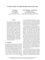
). Figure 1A shows that Alt-C induced signi-
ficant chemotaxis of neutrophils in a concentration-depend-
ent manner. The chemotatic activity of Alt-C (100 n
M
)was
similiar to fMLP (100 n
M
), a classic chemotactic agent.
We also examined the effect of alternagin-C on neutro-
phil chemotaxis induced by fMLP and by itself. Neutrophils
were pretreated with Alt-C (100 n
M
) for 5 min, and the cells
were allowed to migrate toward fMLP or Alt-C in a Boyden
chamber (directional cell movement). In the presence of
Alt-C, the chemotactic response of neutrophils to fMLP was
significantly inhibited, and we observed the same effect in
response to Alt-C (Fig. 1B). In addition, pretreatment of
neutrophils with Alt-C did not affect the random cell
movement.
Effect of ECD-peptide on human neutrophil chemotaxis
To better understand the mechanism of action of
6
Alt-C, the
activity of a synthetic peptide containing the ECD sequence
was examined in chemotaxis assays. This peptide was
synthesized based on the disintegrin-like domain and
cyclized by a disulfide bond between the two cysteines.
Cells were allowed to migrate toward different concentra-
tions of ECD-peptide (1–1000 n
M
), placed in the bottom
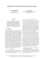
wells of the Boyden chamber. The ECD-peptide showed a
chemotactic effect only at the highest dose used (Fig. 2A;
1000 n
M
).
The inhibition of a chemotactic effect by prior exposure
to structurally related and unrelated chemotactic factors has
been already described for known neutrophil activators
[19,20]. In this regard, the effect of ECD-peptide on
neutrophil chemotaxis induced in vitro by Alt-C or fMLP
was also investigated. In the presence of ECD-peptide,
neutrophil chemotaxis in response to Alt-C (100 n
M
)was
significantly inhibited at all studied doses (Fig. 2B). When
the random cell movement was analysed in the presence of
ECD-peptide, no alterations were observed (Fig. 2C).
However, the chemotactic response of neutrophils to fMLP
was completely blocked by ECD-peptide (Fig. 2C).
Alt-C-induced rearrangement of neutrophil
actin network
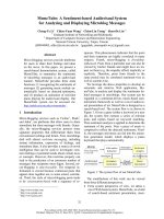
It has been reported that some neutrophil activities, such as
chemotaxis and phagocytosis, are mediated by cytoskeletal
actin polymerization [24]. Neutrophils were incubated with
Ó FEBS 2003 Effects of alternagin-C on neutrophil functions (Eur. J. Biochem. 270) 4801
Alt-C (100 n
M
) and the alterations of neutrophil actin
cytoskeleton network were analyzed by TRITC
7
–phalloidin
staining. Figure 3 shows that Alt-C was able to induce a
marked increase in the F-actin content in these cells
(Fig. 3B) when compared with nonstimulated cells
(Fig. 3A). When the fluorescence intensity was measured,
Alt-C induced 77% more actin polymerization in compar-
ison to the control (nontreated cells: 30.10 ± 4.35; treated
cells: 53.41 ± 6.02; in arbitrary units, P < 0.05) (Fig. 3C).
This suggests that the motile activities of neutrophils
induced by Alt-C are driven by actin cytoskeleton dynamic
rearrangement.
Involvement of tyrosine kinase pathways in neutrophil
activation by Alt-C
We evaluated the involvement of tyrosine kinase pathways
in the neutrophil activation induced by Alt-C. The altera-
tions in protein tyrosine phosphorylation were analyzed by
immunocytochemistry. As shown in Fig. 4B, Alt-C
(100 n
M
) was able to increase the content of phosphotyro-
sine when compared with nonstimulated neutrophils
(Fig. 4A). The immunoreactivity to phosphotyrosine was
80% greater in cells treated with Alt-C when compared to
the control (nontreated cells: 51.56 ± 8.16; treated cells:
93.00 ± 10.05; in arbitrary units, P < 0.05) (Fig. 4C).
Alt-C induced FAK and PI3-kinase activation
Focal adhesion contacts are comprised by integrins, cyto-
skeletal proteins and FAK association followed by FAK
autophosphorylation and activation [7]. By immunopreci-
pitation of FAK, we determined its activation by the
increase in the content of phosphotyrosine. As shown in
Fig. 5A, incubation of neutrophils with Alt-C (100 n
M
)was
able to increase FAK phosphorylation. The tyrosine
phosphorylation of FAK generates docking sites for several
proteins containing Src homology 2 (SH2) domains, as the
p85 regulatory subunit of PI3-kinase [25]. PI3-kinase
activation can modulate some cellular responses such as
cell migration [26]. Activation of PI3-kinase was evaluated
by its p85 subunit association with FAK in cells treated with
Alt-C. Figure 5B shows that Alt-C induced an increase in
the PI3-kinase association with FAK.
Confirming the involvement of the PI3-kinase pathway
in the chemotactic effect of Alt-C on human neutrophil
chemotaxis, a specific PI3-kinase inhibitor, LY294002,
completely blocked Alt-C-induced chemotaxis (Fig. 5C).
In addition to the determination of FAK and PI3-kinase
activation in response to Alt-C-induced chemotaxis, we
analyzed the effect of PI3-kinase inhibitor on FAK
phosphorylation and FAK-associated PI3-kinase upon
stimulation with Alt-C. As shown in Fig. 5A,B, the levels
of FAK phosphorylation and PI3-kinase association with
FAK in LY294002-treated cells were similar to those in
control cells.
Alt-C-induced activation of Erk-2
It has been reported that some disintegrins are able to
increase the activation and translocation of Erk-2 from the
cytoplasm to the nucleus [20]. Here, we investigated whether
Alt-C would be able to induce nuclear translocation of Erk-
2 in human neutrophils. Figure 6A shows that Erk-2 could
be observed in nuclear extracts from control cells. However,
incubation of neutrophils with Alt-C (100 n
M
)for1h
induced a significant increase on Erk-2 nuclear translocation
when compared to nontreated cells (Fig. 6A).
Fig. 1. Effect of Alt-C on human neutrophil chemotaxis. (A) Alt-C
induces chemotaxis in a dose-dependent manner. The cells were
allowed to migrate in a Boyden chamber toward medium alone
(RPMI, control, j), fMLP (100 n
M
, d) or Alt-C (m). Data shows
mean ± SD from three independent experiments. *P <0.05com-
pared to control. (B) Alt-C inhibits fMLP- and Alt-C-induced chemo-
taxis. Cells were incubated in presence or absence of Alt-C (100 n
M
)
for 5 min at 37 °C. Neutrophil chemotaxis was evaluated in a Boyden
chamber toward medium alone (RPMI), fMLP (100 n
M
) or Alt-C
(100 n
M
). Data shows mean ± SD from three independent experi-
ments. *P < 0.05 compared to control.
4802 A. Mariano-Oliveira et al. (Eur. J. Biochem. 270) Ó FEBS 2003
FAK activation mediates the Ras/Raf/MAP kinase
kinase (MEK) signal transduction cascade leading to
Erk-2 activation and regulating cell motility [27]. To
investigate the involvement of the Ras/Raf/MAP kinase
pathway in the effects of Alt-C on neutrophil chemotaxis,
cells were preincubated with PD98059, a MEK inhibitor,
and then allowed to migrate toward Alt-C. Figure 6B
demonstrates that chemotaxis induced by Alt-C was
significantly inhibited by PD 98059 (24% inhibition).
As shown in Fig. 6A, preincubation of neutrophils with
PD98059 abolished Erk-2 nuclear translocation. To provide
further insight into the regulation of Alt-C-induced Erk-2
activation, we determined whether PI3-kinase activity is a
prerequisite for Erk-2 activation. Figure 6A shows that
nuclear translocation of Erk-2 in response to Alt-C was
significantly increased by 38% in neutrophils by exposure
to PI3-kinase inhibitor.
Discussion
Cell adhesion to the ECM is primarily mediated by the
binding of cell surface integrins to the RGD motif found on
ECM proteins [6]. Disintegrins mostly express an RGD
sequence at an integrin-binding loop. The type and position
of the amino acids flanking the RGD motif determine the
selectivity of disintegrin interaction with integrin [15,18].
The understanding of the precise mechanism of action and
structure of disintegrins will provide information about
adhesive ligands and their integrin receptors. We previously
reported that RGD-disintegrins interfered with neutrophil
chemotaxis induced by chemoattractants [19,20]. It has been
postulated that disintegrins are approximately 1000-fold
more potent than linear RGD-containing peptides, being
determined by the conformation of the RGD amino acid
sequence within their structures [13–15]. In the present study
we have investigated the effects of a disintegrin-like protein,
Alt-C, on neutrophil activation and function. Alt-C has
been described to inhibit collagen-induced adhesion of cells
expressing a2b1 integrin in a dose-dependent manner [21].
Alt-C also has a cysteine-rich disulfide-bonding pattern and
the primary structure containing an ECD sequence presents
homology with the disintegrins [21]. The results demonstra-
ted that Alt-C strongly induced human neutrophil chemo-
taxis in vitro. Furthermore, this protein inhibited chemotaxis
of neutrophils induced by fMLP and by itself. We exam-
ined the effect of a synthetic ECD-peptide on neutrophil
Fig. 2. Effect of ECD-peptide on Alt-C-
induced human neutrophil chemotaxis. (A)
ECD-peptide induces chemotaxis. The cells
were allowed to migrate in a Boyden chamber
toward medium alone (RPMI, control, j),
fMLP (100 n
M
, d)orECD(r). Data shows
mean ± SD from three independent experi-
ments. *P < 0.05 in comparison with control.
(B) ECD-peptide inhibits Alt-C-induced che-
motaxis. Neutrophils were preincubated in
presence or absence of ECD-peptide (0.1–
1000 n
M
) for 5 min at 37 °Candthenallowed
to migrate in a Boyden chamber toward
medium alone (RPMI, control, j) or Alt-C
(100 n
M
, m). Data shows mean ± SD from
three independent experiments. *P <0.05in
comparison with Alt-C-incuced chemotaxis.
(C) ECD-peptide inhibits fMLP-induced
chemotaxis. Cells were incubated in absence or
presence of ECD-peptide (1000 n
M
)for5min
at 37 °C. Neutrophil chemotaxis was evalu-
ated in a Boyden chamber toward medium
alone (RPMI) or fMLP (100 n
M
). Data shows
means ± SD from three independent experi-
ments. *P < 0.05 compared to random
movement.
Ó FEBS 2003 Effects of alternagin-C on neutrophil functions (Eur. J. Biochem. 270) 4803
chemotaxis in vitro. The peptide induced neutrophil chemo-
taxis and also had the ability of inhibiting Alt-C-induced
chemotaxis. These data strongly suggest that the chemotac-
tic effect of Alt-C appears to be mediated by ECD sequence
conformation. It has been demonstrated that synthetic
peptides having the sequence RSECD inhibit collagen-
induced platelet aggregation [28]. Thus, our results indicate
that Alt-C may interact with adhesive receptors on the
neutrophil surface inducing cell activation and leading to
desensitization of the receptor to other chemotactic stimuli
after prior stimulation. These findings are in agreement with
previous studies describing that neutrophil migration can be
inhibited or desensitized to a given chemoattractant by prior
exposure to the same agonist (homologous desensitization)
or to unrelated chemotactic factors (heterologous desensi-
tization) [20,29].
Different chemotactic and phagocytic stimuli generate
dynamic alterations in the actin cytoskeleton network in
neutrophils [30]. Integrins induce assembly of actin filaments
and high-order structures, such as focal adhesions, in
response to extracellular stimuli and during cell adhesion
and migration [reviewed in 31]. We previously showed that
RGD-disintegrins are able to induce the integrin activation
and rearrangement of the actin cytoskeleton in human
Fig. 3. Alt-C increases actin cytoskeleton polymerization on human
neutrophils. Neutrophils were incubated with Alt-C (100 n
M
)for5min
at 37 °C and actin filaments were stained with TRITC-phalloidin for
2 h at room temperature. (A) Control and (B) Alt-C incubated neu-
trophils labelled with TRITC-phalloidin. (C) Fluorescence intensity
with mean ± SD from three independent experiments. *P <0.05
compared to control values.
Fig. 4. Involvement of tyrosine kinase activity in the effect of Alt-C on
human neutrophils. Neutrophils were incubated with Alt-C (100 n
M
)
for 5 min at 37 °C, permeabilized with 0.1% (v/v) Triton X-100,
incubated with anti-phosphotyrosine Ig conjugated to biotin followed
by streptavidin-conjugated fluorescein isothiocyanate. (A) Control and
(B) Alt-C incubated FITC-labelled neutrophils. (C) The measure of
fluorescence intensity with mean ± SD from three independent
experiments. *P < 0.05 compared to control values.
4804 A. Mariano-Oliveira et al. (Eur. J. Biochem. 270) Ó FEBS 2003
neutrophils [19,20]. Because Alt-C causes remarkable neu-
trophil chemotaxis, we examined whether Alt-C also
produces changes in cytoskeletal F-actin. Treatment of
neutrophils with Alt-C induced profound alterations in the
actin network with an increase of F-actin content, suggest-
ing that the Alt-C effect on neutrophils could involve
integrin-mediated pathways.
Neutrophil functional responses that require cytoskeletal
reorganization, such as adhesion to the endothelium and
ECM, cell migration and phagocytosis, result in the
activation of protein tyrosine kinases [31–33]. Interaction
between integrins and ligands leads to a profound increase
in tyrosine phosphorylation of several cellular proteins. It is
well established that simple dimerization of integrins is
sufficient to initiate tyrosine phosphorylation events [11].
This has been accomplished with crosslinked anti-integrin
Igs, multimeric integrin ligands [34,35] as well as disinte-
grins, potent inducers of conformational changes in both
subunits of integrins [36]. Results reported here show that
Alt-C induces an increase in tyrosine kinase activity and
tyrosine phosphorylation.
One of the initial events in integrin-mediated signaling is
the activation of FAK, resulting in its autophosphoryla-
tion [7]. This is supported by the findings that distinct
disintegrins binding to integrin stimulate FAK activity [20]
and that activated FAK might mediate signal transduction
in a manner similar to that of integrins. According to this,
the present study demonstrated that Alt-C was able to
induce an increase in phosphotyrosine content of FAK
and that FAK phosphorylation may be directly involved
in the activation of the migratory process in response to
Alt-C. A recent report showed that FAK phosphorylation
is directly required for neutrophil chemotaxis by using a
dominant negative mutant of FAK [37]. Interestingly, it
also has been described that FAK-deficient cells exhibit
an elevated number of focal adhesions accompanied by a
decrease rate of cell migration [38]. Furthermore, FAK, as
a nonreceptor tyrosine kinase that associates with the
cytoplasmic domain of integrins at focal adhesions, might
be critical for cytoskeleton reorganization [7,9]. Earlier
studies have demonstrated that two cytoskeletal proteins,
paxillin and tensin, are substrates for FAK, which could
account for a role of FAK in actin cytoskeleton assembly
and disassembly [7,9]. In the present study we provide
evidence of a link between FAK activation and
rearrangement of the actin cytoskeleton in neutrophils
Fig. 5. Involvement of FAK and PI3-kinase in
the effect of Alt-C on human neutrophils. (A)
Alt-C induces FAK activation. FAK phos-
phorylation was determined in neutrophils
incubated with Alt-C (100 n
M
)for5minat
37 °C in the presence or absence of LY294002
(3 l
M
). Cell lysates were immunoprecipitated
with anti-FAK Ig and blotted with either anti-
phosphotyrosine or anti-FAK Igs. IP, immu-
noprecipitation; WB, Western blotting. Blots
were analyzed by densitometry and the con-
tent of phosphorylated FAK was expressed in
densitometric units. Data show mean ± SD
from three independent experiments. *P <
0.05 compared to cells incubated with medium
alone (Control) (B) Alt-C increases FAK-
associated PI3-kinase. Cell lysates of neu-
trophils incubated with Alt-C (100 n
M
)for
5 min at 37 °C in the presence or absence of
LY294002 (3 l
M
) were immunoprecipitated
withanti-FAKIgandthenblottedwithanti-
FAK or anti-PI3-kinase p85 subunit Igs. IP,
immunoprecipitation; WB, Western blotting.
Blots were analyzed by densitometry and the
content of PI3-kinase associated to FAK was
expressed in densitometric units. Data show
mean ± SD from three independent experi-
ments. *P < 0.05 compared to cells incubated
with medium alone (Control). (C) Alt-C-
induced chemotaxis is reduced by a PI3-kinase
inhibitor. Neutrophils were preincubated for
5 min at 37 °C with LY294002 (3 l
M
)andthen
allowed to migrate in a Boyden chamber to-
ward Alt-C (100 n
M
). Data show mean ± SD
from three independent experiments.
*P < 0.05 compared to control.
Ó FEBS 2003 Effects of alternagin-C on neutrophil functions (Eur. J. Biochem. 270) 4805
immediatelly following stimulation with Alt-C, an ECD-
disintegrin.
FAK is also considered a focal adhesion docking protein
that recruits PI3-kinase and other signaling molecules to
form a multimolecular complex, altering their activities
[10,12,25]. In Alt-C-stimulated neutrophils, PI3-kinase was
found to be associated with FAK. Therefore, it is reasonable
to postulate that this association promotes PI3-kinase
activation, which correlates with a variety of cellular
responses to external stimuli including chemotaxis, which
was completely blocked by a PI3-kinase inhibitor. These
results are in agreement with previous studies showing that
neutrophils lacking PI3-kinase failed to orient toward
different chemotactic stimuli [39,40]. Thus Alt-C-induced
neutrophil chemotaxis could be driven by PI3-kinase
activation, which associates with autophosphorylated
FAK through their SH2 domains.
FAK activation may also trigger the Ras signal trans-
duction cascade [11]. Downstream signal molecules such as
Erk-2 have also been implicated in the regulation of the
neutrophil effector functions [41]. Our study revealed that
Alt-C can induce Erk-2 activation, as observed by its
translocation to the nucleus. Activation of Erk-2 is often
associated with enhanced myosin light chain kinase
9
activity
and increased migration [27]. The effect of Alt-C on
neutrophil chemotaxis was partially reversed by PD98059,
a MEK inhibitor, supporting a role for Erk-2 in Alt-C-
induced neutrophil migration. These findings suggest that
activation of Erk-2 induced by Alt-C may function as a
positive regulator of migration. Recently, some paradoxical
findings have reported the effects of different RGD-disin-
tegrins on cell migration supporting the role for Erk-2 as a
positive or negative effector [20,42]. In addition to Alt-C-
induced Erk-2 activation that accounts for its positive effect
on neutrophil chemotaxis, other cellular responses may be
related to this pathway. Along this line, our results also
demonstrated that PI3-kinase inhibition is accompanied by
an increase of Erk-2 nuclear translocation suggesting a
modulatory role of PI3-kinase signaling pathway on Erk-2
activity. Studies on the expression of cytokines and chemo-
kines and on neutrophil apoptosis are under investigation.
The present study provides evidence that Alt-C, a
disintegrin-like protein presenting an ECD motif, interacts
with neutrophils promoting integrin-mediated signaling and
inducing chemotaxis. Our study elucidates the mechanism
of action of Alt-C, as well as establishes a potential model
for the design of new therapeutic interventions in disorders
involving leukocyte dysfunctions.
Acknowledgements
The authors thank Dr Iolanda M. Fierro (UERJ, Brazil) for the
discussions and critical review of the manuscript. This work was
supported by CAPES, CNPq, FAPERJ, FAPESP, SR-2/UERJ
(Brazil) and IFS (Sweden).
References
1. Brown, E.J. (1986) The interactions of connective tissue proteins
with phagocytic cells. J. Leukoc. Biol. 39, 579–591.
2. Lawrence, M.B. & Springer, T.A. (1991) Leukocytes roll on a
selectin at physiologic flow rates: Distinction from and
prerequisite for adhesion through integrins. Cell 65, 859–873.
3. Berton, G., Yan, S.R., Fugamalli, L. & Lowell, C.A. (1996)
Neutrophil activation by adhesion: Mechanisms and pathophy-
siological implications. Int. J. Clin. Laboratory Res. 26, 160–177.
4. Ruoslahti, E. (1991) Integrins. J. Clin. Invest. 87, 1–5.
5. Hynes, R.O. (1992) Integrins: versatility, modulation, and signa-
ling in cell adhesion. Cell 69, 11–25.
Fig. 6. Involvement of Erk-2 activity in the effect of Alt-C on human
neutrophils. (A) Alt-C induces Erk-2 nuclear translocation. Neu-
trophils were incubated with medium alone (Control) or with Alt-C
(100 n
M
) for 1 h at 37 °C in the presence or absence of LY294002
(3 l
M
) or PD98059 (2 l
M
). The nuclear content of Erk-2 was deter-
mined by immunoblotting using an anti-Erk-2 Ig and quantified by
densitometry. WB, Western blotting. Data show mean ± SD from
three independent experiments. *P < 0.05 compared to cells
incubated with medium alone (Control). (B) Alt-C-induced chemo-
taxis is reduced by a MEK inhibitor. Neutrophils were preincubated
for 5 min at 37 °C with PD98059 (2 l
M
) and then allowed to migrate
in a Boyden chamber toward Alt-C (100 n
M
). Data show mean ± SD
from three independent experiments. *P < 0.05 compared to control.
The # indicates that LY294002 significantly increased Alt-C-induced
Erk-2 nuclear translocation (P <0.05).
4806 A. Mariano-Oliveira et al. (Eur. J. Biochem. 270) Ó FEBS 2003
6. Yamada, K.M. (1991) Adhesive recognition sequences. J. Biol.
Chem. 266, 12809–12812.
7. Burridge, K., Turner, C.E. & Romer, L.H. (1992) Tyrosine
phosphorylation of paxillin and pp125
FAK
accompanies cell
adhesion to extracellular matrix: a role in cytoskeletal assembly.
J. Cell Biol. 119, 893–903.
8. Kornberg, L., Earp, H.S., Parsons, J.T., Schaller, M. & Juliano,
R.L. (1992) Cell adhesion or integrin clustering increases phos-
phorylation of a focal adhesion-associated tyrosine kinase. J. Biol.
Chem. 267, 23439–23442.
9. Parsons, J.T. (1996) Integrin-mediated signalling: regulation by
protein tyrosine kinases and small GTP-binding proteins. Curr.
Opin. Cell Biol. 8, 146–152.
10. Lo
¨
fgren, R., Ng-Sikorski, J., Sjo
¨
lander,A.&Andersson,T.(1993)
b
2
-integrin engagement triggers actin polymerization and phos-
phatidylinositol triphosphate formation in non-adherent human
neutrophils. J. Cell Biol. 123, 1597–1605.
11. Miyamoto, S., Teramoto, H., Coso, O., Silvio, G., Burbelo, P.,
Akiyama, S.K. & Yamada, K.M. (1995) Integrin function:
molecular hierarchies of cytoskeletal and signaling molecules.
J. Cell Biol. 131, 791–805.
12. Zheng, L., Sjo
¨
lander, A., Eckerdal, J. & Andersson, T. (1996)
Antibody-induced engagement of b
2
-integrins on adherent human
neutrophils triggers activation of p21ras through tyrosine phos-
phorylation of the protooncogene product Vav. Proc. Natl. Acad.
Sci. USA 93, 8431–8436.
13. Dennis, M.S., Henzel, W.J., Pitti, R.M., Lipari, M.T., Napier,
M.A., Deisher, T.A., Bunting, S. & Lazarus, R.A. (1990) Platelet
glycoprotein IIb-IIIa protein antagonists from snake venoms: a
family of integrin inhibitory proteins from viper venom. Proc.
Natl. Acad. Sci. USA 87, 2471–2475.
14. Gould,R.J.,Polokoff,M.A.,Fiedman,P.A.,Huang,T F.,Holt,
J.C., Cook, J.J. & Niewiarowski, S. (1990) Disintegrins: a family of
integrin inhibitory proteins from viper venom. Proc. Soc. Exp.
Biol. Medical 195, 168–171.
15. Niewiarowski, S., McLane, M.A., Kloczwiak, M. & Stewart, G.J.
(1994) Disintegrins and other naturally occurring antagonists of
platelet fibrinogen receptors. Sem. Hematol. 31, 289–300.
16. Juliano, D., Wang, Y., Marcinkiewicz, C., Rosenthal, A.L.,
Stewart, G.J. & Niewiarowski, S. (1996) Disintegrin interaction
with a
v
b
3
integrin on human umbilical vein endothelial cells:
expression of ligand-induced binding site on b
3
subunit. Exp. Cell
Res. 225, 132–142.
17. Danen, E.H., Marcinkiewicz, C., Cornelissen, I.M., van Kraats,
A.A., Pachter, J.A., Ruiter, J., Niewiarowski, S. & van Muijen,
G.N. (1998) The disintegrin eristostatin interferes with integrin
alpha 4 beta 1 function and with experimental metastasis of
human melanoma cells. Exp. Cell Res. 238, 188–196.
18. McLane, M.A., Marcinkiewicz, C., Vijay-Kumar, S., Wierzbicka-
Patynowski, I. & Niewiarowski, S. (1998) Viper venom disin-
tegrins and related molecules. Proc. Soc. Exp. Biol. Medical 219,
109–119.
19. Coelho, A.L.J., De Freitas, M.S., Oliveira-Carvalho, A.L.,
Moura-Neto, V., Zingali, R.B. & Barja-Fidalgo, C. (1999) Effects
of jarastatin, a novel snake venom disintegrin, on neutrophil
migration and actin cytoskeleton dynamics. Exp. Cell Res. 251,
379–387.
20. Coelho, A.L.J., De Freitas, M.S., Mariano-Oliveira, A., Oliveira-
Carvalho, A.L., Zingali, R.B. & Barja-Fidalgo, C. (2001)
Interaction of disintegrins with human neutrophils induces
cytoskeleton reorganization, focal adhesion kinase activation and
extracellular-regulated kinase-2 nuclear translocation, interfering
with the chemotactic function. FASEB J. 15, 1643–1645.
21. Souza, D.H.F., Iemma, M.R.C., Ferreira, L.L., Faria, J.P., Oliva,
M.L.V., Zingali, R.B., Niewiarowski, S., Selistre-d. & e-Araujo,
H.S. (2000) The disintegrin-like domain of the snake venom
metalloprotease alternagin inhibits a2b1 integrin-mediated cell
adhesion. Arch. Biochem. Biophys. 384, 341–350.
22. Dooley, D.C., Simpson, J.F. & Merryman, H.T. (1982) Isolation
of large numbers of fully viable human neutrophils: a preparative
technique using Percoll density gradient centrifugation. Exp.
Hematol. 10, 591–599.
23. Bradford, M. (1976) A rapid and sensitive method for quantifi-
cation of microgram quantities of protein utilizing the principle of
protein-dye binding. Anal. Biochem. 72, 248–254.
24. Sheikh, S., Gratzer, W.B., Pinder, J.C. & Nash, G.B. (1997) Actin
polymerization regulates integrin-mediated adhesion as well as
rigidity of neutrophils. Bioch. Biophys. Res. Commun. 238,
910–915.
25. Chen, H.C. & Guan, J.L. (1994) Association of focal adhesion
kinase with its potential substrate phosphatidylinositol 3-kinase.
Proc. Natl. Acad. Sci. USA 91, 10148–10152.
26. Toker, A. & Cantley, L.C. (1997) Signalling through the lipid
products of phosphoinositide-3-OH kinase. Nature 387, 673–676.
27. Klemke, R.L., Cai, S., Giannini, A.L., Gallagher, P.J., de Lane-
rolle, P. & Cheresh, D.A. (1997) Regulation of cell motility by
mitogen-activated protein kinase. J. Cell Biol. 137, 481–492.
28. Jia, L G., Wang, X M., Shannon, J.D., Bjarnasson, J.B. &
Fox, J.W. (1997) Function of disintegrin-like/cysteine-rich
domains of atrolysin A. Inhibition of platelet aggregation by
recombinant protein and peptide antagonists. J. Biol. Chem. 272,
13094–13102.
29. Stanton, K.J., Frewin, M.B. & Gudewicz, P.W. (1999) Hetero-
logous desensitization of IL-8-mediated chemotaxis in human
neutrophils by a cell-binding fragment of fibronectin. J. Leukoc.
Biol. 65, 515–522.
30. Williams, M.A. & Solonkin, J.S. (1999) Integrin-mediated sig-
naling in human neutrophil functioning. J. Leukoc. Biol. 65,
725–735.
31. Schwartz, M.A. (2001) Integrin signaling revisited. Trends Cell
Biol. 11, 466–470.
32. Gaudry, M., Caon, A.C., Gilbert, C., Lille, S. & Naccache, P.H.
(1992) Evidence for the involvement of tyrosine kinases in the
locomotory responses of human neutrophils. J. Leukoc. Biol. 51,
103–108.
33. McGregor, P.E., Agrawal, D.K. & Edwards, J.D. (1994)
Attenuation of human leukocyte adherence to endothelial cell
monolayers by tyrosine kinase inhibitors. Biochem. Biophys. Res.
Commun. 198, 359–365.
34. Huang, M M., Lipfert, L., Cunningham, M., Brugge, J.S.,
Ginsberg, M.H. & Shattil, S.J. (1993) Adhesive ligand binding to
integrin aIIbw3 stimulates tyrosine phosphorylation of novel
protein substrates before phosphorylation of pp125
FAK
. J. Cell
Biol. 122, 473–483.
35. Bhattacharya, S., Fu, C., Bhattacharya, J. & Greenberg, S. (1995)
Soluble ligands of the avb3 integrin mediate enhanced tyrosine
phosphorylation of multiple proteins in adherent bovine
pulmonary artery endothelial cells. J. Cell Biol. Chem. 270,
16781–16787.
36. Bryan Smith, J., Theakston, R.D.G., Coelho, A.L.J., Barja-Fid-
algo, C., Calvete, J.J. & Marcinkiewicz, C. (2002) Characterization
of a monomeric disintegrin, ocellatusin, present in the venom of
the Nigerian carpet viper, Echis ocellatus. FEBS Lett. 512,
111–115.
37.Ilic,D.,Furuta,Y.,Kanazawa,S.,Takeda,N.,Sobue,K.,
Nakatsuji, N., Nomura, S., Fujimoto, J., Okada, M. & Yama-
moto, T. (1995) Reduced cell motility and enhanced focal adhe-
sion contact formation in cells from FAK-deficient mice. Nature
377, 539–544.
38. Feniger-Barish, R., Yron, H., Meshel, T., Matityahu, E. & Ben-
Baruch, A. (2003) Il-8-induced migratory responses thrugh
CXCR1 and CXCR2. Association with phosphorylation and
Ó FEBS 2003 Effects of alternagin-C on neutrophil functions (Eur. J. Biochem. 270) 4807
cellular redistribution of focal adhesion kinase. Biochem. 42,
2874–2886.
39. Li, Z., Jiang, H., Xie, W., Zhang, Z., Smrcka, A.V. & Wu, D.
(2000) Roles of PLC-beta2 and – beta3 and PI3Kgamma in
chemoattractant-mediated signal transduction. Science 287,
1046–1049.
40. Hirsch, E., Katanaev, V.L., Garlanda, C., Azzolino, O., Pirola, L.,
Silengo, L., Sozzani, S., Mantovani, A., Altruda, F. & Wymann,
M.P. (2000) Central role for G protein-coupled phosphoinositide
3-kinase gamma in inflammation. Science 287, 1049–1053.
41. Coffer, P.J., Geijsen, M., M’rabet, L., Schweizer, R.C., Maikoe,
T., Raaijmakers, J.A., Lammers, J.W. & Koenderman, L. (1998)
Comparison of the roles of mitogen-activated protein kinase
kinase and phosphatidylinositol 3-kinase signal transduction in
neutrophil effector function. Biochem. J. 329, 121–130.
42. Ritter, M.R. & Markland, F.S. Jr (2000) Contortrostatin activates
ERK2 and tyrosine phosphorylation events via distinct pathways.
Biochem. Biophys. Res. Commun. 274, 142–148.
4808 A. Mariano-Oliveira et al. (Eur. J. Biochem. 270) Ó FEBS 2003