Báo cáo khoa học: A client-binding site of Cdc37 ppt
Bạn đang xem bản rút gọn của tài liệu. Xem và tải ngay bản đầy đủ của tài liệu tại đây (251.29 KB, 7 trang )
A client-binding site of Cdc37
Kazuya Terasawa and Yasufumi Minami
Department of Biophysics and Biochemistry, and Undergraduate Program for Bioinformatics and Systems Biology, Graduate School of
Science, University of Tokyo, Japan
Molecular chaperones are required for the correct fold-
ing of many proteins inside cells, despite their confor-
mations being predetermined by their own amino acid
sequences, because de novo protein synthesis proceeds
in a directional manner from the N-terminus and
encounters a cellular milieu that is crowded with
macromolecules [1]. Although Hsp90 is abundant and
highly conserved among species, its structure and func-
tional mechanism have been unveiled only quite
recently [2–7]. Whereas Hsp70 and chaperonin act as
general chaperones in the early stage folding of newly
synthesized proteins [1,8,9], Hsp90 takes part in the
folding of client proteins at a later stage of maturation
[2–7]. In addition, Hsp90 client proteins seem to be
restricted to cell-signaling molecules, such as steroid
hormone receptors and protein kinases [2–7].
It is now appreciated that Hsp90 performs the
chaperone function in a manner dependent on its own
ATPase activity, serving as an ATPase-driven molecular
clamp that binds and releases client proteins in a closed
and open state, respectively, this conformational trans-
ition being controlled by ATP binding and hydrolysis
[2–7]. Moreover, this ATPase-dependent chaperone
cycle is cooperatively tuned by various co-chaperones
[2–7]. Cdc37 ⁄ p50 is one Hsp90 co-chaperone and is
characterized as a protein kinase-specific cofactor for
Hsp90 [10–12], because Cdc37 interacts both physically
and genetically with a variety of protein kinases, inclu-
ding pp60
v-src
[13], Raf-1 [14] and Cdk4 [15,16]. Cdc37
binds directly to Hsp90 [17–19]; a recent crystallogra-
phic study found that the C-terminal domain of Cdc37
interacts with the N-terminal ATP-binding domain of
Hsp90 [20]. In the crystal structure, Cdc37 binds to the
open face of the Hsp90 N-terminal domain, interfering
with conformational changes of Hsp90 crucial for its
ATPase activity; this accords well with the finding that
Cdc37 inhibits Hsp90 ATPase activity [21]. Concomit-
ant with the binding to Hsp90, Cdc37 can associate
Keywords
Cdc37, Hsp90, protein kinase, Raf-1
Correspondence
Y. Minami, Department of Biophysics and
Biochemistry, and Undergraduate Program
for Bioinformatics and Systems Biology,
Graduate School of Science, The University
of Tokyo, Hongo 7-3-1, Bunkyo-ku,
Tokyo 113-0033, Japan
Fax: +81 3 5841 3047
Tel: +81 3 5841 3047
E-mail:
(Received 28 June 2005, revised 23 July
2005, accepted 26 July 2005)
doi:10.1111/j.1742-4658.2005.04884.x
The molecular chaperone Hsp90 is distinct from Hsp70 and chaperonin in
that client proteins are apparently restricted to a subset of proteins categor-
ized as cellular signaling molecules. Among these, many specific protein
kinases require the assistance of Hsp90 and its co-chaperone Cdc37 ⁄ p50
for their biogenesis. A series of Cdc37 deletion mutants revealed that all
mutants capable of binding Raf-1 possess amino acid residues between 181
and 200. The 20-residue region is sufficient and, in particular, a five-residue
segment (residue 191–195) is essential for binding to Raf-1. These five resi-
dues are present in one a helix (residues 184–199) in the middle of Cdc37,
which is unexpectedly nested within the Hsp90-interacting domain of
Cdc37, which was recently determined by crystallography, but does not
seem to contribute to direct contact with Hsp90. Furthermore, an N-ter-
minally truncated mutant of Cdc37 composed of residues 181–378 was
shown to bind the N-terminal portion of Raf-1 (subdomains I–IV). This
mutant can bind not only other Hsp90 client protein kinases, Akt1,
Aurora B and Cdk4, but also Cdc2 and Cdk2, which to date have not
been shown to physically interact with Cdc37. These results suggest that a
region of Cdc37 other than the client-binding site may be responsible for
discriminating client protein kinases from others.
Abbreviation
GST, glutathione S-transferase; IP, immunoprecipitation; Knd, kinase domain; WB, western blot.
4684 FEBS Journal 272 (2005) 4684–4690 ª 2005 FEBS
with protein kinases [14–19,22–24], in particular, with
their N-terminal lobes [25–28]. Thus, one role of Cdc37
is thought to be client recruitment to Hsp90; however,
this view is simplistic [11,12]. Cdc37 has the potential
to exhibit chaperone activity independent of Hsp90
[22,23,29–31] and its repertoire of client proteins stret-
ches beyond the protein kinases [26,32,33].
Even though our knowledge of Hsp90 has increased
dramatically and is currently being updated further
[2–7], the whole spectrum of Hsp90 client proteins and
the comprehensive mechanism of the Hsp90 chaperone
cycle remain obscure. To challenge these questions, we
analyzed a set of Cdc37 deletion mutants and eventu-
ally identified a 20-residue region of Cdc37 (residues
181–200) as a client-binding site, in which five residues
(residues 191–195) are important for client binding and
are located on an a helix in the middle of Cdc37. The
helix is embedded in the Hsp90-binding domain of
Cdc37 in the primary structure; however, it is not
involved in interactions with Hsp90 [20]. We found
that an N-terminally truncated mutant of Cdc37 con-
taining residues 181–378, but not the full-length
Cdc37, is able to associate with Cdc2 and Cdk2 (which
have not been reported to physically interact with
Cdc37) in addition to the well-known Hsp90 client
protein kinases, Raf-1, Akt1, Aurora B and Cdk4.
These findings may suggest how Cdc37 ⁄ Hsp90 distin-
guishes a limited set of protein kinases from others.
Results and Discussion
Cdc37 deletion mutants
We analyzed a series of Cdc37 deletion mutants
expressed in COS7 cells (Fig. 1A) to identify the client-
binding site. Both the C- and N-terminally truncated
FLAG-tagged Cdc37 (hereafter called FLAG–Cdc37)
mutants, FLAG–Cdc37(1–200), and FLAG–Cdc37(181–
378), respectively, bind the protein kinase domain
of Raf-1, as shown in Fig. 1B [immunoprecipita-
tion (IP): a-FLAG, middle panel]. Consequently, an
overlapping region (residues between 181 and 200) was
suggested to be the client-binding site of the Raf-1
kinase domain; this was reinforced by the fact that
FLAG–Cdc37(1–180) and FLAG–Cdc37(201–378), nei-
ther of which contain the above-mentioned region, were
unable to bind the kinase domain (Fig. 1B). Further-
more, these observations were corroborated by an
inverse immunoprecipitation experiment using the Raf-1
kinase domain (Fig. 1B, IP: a-Myc, right). However,
C-terminally truncated Cdc37 (residues 1–163) has
previously been reported to bind Raf-1 [18]. We per-
formed a similar experiment using the N- and
C-terminal portions of Cdc37, namely FLAG–Cdc37(1–
163) and FLAG–Cdc37(164–378), respectively, and
found that FLAG–Cdc37(164–378) could bind Raf-1
to a similar extent as the full-length Cdc37, whereas
B
FL
1-276
1-200
1-180
181-378
201-378
IP: α-Myc
whole
FLAG-Cdc37
FL
1-276
1-200
1-180
181-378
201-378
α-FLAG
WB: α-Myc
*
*
IP: α-FLAG
FL
1-276
1-200
1-180
181-378
201-378
A
1 378
FL
1-276
1-200
1-180
181-378
Knd
+
+
+
-
+
-201-378
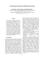
Fig. 1. The residues between 181 and 200 of Cdc37 are required for binding the Raf-1 kinase domain. (A) Primary structures of the full-length
Cdc37 (FL) and its deletion mutants with numbers corresponding to the first and last residue, and their binding activities toward the Raf-1 kin-
ase domain (Knd) are schematically illustrated. The residues between 181 and 200 are shaded. (B) The Myc-tagged kinase domain of Raf-1
and the full-length Cdc37 or each Cdc37 deletion mutant were coexpressed in COS7 cells (whole) and the obtained cell extracts were subjec-
ted to immunoprecipitation with anti-FLAG (IP: a-FLAG) or anti-Myc (IP: a-Myc) monoclonal antibody, followed by immunoblotting with both
anti-Myc and anti-FLAG polyclonal antibodies (WB: a-Myc and a-FLAG). Asterisks indicate nonspecific bands appearing in every lane.
K. Terasawa and Y. Minami A client-binding site of Cdc37
FEBS Journal 272 (2005) 4684–4690 ª 2005 FEBS 4685
FLAG–Cdc37(1–163) was hardly coimmunoprecipita-
ted with Raf-1 (data not shown); these results are consis-
tent with those shown in Fig. 1, however, they do not
agree with previously reported results [18]; we are not
able to interpret this difference at present.
Next, we selected one deletion mutant FLAG–
Cdc37(181–378) to further delineate the tentative client-
binding region of Cdc37. Three protein kinases
(Fig. 2A), Akt1 [34], Aurora B [35] and Cdk4 [15,16],
which have previously been reported to bind to Cdc37,
were all bound to FLAG–Cdc37(181–378) (Fig. 2B).
We repeated the experiment with the kinase domains of
Akt1 and Aurora B instead of the whole molecules,
omitting Cdk4 because it is composed almost solely of
a kinase domain (Fig. 2A). It was clearly shown that
the kinase domains of both Akt1 and Aurora B
bound to FLAG–Cdc37(181–378) (Fig. 2C). Moreover,
endogenous Raf-1 (not ectopically expressed Raf-1) in
COS7 cells interacted with this deletion mutant as
strongly as the full-length Cdc37 (see below).
When the kinase domain of Raf-1 was divided
between subdomains IV and V [36] into the N- and
C-terminal portions and each was fused to Myc–gluta-
thione S-transferase (GST), as shown in Fig. 3A, the
N-terminal portion of Raf-1 (subdomains I–IV), but
not the C-terminal portion (subdomains V–XI), was
bound to FLAG–Cdc37(181–378) (Fig. 3B). Our
results are consistent with previous studies; Cdc37
interacts with protein kinases via their N-terminal
lobes [25–28].
It was shown that the deletion mutant of Cdc37,
FLAG–Cdc37(181–378), is able to bind the client
protein kinases; therefore, it contains a client-binding
site.
B
C
B
a
ror
u
A
4kdC
1tkA
-
IP: α-Myc
whole
Aurora B
Cdk4
Akt1
181-378
*
WB: α-FLAG
α-Myc
Myc-Kinase
IP: α-Mycwhole
WB: α-FLAG
α-Myc
*
B aro
r
uA
1
tkA
-
B a
r
or
uA
1t
k
A
-Myc-Knd
Akt1
Cdk4
Aurora B
3041
5 295
345
1
76 327
4801
149 409
Knd
A
B
a
r
oru
A
4k
d
C
1
tkA
-
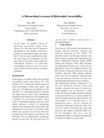
Fig. 2. FLAG–Cdc37(181– 378) binds three known Cdc37 client pro-
tein kinases, Akt1, Aurora B and Cdk4. (A) Primary structures of
Akt1, Aurora B and Cdk4 are schematically drawn with residue
numbers, where in particular, their kinase domains (Knd, light lines)
are discriminated from other regions (dark lines). (B) FLAG–
Cdc37(181–378) was expressed alone (–) or coexpressed with
Myc-tagged kinases (Myc-Kinase) as indicated in COS7 cells and
the cell lysates (whole) were immunoprecipitated with anti-Myc
monoclonal antibody (IP: a-Myc), followed by immunoblotting with
the indicated polyclonal antibodies. Asterisks indicate nonspecific
bands appearing in every lane. (C) Myc-tagged kinase domains
(Myc-Knd) were used instead of their whole molecules, and the
obtained immunoprepitates were analyzed by immunostaining as
described in (B).
α-Myc
Knd
I-IV
Myc-GST
α-FLAG
whole
empty
GST
pull-down
GST
pull-down
V-XI
WB:
B
I-IV V-XI
Myc-GST fusion
fragment
Myc
GST
A
Raf-1 Knd
614349 414/415
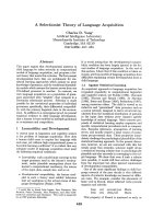
Fig. 3. Cdc37 binds the N-teminal portion of Raf-1. (A) (Upper) Pri-
mary structure of the kinase domain of Raf-1 (Knd), and its N- and
C-terminal portions (I–IV and V–XI, respectively) are schematically
depicted with residue numbers. (Lower) Schematic drawing of the
Myc-GST fusion construct is shown; either Knd, the N- or C-ter-
minal portion of Raf-1 was inserted at a position indicated by ‘frag-
ment’. (B) FLAG–Cdc37(181– 378) and, Myc–GST alone (empty) or
fused with Knd, I–IV or V–XI were coexpressed in COS7 cells. The
cell lysates (whole) were pulled down with glutathione beads (GST
pull-down), after which immunoblotting with anti-FLAG or anti-Myc
polyclonal antibody was performed (WB: a-FLAG and a-Myc).
A client-binding site of Cdc37 K. Terasawa and Y. Minami
4686 FEBS Journal 272 (2005) 4684–4690 ª 2005 FEBS
The 20-residue region of Cdc37 is a client-binding
site
Because the above results infer that the 20-residue
region of Cdc37 is essential for the binding of a client
protein kinase, we tested whether the peptide (residues
181–200 of Cdc37) conjugated to FLAG–GST
(Fig. 4A) was able to bind the kinase domain of Raf-1.
As shown in Fig. 4B, immunoprecipitaton with both
anti-FLAG (IP: a-FLAG; for FLAG–GST–peptide)
and anti-Myc (IP: a-Myc; for a Myc-tagged kinase
domain of Raf-1) monoclonal antibodies proved that
this peptide is capable of binding the Raf-1 kinase
domain, which was further confirmed for the kinase
domains of Akt1 and Aurora B, and full-length Cdk4
(Fig. 4C). Thus, it could be concluded that the 20-resi-
due region of Cdc37 is sufficient for the binding of
client protein kinases.
To specify the required residues in the peptide,
alanine-scanning and deletion mutagenesis of the
two-residue region were performed (Fig. 5A). Alanine-
scanning mutagenesis abolished the ability of mutant
3A to bind the Raf-1 kinase domain, and the ability of
mutant 4A to bind the Raf-1 kinase domain was
remarkably decreased (Fig. 5B). Deletion mutant N10
lost its binding activity, but two mutants, M10 and
C10, retained it (Fig. 5C). Taken together, these results
support the conclusion that a five-residue segment,
VIWCI (residues 191–195), is maximally required for
interaction with the kinase domain of Raf-1.
This segment resides in an a helix composed of
residues between 184 and 199, which was recently
A
181
200
ELVC ETANYLV IWC I DLEVE
FLAG GST
peptide
B
α-FLAG
WB: α-Myc
whole
IP:
α-Myc
IP:
α-FLAG
*
Aurora B-Knd
Cdk4 (FL)
WB: α-FLAG
α-Myc
whole
IP: α-Myc
Akt1-Knd
C
-
*
Aurora B-Knd
Cdk4 (FL)
Akt1-Knd
-
Myc-Knd
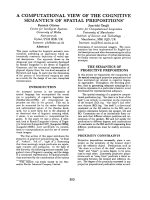
Fig. 4. Cdc37 peptide (residues between 181 and 200) fused with
FLAG–GST binds kinase domains. (A) A primary structure of the
FLAG–GST–peptide fusion is schematically illustrated, with the pep-
tide sequence from 181 to 200 of Cdc37. (B) The Myc-tagged kin-
ase domain of Raf-1 and FLAG–GST fused with nothing (i.e. empty)
(e) or the peptide (p) were coexpressed in COS7 cells. Cell extracts
were prepared (whole) and subjected to immunoprecipitation with
anti-FLAG and anti-Myc monoclonal antibodies (IP: a-FLAG and
a-Myc), followed by immunoblotting with anti-FLAG and anti-Myc
polyclonal antibodies (WB: a-FLAG and a-Myc). Asterisks indicate
nonspecific bands appearing in every lane. (C) FLAG–GST–peptide
fusion protein was expressed alone (–) or coexpressed with
Myc-tagged kinase domains of Akt1 or Aurora B, or Myc-tagged
full-length Cdk4 (FL) in COS7 cells and the obtained immunoprecipi-
tates were analyzed by immunostaining as described in (B).
α-FLAG
WB: α-Myc
whole
FLAG-GST
IP: α-FLAG
B
1A 2A 3A 4A 5Awt 1A 2A 3A 4A 5Awt
whole
IP: α-FLAG
N10 M10 C10wt
C
α-FLAG
WB: α-Myc
FLAG-GST wt N10 M10 C10
A
181 200
wt
1A
2A
3A
4A
5A
N10
M10
C10
LVCEETANYLVIWCIDLEVE
AAAAETANYLVIWCIDLEVE
LVCEAAAAYLVIWCIDLEVE
LVCEETANAAAAWCIDLEVE
LVCEETANYLVIAAAALEVE
LVCEETANYLVIWCIDAAAA
LVCEETANYL
TANYLVIWCI
VIWCIDLEVE
Fig. 5. Five residues of Cdc37 are essential for the binding of the
Raf-1 kinase domain. (A) Peptide sequences fused to FLAG–GST
are shown; wt: a wild-type peptide; 1A)5A: five different alanine-
scanning mutant peptides (four consecutive residues changed to
alanine are underlined); N10, M10 and C10: 10-residue truncation
mutant peptides. The five most important residues, VIWCI, are sha-
ded. (B, C) The Myc-tagged kinase domain of Raf-1 and each
FLAG–GST–peptide indicated were coexpressed in COS7 cells. The
obtained cell lysates (whole) were immunoprecipitated with anti-
FLAG monoclonal antibody (IP: a-FLAG) and subsequently immuno-
stained with anti-Myc (for Myc-Knd) and anti-FLAG (for FLAG–GST–
peptide) polyclonal antibodies (WB: a-Myc and a-FLAG).
K. Terasawa and Y. Minami A client-binding site of Cdc37
FEBS Journal 272 (2005) 4684–4690 ª 2005 FEBS 4687
determined using crystallography [20], and unexpect-
edly, is nested within the Hsp90-binding region in the
primary structure. However, the helix does not partici-
pate in physical interaction with Hsp90 [20].
The N-terminally deleted mutant of Cdc37 binds
Cdc2 and Cdk2
We wondered whether the N-terminally deleted mutant
of Cdc37, FLAG–Cdc37(181–378), would bind protein
kinases other than well-known client protein kinases
such as Raf-1. Yeast Cdc28 (Cdc2 homolog) has been
reported to interact genetically with Cdc37 [37,38] and
their interaction was shown in a yeast two-hybrid sys-
tem [25], however, Cdc2 did not appear to physically
associate with Cdc37 [16]. As shown in Fig. 6 [western
blot (WB): a-Cdc2], FLAG–Cdc37(181– 378) was able
to bind endogenous Cdc2 in COS7 cells; however, full-
length Cdc37 could not, which is compatible with
the above study [16]. More surprisingly, FLAG–
Cdc37(181–378) bound endogenous Cdk2 (Fig. 6, WB:
a-Cdk2), whose interaction with Cdc37 was not detec-
ted in a previous study [16]; indeed, Cdk2 was invisible
in the immunoprecipitate of the full-length Cdc37 in
this study also (Fig. 6).
Thus, although Cdc37 selectively binds a subset
of protein kinases, its N-terminally deleted mutant
FLAG–Cdc37(181–378), which retains a binding site
toward the known client protein kinases, becomes
competent to bind protein kinases that do not appear
to interact with full-length Cdc37. The data imply that
many, if not all, protein kinases may possess similar
sequences capable of interacting with the Cdc37 client-
binding site determined in this study; this is conceiv-
able because protein kinases are quite similar as far as
the architecture of the catalytic domains is concerned
[39]. Therefore, it must be clarified how Cdc37 prefer-
ably distinguishes a limited set of protein kinases from
others. The truncated region of FLAG–Cdc37(181–
378), i.e. the N-terminal portion of Cdc37 (residues
1–180), might be critically committed to its client selec-
tion and binding, which is possibly in line with other
studies [24,40–43].
Experimental procedures
Cell culture and transfection
COS7 cells were cultured at 37 °C in Dulbecco’s modified
Eagle’s medium containing 10% (v ⁄ v) fetal bovine serum.
Cells were transfected with Lipofectamine Plus (Invitrogen,
Carlsbad, CA, USA), according to the manufacturer’s
protocol.
Plasmid construction
Full-length cDNAs of human Cdc37, Aurora B and Cdk4
were synthesized by PCR from mRNA isolated from HeLa
cells. Plasmids used in this study (pcDNA3Myc1, pcDNA3-
FLAG1, SRa and SRa-MycGST) were supplied by
E. Nishida (Kyoto University, Japan). Full-length and var-
ious mutant constructs of Cdc37, Aurora B and Cdk4 were
produced by PCR with the addition of a BamHI site at the
5¢-end and an EcoRI site following a stop codon (TGA) at
the 3¢-end, and each was ligated to either the pcDNA3-
Myc1 or pcDNA3FLAG1 plasmid cut with both BamHI
and EcoRI. The BamHI fragments of human Raf-1 cDNA
(provided by E. Nishida) and human Akt1 cDNA (provi-
ded by Y. Gotoh of The University of Tokyo, Japan) were
inserted into the BamHI site of the pcDNA3Myc1 plasmid.
Full-length, and the N- and C-terminally divided portions
of Raf-1 (subdomains I–IV and V–XI) were produced by
PCR with the addition of a BamHI site at the 5¢-end and
an EcoRI site following a stop codon (TGA) at the 3¢-end,
and each was ligated to the SRa–MycGST plasmid cut with
both BglII and EcoRI. The oligonucleotide for a FLAG
epitope tag was introduced into the SRa plasmid to obtain
SRa–FLAG1. A coding region of GST was produced by
PCR with the SRa–MycGST plasmid used as a template,
concomitantly adding a BamHI and BglII site at the
5¢- and 3¢-end, respectively, and then was ligated to the
BglII site of the SRa–FLAG1 plasmid, yielding SRa–
FLAG–GST. To make constructs for GST–peptide fusion
proteins, oligonucleotides corresponding to peptide
sequences were inserted into the SRa–FLAG–GST plasmid.
All constructs were confirmed by DNA sequencing.
WB: α-Raf-1
α-Cdc2
α-Cdk2
whole
IP:
α-FLAG
FL
181-378
α-FLAG
FL FL
181-
378
181-
378
FLAG-Cdc37
Fig. 6. FLAG–Cdc37(181– 378) binds endogenous Raf-1, Cdc2 and
Cdk2. FLAG-tagged full-length Cdc37 (FL) and FLAG–Cdc37(181–
378) were expressed in COS7 cells and the cell lysates (whole)
were immunoprecipitated with anti-FLAG monoclonal antibody (IP:
a-FLAG), followed by immunoblotting with the indicated polyclonal
antibodies.
A client-binding site of Cdc37 K. Terasawa and Y. Minami
4688 FEBS Journal 272 (2005) 4684–4690 ª 2005 FEBS
Immmunoprecipitation and immunoblotting
Cells were lyzed with lysis buffer containing 20 mm Hepes,
pH 7.5, 1 mm MgCl
2
,1mm EGTA, 150 mm NaCl, 1%
(v ⁄ v) Nonidet P-40 and 1% (v ⁄ v) Proteinase Inhibitor
Cocktail (Sigma, St. Louis, MO). To immunoprecipitate
Myc-tagged proteins, cell lysates were mixed with c-Myc
(9E10) antibody (Santa Cruz Biotechnology, Santa Cruz,
CA) for 30 min at 4 °C and further incubated in the pres-
ence of protein G Sepharose (Amersham Biosciences,
Piscataway, NJ) with gentle rotation for 2 h at 4 °C.
FLAG-tagged proteins were immunoprecipitated by incuba-
tion with anti-FLAG M2-Agarose (Sigma) for 2 h at 4 °C.
GST-tagged proteins were pulled down by incubation with
glutathione Sepharose 4B (Amersham Biosciences) for 2 h
at 4 °C. The beads were collected by centrifugation and
washed three times with lysis buffer. The obtained proteins
were separated by SDS ⁄ PAGE and analyzed by immuno-
blotting. Anti-Myc (A-14), anti-Raf-1 (C12), anti-Cdc2 p34
(PSTAIRE) and anti-CdK2 (M2) polyclonal antibodies
were from Santa Cruz Biotechnology, anti-FLAG polyclo-
nal antibody was from Sigma, and anti-Cdc37 polyclonal
antibody was from Neomarkers (Fremont, CA).
Acknowledgements
We wish to thank Drs E. Nishida and Y. Gotoh for
kindly providing plasmid DNAs. We also thank mem-
bers of our laboratory for their technical assistance
and helpful discussion. This study was supported by
grants-in-aid for Scientific Research on Priority Areas
to YM, Special Coordination Funds for Promoting
Science and Technology to KT and YM from the Mini-
stry of Education, Culture, Sports, Science and Tech-
nology of Japan, and Research on Health Sciences
Focusing on Drug Innovation to YM from The Japan
Health Sciences Foundation.
References
1 Hatl FU & Hayer-Hartl M (2002) Molecular chaper-
ones in the cytosol: from nascent chain to folded pro-
tein. Science 295, 1852–1858.
2 Pearl LH & Prodromou C (2000) Structure and in vivo
function of Hsp90. Curr Opin Struct Biol 10, 46–51.
3 Young JC, Moarefi I & Hartl FU (2001) Hsp90: a spe-
cialized but essential protein-folding tool. J Cell Biol
154, 267–273.
4 Picard D (2002) Heat-shock protein 90, a chaperone for
folding and regulation. Cell Mol Life Sci 59, 1640–1648.
5 Pratt WB & Toft DO (2003) Regulation of signaling
protein function and trafficking by the hsp90 ⁄ hsp70-
based chaperone machinery. Exp Biol Med 228, 111–
133.
6 Wegele H, Mu
¨
ller L & Buchner J (2004) Hsp70 and
Hsp90 – a relay team for protein folding. Rev Physiol
Biochem Pharmacol 151 , 1–44.
7 Terasawa K, Minami M & Minami Y (2005) Con-
stantly updated knowledge of Hsp90. J Biochem 137,
443–447.
8 Bukau B & Horwich AL (1998) The Hsp70 and Hsp60
chaperone machines. Cell 92, 351–366.
9 Young JC, Agashe VR, Siegers K & Hartl FL (2004)
Pathways of chaperone-mediated protein folding in the
cytosol. Nat Rev Mol Cell Biol 5, 781–791.
10 Hunter T & Poon RYC (1997) Cdc37: a protein kinase
chaperone? Trends Cell Biol 7, 157–161.
11 MacLean M & Picard D (2003) Cdc37 goes beyond
Hps90 and kinases. Cell Stress Chaperones 8, 114–119.
12 Pearl LH (2005) Hsp90 and Cdc37 – a chaperone cancer
conspiracy. Curr Opin Genet Dev 15, 55–61.
13 Brugge JS (1986) Interaction of the Rous sarcoma virus
protein pp60
src
with the cellular proteins pp50 and
pp90. Curr Top Microbiol Immunol 123, 1–22.
14 Stancato LF, Chow YH, Hutchison KA, Perdew GH,
Jove R & Pratt WB (1993) Raf exists in a native hetero-
complex with hsp90 and p50 that can be reconstituted
in a cell-free system. J Biol Chem 268, 21711–21716.
15 Dai K, Kobayashi R & Beach D (1996) Physical inter-
action of mammalian CDC37 with CDK4. J Biol Chem
271, 22030–22034.
16 Stepanova L, Leng X, Parker SB & Harper JW (1996)
Mammalian p50
Cdc37
is a protein kinase-targeting sub-
unit of Hsp90 that binds and stabilizes Cdk4. Genes Dev
10, 1491–1502.
17 Silverstein AM, Grammatikakis N, Cochran BH, Chin-
kers M & Pratt WB (1998) p50
cdc37
binds directly to the
catalytic domain of Raf as well as to a site on hsp90
that is topologically adjacent to the tetratricopeptide
repeat binding site. J Biol Chem 273, 20090–20095.
18 Grammatikakis N, Lin JH, Grammatikakis A, Tsichlis
PN & Cochran BH (1999) p50
cdc37
acting in concert
with Hsp90 is required for Raf-1 function. Mol Cell Biol
19, 1661–1672.
19 Hartson SD, Irwin AD, Shao J, Scroggins BT, Volk L,
Huang W & Matts RL (2000) p50
cdc37
is a nonexclusive
Hsp90 cohort which participates intimately in Hsp90-
mediated folding of immature kinase molecules.
Biochemistry 39, 7631–7644.
20 Roe SM, Ali MMU, Meyer P, Vaughan CK, Panaretou
B, Piper PW, Prodromou C & Pearl LH (2004) The
mechanism of Hsp90 regulation by the protein kinase-
specific cochaperone p50
cdc37
. Cell 116, 87–98.
21 Siligardi G, Panaretou B, Meyer P, Singh S, Woolfson
DN, Piper PW, Pearl LH & Prodromou C (2002) Regu-
lation of Hsp90 ATPase activity by the co-chaperone
Cdc37 ⁄ p50
cdc37
. J Biol Chem 277, 20151–20159.
22 Scholz G, Hartson SD, Cartledge K, Hall N, Shao J,
Dunn AR & Matts RI (2000) p50
cdc37
can buffer the
K. Terasawa and Y. Minami A client-binding site of Cdc37
FEBS Journal 272 (2005) 4684–4690 ª 2005 FEBS 4689
temperature-sensitive properties of a mutant of Hck.
Mol Cell Biol 20, 6984–6995.
23 Lee P, Rao J, Fliss A, Yang E, Garrett S & Caplan AJ
(2002) The Cdc37 protein kinase-binding domain is suf-
ficient for protein kinase activity and cell viability.
J Cell Biol 159, 1051–1059.
24 Shao J, Irwin A, Hartson SD & Matts RL (2003) Func-
tional dissection of Cdc37: characterization of domain
structure and amino acid residues critical for protein
kinase binding. Biochemistry 42, 12577–12588.
25 Mort-Bontemps-Soret M, Facca C & Faye G (2002)
Physical interaction of Cdc28 with Cdc37 in Saccharo-
myces cerevisiae. Mol Genet Genomics 267, 447–458.
26 Scroggins BT, Prince T, Shao J, Uma S, Huang W,
Guo Y, Yun BG, Hedman K, Matts RL & Hartson SD
(2003) High affinity binding of Hsp90 is triggered
by multiple discrete segments of its kinase clients.
Biochmeistry 42, 12550–12561.
27 Zhao Q, Boschelli F, Caplan AJ & Arndt KT (2004)
Identification of a conserved sequence motif that pro-
motes Cdc37 and cyclin D1 binding to Cdk4. J Biol
Chem 279, 12560–12564.
28 Prince T & Matts RL (2004) Definition of protein
kinase sequence motifs that trigger high affinity binding
of Hsp90 and Cdc37. J Biol Chem 279, 39975–39981.
29 Kimura Y, Rutherford SL, Miyata Y, Yahara I, Free-
man BC, Yue L, Morimoto RI & Lindquist S (1997)
Cdc37 is a molecular chaperone with specific functions
in signal transduction. Genes Dev 11, 1775–1785.
30 Rao J, Lee P, Benzeno S, Cardozo C, Albertus J,
Robins DM & Caplan AJ (2001) Functional interaction
of human Cdc37 with the androgen receptor but not
with the glucocorticoid receptor. J Biol Chem 276,
5814–5820.
31 Tatebe H & Shiozaki K (2003) Identification of Cdc37
as a novel regulator of the stress-responsive mitogen-
activated protein kinase. Mol Cell Biol 23, 5132–5142.
32 Fliss AE, Fang Y, Boschelli F & Caplan AJ (1997) Dif-
ferential in vivo regulation of steroid hormone receptor
activation by Cdc37. Mol Biol Cell 8, 2501–2509.
33 Wang X, Grammatikakis N & Hu J (2002) Role of
p50 ⁄ CDC37 is hepadnavirus assembly and replication.
J Biol Chem 277, 24361–24367.
34 Basso AD, Solit DB, Chiosis G, Giri B, Tsichlis P &
Rosen N (2002) Akt forms an intracellular complex
with heat shock protein 90 (Hsp90) and Cdc37 and is
destabilized by inhibitors of Hsp90 function. J Biol
Chem 277, 39858–39866.
35 Lange BMH, Rebollo E, Herold A & Gonza
´
lez C
(2002) Cdc37 is essential for chromosome segregation
and cytokinesis in higher eukaryotes. EMBO J 21,
5364–5374.
36 Hanks SK & Hunter T (1995) The eukaryotic protein
kinase superfamily: kinase (catalytic) domain structure
and classification. FASEB J 9, 576–596.
37 Gerber MR, Farrell A, Deshaies RJ, Herskowitz I &
Morgan DO (1995) Cdc37 is required for association of
the protein kinase Cdc28 with G
1
and mitotic cyclins.
Proc Natl Acad Sci USA 92, 4651–4655.
38 Farrell A & Morgan DO (2000) Cdc37 promotes the
stability of protein kinases Cdc28 and Cak1. Mol Cell
Biol 20, 749–754.
39 Nolen B, Taylor S & Ghosh G (2004) Regulation of
protein kinases: controlling activity through activation
segment conformation. Mol Cell 15, 661–675.
40 Cutforth T & Rubin GM (1994) Mutations in Hsp83
and cdc37 impair signaling by the sevenless receptor
tyrosine kinase in Drosophila. Cell 77, 1027–1036.
41 Bandhakavi S, McCann RO, Hanna DE & Glover CVC
(2003) A positive feedback loop between protein kinase
CKII and Cdc37 promotes the activity of multiple pro-
tein kinases. J Biol Chem 278, 2829–2836.
42 Shao J, Prince T, Hartson SD & Matts RL (2003) Phos-
phorylation of serine 13 is required for the proper func-
tion of the Hsp90 co-chaperone, Cdc37. J Biol Chem
278, 38117–38120.
43 Miyata Y & Nishida E (2004) CK2 controls multiple
protein kinases by phosphorylating a kinase-targeting
molecular chaperone, Cdc37. Mol Cell Biol 24, 4065–
4074.
A client-binding site of Cdc37 K. Terasawa and Y. Minami
4690 FEBS Journal 272 (2005) 4684–4690 ª 2005 FEBS