Báo cáo khoa học: In situ proton NMR analysis of a-alkynoate biotransformations ppt
Bạn đang xem bản rút gọn của tài liệu. Xem và tải ngay bản đầy đủ của tài liệu tại đây (256.87 KB, 6 trang )
In situ
proton NMR analysis of a-alkynoate biotransformations
From ‘invisible’ substrates to detectable metabolites
Lothar Brecker
1,
*, Julia Petschnigg
1
, Nicole Depine
´
1
, Hansjo¨ rg Weber
1
and Douglas W. Ribbons
1,2,†
1
Institute of Organic Chemistry, University of Technology Graz, Austria;
2
Institute of Biotechnology,
University of Technology Graz, Austria
Only 2% of the known natural products with acetylenic
bonds are a-alkynoates. Their polarized, conjugated triple
bond is an optimal target for an enzymic hydration.
Therefore they are good substrates for the enzymes
involved in metabolism of acetylenic compounds, resulting
in products that are suitable for bacterial growth. We
isolated a Pseudomonas putida strain growing on
2-butynedioate as well as on propynoate, and determined
the metabolic pathways of these two a-alkynoates. The
triple bonds in both compounds were initially hydrated
and 2-ketobutandioate as well as 3-ketopropanoate were
formed. These two b-keto acids were decarboxylated
resulting in pyruvate and acetaldehyde, respectively.
Pyruvate was further hydrolysed mainly to acetate and
formate, whereas minor amounts were reduced to lactate.
In the other biotransformation, acetaldehyde was oxidized
to acetate accompanied by the reduction of 3-ketopro-
panoate to 3-hydroxypropanoate. Analyses of these meta-
bolic processes were performed by in situ
1
H-NMR
spectroscopy in
1
H
2
O, although the substrates, propynoate
and 2-butynedioate, carried only one or even no detectable
protons, respectively. However, while protons from the
solvent are incorporated in the course of the pathway, the
metabolites can be detected and identified. Therefore a
detailed determination of the metabolic process is possible.
Keywords: 2-butynedioate; in situ
1
H-NMR; Pseudomonas
putida; propynoate; triple bond.
Acetylenic bonds are quite rare in natural compounds
compared with vinyl bonds, carbonyl groups, carboxylates
or aromatic rings, but they are found in more natural
products than bromides, chlorides, or nitriles [1]. Com-
pounds containing triple bonds are widely distributed in
plants, fungi, bacteria, and other living organisms [1]. In
addition to these natural compounds, several synthetic
chemicals contain triple bonds. The amount of these
products released to the natural environment in the form
of drugs, pesticides, or even accidentally can only be
speculated upon. While several natural and unnatural
acetylenic compounds possess high toxic potential [1], it is
of interest to elucidate their pathways in bacterial metabo-
lism and detoxification. However, these biodegradations
have not yet been generally studied and therefore only a
small number of enzymes metabolizing triple bonds have
been described. Acetylene, the simplest compound with a
triple bond, is reported to be reduced to ethylene by
nitrogenases (E.C. 1.18.6.1 [2]), or to be hydrated by
acetylene hydratase (E.C. 4.2.1.71 [3]). Triple bonds in
other substrates, however, are isomerized to conjugated
allenes (E.C. 5.3.3.8 [4–6]), or hydrated (E.C. 4.2.1.71
[7–10]).
In searching for organisms that degrade a-alkynoates, we
isolated a Pseudomonas putida strain growing on 2-butyne-
dioate or propynoate as sole carbon source. To investigate
the acetylene bond biodegradation, we used in situ proton
nuclear magnetic resonance (
1
H-NMR) in water (
1
H
2
O) as
a versatile analytical method [11–15]. This technique allows
1
H-NMR spectra to be directly recorded at any stage of a
biotransformation with or without sampling the culture,
and enables the identification of metabolites, examination of
metabolic pathways, and analysis of the fermentation time
courses [11–15]. While, except in propynoate, the triple
bond in a-alkynoates do not carry a proton, the substrates
are ÔinvisibleÕ in
1
H-NMR. The incorporation of protons
from the solvent in the pathway, however, makes the
metabolites detectable. Therefore additional defined
amounts of deuterium (D
2
O) in water (
1
H
2
O) can easily
be detected in the metabolites and allow conclusions about
detailsofthepathway[16].Herewedescribethein situ
1
H-NMR analysis of the 2-butynedioate and propynoate
biotransformations by the isolated P. putida strain. Both
pathways were found to be initiated by a hydrolysis of the
triple bond.
Correspondence to L. Brecker, Institute of Organic Chemistry,
University of Technology Graz, Stremayrgasse 16, A-8010 Graz,
Austria. Fax: + 43 316 873 8740, Tel.: + 43 316 873 8250,
E-mail: or
Enzymes: acetylene hydratase (acetylenecarboxylate hydratase)
[E.C. 4.2.1.71]; acetylene isomerase (dodecenoyl-CoA delta-isomerase)
[E.C. 5.3.3.8]; nitrogenase [E.C. 1.18.6.1].
*Present address: Institute of Organic Chemistry, University Vienna,
Wa
¨
hringer Straße 38, A-1090 Wien, Austria.
Note: Deceased October 7, 2002. This paper is dedicated to his
memory. His enthusiasm, insights, and unique perspective were an
inspiration to many, and his presence is greatly missed.
(Received 21 October 2002, revised 5 January 2003,
accepted 14 January 2003)
Eur. J. Biochem. 270, 1393–1398 (2003) Ó FEBS 2003 doi:10.1046/j.1432-1033.2003.03460.x
Materials and methods
Chemicals
All chemicals were purchased from Sigma-Aldrich Chemi-
cal Co. in the highest available purity and used without
further purification.
Strain and media
A strain was isolated from rotten fruits by growth on
mineral medium at pH 7 and 30 °C [17] using 2-butyne-
dioate (3 m
M
) as sole carbon source. It was identified as
P. putida (DSMZ ID 99–842) by ÔDeutsche Stammsam-
mlung von Mikroorganismen, Braunschweig, GermanyÕ.
Growth on other substrates was tested on mineral agar
plates [17] containing the substrates (5 m
M
)atpH7.
Liquid cultures were grown on a rotary shaker at 30 °C
and pH 7 in mineral medium [17] supplemented with
5m
M
2-butynedioate or 5 m
M
propynoate, respectively.
Samples were taken every hour for determination of the
optical density (D
570
). After 24 h cells were centrifuged,
washed, and resuspended in 100 m
M
phosphate buffer in
1
H
2
O (pH 7) or
1
H
2
O/D
2
O (1 : 1, pH 7) to a suspen-
sion with a D
570
of 0.3–0.5. Aerobic biotransformations
to determine the metabolism time courses at 30 °C
were started by addition of the substrate (5 m
M
)to
these suspensions. They were sampled at every 2 h for
UV measurements and then every 12 h for
1
H-NMR
analysis.
UV spectroscopy
UV spectra were taken on a Spectronic Genesis 2PC,
Thermo Spectronic, Rochester, USA and with a Shimadzu
240, Shimadzu, Kyoto, Japan. Bacterial growth rates were
determined by measuring the optical density of cell
cultures at 570 nm (D
570
). Substrate consumption was
determined from the supernatant of the culture after
centrifugation of the cells. Spectra were measured from
220 nm to 320 nm.
1
H-NMR spectroscopy
All
1
H-NMR spectra were recorded on a 200-MHz
narrow bore magnet (Gemini 2000, Varian, Palo Alto,
CA, USA) equipped with a 5-mm broadband probe head.
For a lock a D
2
O vortex capillary was added to the
NMR tube to avoid
1
H/D exchange reactions. During
measurements the tube was rotated at 20 rev.Æs
)1
.The
huge water signal was suppressed using the presaturation
method [18,19]. The following measurement parameters
were adjusted: presaturation duration, 1.0 s;
1
H pulse
angle, 90°; acquisition time, 2.0 s; relaxation delay, 1.5 s.
A total of 128 scans was accumulated and after a zero
filling to 32 768 data points the free induction decay was
Fourier transformed. The 3-(trimethylsilyl)-propionic-
2,2,3,3-d
4
acid (TSP) signal (
1
H d: 0.00 p.p.m) was used
as an external reference. For in situ
1
H-NMR measure-
ment we analysed the supernatant of the cell cultures. All
metabolites were identified by addition of standard
solutions to the sample.
Results
Isolation and characterization of the strain
We isolated a strain from rotten fruits by aerobic enrichment
cultures in liquid minimal medium containing 2-butynedio-
ate as the sole carbon source. The bacterial strain was
classified as P. putida (DSMZ ID 99–842) by ÔDeutsche
Stammsammlung von Mikroorganismen, Braunschweig,
GermanyÕ.TheP. putida strain accepted several other
acetylenic and nonacetylenic compounds as substrates for
growth. Apart from the triple bonds and carboxylate
functions these compounds additionally contained hydroxyl
groups, keto groups, or an aromatic ring (Table 1). None of
these chemical functionalities significantly inhibited bacterial
growth, which was monitored by following D
570
(cell density)
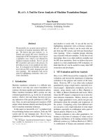
(Fig. 1). Growth rates are comparable for both investigated
aerobic biotransformations, indicating that the presence of
one or two carboxyl groups does not considerably influence
the substrate acceptance. To determine the metabolic
Table 1. Growth of P. putida on different substrates, measured on agar
plates.
Substrate Structure Growth
a
Propynoate +
2-Butynedioate
+
Propynol
++
But-3-yn-1-ol
+
Pent-3-yn-1-ol
+
Phenylethyne
b
+
Pyruvate
+
Succinate
+++
Citrate
+++
Glucose
+++
a
+, Comparable to growth rate on 2-butyndioate; ++, 2–3 times
faster growth rate than on 2-butyndioate; +++, 5–10 times faster
growth rate than on 2-butyndioate.
b
Phenylethynes are substrates
for ring dioxygenases in other bacteria [7].
1394 L. Brecker et al. (Eur. J. Biochem. 270) Ó FEBS 2003
pathways of the two acetylenic compounds UV spectroscopy
and in situ
1
H-NMR [11–15] were used.
Biotransformation of propynoate
The metabolism of propynoate (k
max
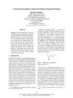
¼ 263 nm) was first
analysed by UV indicating that the substrate concentration
(5 m
M
) in the supernatant decreased during the first 12 h
and that no significant accumulation of a UV absorbing
metabolite occurred (Fig. 2). The substrate consumption
was confirmed by in situ
1
H-NMR analysis (
1
H
d: 3.06 p.p.m) in
1
H
2
O. Spectra indicated the additional
presence of a small amount of 3-ketopropanoate (
1
H
d: 3.48 p.p.m) and acetaldehyde (
1
H d: 1.17 p.p.m) during
the propynoate consumption. Both compounds were only
identified in the keto form, as the corresponding hydrates
were present in concentrations below the limit of detection.
The detected transient metabolites were present at 0.2–
0.4 m
M
(Fig. 3a and b). In parallel the amount of acetate
formed (
1
H d: 1.90 p.p.m) and 3-hydroxypropanoate (
1
H
d: 2.42 p.p.m) increased up to 2.5 m
M
while propynoate was
metabolized completely. Whereas acetate was further con-
sumed and metabolized to non-detectable products during
the following 64 h, 3-hydroxypropanoate had not been
accepted as a substrate and was present at a constant
concentration (Fig. 3c).
These findings indicate an initial hydration of the triple
bond in propynoate forming 3-hydroxyprop-2-enoate acid.
This metabolite spontaneously isomerizes to 3-ketopropa-
noic acid, which is than partly decarboxylated forming
acetaldehyde and gaseous carbon dioxide. In the following
step, acetaldehyde was oxidized to acetate accompanied by
the reduction of 3-ketopropanoate to 3-hydroxypropanoate.
These parallel reactions suggest that hydrogen atoms from
acetaldehyde are incorporated into the alcohol formation in
the other metabolite. The assumption was proved by adding
acetaldehyde (0.5 m
M
and 5 m
M
) to the biotransformation.
The 0.5 m
M
amount led to a direct consumption of the
acetaldehyde added and the production of equal amounts of
3-hydroxypropanoate, while the concentration of transient
3-ketopropanoate was too low to be detected. Addition of
5m
M
acetaldehyde obviously induced other enzymes that
metabolise this substrate in a different way.
Performing the propynoate biotransformation in
1
H
2
O/
D
2
O (1 : 1) led to an incorporation of 50% deuterium in all
metabolites. The addition of 0.5 m
M
acetaldehyde to this
biotransformation in 50% D
2
Ocausedan 10% higher
amount of hydrogen in the acetate, as it is formed directly
from the acetaldehyde. The incorporation of a higher
hydrogen amount in position three of 3-hydroxypropanoate
was not determined, probably due to isotopic exchange
during the reduction/oxidation reactions. The propynoate
pathway in P. putida isshowninFig.4.
Biotransformation of 2-butynedioate
The UV spectrophotometric analysis of the 2-butynedioate
metabolism provided scattered absorptions at k
max
¼
265 nm during the first 24 h of the biotransformation.
Furthermore the consumption of 2-butynedioate could not
be monitored by
1
H-NMR due to the lack of protons in
this substrate. However,
1
H-NMR clearly indicates the
formation of pyruvic acid [
1
H d: 2.37 p.p.m. (keto form);
Fig. 1. Aerobic growth of isolated P. putida on 2-butynedioate (j) and
propynoate (d).
Fig. 2. UV Spectra taken from the supernatant of the propynoate fer-
mentation. Spectra were taken every 2 h and indicate metabolism of the
conjugated system in propynoate.
Ó FEBS 2003
1
H-NMR of a-alkynoate biotransformations (Eur. J. Biochem. 270) 1395
1.43 p.p.m. (hydrate form)] during this time period. The
accumulation of this metabolite explains the UV results,
because it absorbs in the same spectral range as 2-butyne-
dioate. While no intermediate with four carbon atoms was
detected, we assumed that pyruvate was formed by decarb-
oxylation of 2-ketobutandioate, the product of a triple-bond
hydrolysis of 2-butynedioate. An initial decarboxylation
was excluded, as neither propynoate, nor its metabolite
3-hydroxypropanoate were detected. Control experiments
using 2-ketobutandioate as substrate confirmed this
assumption. The decarboxylation by the P. putida strain
was about 10 times faster than the spontaneous decarboxy-
lation, indicating the presence of an induced decarboxylase
in the organism. The accumulated pyruvate was mainly
hydrolysed to equal amounts of acetate (
1
H d: 1.87 p.p.m)
and formate (
1
H d: 8.36 p.p.m), which were both further
slowly metabolized to nondetectable products (Fig. 5).
The small shift differences of the acetate signal in the two
biotransformations were due to variations in the salt
concentrations and the pH value [16]. About 10% of the
pyruvate was transformed to lactate (
1
H d: 1.29 p.p.m.;
Fig. 5), indicating an incorporation of hydrogen from
formate degradation. A metabolism of pyruvate via a
dehydrogenase might also be possible in small amounts.
Fig. 6 shows the 2-butynedioate metabolic pathway in
P. putida.
Discussion
Of the variety of isolated natural products with acetylenic
bonds only 2% are a-alkynoates [1]. As these compounds
are seldomly accumulated, they seem to be good substrates
for metabolism of the triple bond. So far only one hydratase
from a Pseudomonas strain has been described to act directly
on a-alkynoates [9,10]. It is reported to accept 2-butyne-
dioate and propynoate as substrates. One b-alkynoate
(3-butynoate) has also been described to be hydrated by
another hydratase from Pseudomonas BB1 [8]. However,
none of these hydratases has been purified.
Our isolation again resulted in a Pseudomonas strain that
grew on 2-butynedioate and propynoate. Although using
strictly aerobic conditions in both cases, the triple bonds
were hydrolysed, and not oxidized. The two triple bonds in
the substrates were probably hydolysed by the same enzyme,
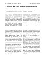
Fig. 4. Propynoate metabolic pathway in the isolated P. putida strain.
An initial hydrolysis formed 3-ketopropanoate, which was then partly
decarboxylated to acetaldehyde. The latter was dehydrogenated to
acetate, whereas the 3-ketopropanoate was hydrated to 3-hydroxy-
propanoate.
Fig. 3. Selected
1
H-NMR spectra from propynoate metabolism. (A)
Spectrum of the substrate. (B) Spectrum after 36 h. Small amounts of
acetealdehyde and 3-ketopropanoate were identified by the addition of
standard solutions. Larger amounts of acetate and 3-hydroxypro-
panoate were detected directly from the spectra. Unidentified minor
by-products are indicated with asterisks (*). (C) Spectrum after 128 h.
Starting material and intermediates were completely consumed and
3-hydroxypropanoate accumulated; some acetate was left and slowly
further metabolized.
1396 L. Brecker et al. (Eur. J. Biochem. 270) Ó FEBS 2003
resulting in different metabolites. Whereas the metabolites
of the dicarboxylate were completely consumed, half of the
substrate with the terminal acetylenic bond was transformed
to a compound that was not further accepted as a substrate.
This finding seems to be in contrast with the bacterial growth
on the two substrates, which resulted in a similar optical
density. However, the discrepancy is explainable considering
that a-alkynoates are probably not the natural growth
substrates. Rather these compounds were metabolized by
thestraintodetoxifyitsenvironmentandtheresulting
metabolites have been occasionally used for growth up to
the stationery phase in both biotransformations.
The suggested pathways, however, are based solely on
metabolic data. Therefore it is necessary to verify the
presence of the postulated enzymes by protein biochemical
or genetic analyses. As until now no other isolated acetylene
hydratase has been described, a purification, sequencing,
and protein biochemical characterization of the initial
acetylene hydratase is inevitable. In case of the other, more
common enzymes, which are involved a genomic sequence
analysis and a comparison to the genome of other strains
can also provide valuable information and enable protein
identification.
Apart from the hydrolyses investigated very little is known
about microbial metabolism in other organisms that detoxify
the environment from acetylenic compounds. To get a deeper
insight into such biotransformations in situ
1
H-NMR
analysis in
1
H
2
O is a valuable analytical method, although
the substrates themselves are often ÔinvisibleÕ. Several
metabolites can be detected, identified, and quantified
directly from the cell culture or from the supernatant in
concentrations > 0.2 m
M
. The use of natural
1
H
2
O excludes
virtual reactions and does not affect growth rates by means of
isotopic effects [16]. Addition of defined amounts of D
2
O,
however, is useful to determine the incorporation of protons
from the solvent into the products. Therefore this analytical
technique allows a detailed analysis of the acetylene bond
biodegradation in several organisms.
Acknowledgements
H. Griengl (Graz) and W. Steiner (Graz) are gratefully acknowledged
for substantial contribution to this project. We thank G. Straganz
(Graz) for valuable help and support performing the biochemical work.
L. B. gratefully acknowledges Whiteknight Technologies, Ltd. (Exeter,
GB) for financial support.
References
1. Beilstein Crossfire, Version 3.1. Database PS0201PR. (1996–2002)
Beilstein Information Systems GmbH, Frankfurt, Germany.
2. Benton, P.M.C., Christiansen, J., Dean, D.R. & Seefeldt, L.C.
(2001) Stereospecificity of acetylene reduction catalyzed by
nitrogenase. J. Am. Chem. Soc. 123, 1822–1827.
Fig. 5.
1
H-NMR spectrum of 2-butyndioate metabolism, taken from the supernatant after 36 h. This shows the intermediate pyruvate, the main
products acetate and formate, as well as the small amount of the by-product lactate.
Fig. 6. 2-Butynedioate metabolic pathway in the isolated P. putida
strain. The substrate was initially hydrolysed to 2-ketobutandioate and
its appropriate enol isomer. This intermediate was decarboxylated to
carbon dioxide and pyruvate. About 90% of the latter metabolite was
than further hydrolysed to acetate and formate. Approximately 10%
of the pyruvate was reduced to lactate, probably incorporating
hydrogen from the formate, which was metabolized further.
Ó FEBS 2003
1
H-NMR of a-alkynoate biotransformations (Eur. J. Biochem. 270) 1397
3. Rosner, B.M., Rainey, F.A., Kroppenstedt, R.M. & Schink, B.
(1997) Acetylene degradation by new isolates of aerobic bacteria
and comparison of acetylene hydratase enzymes. FEMS Micro-
biol. Lett. 148, 175–180.
4. Walsh, C. (1977) Recent developments in suicide substrates and
other active site-directed inactivating agents of specific target
enzymes. Horizons Biochem. Biophys. 3, 36–81.
5. Marcotte, P. & Walsh, C. (1978) Sequence of reactions which
follow enzymic oxidation of propargylglycine. Biochemistry 17,
5613–5619.
6. Miesowicz, F.M. & Bloch, K. (1979) Purification of hog liver
isomerase. Mechanism of isomerization of 3-alkenyl and 3-alkynyl
thioesters. J. Biol. Chem. 254, 5868–5877.
7. Ahmed, S. (1991) Microbial oxidative reactions of arenes
1
PhD
Thesis. Imperial College, London, UK.
8. van den Tweel, W.J.J. & De Bont, J.A.M. (1985) Metabolism of
3-butyn-1-ol by Pseudomonas BB1. J. Gen. Microbiol. 131, 3155–
3162.
9. Yamada, E.W. & Jakoby, W.B. (1958) Enzymatic utilization of
acetylenic compounds–I. An enzyme converting acetylenedicarb-
oxylic acid to pyruvate. J. Biol. Chem. 233, 706–711.
10. Yamada, E.W. & Jakoby, W.B. (1958) Enzymatic utilization of
acetylenic compounds – II. Acetylenemonocarboxylic acid hyd-
rase. J. Biol. Chem. 233, 941–945.
11. Brecker, L. & Ribbons, D.W. (2000) Biotransformations moni-
tored in situ by proton nuclear magnetic resonance spectroscopy.
Trends Biotechnol. 18, 197–202.
12. Weber, H. & Brecker, L. (2000) Online NMR for monitoring
biocatalysed reactions. Curr. Opin. Biotechnol. 11, 572–578.
13. Gillies, R.J. (1994) NMR in Physiology and Biomedicine.Academic
Press, New York.
14. Barbotin, J.N. & Portais, J.C. (2000) NMR in Microbiology.
Theory and Applications. Horizon Scientific Press, Wymondham,
UK.
15. Field, L.D. & Sternhell, S. (1989) Analytical NMR. John Wiley &
Sons, New York, USA.
16. Brecker, L., Weber, H., Griengl, H. & Ribbons, D.W. (1999)
In situ proton-NMR analyses of Escherichia coli HB101 fermen-
tations in
1
H
2
OandinD
2
O. Microbiology 145, 3389–3397.
17. Morawski, B., Eaton, R.W., Rossiter, J.T., Guoping, S., Griengl,
H. & Ribbons, D.W. (1997) 2-Naphthoate catabolic pathway in
Burkholderia strain. JT 1500. J. Bacteriol. 179, 115–121.
18. Gue
´
ron, M., Plateau, P. & Decorps, M. (1991) Solvent signal
suppression in NMR. Prog. NMR Spectrosc. 23, 135–209.
19. Hore, J.P. (1989) Solvent suppression. In Methods in Enzymology
(Oppenheimer, N.J. & James, J.T., eds), Vol. 176, pp. 64–77.
Academic Press, Inc., San Diego, CA, USA.
1398 L. Brecker et al. (Eur. J. Biochem. 270) Ó FEBS 2003