Báo cáo khoa học: Local changes in the catalytic site of mammalian histidine decarboxylase can affect its global conformation and stability pptx
Bạn đang xem bản rút gọn của tài liệu. Xem và tải ngay bản đầy đủ của tài liệu tại đây (670.29 KB, 12 trang )
Local changes in the catalytic site of mammalian histidine
decarboxylase can affect its global conformation and stability
Carlos Rodrı
´
guez-Caso
1
, Daniel Rodrı
´
guez-Agudo
1
, Aurelio A. Moya-Garcı
´
a
1
, Ignacio Fajardo
1
,
Miguel A
´
ngel Medina
1
, Vinod Subramaniam
2,
* and Francisca Sa
´
nchez-Jime
´
nez
1
1
Department of Molecular Biology and Biochemistry, Faculty of Sciences, Ma
´
laga, Spain;
2
Max Planck Institute for Biophysical
Chemistry, Goettingen, Germany
Mature, active mammalian histidine decarboxylase is a di-
meric enzyme of carboxy-truncated monomers ( 53 kDa).
By using a biocomputational approach, we have generated a
three-dimensional model of a recombinant 1/512 fragment
of the rat enzyme, which shows kinetic constants similar to
those of the mature enzyme purified from rodent tissues.
This model, together with previous spectroscopic data, al-
lowed us to postulate that the occupation of the catalytic
center by the natural substrate, or by substrate-analogs,
would induce remarkable changes in the conformation of
the intact holoenzyme. To investigate the proposed con-
formational changes during catalysis, we have carried out
electrophoretic, chromatographic and spectroscopic analy-
ses of purified recombinant rat 1/512 histidine decarboxylase
in the presence of the natural substrate or substrate-analogs.
Our results suggest that local changes in the catalytic
site indeed affect the global conformation and stability of
the dimeric protein. These results provide insights for
new alternatives to inhibit histamine production efficiently
in vivo.
Keywords: histidine decarboxylase; histamine; a-fluoro-
methylhistidine;
L
-histidine methyl ester; pyridoxal phos-
phate-dependent enzymes.
Mammalian histidine decarboxylase (HDC), the enzyme
responsible for the biosynthesis of histamine, is a pyridoxal
5¢-phosphate (PLP)-dependent enzyme that belongs to the
evolutionary group II of
L
-amino acid decarboxylases [1–3].
Histamine is involved in several physiological responses
(immune responses, gastric acid secretion, neurotransmis-
sion, cell proliferation, etc.) and is also implicated in widely
spread human pathologies (inflammation-related diseases,
neurological disorders, cancer and invasion) [4–8]. In spite
of the importance of these pathologies, HDC has not been
fully characterized, and important questions about the
regulation of the enzyme expression, sorting, processing,
structural characterization and turnover remain unan-
swered [9–15].
Mature HDC purified from mammalian tissues has been
reported to be a dimer. Although the exact sequence of each
monomer is not known, it is generally believed that the
74 kDa precursor is processed to a carboxy-truncated form
of 53–58 kDa [16,17]. The N-terminus of the polypeptide
(residues 1–480) exhibits a moderately high degree of
identity with the porcine DOPA decarboxylase (DDC),
another dimeric group II
L
-amino acid decarboxylase for
which an X-ray structure has been solved [18]. Recently, we
have characterized the catalytic mechanism of a recombin-
ant carboxy-truncated form of the rat enzyme (fragment
1–512, also named HDC 1/512) [19], which shows kinetic
constants similar to those of the mature enzyme purified
from rodent tissues [16,17].
Mammalian HDC and DDC appear to share several
catalytic features [19,20]. First, the PLP-enzyme internal
Schiff base consists mainly of an enolimine tautomeric form
(free holoenzyme). Second, Michaelis complex formation
leads to a polarized ketoenamine form of the Schiff base.
Third, after transaldimination, the coenzyme–substrate
Schiff base exists mainly as an unprotonated aldimine.
Finally, decarboxylation occurs and the free holoenzyme is
recovered after protonation and a reverse transaldimination
that releases the amine product. In spite of these shared
features, the following observations [19] suggest that key
structural differences must exist between the mammalian
HDC catalytic site and those of the other group II
L
-amino
acid decarboxylases: (a) HDC is the least efficient enzyme of
its group; (b) mammalian HDC activity is less sensitive to the
presence of thiol reducing compounds in the medium than
other homologous and nonhomologous PLP-dependent
decarboxylases; and (c) transaldimination from the internal
to the external aldimine involves a higher degree of rotation
in the torsion angle (v) than those observed for other
homologous enzymes. The last observation is in agreement
with the remarkably low catalytic efficiency of mammalian
Correspondence to F. Sa
´
nchez Jime
´
nez, Departamento de Biologı
´
a
Molecular y Bioquı
´
mica, Facultad de Ciencias, Universidad de
Ma
´
laga, 29071 Ma
´
laga, Spain.
Fax: + 34 95 2132000, Tel.: + 34 95 2131674,
E-mail:
Abbreviations:DDC,aromatic
L
-amino acid decarboxylase or DOPA
decarboxylase (EC 4.1.1.28); DOPA,
L
-3,4-dihydroxyphenylalanine;
a-FMH, alpha-fluoromethylhistidine; a-FMHA, alpha-fluoromethyl-
histamine; HDC, histidine decarboxylase (EC 4.1.1.22); HisOMe,
L
-histidine methyl ester; PLP, pyridoxal 5¢-phosphate.
Enzymes:aromatic
L
-amino acid decarboxylase or DOPA decarb-
oxylase (EC 4.1.1.28); histidine decarboxylase (EC 4.1.1.22).
*Present address: Advanced Science and Technology Laboratory,
AstraZeneca R & D Charnwood, Loughborough, UK.
(Received 7 August 2003, revised 9 September 2003,
accepted 12 September 2003)
Eur. J. Biochem. 270, 4376–4387 (2003) Ó FEBS 2003 doi:10.1046/j.1432-1033.2003.03834.x
HDC, as this angle defines the degree of conjugation
between the pyridine ring and the imine bond (maximum at
v ¼ 0), which is important for catalytic efficiency of PLP-
dependent enzymes [21]. In fact, the catalytic steps after
the first transaldimination are rate-limiting in the entire
catalytic reaction of mammalian HDC [19].
On the other hand, the crystal structure of DDC has
shown that the catalytic sites of this enzyme are located in
the interface between monomers [18]. Assuming that the
catalytic sites of HDC are also in the interface between
monomers, we postulated the hypothesis that the observed
high degree of rotation of the torsion angle v induced by the
presence of the substrate could produce a remarkable
conformational change in the whole holoenzyme.
In this work, we have used biocomputational methods
to locate the catalytic center and predict the shape of the
dimeric protein. The model reinforced our hypothesis
proposed above. To further address this hypothesis, we
have characterized the protein conformational changes
duringcatalysis,byusingastrategysimilartothatused
previously for the catalytic mechanism characterization
[19]. Substrate analogs capable of blocking the HDC
catalytic site at different catalytic steps are known, allowing
the detection and analysis of the respective conformational
states of the protein during the reaction. Thus, electro-
phoretic, chromatographic and spectroscopic analyses
carried out with purified preparations of the recombinant
HDC 1/512 version in the presence and absence of substrate
or substrate analogs have allowed us to characterize the
conformational changes of the holoenzyme whenever a
PLP substrate- or PLP product-like adduct is inside the
catalytic center. We have also evaluated the stability of
these conformational states against several agents that
disrupt structure, i.e. detergent, thiol reductants, and
temperature.
Materials and methods
Biocomputational analyses
An initial model of the target protein (residues 5–479) was
generated from the rat HDC sequence (Swiss-Prot accession
number P16453) using the automated comparative protein
modeling server
SWISS
-
MODEL
[22–24] in First Approach
mode. The two pig DDC structures obtained from the
Protein Data Bank (PDB ID 1JS3 and 1JS6) were used as
templates.
The docking program
GRAMM
[25] was used to build the
structure of the dimeric rat HDC from the coordinate file
provided by
SWISS
-
MODEL
. A low resolution docking was
performed with a grid step of 6.8 A
˚
and 20° rotation
increments. This type of docking is designed to overcome
the problems of conformational flexibility and induced fit
movements inherent to the formation of a protein–protein
complex. The lowest energy docking solution was selected
as representative of the dimer structure. Secondary structure
of the model was calculated with the
DSSP
program
[26]. Energy minimization calculations were performed with
the program
XPLOR
[27] in an SGI Altix 3000 under GNU/
L
INUX
R
ED
H
AT
7.2.
Three-dimensional visualization and analysis were per-
formed with
RASMOL
[28] and
SWISS
-
PDBVIEWER
[22].
Molecular graphics were generated using
MOLSCRIPT
[29],
RASTER
3
D
[30] and
PYMOL
[31].
Recombinant HDC purification procedures
and enzyme activity assay
The DNA encoding for residues 1–512 of rat HDC [32] was
subcloned in the pET-11a vector (Novagen, USA). The
recombinant plasmid transformed into the Escherichia coli
BL21(DE3)pLysS strain. Transformed cultures were
induced to express the HDC 1/512, which was purified by
applying three chromatographic steps (Phenyl-Sepharose
CL-4B, DEAE interchange, and hydroxyapatite). The final
preparations were dissolved in 50 m
M
potassium phosphate,
0.1 m
M
PLP, pH 7.0. Purity of the HDC 1/512 construct
was checked by Coomassie blue staining and Western
blotting, and was higher than 95% in the final preparations.
HDC activity was assayed by following
14
CO
2
release from
L
-His-[U-
14
C] (American Radiolabeled Chemicals, USA).
Analogs and histidine were provided by Sigma-Aldrich
(Spain). All of these procedures (overexpression, purifica-
tion and analysis) are described extensively elsewhere [19].
When required, the enzyme was concentrated in different
Amicon (USA) ultrafiltration systems (cut-off between 10
and 30 kDa) depending on the initial volume. To avoid
interference by free PLP, the final preparation was subjected
to size-exclusion gel chromatography by using a Sephadex
G25 column or, alternatively, a Sephacryl HiPrep S-200
column (Pharmacia Biotech, Sweden) in 50 m
M
potassium
phosphate buffer (pH 7) immediately before starting
spectroscopic analyses.
Chromatographic analysis
Purified HDC preparations were incubated at room tem-
perature in either the presence or absence of 1 m
M
alpha-
fluoromethylhistidine (a-FMH) for 1 h. After incubation,
protein was subjected to size-exclusion gel chromatography
on a HiPrep Sephacryl S-200 high-resolution column, pre-
equilibrated with 50 m
M
potassium phosphate and coupled
to a FPLC system (Pharmacia). Absorbance at 280 nm was
monitored continuously to detect protein peaks. M
r
swere
calculated after calibration of the column with the following
molecular-mass standards (all from Sigma): alcohol dehy-
drogenase (M
r
14 2000), bovine serum albumin (M
r
65 000), chymotrypsinogen A (M
r
25 000) and cyto-
chrome c (M
r
12 400).
Electrophoretic and Western blotting analysis
Aliquots of the purified enzyme (1–3 lg) were incubated at
room temperature for 1 h in the presence of either 1 m
M
histidine-analogs or 10 m
M
histidine, or in their absence
(untreated enzyme). All solutions were adjusted to pH 7.
Ten millimolar histidine (more than 20-fold the previously
reported K
m
value) was chosen to maximize the percentage
of enzyme taking part in the enzyme–substrate complex.
When indicated, 2-mercaptoethanol was added after 55 min
of incubation to yield a final concentration of 80 m
M
,
followed immediately by loading buffers. For conventional
denaturing SDS/PAGE experiments, the protocol described
by Laemmli was followed [33]. Samples were boiled and
Ó FEBS 2003 Conformational changes of histidine decarboxylase (Eur. J. Biochem. 270) 4377
stored at )20 °C until their use. However, for semidena-
turing SDS/PAGE experiments, samples were not boiled
and the loading buffers lacked 2-mercaptoethanol. Two
different loading buffers were used. Loading buffer A
(pH 7.4) lacked SDS, while loading buffer B (pH 6.8)
contained 0.7% SDS. Immediately after addition of the
respective loading buffer, electrophoresis was performed at
4 °C in a 7% acrylamide SDS-free PAGE gel (semidena-
turing condition A) or in a 7% acrylamide 0.1% SDS/
PAGE gel (semidenaturing conditions B), both lacking
stacking gel. The running buffer always contained 0.1%
SDS. To avoid problems with any minor contaminant band
in the purified preparations, electrophoretic results were
visualized by Western blotting, following a protocol
described elsewhere [15]. The anti-HDC K9503 serum was
generously supplied by L. Persson (Lund University,
Sweden). Recombinant Strep-tag unstained standards
161–0362 (Bio-Rad, USA) were used as references for
electrophoretic migrations. In each experiment, treatments
were carried out in parallel on aliquots of the same purified
enzyme preparations. The results of semidenaturing gels
shown here are representative of at least four different
independent experiments.
Spectroscopic analysis
Absorption spectra were obtained using a HP8452A diode
array spectrophotometer (Hewlett-Packard, USA). The
acquisition time for each absorption spectrum was 2 s.
Fluorescence spectra were obtained in a QuantaMaster
SE spectrofluorimeter (Photon Technology International
Inc., USA). Integration time was 0.1 sÆnm
)1
; three spectra
were averaged. CD spectroscopy was carried out with a
Jasco J-715 spectropolarimeter at a scan speed of
50 nmÆmin
)1
; 10 spectra were averaged. Analogs were
used at the specified final concentrations. Unless otherwise
indicated, all spectroscopic measurements were carried out
at room temperature. Fluorescence (300–400 nm, excita-
tion at 275 nm) was not detectable from solutions of
10 m
M
histidine or 1 m
M
analogsinanenzyme-free
buffer. CD signals (195–250 nm) were not detectable from
solutions of 1 m
M
a-FMH, 14 l
M
PLP or both together
in an enzyme-free buffer.
Results and discussion
Protein modeling of rat HDC dimer predicts
the location of its catalytic site
We have previously observed that transaldimination from
the internal to the external aldimine of HDC involves a
higher degree of rotation in the torsion angle (v)thanin
other homologous enzymes [19]. This observation led us
to postulate that the interaction of HDC with its substrate
could induce significant conformational changes, at least
in the catalytic center environment. In addition, this
experimental observation also indicated that some differ-
ences must exist in the relative position of the cofactor–
substrate adduct when compared with homologous
enzymes.
The moderately high degree of identity between mam-
malian HDC and DDC (> 50%), the published structural
models [2,3] and the reported crystal structure of porcine
DDC [18], in combination make it possible to apply
comparative modeling methods to overcome the lack of
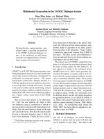
reported crystal structures for mammalian HDC. Figure 1
shows the lowest energy predicted 3D structure of the rat
HDC monomer and its quaternary structure, obtained after
docking and energy minimization calculations carried out
as described in the Materials and methods section. One
thousand dimer structures were generated without any
manual fitting at all. At least the first 200 were examined
and found to be very similar. They are conformed as
twofold axial symmetry dimers similar to those described
for the crystal structures of pig DDC [18]. Nevertheless, it
predicts a more occluded catalytic center when compared
with the DDC crystal structure. This is not surprising, as
HDC has a more restrictive catalytic center, and it could be
suspected from experimental data previously shown [19].
Figure 2 shows the alignment of rat HDC and pig DDC
primary sequences, as well as the distribution of a-helices
and b-sheets in both the crystal structure of pig DDC and
that estimated from the rat HDC 3D model. This figure
stresses that the pattern of secondary structures in both
enzymes is very similar, in spite of their differences in
primary structure, as expected. The complete distribution of
consensus secondary structure estimated from the model is
as follows: 39% of a-helices, 9% of b-sheets, 12% of turns,
21% of random coil and 19% of other structures. These
estimations are similar to those obtained from rat HDC
primary sequence, and they are consistent with estimations
from far-UV CD spectra (controls at 20 °CinFig.9,and
not shown here to avoid redundancy).
In spite of the lack of overall sequence identity, a
common PLP-binding motif consisting of clusters of
conserved residues is present in decarboxylases belonging
to groups I, II and III [2]. The PLP-binding site of
Morganella morganii AM-15 HDC was experimentally
located in its K233 residue [34]. This lysine residue is
extremely conserved, and corresponds to K303 of pig and
rat DDC, also previously shown to play this role [18,35],
and to K308 in rat HDC. A histidine residue, corres-
ponding to H197 in rat HDC, also is strictly conserved in
group II of mammalian
L
-amino acid decarboxylases, in
which it seems to be stacked in front of the cofactor
pyridine ring [18]. Thus, our model combined with the
previously reported structural and mechanistic character-
ization of these enzymes [19,20,36] allowed us to locate
the HDC catalytic center at the interface between the
monomers (Fig. 1), as is the case of DDC [18]. One of the
monomers (monomer A) would contain the major part of
the catalytic site pocket, including K308 and H197 in rat
HDC.
Figure 3 shows the most important residues close to
H197 and K308, as predicted by our 3D model. The
predicted catalytic center contains a number of polar
residues (D276, N305, H197 and K308). All of these
residues are strictly conserved in mammalian DDC (see
Fig. 2), where they take similar positions to those
predicted in HDC. In mammalian DDC, D271 and
N300 (counterparts of D276 and N305 in rat HDC) are
predicted to interact with the imidazole ring and the
phosphate group of PLP, respectively [18,36,37]. It is
noteworthy that in spite of the high flexibility of the
4378 C. Rodrı
´
guez-Caso et al. (Eur. J. Biochem. 270) Ó FEBS 2003
fragments (see Fig. 2) containing most of these residues,
our prediction located them very close in space and at
similar positions to those described for DDC [18].
Therefore, it seems to be likely that they play a similar
role in mammalian HDC, delimiting what could be
termed as the PLP interaction region (PLP-IR, see
Fig. 3).
After formation of the holoenzyme, the substrate (histi-
dine) should enter the catalytic site from the bottom part of
Fig. 3 through a space delimited by the PLP-IR and a
region in which our model predicts the location of several
residues of both monomers able to establish hydrophobic
interactions; for instance, Y84 (Fig. 3) and the fragment
PAL 85–87 from monomer A (the latter not shown in Fig. 3
for clarity), and F331, I364 and L356 from monomer B.
These predictions are in agreement with previous bio-
physical and kinetic studies from our laboratory indicating
that, in the internal aldimine form, the catalytic site of HDC
is enriched in hydrophobic residues, leading to an enolimine
tautomeric form of PLP [19]. A hydrophobic channel for
the substrate has also been proposed for DDC [38,39].
However, the specific hydrophobic residues of monomer B
contributing to this region of DDC (I101 and F103, as
deduced from data in reference [18]) are different, as
expected from the structural differences between their
respective substrates. It is also noteworthy that some of
the closest hydrophobic residues of monomer B (for
instance, F331) are part of or close to the Ôflexible loopÕ
described for mammalian DDC, which could not be solved
from the crystal structure (residues 328–339 in Fig. 2). In pig
DDC, a conformational change of this loop in response to
substrate binding has been demonstrated [18,37]. A similar
role of its counterpart in mammalian HDC in relation to the
conformational change described in the present work could
be suspected.
Finally, from this model we have predicted that the
occupation of the catalytic center by the polar substrate or
an analog through a hydrophobic channel could induce
drastic conformational changes of the holoenzyme that
would probably affect, at least locally, interactions at the
monomer interface and vice versa. This reinforces our
starting hypothesis.
Fig. 1. Three-dimensional model of rat HDC
structure. The model was generated from res-
idues 2–475 of the primary sequence, as des-
cribed in Material and methods section. (A)
and (B) Surface representations of the dimeric
form, one monomer in white and the other in
red. (C) Surface representation of one mono-
mer in white. The predicted interface between
monomers is shown in red. The active site
residues, K308 and H197, are shown in blue.
W and Y residues are depicted in green. In (A)
and(C),thewhitemonomerisshowninthe
same position. (B) is left-twisted around the
z-axis with respect to (A) to show the
localization of K308 and H197 within the
monomer interface. A double-headed arrow
in (A) indicates the maximum distance
determining the Stokes’ radius predicted for
the dimer.
Ó FEBS 2003 Conformational changes of histidine decarboxylase (Eur. J. Biochem. 270) 4379
Active HDC1/512 is a dimer and the presence
of a substrate analog in its active site diminishes
its Stokes’ radius
HDC1/512 is a recombinant carboxy-truncated form of the
rat enzyme that we have previously used to study structure/
function relationships of the mature HDC [10,13,14,19,40].
We have recently shown that HDC1/512 has kinetic
constants similar to those of the mature enzyme purified
from rodent tissues [19]. Figure 4 shows the results of size-
exclusion gel chromatography of purified HDC1/512. A
major peak (M
r
107 000) was observed for the untreated
enzyme. Some inactive higher molecular weight HDC
aggregates were detected by Western blots (data not shown).
In fact, the purified enzyme preparations slowly tend to
form inactive aggregates when incubated at room tempera-
ture or higher (C. Rodrı
´
guez-Caso, D. Rodrı
´
guez-Agudo,
A. A. Moya-Garcı
´
a, M. A
´
. Medina, V. Subramanian &
F. Sa
´
nchez-Jime
´
nez, unpublished observations). Enzymatic
activity was only detectable in fractions corresponding to
the major peak. These results indicated that the quaternary
Fig. 2. Alignment of rat HDC and pig DDC sequences. The 5–479
fragment of rat HDC (Swiss-Prot accession number P16453) and pig-
DDC sequence from the Protein Data Bank (PDB ID 1JS6) were
aligned using the ProdModII method in Swiss-Model server (http://
www.expasy.org/swissmod/SWISS-MODEL.html). The secondary
structure of the crystallized pig DDC and that predicted from the rat
HDC 3D model are shown below the aligned sequences: h, helix;
s, sheet; ?, unpredicted conformation in crystallized pig-DDC.
Fig. 3. Structural neighborhood of the PLP-binding site. The most
relevant residues closer than 7 A
˚
to H197 or K308, as predicted by our
3D apoenzyme model, are depicted. The line delimits the PLP inter-
action region (PLP-IR). The putative entrance for the substrate
between the PLP-IR and the hydrophobic region is marked with a star.
Fig. 4. Size-exclusion gel chromatography of the free-holoenzyme and
the a-FMH-treated enzyme. Purified enzyme was incubated for 1 h in
the presence or absence of 1 m
M
a-FMH and submitted to size-
exclusion gel chromatography, using a FPLC system as described in
the Material and methods. No monomer was observed. Fractions 1 to
38 represented void volume. For the free-holoenzyme extracts, enzyme
activity was coincident with the major peak. Arrows indicate the peaks
of the M
r
standards: 1, alcohol dehydrogenase (M
r
142 000); 2, bovine
serum albumin (M
r
65 000); 3, chymotrypsinogen A (M
r
25 000); 4,
cytochrome c (M
r
12 400).
4380 C. Rodrı
´
guez-Caso et al. (Eur. J. Biochem. 270) Ó FEBS 2003
structure of the active recombinant purified enzyme used in
this work is, indeed, a dimer, as also deduced for the native
enzyme purified from natural sources [17].
Figure 5 shows a scheme of the HDC reaction and the
specific steps interfered by the substrate analogs histidine
methyl ester (HisOMe) and a-FMH, deduced from previous
reports in the literature [19,41]. HisOMe, a reversible
competitive inhibitor, blocks the reaction after formation
of an external aldimine tautomeric form very similar to that
of the PLP–histidine adduct (Fig. 3 and [19]). By using the
substrate analog a-FMH, the reaction can proceed (inclu-
ding the decarboxylation step) to form a-fluoromethylhis-
tamine (a-FMHA; Fig. 5 and [41]). In the case of fetal rat
HDC, this reaction has been reported to proceed much
slower than with the natural substrate histidine [42].
Nevertheless, after decarboxylation and elimination of the
fluoride, a reverse transaldimination can occur, so that an
enamine form of the product can either leave the catalytic
site or react again with the internal aldimine to form a PLP-
adduct covalently attached to the catalytic center. It has
been proposed that the occurrence frequency of these two
possibilities depends on how long the enamine remains in
the active site and its rate of attack on the aldimine bond,
and can be modified by slight differences in the position of
the residues [41]. As covalent binding was proposed to
occur, at least partially, between PLP–a-FMHA derivatives
and the enzyme, this analog is considered as a suicide
inhibitor of PLP-dependent HDC [43]. Complexes III and
VII in Fig. 5 would correspond to the major final forms
stabilized at short-term in the catalytic site after the
reactions with HisOMe and a-FMH, respectively.
Based on the shape of the predicted dimeric HDC
(Fig. 1), a conformational change affecting the monomer
interface would change the Stokes’ radius of the protein, as
the major diameter of the dimer is predicted to be given by
the distance between the carboxy termini of both
monomers (from the lower left to the upper right of
Fig. 1B), which in turn is dependent on the dimerization
surface conformation. Gel filtration is a validated method
to distinguish changes in the Stokes’ radius of an oligomeric
enzyme. Among the different compounds tested (the
natural substrate and substrate-analogs), a-FMH is the
only one that can accumulate a stable PLP-adduct cova-
lently bound to the enzyme (Fig. 5), thus being able to
withstand a gel filtration procedure. An apparent reduction
of the M
r
was indeed observed in the a-FMH-treated
samples (Fig. 4), suggesting a treatment-induced change of
the dimeric structure to a conformation with diminished
Stokes’ radius.
Analog-treated HDC shows altered electrophoretic
mobility under semidenaturing conditions
It is well known that quaternary structure of proteins is
frequently established, at least partially, through hydropho-
bic interactions that can be weakened by SDS and other
detergents. Thus, electrophoresis of the samples carried out
in the presence of SDS as the only denaturing agent could
reveal: (a) reinforcements of monomer associations, as only
the strongest associations could survive the denaturing
agent; and (b) any change in the volume of a single
polypeptide (or a polypeptide association). We analyzed
HisOMe- and a-FMH-treated samples under the semide-
naturing conditions described in the Material and methods
section. Figure 6 shows that treatments with analogs change
the relative electrophoretic mobility of untreated HDC
under semidenaturing conditions, supporting our hypothe-
sis on global conformational changes of HDC induced by
the presence of analogs in the active center. Furthermore,
these findings also seem to indicate that the analogs can
Fig. 5. Scheme of the HDC reactions with the
natural substrate histidine and the histidine-
analogs HisOMe and a-FMH. This scheme
was built from the major forms for each step
mentioned in the text deduced from the pre-
vious information ([18,19] and the present
results). The absorption spectrum maxima
described for the tautomeric forms mentioned
in the text are indicated in brackets. The pro-
posed major forms reached with HisOMe and
a-FMH are shown inside dashed boxes.
T, transaldimination steps.
Ó FEBS 2003 Conformational changes of histidine decarboxylase (Eur. J. Biochem. 270) 4381
induce changes of the enzyme to conformational states more
resistant to denaturation by detergents, especially in the case
of conformations adopted during the external aldimine state
(HisOMe-treated samples).
Absorption spectra of HDC during reaction with histidine
and analogs reveal details of the catalytic mechanism
Taking into account the extremely low reaction rate
reported for mammalian HDC [19], and especially in the
presence of a-FMH [42], it is expected that different steps
of the reaction can be distinguished as a function of time
in a conventional UV-visible spectrophotometer. Before
Fig. 6. Western blots of the free-holoenzyme and analog-treated samples
under different semidenaturing and denaturing conditions. Aliquots of
the same purified preparation were incubated for 1 h in the absence
(control, C) or presence of 1 m
M
histidine analogs (histidine methyl
ester, HisOMe; a-fluoromethylhistidine, a-FMH) and submitted to the
semidenaturing conditions A (A, loading buffer A) and B (B) and (C)
described in the Materials and methods section. In (C), samples had
been treated for 5 min with 2-mercaptoethanol. (D) corresponds to a
conventional denaturing SDS/PAGE electrophoresis. In this case,
molecular mass standards are shown as M
r
· 10
)3
. In (A–C), bands
are designed according to their relative electrophoretic mobilities as F
(the fastest mobility), S (the slowest mobility) and I (intermediate
mobilities).
Fig. 7. Changes with time of the absorption spectra of HDC in the
presence of the natural substrate histidine or histidine-analogs. Con-
centrated, neutralized stocks (6.36 lL) of the natural substrate histi-
dine (A), HisOMe (B) or a-FMH (C) were added to 70 lLofa
13–14 l
M
solution of purified and gel filtered protein to reach final
concentrations of 10 m
M
histidine or 1 m
M
analogs.
4382 C. Rodrı
´
guez-Caso et al. (Eur. J. Biochem. 270) Ó FEBS 2003
addition of any substrate, we observed the same PLP
absorption profile previously reported for the free holo-
enzyme (Fig. 7, untreated samples in all panels, and [19]):
a major enolimine form (maximum at 335 nm, complex
I in Fig. 5) and a minor ketoenamine form (maximum at
420 nm) of the internal aldimine. However, a few
seconds after substrate or analog addition, a new peak
arose at 390 nm (Fig. 7, all panels), which must corres-
pond to accumulation of enzyme molecules at the external
aldimine stage, as reported previously ([19], see also
complex III in Fig. 5). The peak was observed not only
with histidine but also with both analogs, corresponding
to their reported action mechanisms. After 1 min, this
390-nm peak could still be observed in all cases. As
mentioned before, this external aldimine complex (com-
plex III) is the final product of the reaction with HisOMe.
In fact, after 5 min, the spectra of the HisOMe-treated
samples had stabilized with the 390-nm peak as the major
and final one. In the reaction in the presence of an excess
of the natural substrate histidine, a shoulder around
430 nm is also observed (Fig. 7A), which must correspond
to Michaelis complexes (complex II in Fig. 5) and/or to
ketoenamine forms of external aldimines (that is to say, to
other stages of the reaction), many of them having typical
absorption maxima around 430 nm [44].
On the other hand, when using a-FMH, accumulation
of a PLP-derivative with an absorption maximum at
345 nm can be clearly observed after the reaction had
passed through a maximum concentration of the external
aldimine complex (Fig. 7C). From the first minutes on,
this peak became the major one clearly observed in the
a-FMH-treated enzyme, suggesting that it corresponds to
a major molecular form of the PLP–a-FMHA derivative
that was rather stable for at least the first hour of
treatment (complex VII in Fig. 5). Nevertheless, as the
absorption at wavelengths higher than 400 nm was even
increasing with the treatment period, other noncovalently
bound PLP adducts cannot be ruled out. Bhattacharjee
and Snell [41], working with the bacterial M. morganii
HDC enzyme, proposed that the covalently bound adduct
can be converted slowly into other PLP adducts with
absorption maxima higher than 400 nm (not shown in
Fig. 5), which can be removed from the catalytic center by
dialysis and boiling. The absorption maximum observed
at 345 nm, as well as the shape of the final spectra, are
extremely similar to that reported [41] for the product of
Fig. 8. Fluorescence emission (excitation at 274 nm) of the free-HDC holoenzyme and the enzyme treated with the natural substrate or analogs. Ten
microliters of 50 m
M
potassium phosphate pH 7 (control condition), or 10 lL of concentrated neutralized stocks of the natural substrate histidine
(A), HisOMe (B) or a-FMH(CandD)wereaddedto90lLofa6.5–7 l
M
solution of purified and gel filtered protein to reach final concentrations
of 10 m
M
histidine or 1 m
M
analogs. Stability of the control spectra were assessed by three different determinations.
Ó FEBS 2003 Conformational changes of histidine decarboxylase (Eur. J. Biochem. 270) 4383
the enamine reaction with the internal aldimine, that is, a
PLP-a-FMHA derivative covalently bound to the enzyme
of Gram negative microorganisms (also Fig. 5, complex
VII). As far as we know, this is the first time that the
spectra of the PLP-adducts during the reaction of a
mammalian enzyme with a-FMH are recorded. Previous
studies of the reaction were carried out on partially
purified extracts of the rat enzyme, working with
radiolabeled a-FMH [42]. These authors deduced that
one-third of the decarboxylated a-FMH products
seemed to be covalently attached to the enzyme. Our
data are also consistent with this proposal. As the
inhibitor is in excess with respect to the enzyme, successive
decarboxylations would accumulate the covalent adduct in
the assay period, thus becoming the major form detected
in the spectra.
Summarizing, data shown in Fig. 7 reinforce our previ-
ous findings on the tautomeric forms of the internal
aldimine in the free holoenzyme [19] and the reaction
carried out with the suicide analog a-FMH [40].
The conformational changes induced in HDC
by histidine and histidine-analogs involve alterations
in the environment of the aromatic amino acid
residues of the protein
Conformational changes of proteins can modify the intrin-
sic fluorescence from their aromatic residues. Both HisOMe
and a-FMH block the HDC catalytic center after the first
transaldimination step (after external aldimine formation),
leading to catalytic sites occupied by different PLP-adducts.
HDC 1/512 contains 11 W and 13 Y residues. From them,
six W and seven Y residues are predicted to be in the
monomer interface (Fig. 1). These represent most of the W
(86%) and Y (64%) residues predicted to be exposed to the
monomer surface (seven W and 11 Y). Thus, it seemed likely
that a conformational change in the monomer interaction
surface could be reflected in the fluorescence measurements
of the free holoenzyme and the enzyme after addition of
histidine and histidine analogs. An increase in W fluores-
cence intensity is most commonly associated with reduced
exposure of the W residues to the solvent, i.e. a transition
from a predominantly solvent-exposed to a more hydro-
phobic situation brought about by a conformational
change. Although an increase in interaromatic energy
transfer is also possible, this is not the most likely reason
for a fluorescence increase. Indeed, when there are several
aromatic residues in close proximity, interactions quenching
the fluorescence tend to occur [45].
Figure 8 shows the fluorescence emission spectra (from
300 to 400 nm, excitation at 274 nm) of HDC holoenzyme
obtained before and after substrate or analog addition. In all
cases, increases of fluorescence were observed to occur within
the first minute after the compound addition, suggesting that,
indeed, all of them are able to induce structural changes
that shield aromatic residues from solvent interactions. It is
Fig. 10. Thermal denaturation profile at 222 nm of the free HDC
holoenzyme and the analog-treated samples. Aliquots of a 3-l
M
solution
of purified and gel filtered enzyme were treated with or without the
histidine-analogs at 1 m
M
final concentration. CD spectra were
recorded after stabilization of the samples at the different assayed
temperatures. Stabilization times were 2–5 min.
Fig. 9. Thermal denaturation of the free HDC holoenzyme and the
a-FMH-treated enzyme. Aliquots of a 3-l
M
solution of purified and gel
filtered enzyme solution were treated (B) or not (A, free holoenzyme)
with 1 m
M
a-FMH. CD spectra were recorded after stabilization of the
samples at the different assayed temperatures. Stabilization times were
2–5 min.
4384 C. Rodrı
´
guez-Caso et al. (Eur. J. Biochem. 270) Ó FEBS 2003
noteworthy that these conformational changes take place
within the time period in which most of the enzyme is passing
through the external aldimine state, irrespective of the
substrate or analog added (Fig. 7). At least with both
histidine and HisOMe, a shift to the blue in the emission
maximum could be clearly observed, supporting a transition
to a more hydrophobic environment.
On the other hand, an increased, red-shifted fluorescence
is observed in the a-FMH-treated enzyme after 30 min of
the reaction (Fig. 8), when most of the enzyme is forming
the covalently bound adduct with the PLP-a-FMHA
derivative (Fig. 7). These observations suggest that the
conformational change involves alterations in the environ-
ment of the aromatic amino acid residues of the protein.
Resistance of the different conformational states
to thermal denaturation
Results obtained with semidenaturing electrophoresis seem
to indicate that histidine analogs can induce changes in the
enzyme to conformational states more resistant to dena-
turation by detergents. To test whether these analog-
induced conformational states are also more resistant to
thermal denaturation, we carried out CD analysis of HDC
under several treatments and at different temperatures.
Changes in the secondary structure of the enzyme during
temperature-induced unfolding can be deduced by using
this approach. Figure 9 shows far-UV CD spectra
(190–250 nm) of the free-holoenzyme (Fig. 9A) and the
a-FMH-treated enzyme (Fig. 9B) incubated at different
temperatures after treatment. Unfolding of the protein can
be deduced from changes in the spectra observed with
increasing temperatures (Fig. 9), as well as from the changes
observed in the ellipticity at 222 nm (Fig. 10). Nevertheless,
these temperature-induced changes were more evident for
the free holoenzyme than for the a-FMH-treated enzyme
sample, indicating that the enzyme that had a covalently
bound PLP-adduct was more resistant to temperature-
induced denaturation. Scarce 2D structural information can
be obtained from similar experiments made with HisOMe,
due to the basal CD absorption of this compound.
However, the observed increasing trend in the ellipticity at
222 nm of the HisOMe-treated enzyme preparations
(Fig. 10) suggests that the reversible inhibitor was not able
to protect the enzyme against thermal denaturation.
The increased resistance of the a-FMH-treated protein to
thermal denaturation would indicate that the covalent
binding of the adduct to the catalytic center could fix some
secondary structure in the enzyme, suggesting several
interaction points for the adduct within the catalytic center
in addition to those established by the internal aldimine
alone. From the shape of the a-FMH-treated protein
spectra, stabilization of some a-helix by the cofactor adduct
could be suspected (Fig. 9). It is noteworthy that most of the
catalytic site is predicted to adopt a-helix and random coil
secondary structures (predictions not shown, also derived
from Figs 1 and 2).
Concluding remarks
Since the initial suggestions by Pauling in 1948 and the later
formulation of the induced-fit hypothesis by Koshland, it is
well established that the interactions between an enzyme
and its substrate induce conformational changes at the
active site. In most cases, these conformational changes are
only local and relatively small. However, for some enzymes
these changes may be remarkable. This seems to be the case
of mammalian HDC.
We had previously observed that transaldimination from
the internal to the external aldimine of HDC involves a
higher degree of rotation in the torsion angle (v)than
those observed in other homologous enzymes [19]. This
observation allowed us to postulate the hypothesis that the
interaction of HDC with its substrate could induce signi-
ficant conformational changes, at least, in the catalytic
center environment. By using biocomputational tools, we
have located the catalytic center of this enzyme in the
interface between the monomers (Fig. 1). Furthermore, we
have predicted that the occupation of the catalytic center by
the substrate or an analog could induce global conforma-
tional changes in the intact holoenzyme, reinforcing our
starting hypothesis. Evidence has also been obtained
suggesting differences between these conformations before
and after decarboxylation, revealed by using HisOMe and
a-FMH, respectively.
As the catalytic site of HDC is located at the dimer
interface, small changes in the interaction surface between
monomers could affect the exposure of the catalytic pocket
content to the medium. During reaction, PLP-substrate
and PLP-product adducts are not covalently bound to the
enzyme. Thus, it would make sense that conformational
changes occur to keep the reaction intermediates within the
catalytic site, at least for several seconds, which is the time
reported to complete the decarboxylation reaction of a
single histidine molecule [16,19,46].
It is worthwhile mentioning that some conformational
changes have also been suggested for homologous enzymes
during the decarboxylation reaction. For instance, it has
been proposed that the fragment 328–339, which contains
residues proven to be important for the activity of the
enzyme and which cannot be properly resolved by X-ray
diffraction studies, could be a flexible part of the molecule
that changes its conformation during catalysis [18,37,38].
More recently, Hayashi et al. [47] have reported an
important conformational change in aspartate aminotrans-
ferase after substrate binding, which promotes the catalytic
reaction, as it favors maximum imine–pyridine conjugation.
Aspartate aminotransferase is also a dimeric PLP-depend-
ent enzyme with a similar fold and some catalytic properties
in common with both DDC and HDC [19,21,48]. The exact
nature of the mammalian HDC conformational change is
still unknown; nevertheless, a process similar to that
occurring in the transaminase could lead to a more severe
rotation in angle v up to negative values [19], so that these
conformational changes are related to the catalytic effi-
ciency of the enzyme.
Our results indicate that mammalian HDC adopts, at
least two well-differentiated conformations during the
catalytic reaction. The one corresponds to the fully active
internal aldimine of the enzyme, and the second takes place
during the presence of a PLP-adduct (PLP-substrate or
PLP-product) in the catalytic site. These conformational
changes are part of the HDC reaction with its natural
substrate. The latter conformation represents inactive forms
Ó FEBS 2003 Conformational changes of histidine decarboxylase (Eur. J. Biochem. 270) 4385
of the enzyme, which occur during the rate-limiting steps of
mammalian HDC catalysis [19]. Therefore, we suggest that
the full molecular characterization of these conformational
changes could be useful to look for new strategies to inhibit
specifically and efficiently histamine production in vivo. This
alternative strategy, blocking the enzyme in the second type
of conformation, would not necessarily have to involve the
entrance of any inhibitor into the catalytic site, as the
conformational change seems to affect the whole enzyme
structure. In addition, the present results and experimental
approaches could also be interesting for groups working in
other enzymes with a similar folding model (i.e.
L
-amino
acid decarboxylases, amino transferases).
Acknowledgements
This work was supported by Grants SAF2002-2586 (MCYT, Spain),
REMA (ISCIII, Spain), Fundacio
´
nRamo
´
n-Areces and Junta de
Andalucı
´
a (CIV-267). CRC received a FPU fellowship from MCED
(Spain) and funds for a short stay in the Department of Molecular
Biology, Max Planck Institute for Biophysical Chemistry, Go
¨
ttingen,
Germany. We are indebted to Dr T. M. Jovin (Max Planck Institute,
Go
¨
ttingen) for accepting CRC in his department, to Dr J. L. Urdiales
(University of Malaga) and Dr J. V. Fleming (University of
Massachusetts Medical Center) for their valuable comments, and to
Drs J.A. Ranea and A. Valencia (National Centre of Biotechnology,
Madrid, Spain) for advice during 3D structure prediction of rat HDC.
Thanks are due to the Department of Architecture of Computers
(University of Ma
´
laga) for allowing us to get access to its computing
facilities.
References
1. Sandmeier, E., Hale, T.I. & Christen, P. (1994) Multiple evolu-
tionary origin of pyridoxal-5¢-phosphate-dependent amino acid
decarboxylases. Eur. J. Biochem. 221, 997–1002.
2. Momany, C., Ghosh, R. & Hackert, M.L. (1995) Structural motifs
for pyridoxal-5¢-phosphate binding in decarboxylases: an analysis
based on the crystal structure of the Lactobacillus 30a ornithine
decarboxylase. Protein Sci. 4, 849–854.
3. Jansonius, J.N. (1998) Structure, evolution and action of vitamin
B6-dependent enzymes. Curr.Opin.Struct.Biol.8, 759–769.
4. Ohtsu, H., Tanaka, S., Terni, T., Hori, Y., Makabe-kobayahi, Y.,
Pejler,G.,Tchougonova,E.,Hellman,L.,Gutsenstein,M.,
Hirasawa,N.,Sakurai,E.,Buzas,E.,Kovacs,P.,Casaba,Gy,
Kittel,A.,Okada,M.,Hara,M.,Mai,L.,Numayama-Tsuruta,
K.,Ishigaki-Suzuki,S.,Ohuchi,K.,Ichikawa,A.,Falus,A.,
Watanabe, T. & Nagy, A. (2001) Mice lacking histidine dec-
arboxylase exhibit abnormal mast cells. FEBS Lett. 502, 53–56.
5. Ghosh, A.K., Hirasawa, N., Ohtsu, H., Watanabe, T. & Ohuchi,
K. (2002) Defective angiogenesis in the inflammatory granulation
tissue in histidine decarboxylase-deficient mice but not in mast cell-
deficient mice. Exp. Med. 195, 973–982.
6. Graff,L.,Frungieri,M.,Zanner,R.,Pohlinger,A.,Prinz,C.&
Gratzl, M. (2002) Expression of histidine decarboxylase and
synthesis of histamine by human small cell lung carcinoma. Am. J.
Pathol. 160, 1561–1565.
7. Nathan, C. (2002) Points of control in inflammation. Nature 420,
846–852.
8. Medina, M.A., Urdiales, J.L., Rodrı
´
guez-Caso, C., Ramı
´
rez, F.J.
&Sa
´
nchez-Jime
´
nez, F. (2003) Biogenic amines and polyamines:
similar biochemistry for different physiological missions and bio-
medical applications. Crit.Rev.Biochem.Mol.Biol.38, 23–59.
9. Viguera,E.,Trelles,O.,Urdiales,J.L.,Mate
´
s, J.M. & Sa
´
nchez-
Jime
´
nez, F. (1994) Mammalian 1-amino acid decarboxylases
producing 1,4-diamines: analogies among differences. Trends
Biochem. Sci. 19, 318–319.
10. Engel, N., Olmo, M.T., Coleman, C.S., Medina, M.A., Pegg, A.E.
&Sa
´
nchez-Jime
´
nez, F. (1996) Experimental evidence for
structure/function features in common between mammalian his-
tidine decarboxylase and ornithine decarboxylase. Biochem. J.
320, 365–368.
11. Dartsch, C., Chen, D., Hakanson, R. & Persson, L. (1999) Mul-
tiple forms of rat stomach histidine decarboxylase may reflect
post-translational activation of the enzyme. Regul. Peptides 81,
41–48.
12. Fleming, J.W. & Wang, T.C. (2000) Amino and carboxyterminal
PEST domain mediate gastrin stabilization of rat 1-histidine
decarboxylase isoforms. Mol. Cell Biol. 20, 4932–4947.
13. Olmo,M.T.,Urdiales,J.L.,Pegg,A.E.,Medina,M.A.&Sa
´
nchez-
Jime
´
nez, F. (2000) In vitro study of proteolytic degradation of rat
histidine decarboxylase. Eur. J. Biochem. 267, 1527–1531.
14. Rodrı
´
guez-Agudo, D., Sa
´
nchez-Jime
´
nez, F. & Medina, M.A.
(2000) Rat histidine decarboxylase is a substrate for m-calpain
in vitro. Biochem. Biophys. Res. Commun. 271, 777–781.
15. Fajardo, I., Urdiales, J.L., Medina, M.A. & Sa
´
nchez-Jime
´
nez, F.
(2001) Effects of phorbol ester and dexamethasone treatment on
histidine decarboxylase and ornithine decarboxylase in basophilic
cells. Biochem. Pharmacol. 61, 1101–1106.
16. Taguchi, Y., Watanabe, T., Kubota, H., Hayashi, H. & Wada, H.
(1984) Purification of histidine decarboxylase from the liver of
fetal rats and its immunochemical and immunohistochemical
characterization. J. Biol. Chem. 259, 5214–5221.
17. Ohmori,E.,Fukui,T.,Imanishi,N.,Yatsunami,K.&Ichikawa,
A. (1990) Purification and characterization of 1-histidine dec-
arboxylase from mouse mastocytoma P-815 cells. J. Biochem. 107,
834–839.
18. Burkhard, P., Dominici, P., Borri-Voltattorni, C., Jansonius, J.N.
& Malashkevich, V.N. (2001) Structural insight into Parkinson’s
disease treatment from drug-inhibited DOPA decarboxylase. Nat.
Struct. Biol. 8, 963–967.
19. Olmo,M.T.,Sa
´
nchez-Jime
´
nez, F., Medina, M.A. & Hayashi, H.
(2002) Spectroscopic analysis of recombinant rat histidine dec-
arboxylase. J. Biochem. 132, 433–439.
20. Hayashi, H., Mizuguchi, H. & Kagamiyama, H. (1993) Rat liver
aromatic 1-aromatic acid decarboxylase: spectroscopic and kinetic
analysis of the coenzyme and reaction intermediates. Biochemistry
32, 812–818.
21. Hayashi, H., Mizugushi, H. & Kagamiyama, H. (1998) The imine-
pyridine torsion of the pyridoxal 5¢-phosphate Schiff base of
aspartate aminotransferase lowers its pK
a
in the unliganded
enzyme and is crucial for the successive increase in the pK
a
during
catalysis. Biochemistry 37, 15076–15085.
22. Guex, N. & Peitsch, M.C. (1997)
SWISS
-
MODEL
and the
SWISS
-
PDBVIEWER
: an environment for comparative protein modeling.
Electrophoresis 18, 2714–2723.
23. Peitsch, M.C. (1996)
PROMOD
and
SWISS
-
MODEL
: Internet-based
tools for automated comparative protein modelling. Biochem. Soc.
Trans. 24, 274–279.
24. Peitsch, M.C. (1995) Protein modeling by E-mail. Bio/Technology
13, 658–660.
25. Vakser, I.A. (1995) Protein docking for low resolution structures.
Protein Eng. 8, 371–373.
26. Kabsch, W. & Sander, C. (1983) Dictionary of protein secondary
structure: pattern recognition of hydrogen bonded and geo-
metrical features. Biopolymers 22, 2577–2637.
27. Brunger, A.T. (1992)
X
-
PLOR
, Version 3.1. A System for X-Ray
Crystallography and NMR. Yale University Press, New Haven,
CT.
28. Sayle, R. & Milner-White, E.J. (1995) RASMOL: biomolecular
graphics for all. Trends Biochem. Sci. 20,374.
4386 C. Rodrı
´
guez-Caso et al. (Eur. J. Biochem. 270) Ó FEBS 2003
29. Kraulis, P.J. (1991) MOLSCRIPT: a program to produce both
detailed and schematic plots of protein structures. J. Appl. Crys-
tallogr. 24, 946–950.
30. Merritt, E.A. & Bacon, D.J. (1997) Photorealistic molecular gra-
phics. Methods Enzymol. 277, 505–524.
31. DeLano, W.L. (2002) The PyMOL Molecular Graphics System.
DeLano Scientific, San Carlos, CA, .
32. Joseph, D.R., Sullivan, P.M., Wang, Y.M., Kozak, C., Fenster-
macher, D.A., Behrendsen, M.E. & Zahnow, C.A. (1990) Char-
acterization and expression of the complementary DNA encoding
rat histidine decarboxylase. Proc. Natl Acad. Sci. USA 87,733–
737.
33. Laemmli, U.K. (1970) Cleavage of structural proteins during
the assembly of the head of bacteriophage T4. Nature 227, 680–
685.
34. Hayashi, H., Tanase, S. & Snell, E.E. (1986) Pyridoxal 5¢-phos-
phate-dependent histidine decarboxylase. J. Biol. Chem. 261,
11003–11009.
35. Nishino, J., Hayashi, H., Ishii, S. & Kagamiyama, H. (1997) An
anomalous side reaction of the Lys303 mutant aromatic 1-amino
acid decarboxylase unravels the role of the residue in catalysis.
J. Biochem. 121, 604–611.
36. Poupon, A. & Mornon, J.P. (1998) Populations of hydrophobic
amino acids within protein globular domains: identification of
conserved ÔtopohydrophobicÕ positions. Proteins 33, 329–342.
37. Ishii, S., Hayashi, H., Okamoto, A. & Kagamiyama, H. (1998)
Aromatic 1-amino acid decarboxylase: conformational change in
the flexible region around Arg334 is required during the trans-
aldimination process. Protein Sci. 7, 1802–1810.
38. Bertoldi,M.,Gonsalvi,M.,Contestabile,R.&Voltattorni,C.B.
(2002) Mutation of tyrosine 332 to phenylalanine converts
dopa decarboxylase into a decarboxylation-dependent oxidative
deaminase. J. Biol. Chem. 277, 36357–36362.
39. Bertoldi, M., Castellani, S. & Voltattorni, C.B. (2001) Mutation of
residues in the coenzyme binding pocket of Dopa decarboxylase.
Eur. J. Biochem. 268, 2975–2981.
40. Rodrı
´
guez-Caso, C., Rodrı
´
guez-Agudo,D.,Sa
´
nchez-Jime
´
nez, F.
& Medina, M.A. (2003) Green tea epigallocatechin-3-gallate is an
inhibitor of mammalian histidine decarboxylase. Cell.Mol.Life
Sci. 60, 1760–1763.
41. Bhattacharjee, M.K. & Snell, E.E. (1990) Pyridoxal 5¢-phosphate-
dependent histidine decarboxylase: mechanism of inactivation by
alpha-fluoromethylhistidine. J. Biol. Chem. 265, 6664–6668.
42. Kubota, H., Hayashi, H., Watanabe, T., Taguchi, Y. & Wada, H.
(1984) Mechanism of inactivation of mammalian 1-histidine
decarboxylase by (S)-a-fluoromethylhistidine. Biochem. Pharma-
col. 33, 983–990.
43. Colucci, R., Fleming, J.V., Xavier, R. & Wang, T.C. (2001)
1-Histidine decarboxylase decreases its own transcription through
downregulation of ERK activity. Am. J. Physiol. Gastrointest.
Liver Physiol. 281, G1081–G1091.
44. Kallen, R.G., Korpela, T., Martell, A.E., Matsushima, Y., Metzler,
C.M.,Metzler,D.E.,Morozov,Y.V.,Ralston,I.M.,Savin,F.A.,
Torchinsky, Y.M. & Ueno, H. (1985) Transaminases (Metzler, D.E.
& Christen, P., eds), pp. 37–108. John Wiley & Sons, New York.
45. Schmid, F.X. (1990) Spectral methods of characterizing protein
conformation and conformational changes. In Protein Structure.
A Practical Approach (Creighton, E.T., ed). IRL Press, Oxford.
46. Tanase, S., Guirard, B.M. & Snell, E.E. (1985) Purification and
properties of a pyridoxal 5¢-phosphate-dependent histidine
decarboxylase from Morganella morganii AM-15. J. Biol. Chem.
260, 6738–6746.
47. Hayashi, H., Mizugushi, H., Miyahara, I., Nakajima, Y., Hiro-
tsu, K. & Kagamiyama, H. (2003) Conformational change in
aspartate aminotransferase on substrate binding induces strain in
the catalytic group and enhances catalysis. J. Biol. Chem. 278,
9481–9488.
48. Hayashi, H. (1995) Pyridoxal enzymes: mechanistic diversity and
uniformity. J. Biochem. 118, 463–473.
Ó FEBS 2003 Conformational changes of histidine decarboxylase (Eur. J. Biochem. 270) 4387