Báo cáo Y học: Transcriptional regulation of erythropoiesis Fine tuning of combinatorial multi-domain elements ppt
Bạn đang xem bản rút gọn của tài liệu. Xem và tải ngay bản đầy đủ của tài liệu tại đây (323.39 KB, 12 trang )
REVIEW ARTICLE
Transcriptional regulation of erythropoiesis
Fine tuning of combinatorial multi-domain elements
Chava Perry
1,2
and Hermona Soreq
1
1
Department of Biological Chemistry, The Institute of Life Sciences, The Hebrew University of Jerusalem, Israel;
2
Department of Hematology, The Tel Aviv Sourasky Medical Center, Tel Aviv and Tel Aviv University, Israel
Haematopoiesis, the differentiation of haematopoietic stem
cells and progenitors into various lineages, involves complex
interactions of transcription factors that modulate the
expression of downstream genes and mediate proliferation
and differentiation signals. Commitment of pluripotent
haematopoietic stem cells to the erythroid lineage induces
erythropoiesis, the production of red blood cells. This pro-
cess involves a concerted progression through an erythroid
burst forming unit (BFU-E), an erythroid colony forming
unit (CFU-E), proerythroblast and an erythroblast. The
terminally differentiated erythrocytes, in mammals, lose
their nucleus yet function several more months. A well-
coordinated cohort of transcription factors regulates the
formation, survival, proliferation and differentiation of
multipotent progenitor into the erythroid lineage. Here, we
discuss broad-spectrum factors essential for self-renewal
and/or differentiation of multipotent cells as well as specific
factors required for proper erythroid development. These
factors may operate solely or as part of transcriptional
complexes, and exert activation or repression. Sequence
comparisons reveal evolutionarily conserved modular com-
position for these factors; X-ray crystallography demon-
strates that they include multidomain elements (e.g. HLH or
zinc finger motifs), consistent with their complex interactions
with other proteins. Finally, transfections and genomic
studies show that the timing of each factor’s expression
during the hematopoietic process, the cell lineages affected
and the existing combination of other factors determine the
erythroid cell fate.
Keywords: transcriptional regulation; hematopoiesis; ery-
thropoiesis; DNA binding motifs; acetylcholinesterase.
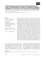
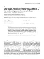
EMBRYONIC ERYTHROPOIESIS
In vertebrates, embryonic hematopoiesis involves primitive
and definitive steps [1–3] (Fig. 1). Primitive, large nucleated
erythroblasts that synthesize embryonic globin forms arise in
blood islands that emerge from extraembryonic mesoderm in
the yolk sac, at murine embryonic day 7.5 (E7.5) or day 15–18
in humans [4,5]. Definite hematopoiesis is established in the
fetal liver beginning at mouse E9.5; it is multilineage,
generating well-defined erythrocytes that synthesize adult
forms of globin and become enucleated, as well as myeloid,
megakaryocyte and lymphoid cells [6]. It is generally believed
to initiate in the aorto-gonad-mesonephros (AGM) region
[7], though a recent study suggests that the yolk sac is the
predominant source of both primitive and definitive hema-
topoietic progenitors [4]. Hematopoietic progenitors migrate
through the blood stream to seed the fetal liver. Late in fetal
life, bone marrow assumes hematopoietic activity and
becomes the predominant hematopoietic organ in postnatal
life [4,8]. Both embryonic and adult erythropoiesis require
broad spectrum as well as erythroid transcription factors.
Figure 1 presents the plethora of these factors within the
context of the hematopoietic process.
BROAD SPECTRUM FACTORS
Stem cell leukemia (SCL)
Originally identified in a chromosomal translocation in
T-cell acute lymphoblastic leukemia (ALL), the stem cell
leukemia (SCL) gene on chromosome 1p32–33 encodes a
basic helix-loop-helix (bHLH) transcription factor [9,10].
SCL binds E-box (CAGGTG) DNA elements as a
heterodimer in complex with E12/E47, the bHLH alternat-
ively spliced products of the E2A gene [11,12]. It also
participates in a DNA-bound complex containing the
transcription factors E12/E47, GATA-1, Ldb-1 and
LMO2 [13].
SCL is detected in early hematopoietic progenitors and in
more mature megakaryocytes, erythroid and mast cells as
Correspondence to H. Soreq, Department of Biological Chemistry,
The Institute of Life Sciences, The Hebrew University of Jerusalem,
91904, Israel.
Fax: + 972 2 6520258, Tel.: + 972 2 6585109,
E-mail:
Abbreviations: ALL, acute lymphoblastic leukemia; AGM, aorto-
gonad-mesonephros; BFU-E, erythroid burst forming unit; CFU-E,
erythroid colony forming unit; ES, embryonic stem; SCL, Stem cell
leukemia; Epo, erythropoietin; FOG, friend of GATA; EKLF,
erythroid Kruppel-like factor; BKLF, basic Kruppel-like factor;
AChE, acetylcholinesterase; LCR, locus control region. HRD,
hematopoietic regulatory domain; CF and NF, C-terminal and the
N-terminal zinc-fingers, respectively; HS, hypersensitive domains; Rb,
retinoblastoma; Stat, signal transducer and activator of transcription;
HERF1, hematopoietic RING finger 1.
Definitions: embryonic age is written as Ex,wherex represents the
number of days post-conception.
(Received 14 March 2002, revised 2 May 2002, accepted 16 May 2002)
Eur. J. Biochem. 269, 3607–3618 (2002) Ó FEBS 2002 doi:10.1046/j.1432-1033.2002.02999.x
well as in the mesencephalon, metencephalon, embryonic
skeleton, endothelial cells and neurons [14–16]. Its expres-
sion increases during erythroid differentiation, where it
evokes enhanced proliferation and differentiation. SCL
confers proliferation advantage while repressing differenti-
ation in myeloid progenitors, and is absent from most
mature myeloid and lymphoid cells [17].
SCL null mice die in utero at about E8.5, showing no
evidence of blood formation. SCL null embryonic stem (ES)
cells fail to give rise to any hematopoietic lineage, suggesting
that SCL is crucial for primitive hematopoiesis/erythropoi-
esis [18,19]. Clonogenic assays show failure in myelopoiesis,
pointing at SCLs critical role in very early hematopoietic
differentiation [11].
LIM-only protein 2 (LMO2)
Also known as Rbtn2 and TTG2, LMO2 includes two
cysteine-rich LIM domains homologous to the DNA
binding domain of GATA transcription factors. Localized
to chromosome 11p13, the LMO2 gene is involved in the
11;14 translocation of childhood T cell ALL [20]. Highest
LMO2 expression levels are found in hematopoietic tissues
[21]. LMO2 does not bind DNA by itself, but acts as a
bridge between DNA-binding transcription factors such as
SCL and GATA-1. Over half of the erythroid LMO2
protein associates with SCL [11,13]. LMO2 null mice die
around E9 of severe anemia, with lack of any yolk sac
hematopoiesis, identifying an essential role for LMO2 in
early hematopoiesis (Table 1) [22,23]. However, LMO2
may also participate in the lineage-specific mechanisms that
regulate erythropoiesis, as it takes part in an erythroid
transcription-activation complex, together with SCL, E2A,
GATA-1 and Lbd1. The complex recognizes an E box motif
approximately one helix turn (10 bp) upstream from a
GATA site. The GATA-1 gene itself includes sites promo-
ting formation of this multimeric erythroid complex [13,24].
LMO2 on its own, like SCL and GATA-1, had little
effect on developing Xenopus embryos. However, ectopic
coexpression of LMO2, SCL1 and GATA-1 in Xenopus
embryos enlarged the ventral blood islands at the expense of
dorsal mesoderm (muscle and notochord) embryogenesis
[25]. Ectopic expression of LMO2 in Xenopus pole explants
treated with basic fibroblast growth factor (bFGF) resulted
in erythroid differentiation and extensive globin gene
expression. LMO2, SCL1 and GATA-1 overexpression in
activin-treated Xenopus pole explants further increased the
production of hemoglobinized cells. This suggests the
Fig. 1. Embryonic erythropoiesis. Shown are developmental stages in primitive and definitive hematopoiesis up to erythroid commitment.
3608 C. Perry and H. Soreq (Eur. J. Biochem. 269) Ó FEBS 2002
Table 1. Effects of Hematopoietic/Erythroid transcription factors.
Gene Motifs
Phenotype of
genomic disruption
Lethality
(mouse
embryonic day) Overexpression
DNA binding
sequence Reference
SCL bHLH Bloodless mice-absence of yolk sac E8.5 Myeloid proliferation; reduced differentiation response CAGGTG [19]
hematopoiesis Erythroid proliferation and differentiation (E box)
LMO2 LIM domain Bloodless mice-absence of yolk sac
hematopoiesis E8.5–9 Erythroid differentiation and globin gene expression in
Xenopus pole explants, not in whole embryo
– [22]
GATA-2 Zinc finger Decreased embryonic erythrocytes
(primitive and definitive);
poorly proliferating multipotent
progenitors; absence of mast cells
E10-11 Promotes proliferation and blocks erythroid
differentiation in erythroid precursors
T/AGATAA/G [16]
c-Myb Helix- Normal primitive but E15 Inhibits erythroid differentiation TAACGG [11,95,96]
turn-helix severely impaired
Leucine- zipper definitive erythropoiesis
region
GATA-1 Zinc finger Ablated embryonic erythropoiesis due to E11.5 Promotes megakaryocytic differentiation in an early T/AGATAA/G [28,36]
blocked maturation at proerythroblasts myeloid cell line
(with apoptosis); arrested megakaryocyte
development (with hyperproliferation)
FOG Zinc finger Blocked erythroid maturation at E12.5 Inhibits red cell formation and maturation in whole – [42]
proerythroblasts; ablated
megakaryocytopoiesis
Xenopus embryos (mFOG2); represses GATA-1-
induced activity (FOG1)
EKLF Zinc finger Severe anemia; b-globin
deficiency
E16 Earlier switch from fetal to adult type globin;
enhanced differentiation and hemoglobinization,
reduced proliferation (in EKLF null cells)
CACC
GC rich
[56]
BKLF Zinc finger Myeloproliferative disorder – – CACC [51]
Stat5 – Transient anemia due to fetal liver – – TTCC(A > T)GGAA [77,97]
erythroid progenitors apoptosis
at E13.5; mild anemia,
exacerbated by stress, at adult life
PU.1 Winged helix-turn-helix Blocked erythropoiesis (relieved by GATA-1) [74]
Fli-1 Winged helix-
turn-helix
Inhibited erythroid differentiation, impaired
ability to respond to specific erythroid inducers
and reduced levels of GATA-1
[69]
Ó FEBS 2002 Transcriptional regulation of erythropoiesis (Eur. J. Biochem. 269) 3609
formation of synergistic multiprotein complexes that pro-
mote red cell formation and differentiation during embryo-
genesis, in addition to SCL and LMO2s crucial role in
early hematopoiesis [25]. A pentameric complex of LMO2,
SCL, E2A, Lbd1 and pRb was shown to repress gene
expression in erythroblasts [26], likely counteracting tran-
scriptional activation to limit erythroid differentiation [25].
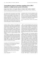
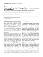
Figure 2 presents a scheme of the erythroid transcription-
activation complex along promoters of erythropoietically
active genes.
GATA-2
All members of the GATA family of transcription factors
contain two homologous zinc-finger domains and bind to
the DNA GATA-consensus sequence (T/AGATAA/G),
present in regulatory elements of many erythroid genes [e.g.
globins, band 3, EKLF, FOG, erythropoietin receptor
(EpoR) and heme biosynthetic enzymes] [27,28].
GATA-2, a member of the GATA family, is expressed in
hematopoietic and ES cells and endothelial cells. Its forced
expression in erythroid precursors promotes proliferation
and blocks erythroid differentiation [11]. Expression of
GATA-2 precedes that of another family member, GATA-1,
and must decrease as GATA-1 expression increases to
enable erythroid differentiation. GATA-2 null mice are
embryonic lethal, due to severe anemia during the early
phase of yolk sac hematopoiesis (E10–11) [16]. The most
pronounced decrease occurs in the frequency of primitive
and definitive erythroid and mast cell colonies, differenti-
ating from GATA-2 null ES cells. Multipotential progen-
itors arising from GATA-2 null ES cells proliferate poorly
and undergo excessive apoptosis [11,16], suggesting that
GATA-2 is essential for appropriate expansion and survival
of early hematopoietic cells, at the expense of differenti-
ation.
The proto-oncogene c-Myb
c-Myb is abundantly expressed in immature hematopoietic
cells of erythroid, myeloid and lymphoid lineages but
decreases as they differentiate. Moreover, its forced expres-
sion inhibits erythroid differentiation [11]. c-Myb is required
for early definitive cellular expansion and, like GATA-2, it
needs to be downregulated to allow terminal differentiation
[11,29].
c-Myb null mice exhibit normal primitive but severely
impaired definitive hematopoiesis, resulting in death at E15.
Mature circulating definitive erythrocytes as well as other
lineage progenitors are decreased, while megakaryocytes,
granulocytes and monocytes appear to be normal.
The v-Myb gene, transduced by avian myeloblastosis
virus (AMV), is an oncogene that specifically blocks
terminal differentiation in macrophage precursors, activates
their self-renewal capacities and determines the commitment
of progenitors to macrophages while suppressing develop-
ment of other lineages [30,31]. This specificity, distinct from
the multilineage effects of c-Myb, likely reflects the loss of
some c-Myb functions due to deletions and point mutations.
The macrophage precursor-restricted activity of v-Myb
resides in its leucine-zipper region (LZR), mutation of which
enables v-Myb to affect uncommitted progenitors, support-
ing development of erythroid cells, granulocytes and
megakaryocytes [32]. v-Myb induces myeloid factors
(PU.1, C/EBP), while the v-Myb mutant induces SCL and
GATA-1 in transformed blastoderm cells [32]. The c-Myb
C-terminus can interact with its own N-terminus [33], likely
affecting LZR accessibility for myeloid factors, activating
myeloid-specific genes. Inaccessible Myb-LZR might favor
formation of c-Myb complexes with erythroid factors,
activating erythroid-specific genes. This molecular switch
thus directs hematopoietic progenitors into lineage-specific
development [32].
ERYTHROID TRANSCRIPTION FACTORS
GATA-1
The GATA-1 gene, located on chromosome Xp11.23 [34], is
expressed in erythroid cells, megakaryocytes, eosinophils,
mast cells and Sertoli cells in the testis [35]. GATA-1 null
mice show complete ablation of embryonic erythropoiesis
due to arrested maturation and apoptosis of erythroid
precursors at the proerythroblast stage [36], supporting its
key role in erythroid commitment (Table 1). These mice
also present blocked megakaryocyte development in mid-
maturation and die by E11.5. However, GATA-1-negative
ES cells can develop into other hematopoietic lineages.
Forced expression of GATA-1 in an early myeloid cell line
promotes megakaryocytic differentiation, suggesting
that GATA-1 may affect both lineage selection and late
erythroid maturation [11,37,38].
GATA-1 is expressed as two distinct transcripts in
hematopoietic cells and in the testis, directed by different
first exons/promoters. The coding exons are common to
both transcripts [28].
In primitive erythroid cells, GATA-1 expression is
regulatedbya5¢ enhancer, whereas its expression in
definitive erythroid cells requires an additional element
located in the first intron. Together, these two elements form
the GATA-1 locus hematopoietic regulatory domain
(HRD) [28].
Fig. 2. The erythroid transcription-activation complex. SCL and E47 bind an E box (CAGGTA), about 10 bp upstream from a GATA motif.
LMO2 and Lbd1 bridge between SCL1 and GATA-1. However, GATA-1 binds DNA more commonly in a nonspecific orientation, with FOG as
its cofactor.
3610 C. Perry and H. Soreq (Eur. J. Biochem. 269) Ó FEBS 2002
The C-terminal and the N-terminal zinc-fingers (CF and
NF, respectively) in GATA-1 are required for recognition of
the GATA motif and DNA binding as well as for physical
interaction with other transcription factors. The highly
conserved NF is essential for interaction with the GATA-1
coactivator FOG (Friend Of GATA) as well as with EKLF,
LMO2 and C/REB binding protein (CBP), and enhances
the specificity and stability of binding of the two-finger
DNA binding domains to palindromic GATA recognition
sequences [27,39]. CF is indispensable for GATA-1 func-
tion, while NF is indispensable for definitive but not for
primitive erythropoiesis. This suggests that different
GATA-1 functional domains are required for target gene
activation in primitive and definitive erythropoiesis [28].
Thus, both transcriptional regulatory elements and protein
functional domains may ensure proper lineage specification
in primitive and definitive erythropoiesis.
GATA motifs may appear by themselves or occur in a
specific orientation from an E box motif. Thus, the genomic
orientation of GATA motifs and their neighboring
sequences bears important functional implications. It has
been speculated that GATA-1 binds isolated GATA motifs
in a nonspecific orientation, in which FOG is the cofactor.
In addition, GATA-1 binds GATA-E box elements, in
which SCL and other components cooperate with GATA-1
(Fig. 2) [27]. For example, the pentameric erythroid tran-
scription-activation complex includes SCL and E12/E47
that binds an E-box, about 10 bp upstream from a GATA
motif, as well as LMO2 and Lbd1 bridging between SCL1
and GATA-1 [13].
GATA-3 is normally restricted to lymphoid precursors
and committed T cells. Its overexpression in murine
hematopoietic stem cells arrests proliferation, induces
erythroid and megakaryocyte differentiation and inhibits
development of myeloid and lymphoid precursors. This
apparent functional redundancy among the GATA proteins
suggests that lineage determination by individual GATA
proteins is developmental-stage dependent [40].
Friend of GATA (FOG)
FOG is a complex zinc-finger protein. It associates with
GATA-1 NF through at least one of its nine fingers (usually
finger 6). FOG is coexpressed with GATA-1 in fetal liver,
embryonic erythroblasts, mast cells, megakaryocytes and
adult spleen [41] and cooperates with GATA-1 to promote
erythroid and megakaryocytic differentiation. Mutated
GATA-1 that is unable to interact with FOG, fails to
support terminal erythroid maturation due to deregulated
expression of multiple GATA-1 target genes, such as the
a-andb-globins and band 3, but not EKLF or FOG itself
[27]. FOG does not modulate GATA-1 DNA binding
specificity, or activation properties. Rather, it recruits
additional nuclear factors, perhaps via its other fingers.
Mice lacking FOG exhibit blocked erythripoiesis, similar
to GATA-1-deficient mice. However, the NF domain,
which mediates GATA)1 interactions with coactivators
such as FOG, was found to be dispensable in primitive
erythropoiesis. Therefore, FOGs contribution to primitive
erythropoiesis appears to be independent of GATA-1. FOG
null mice also display ablation of the megakaryocytic
lineage, unlike loss of GATA-1 which blocks megakaryo-
cyte development in midmaturation. This points at addi-
tional, GATA-1 independent role of FOG during the
earliest stages of megakaryocyte development. Thus, the
early, independent functions of FOG differ from its later,
GATA-1 dependent role during erythroid and megakaryo-
cyte maturation [42].
FOG may also function as a repressor. A FOG homo-
logue in Drosophila, u-shaped, was found to repress the
action of a GATA-like factor, pannier. A second mamma-
lianFOG,mFOG2,isexpressedinheart,neuronsand
gonads in the adult with somewhat broader expression
during embryogenesis [43]. Ectopic expression of mFOG2
inhibits red cell formation and maturation in intact Xenopus
embryos and reduces xGATA-1 and xSCL levels in ventral
marginal zone explants, while xGATA-2 levels remain
unchanged [43,44] (Table 1). In murine erythroleukemia
cells, FOG1 represses the GATA-1-induced activity of the
transferrin receptor-2 (TfR-2)-promoter [45].
A Xenopus FOG homologue, xFOG, contains a short
peptide motif (PIDLSK), which is highly conserved among
FOG proteins and mediates interactions with the transcrip-
tion corepressor CtBP [46]. In Xenopus embryos, FOG2
with a mutated CtBP binding site stimulated red cell
formation dramatically [44], although, knock-in mice
expressing a FOG1 variant, which is unable to bind CtBP
have normal erythropoiesis [47]. It was suggested that
FOG:GATA-1 complexes may repress transcription of
GATA-2, which promotes progenitor proliferation over
differentiation in committed erythroblasts, limiting the
number of cells with erythropoietic fate and preventing
depletion of pluripotent stem cells. In the absence of FOG,
GATA-1 might fail to shut off GATA-2 transcription and
erythropoiesis might be stalled at a blast-phase. Once cells
are committed, FOG may cooperate with GATA-1 in
erythroid maturation.
Familial X-linked dyserythropoietic anemia due to a
substitution of methionine for valine at residue 205, in a
highly conserved region of GATA-1 NF, interrupts the
GATA)1:FOG1 interaction and inhibits the ability of
GATA-1 to rescue erythroid differentiation in a GATA-1
deficient erythroid cell line [48]. This results in severe fetal
anemia and anemia with severe thrombocytopenia at birth
and thereafter, as well as cryptorchidism, in the male
offspring. The substitution Ser208 fi Gly or Gly218 fi Asp
in GATA-1 NF domain, was reported in families with
recessive X-linked thrombocytopenia and X-linked macro-
thrombocytopenia, respectively. The replaced residues are
involved in GATA-1:FOG1 direct interactions and the
mutation partially disrupts this interaction [49,50]. Table 2
lists these mutations and their clinical consequences, which
together confirm the vital role played by specific domains in
the corresponding transcription factors during in vivo
erythroid and megakaryocyte development.
Erythroid Kruppel-like factor (EKLF)
This zinc finger protein plays an essential role in the
regulation of b-globin gene expression [51].
The b-globin locus regulation has recently been exten-
sively reviewed [52–54]. The b-globin gene is part of the
globin cluster, the genes of which are arranged in the order
of their expression during development. Regulation of the
b-globin tissue- and developmental stage-specific expression
ismediatedbyitspromoteraswellasbydistalregulatory
Ó FEBS 2002 Transcriptional regulation of erythropoiesis (Eur. J. Biochem. 269) 3611
sequences, the most prominent of which is the locus control
region (LCR). The LCR consists of several DNase 1
hypersensitive domains (HS sites). In erythroid cells, where
the gobin genes are transcriptionally active, the locus shows
higher DNase 1 sensitivity, indicating an open and access-
ible chromatin structure (euchromatin). Tissue- and stage-
specific expression of the various globin genes is determined
by the interactions between the LCR and the specific globin
gene promoters, interactions mediated by recruiting chro-
matin modifying, coactivators and transcription complexes
[52].
EKLF expression is remarkably restricted to erythroid,
megakaryocytic and mast cells [55]. The human EKLF gene
was located to chromosome 19p13, a region deleted in some
cases of human erythroleukemia [56].
Human EKLF encodes a 362 residues protein that
includes three C
2
H
2
type zinc fingers at its C-terminus. It
shares 69% overall identity and 93% identity with the
three C-terminal zinc finger domains of mouse EKLF.
Each finger includes three key amino acids that form
sequence-specific contacts with three DNA residues. The
N-terminal of the protein is rich in proline and acidic
residues [57].
EKLF, like other members of the Kruppel family, binds a
CACC consensus-sequence in regulatory elements of many
erythroid-specific genes, including adult b-globin, often
closely spaced from a GATA site (Fig. 2). GATA proteins
interact physically and functionally with Kruppel-like
proteins to regulate gene expression [58].
Competition assays show that EKLF favors binding to
the human and murine adult type of b-globin CACC
element over the CACC elements in the murine fetal bh1-
globin, human c-globin or the erythropoietin receptor
(EpoR) gene promoters. Naturally occurring adult type
b-globin CACC box mutations result in reduced b-globin
expression and b-thalassemia, due to poor EKLF binding
(Table 2) [57,59].
EKLF null mice die before E16 of severe anemia and
b-globin deficiency. Embryonic erythropoiesis and embry-
onic e and f globin genes expression is normal [60],
demonstrating the pivotal role of EKLF in the activation
of the adult b-globin gene in the late stages of erythropoiesis.
Overexpressing EKLF induces an earlier switch from fetal
to adult type globin [61]. EKLF deficient mice that carry a
complete copy of the human b-globin locus display elevated
levels of the human fetal c-globin mRNA, in addition to
b-globin deficiency (Table 1). Elevated fetal type c-globin
levels, in adult life, were reported in carriers of point
mutations within the b-globin promoter CACC box [57,59].
This may indicate a role for EKLF in silencing c-globin
expression, or in the c-tob-globin switching process.
EKLF activation of the b-globin gene is dramatically
enhanced in the presence of the DNase 1 HS2 of the gene
LCR [62]. Within the LCR, EKLF was found to activate
HS3 directly. One model for the globin chromatin opening
proposes that factor binding at HS3 initiates the process,
allowing the spreading of open chromatin, binding of other
trans-acting factors throughout the LCR, and looping out
intervening DNA to establish the LCR holocomplex [53]. A
protein complex that can activate transcription of a
chromatin-assembled b-globin, in an EKLF-dependent
fashion, was purified and named EKLF coactivator
remodeling complex-1 (E-RC1) [63]. This suggests that the
function of EKLF as an activator of transcription is to
attract the complex to the b-globin promoter.
Reintroducing EKLF into an EKLF-null erythroid cell
line, which harbors a copy of the human b-globin locus,
resulted in enhanced differentiation and hemoglobinization,
as well as reduced proliferation. This may point to a role for
EKLF in cell cycle regulation and hemoglobinization, in
addition to regulation of b-globin gene expression [64].
J2E cells transfected with antisense EKLF cDNA show
normal proliferation but reduced expression of b-globin and
two rate-limiting heme synthesis enzymes as well as defective
hemoglobinization in response to erythropoietin stimulation
[65]. This may suggest EKLF regulation of other genes
involved in hemoglobin synthesis.
Basic Kruppel-like factor (BKLF)
The BKLF protein is found in erythroid cells, fibroblasts
and brain. It binds CACC motifs through three highly
conserved C-terminal Kruppel-like zinc fingers and interacts
with the corepressor CtBP to repress EKLF promoter
activation in vitro [46,66]. BKLF erythroid expression
depends on EKLF, so that EKLF deficient mice express
significantly reduced levels of BKLF in erythroid cells and
normal BKLF levels in the brain [66].
Table 2. Translocated or mutated transcription factor genes in human pathologies.
Gene Motif Pathology Molecular background
SCL bHLH T cell acute lymphoblastic
leukemia (ALL)
t1; 14, t1; 3, t1; 5, t1; 7
LMO2 LIM domain Childhood T cell ALL t11; 14
GATA-1 Zinc finger Familial dyserythropoietic
anemia (with cryptorchidism)
V205M at GATA)1 (interrupting
interaction with FOG)
Recessive X-linked
thrombocytopenia
G208S at GATA)1 (interrupting
interaction with FOG)
EKLF and
target genes
Zinc finger b-Thalassemia b-globin promoter CACC box
mutations
Erythroleukemia del 19p13
SHP-1
(BKLF-activated?)
Polycythemia vera SHP-1 is down-regulated in
CFU-E; hematopoietic
progenitor hyper-susceptible to
growth factors?
3612 C. Perry and H. Soreq (Eur. J. Biochem. 269) Ó FEBS 2002
EKLF null mice express elevated levels of fetal globins,
perhaps due to missing EKLF upregulation of BKLF in
erythroid cells. This suggests that BKLF represses the
expression of embryonic and fetal globin genes, both of
which contain a CACC box in their promoters [55].
BKLF deficient mice display a myeloproliferative disorder
and an overall phenotype that resembles that of mice
mutated for the protein tyrosine phosphatase SHP-1,
suggesting a role for BKLF in regulation of SHP-1
expression [55]. SHP-1 is expressed in erythroid progenitors,
and is downregulated during terminal differentiation. It
inactivates complexes of growth factors and their receptors,
including factors known to guide proliferation and differen-
tiation in erythroid progenitors. Polycytemia vera is a clonal
myeloproliferative disorder, leading to hyperproliferation of
erythroid, myeloid and megakaryocytic cells. Sixty percent
of polycytemia vera patients have diminished expression of
SHP-1 in CFU-E populations (Table 2), suggesting that
repression of this inactivator of growth factor complexes
renders the hematopoietic progenitors in polycytemia vera
patients more susceptible to growth effects [67].
Neptune
and other KLF family members
Neptune,aXenopus member of the Kruppel-like factor
(KLF) family of zinc-finger transcription factors, can bind
CACC as well as GC-rich DNA elements. Neptune shares
91% of its sequence, at the nuclear localization signal and
zinc finger region, with another family member, the gut
KLF-GKLF, and 76% with EKLF [68].
Neptune appears at sites of primitive erythropoiesis prior
to xGATA-1. It is expressed in the ventral blood islands, in
cells committed to primitive erythropoiesis, cranial ganglia
and hatching and cement glands, as well as in peripheral red
blood cells and spleen.
Neptune specifically binds to CACC elements in the
promoters of both embryonic and adult mouse b-globin
genes, with minimal binding to CACC elements in the fetal
c-globin gene promoter. Similarly to EKLF, neptune
activates the human b-globin promoter and cooperates
with xGATA-1 to enhance globin induction in animal cap
explants, though by itself it fails to induce globin produc-
tion. Globin gene regulation by xGATA-1 depends on
neptune function in ventral marginal zones and animal caps,
both sites of primitive erythropoiesis [68].
biklf, the zebrafish ortholog of neptune, is required for
erythroid cell differentiation. biklf is expressed in the
hatching gland and in the zabrafish homologue of the
Xenopus ventral blood islands. Repressing biklf expression
in zebrafish embryos results in embryonic anemia, sup-
pressed expression of the embryonic globin and inhibition of
GATA-1 expression, demonstrating conservation of func-
tion during vertebrate evolution [69].
FKLF (human Fetal KLF) [70] activates embryonic (e)
globin expression, and to a lesser extent the fetal (c) globin
genes, through its interaction with these genes’ CACC
boxes, but fails to activate other CACC box-containing
erythroid genes.
Murine FKLF-2 increases c-globin expression 100-fold.
It activates the promoters of e-andb-globins, GATA-1 and
heme synthesis enzyme genes to a much lower degree [71].
Thus, all globin genes contain CACC boxes in their
regulatory domains, yet FKLF, FKLF-2 and EKLF
activate the embryonic, fetal and adult globin genes,
respectively.
A four-step model for human b globin gene regulation
has been suggested [52]; the first step involves partial
unfolding of globin chromatin structure and generation of
highly accessible LCR. It is mediated by erythroid-specific
proteins, which bind to sequences throughout the globin
locus. GATA-1, which is known to associate with histone
acetyl-transferases, may be involved in this step. The
disruption of the LCR chromatin structure allows binding
of transcription factors such as EKLF and other KLF
family members, GATA family members and the HLH
proteins to the LCR HS sites, and the recruitment of
chromatin-remodeling complexes and coactivators. In the
third step, chromatin domains permissive for transcription
are being established. Intergenic transcription was suggested
to modify chromatin structure of an active gene domain,
distinguishing it from an accessible but inactive one, that
way separating the globin gene into developmental stage-
specific chomatin domains. Finally, transcription complexes
are being transferred from the LCR to individual glo-
bin gene promoters within transcriptionally permissive
domains, allowing the developmental stage-specific pattern
of globin gene expression.
The
Fli-1
oncogene
A member of the Ets family of transcription factors, Fli-1,
was identified in Friend virus-induced erythroleukemia and
affects the self-renewal of erythroid progenitor cells [72]. In
pluripotent human hematopoietic cells, differentiation is
followed by reduced Fli-1 expression and over expressing
Fli-1 inhibits erythroid differentiation, impairs the cells’
ability to respond to specific erythroid inducers, such as
hemin, and reduces the levels of GATA-1 [73].
In the erythroblastic cell line, HB60, Fli-1 expression is
downregulated by erythropoietin (Epo), which induces
terminal erythroid differentiation. Constitutive expression
of Fli-1 blocks Epo-induced differentiation and enhances
cell proliferation in HB60 cells, suggesting that Fli-1 targets
erythroid cells to either proliferation or differentiation, in
response to Epo [74].
Fli-1 binds a cryptic Ets consensus site within the
retinoblastoma (Rb) gene promoter, repressing Rb expres-
sion, which results in impaired terminal erythroid matur-
ation and continuous presence of nucleated erythrocytes in
peripheral blood [75]. Negative regulation of Rb by Fli-1
could destine erythroid progenitors to self-renewal, while
Epo-induced repression of Fli-1 expression will enable
differentiation [74].
PU.1
The putative oncogene Spi-1 (PU.1) protein product is a
hematopoietic-specific Ets factor, promoting differentiation
of lymphoid and myeloid lineages [76]. PU.1 expression in
erythroid progenitors can induce erythroleukemia in mice.
Like Fli-1, PU.1 blocks erythroid differentiation and
restoration of terminal erythroid differentiation in murine
erythroleukemia (MEL) cells requires PU.1 suppression
[77,78].
PU.1 can interact directly with GATA-1 and repress
GATA-1 mediated transcriptional activation. Both the
Ó FEBS 2002 Transcriptional regulation of erythropoiesis (Eur. J. Biochem. 269) 3613
PU.1 DNA binding domain and transactivation domain are
required for GATA-1 suppression and for blocking terminal
differentiation in MEL cells. PU.1 does not seem to affect
binding of other factors, such as FOG, to GATA-1, nor
does it prevent GATA-1 DNA binding [78]. It is likely that
PU.1 binds to assembled, DNA-bound GATA-1 complexes
and represses their activity.
Ectopic expression of PU.1 in Xenopus embryos blocks
erythropoiesis. Exogenous GATA-1 is able to relieve this
blockage of erythroid differentiation in MEL cells as well as
in Xenopus embryos and explants, suggesting that lineage
commitment decisions are regulated by their relative levels
[78].
PU.1 can also bind to GATA-2 and EKLF, in vitro.As
both PU.1 and GATA-2 are capable of blocking terminal
erythroid differentiation, it is possible that these factors
cooperate to stimulate self-renewal in early erythroid
progenitors.
Fli-1, known to block erythroid differentiation and
suppress GATA-1 expression, was identified as a PU.1
target gene [73,79].
Signal transducer and activator of transcription
(Stat) 5
Epo binding to its receptor (EpoR) leads to rapid activation
of the transcription factor Stat5. Tyrosine phosphorylation
of EpoR-bound Stat5 dimerizes the complex and translo-
cates it to the nucleus, where it can induce the immediate
early expression of the antiapoptotic gene bcl-x. Stat5
confers an antiapoptotic effect over erythroid cell lines;
repressing stat5 expression increases apoptosis and inhibits
growth of fetal liver erythroid precursors [80,81].
Decreased bcl-x expression and increased apoptosis
in early erythroblasts suggests that Stat5 and bcl-x
mediate the Epo antiapoptotic effect on erythroid pro-
genitors [81]. Stat5a- and Stat5b-deficient mice are severely
anemic due to decreased survival of fetal liver erythroid
progenitors and show a marked increase in apoptosis at
E13.5, when fetal liver cells are cultured with Epo. This is
consistent with Stat5 mediating an Epo-dependent anti-
apoptotic effect in fetal erythroid progenitors [81]. The
anemia resolves during adult life in about half of Stat5-
mutated mice, which then have near-normal hematocrit.
However, they are deficient in generating high erythro-
poietic response to hemolysis-induced stress and have
persistent anemia despite compensatory expansion of
their erythropoietic tissue, with erythroblasts failing to
differentiate.
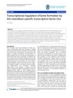
Hematopoietic RING finger 1 (HERF1)
During the initial development of definitive hematopoietic
progenitors, the expression of HERF1 coincides with the
appearance of definitive erythropoiesis. In adult mice, it is
restricted to erythroid cells. Inhibition of HERF1 expression
blocks terminal erythroid differentiation, whereas its over-
expression induces erythroid maturation in MEL cells [82].
This suggests that HERF1 may have a role in the
development of mature erythroid cells. Figure 3 lists some
Fig. 3. Differentiation of committed erythroid progenitors. Shown are the transcription factors that affect erythrocyte precursors through their
differentiation into erythroblasts. The exerted effects are marked in brackets.
3614 C. Perry and H. Soreq (Eur. J. Biochem. 269) Ó FEBS 2002
of these key transcriptional regulators of the erythropoietic
process and notes at least part of their multi-element
interactions during erythroid differentiation.
DOWNSTREAM TARGET GENES
Transcriptional regulation of erythropoietin
Epo, a glycoprotein hormone, is not a transcription factor
but activates intracellular signaling through binding to its
receptor, EpoR. This stimulation upregulates the expression
of globins, transferrin receptor and some membrane pro-
teins that are characteristic of erythrocytes. It enhances the
viability and maturation of erythroid progenitor cells, while
Epo deprivation results in increased apoptosis [83]. Epo null
mice die at E13.5 of severe anemia, when primitive
erythroblasts die and are not being replaced by definitive
erythropoiesis, accompanied by a dramatic increase in cell
death. All this points at Epo’s major contribution to the
survival, proliferation and differentiation of definitive
erythroid progenitors [84].
The primary regulator of Epo expression in late fetal and
postnatal life is oxygen tension. A hypoxia sensing mech-
anism results in activation of the transcription factor
Hypoxia inducible factor (HIF)1, which binds a 3¢ enhancer
of the Epo gene, initiating its expression [85]. The mouse
Epo 3¢ enhancer contains a DR2 element, a direct repeat of
the hexameric sequence TGACC(C/T), adjacent to the
HIF1 binding site. Coupled HIF1–DR2 sequences augment
hypoxic induction of Epo gene reporter constructs, prob-
ably through hepatocyte nuclear factor (HNF)4 [84,86].
During early erythropoiesis, the Epo gene is a direct
transcriptional target of the retinoic acid receptor RXRa.
Mouse embryos lacking RXRa are deficient in erythroid
differentiation. Their Epo mRNA levels are reduced at
E10.25 and E11.25 but can be induced by retinoic acid.
The Epo gene enhancer was found to contain a DR2
element. DR2 represents a retinoic acid receptor binding
site and a retinoic acid receptor transcriptional response
element [84].
Surprisingly, the erythropoietic deficiency in RXRa null
mice is transient. Epo is expressed at normal levels by E12.5
and erythropoiesis reaches normal levels by E14.5. HNF4,
abundant in fetal liver hepatocytes, was shown to compete
with RXRa for binding to the Epo gene enhancer DR2
element. Thus, Epo expression may be regulated by RXRa
during early fetal erythropoiesis and then by HNF4 activity,
a transition that may be responsible for switching the
regulation of Epo expression from paracrine, retinoic acid
control to hypoxic, HNF4-related control [84].
Acetylcholinesterase, a potential hematopoiesis/
erythropoiesis regulator
A case study for a downstream regulator may be that of
acetylcholinesterase (AChE). Primarily known to hydrolyze
acetylcholine at brain synapses and neuromuscular junc-
tions, its extended biological roles involve contribution to
cell proliferation and differentiation in multiple tissues
(reviewed in [87]). These include sites of hematopoiesis and
osteogenesis, both known to share a common progenitor, as
well as different tumor types [88–91]. One of the alternat-
ively spliced transcripts of AChE, the ÔreadthroughÕ isoform
(AChE-R), which is known to be upregulated in response to
psychological and chemical stress, is induced by cortisol
in CD
34+
hematopoietic progenitor cells. This cortisol-
induced expression of AchE-R correlates with hematopoi-
etic expansion, perhaps implying a role for AChE in bone
marrow adaptive responses to stress [92].
AChE is also expressed in immature human megakaryo-
cytes, where it is surprisingly localized to the nucleus [93].
Induction of differentiation in human megakaryoblasts
suppresses AChE expression, as was reported for GATA-1
[93,94]. Transient suppression of ACHE gene expression in
mouse hematopoietic multipotential progenitors, using an
antisense oligonucleotide, induced AChEmRNA overex-
pression, followed by cell expansion and suppressed apop-
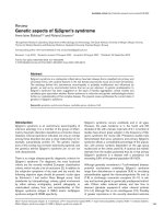
tosis [95]. Consensus DNA binding sites for hematopoietic
transcription factors are extremely abundant along the three
known regulatory domains in the ACHE locus. Among
others, they include E2 and CACC boxes, glucocorticoid
response elements and consensus binding sites for GATA-1,
C/EBP and Stat5 [89,96–98]. Binding sites for the LMO2
complex with adjacent GATA-1 (though not 10 bp apart)
and KLF motifs are found in the upstream enhancer,
proximal promoter and intronic enhancer of the ACHE
locus, suggesting multileveled control over its hematopoietic
expression (Fig. 4).
All this suggests that AChE may be either a downstream
target for hematopoietic and/or erythroid-specific transcrip-
tion factors or, in view of its surprising nuclear localization,
that it is a transcription modifier by itself, affecting fate-
determining crossroads. The apparent regulatory role of
AChE in hematopoietic proliferation and differentiation at
early developmental stages may be accompanied by a
capacity for inducing proliferation at later, erythroid-
commited stages, as was shown in megakaryoblasts [93].
Finally, this is a promising candidate for involvement with
stress responses that induce erythropoietic development
In conclusion, erythropoiesis is a highly complex process
that is regulated by a finely tuned combination of
transcription factors in a stage-specific and context-depend-
ent manner. Several key characteristics of transcriptional
regulation of erythropoiesis may be pointed out:
Fig. 4. Erythroid transcription factor binding sites across the ACHE
locus. Depicted is the reverse sequence of the cosmid inset (accession
no. AF002993) including the ACHE gene and 22 kb of its upstream
sequence. Exons (numbered above) and introns (numbered below) are
marked. Arrows designate positions of the ACHE regulatory domains:
distal enhancer domain (D.D), the proximal promoter (P.P) and the
intronic enhancer (I.E), along the cosmid reverse sequence (nt 22 465
being the ACHE transcription start site). Consensus binding sites for
the noted transcription factors are represented by wedges. LMO2
Complex ¼ LMO2 associated with DNA-bound SCL1-E47 and able
to bridge binding to DNA-bound GATA-1 in the erythroid tran-
scription-activation complex (see Fig. 2).
Ó FEBS 2002 Transcriptional regulation of erythropoiesis (Eur. J. Biochem. 269) 3615
A single transcription factor may exert different effects on
cell fate when expressed at different developmental stages.
For example, SCL exerts a self-renewal, proliferative effect
when expressed in early progenitors, but induces differen-
tiation when expressed in more mature cells. Expression of a
specific transcription factor at different developmental
stages may also modulate lineage-commitment, and facili-
tate interactions with different partner proteins.
An intriguing compromise or antagonism between pro-
liferation and differentiation emerges at several stages of the
erythropoietic process. Determination of cell fate depends
not only on the ability to express certain pro-differentiation
factors, but also on the ability to repress other survival/
proliferation-inducing transcription factors (GATA-2,
c-Myb, PU.1, Fli-1) at developmental crossroads. Both of
these abilities are essential for erythroid differentiation.
Genomic orientation and neighboring sequences may
have functional implications for the interactions among the
transcription factors.
Many of the erythropoietic transcription factors are
complex, multidomain proteins. Different domains of the
same protein may be required to activate various target
genes at different developmental stages. Therefore, the
interplay among the various transcription factors involving
their stage of expression, type of cells expressing them, the
combination of factors present at a certain time point and
the multidomain structure of many of these factors variegate
the complex regulation of erythropoiesis.
ACKNOWLEDGEMENTS
Chava Perry MD, is the incumbent of a basic research fellowship from
the Israel Ministry of Health. The study was supported by the US Army
Medical Research and Materiel Command (DAMD 17-99-1-9547) and
by Ester Neuroscience Ltd.
REFERENCES
1. Palis, J. & Segel, G.B. (1998) Developmental biology of ery-
thropoiesis. Blood Rev. 12, 106–114.
2. Migliaccio, A.R. & Migliaccio, G. (1998) The making of an ery-
throid cell. Molecular control of hematopoiesis. Biotherapy 10,
251–268.
3. Dame, C. & Juul, S.E. (2000) The switch from fetal to adult ery-
thropoiesis. Clin. Perinatol. 27, 507–526.
4. Palis, J., Robertson, S., Kennedy, M., Wall, C. & Keller, G. (1999)
Development of erythroid and myeloid progenitors in the yolk sac
andembryoproperofthemouse.Development 126, 5073–5084.
5. Orkin, S.H. (1996) Development of the hematopoietic system.
Curr. Opin. Genet Dev. 6, 597–602.
6. Dzierzak, E. & Medvinsky, A. (1995) Mouse embryonic hema-
topoiesis. Trends Genet. 11, 359–366.
7. Dzierzak, E., Medvinsky, A. & de Bruijn, M. (1998) Qualitative
and quantitative aspects of haematopoietic cell development in the
mammalian embryo. Immunol. Today. 19, 228–236.
8. Medvinsky, A. & Dzierzak, E. (1996) Definitive hematopoiesis is
autonomously initiated by the AGM region. Cell 86, 897–906.
9. Hershfield, M.S., Kurtzberg, J., Harden, E., Moore, J.O., Whang-
Peng,J.&Haynes,B.F.(1984)Conversionofastemcellleukemia
from a T-lymphoid to a myeloid phenotype induced by the ade-
nosine deaminase inhibitor 2¢-deoxycoformycin. Proc. Natl Acad.
Sci. USA 81, 253–257.
10. Begley, C.G., Visvader, J., Green, A.R., Aplan, P.D., Metcalf, D.,
Kirsch, I.R. & Gough, N.M. (1991) Molecular cloning and
chromosomal localization of the murine homolog of the
human helix-loop-helix gene SCL. Proc. Natl Acad. Sci. USA 88,
869–873.
11. Shivdasani, R.A. & Orkin, S.H. (1996) The transcriptional control
of hematopoiesis. Blood 87, 4025–4039.
12. Hsu, H.L., Wadman, I. & Baer, R. (1994) Formation of in vivo
complexes between the TAL1 and E2A polypeptides of leukemic
Tcells.Proc.NatlAcad.Sci.USA91, 3181–3185.
13. Wadman, I.A., Osada, H., Grutz, G.G., Agulnick, A.D.,
Westphal, H., Forster, A. & Rabbitts, T.H. (1997) The LIM-only
protein Lmo2 is a bridging molecule assembling an erythroid,
DNA-binding complex which includes the TAL1, E47, GATA-1
and Ldb1/NLI proteins. EMBO J. 16, 3145–3157.
14. Green,A.R.,Lints,T.,Visvader,J.,Harvey,R.&Begley,C.G.
(1992) SCL is coexpressed with GATA-1 in hemopoietic cells but
isalsoexpressedindevelopingbrain.Oncogene 7, 653–660.
15. Drake, C.J., Brandt, S.J., Trusk, T.C. & Little, C.D. (1997) TAL1/
SCL is expressed in endothelial progenitor cells/angioblasts and
defines a dorsal-to-ventral gradient of vasculogenesis. Dev. Biol.
192, 17–30.
16. Shivdasani, R.A. (1997) Stem cell transcription factors. Hematol.
Oncol. Clin. North Am. 11, 1199–1206.
17. Begley, C.G. & Green, A.R. (1999) The SCL gene: from case
report to critical hematopoietic regulator. Blood 93, 2760–2770.
18. Porcher, C., Swat, W., Rockwell, K., Fujiwara, Y., Alt, F.W. &
Orkin, S.H. (1996) The T cell leukemia oncoprotein SCL/tal-1 is
essential for development of all hematopoietic lineages. Cell 86,
47–57.
19. Shivdasani, R.A., Mayer, E.L. & Orkin, S.H. (1995) Absence of
blood formation in mice lacking the T-cell leukaemia oncoprotein
tal-1/SCL. Nature 373, 432–434.
20. Rabbitts, T.H. (1998) LMO T-cell translocation oncogenes typify
genes activated by chromosomal translocations that alter
transcription and developmental processes. Genes Dev. 12,
2651–2657.
21. Foroni, L., Boehm, T., White, L., Forster, A., Sherrington, P.,
Liao, X.B., Brannan, C.I., Jenkins, N.A., Copeland, N.G. &
Rabbitts, T.H. (1992) The rhombotin gene family encode related
LIM-domain proteins whose differing expression suggests multi-
ple roles in mouse development. J. Mol. Biol. 226, 747–761.
22. Warren, A.J., Colledge, W.H., Carlton, M.B., Evans, M.J., Smith,
A.J. & Rabbitts, T.H. (1994) The oncogenic cysteine-rich LIM
domain protein rbtn2 is essential for erythroid development. Cell
78, 45–57.
23. Yamada, Y., Warren, A.J., Dobson, C., Forster, A., Pannell, R. &
Rabbitts, T.H. (1998) The T cell leukemia LIM protein Lmo2 is
necessary for adult mouse hematopoiesis. Proc. Natl Acad. Sci.
USA 95, 3890–3895.
24. Vyas, P., McDevitt, M.A., Cantor, A.B., Katz, S.G., Fujiwara, Y.
& Orkin, S.H. (1999) Different sequence requirements for
expression in erythroid and megakaryocytic cells within a reg-
ulatory element upstream of the GATA-1 gene. Development 126,
2799–2811.
25. Mead, P.E., Deconinck, A.E., Huber, T.L., Orkin, S.H. & Zon,
L.I. (2001) Primitive erythropoiesis in the Xenopus embryo: the
synergistic role of LMO-2, SCL and GATA-binding proteins.
Development 128, 2301–2308.
26. Vitelli, L., Condorelli, G., Lulli, V., Hoang, T., Luchetti, L.,
Croce, C.M. & Peschle, C. (2000) A pentamer transcriptional
complex including tal-1 and retinoblastoma protein down-
modulates c-kit expression in normal erythroblasts. Mol. Cell.
Biol. 20, 5330–5342.
27. Crispino, J.D., Lodish, M.B., MacKay, J.P. & Orkin, S.H. (1999)
Use of altered specificity mutants to probe a specific protein–
protein interaction in differentiation: the GATA-1: FOG complex.
Mol. Cell. 3, 219–228.
28. Shimizu, R., Takahashi, S., Ohneda, K., Engel, J.D. & Yama-
moto, M. (2001) In vivo requirements for GATA-1 functional
3616 C. Perry and H. Soreq (Eur. J. Biochem. 269) Ó FEBS 2002
domains during primitive and definitive erythropoiesis. EMBO J.
20, 5250–5260.
29. Mucenski, M.L., McLain, K., Kier, A.B., Swerdlow, S.H.,
Schreiner,C.M.,Miller,T.A.,Pietryga,D.W.,Scott,W.J.Jr&
Potter, S.S. (1991) A functional c-myb gene is required for normal
murine fetal hepatic hematopoiesis. Cell 65, 677–689.
30. Ganter, B. & Lipsick, J.S. (1999) Myb and oncogenesis. Adv.
Cancer Res. 76, 21–60.
31. Lipsick, J.S. & Wang, D.M. (1999) Transformation by v-Myb.
Oncogene 18, 3047–3055.
32. Karafiat, V., Dvorakova, M., Pajer, P., Kralova, J., Horejsi, Z.,
Cermak,V.,Bartunek,P.,Zenke,M.&Dvorak,M.(2001)The
leucine zipper region of Myb oncoprotein regulates the commit-
ment of hematopoietic progenitors. Blood 98, 3668–3676.
33. Dash, A.B., Orrico, F.C. & Ness, S.A. (1996) The EVES motif
mediates both intermolecular and intramolecular regulation of
c-Myb. Genes Dev. 10, 1858–1869.
34. Zon, L.I., Tsai, S.F., Burgess, S., Matsudaira, P., Bruns, G.A.
& Orkin, S.H. (1990) The major human erythroid DNA-
binding protein (GF-1): primary sequence and localization of the
gene to the X chromosome. Proc.NatlAcad.Sci.USA87, 668–
672.
35. Yamamoto, M., Takahashi, S., Onodera, K., Muraosa, Y. &
Engel, J.D. (1997) Upstream and downstream of erythroid tran-
scription factor GATA-1. Genes Cells. 2, 107–115.
36. Fujiwara, Y., Browne, C.P., Cunniff, K., Goff, S.C. & Orkin, S.H.
(1996) Arrested development of embryonic red cell precursors in
mouse embryos lacking transcription factor GATA-1. Proc. Natl
Acad.Sci.USA93, 12355–12358.
37. Visvader, J.E., Elefanty, A.G., Strasser, A. & Adams, J.M. (1992)
GATA-1 but not SCL induces megakaryocytic differentiation in
an early myeloid line. EMBO J. 11, 4557–4564.
38. Shivdasani, R.A., Fujiwara, Y., McDevitt, M.A. & Orkin, S.H.
(1997) A lineage-selective knockout establishes the critical role of
transcription factor GATA-1 in megakaryocyte growth and pla-
telet development. EMBO J. 16, 3965–3973.
39. Trainor, C.D., Omichinski, J.G., Vandergon, T.L., Gronenborn,
A.M.,Clore,G.M.&Felsenfeld,G.(1996)Apalindromicreg-
ulatory site within vertebrate GATA-1 promoters requires both
zinc fingers of the GATA-1 DNA-binding domain for high-affi-
nity interaction. Mol. Cell. Biol. 16, 2238–2247.
40. Chen, D. & Zhang, G. (2001) Enforced expression of the GATA-3
transcription factor affects cell fate decisions in hematopoiesis.
Exp. Hematol. 29, 971–980.
41. Tsang, A.P., Visvader, J.E., Turner, C.A. & Fujiwara, Y., YuC.,
Weiss, M.J., Crossley, M. & Orkin, S.H. (1997) FOG, a multitype
zincfingerprotein,actsasacofactorfortranscriptionfactor
GATA-1 in erythroid and megakaryocytic differentiation. Cell 90,
109–119.
42. Tsang, A.P., Fujiwara, Y., Hom, D.B. & Orkin, S.H. (1998)
Failure of megakaryopoiesis and arrested erythropoiesis in mice
lacking the GATA-1 transcriptional cofactor FOG. Genes Dev. 12,
1176–1188.
43. Tevosian, S.G., Deconinck, A.E., Cantor, A.B., Rieff, H.I.,
Fujiwara, Y., Corfas, G. & Orkin, S.H. (1999) FOG-2: a novel
GATA-family cofactor related to multitype zinc-finger proteins
Friend of GATA-1 and U-shaped. Proc. Natl Acad. Sci. USA 96,
950–955.
44. Deconinck, A.E., Mead, P.E., Tevosian, S.G., Crispino, J.D.,
Katz, S.G., Zon, L.I. & Orkin, S.H. (2000) FOG acts as a
repressor of red blood cell development in Xenopus. Development
127, 2031–2040.
45. Kawabata,H.,Germain,R.S.,Ikezoe,T.,Tong,X.,Green,E.M.,
Gombart, A.F. & Koeffler, H.P. (2001) Regulation of expression
of murine transferrin receptor 2. Blood 98, 1949–1954.
46. Turner, J. & Crossley, M. (1998) Cloning and characterization of
mCtBP2, a co-repressor that associates with basic Kruppel-like
factor and other mammalian transcriptional regulators. EMBO J.
17, 5129–5140.
47. Katz, S.G., Cantor, A.B. & Orkin, S.H. (2002) Interaction
between FOG-1 and the corepressor C-terminal binding protein is
dispensable for normal erythropoiesis in vivo. Mol. Cell. Biol. 22,
3121–3128.
48. Nichols, K.E., Crispino, J.D., Poncz, M., White, J.G., Orkin,
S.H., Maris, J.M. & Weiss, M.J. (2000) Familial dyserythropoietic
anaemia and thrombocytopenia due to an inherited mutation in
GATA1. Nat Genet. 24, 266–270.
49. Freson, K., Devriendt, K., Matthijs, G., Van Hoof, A., De Vos,
R.,Thys,C.,Minner,K.,Hoylaerts,M.F.,Vermylen,J.&Van
Geet, C. (2001) Platelet characteristics in patients with X-linked
macrothrombocytopenia because of a novel GATA1 mutation.
Blood 98, 85–92.
50. Mehaffey,M.G.,Newton,A.L.,Gandhi,M.J.,Crossley,M.&
Drachman, J.G. (2001) X-Linked thrombocytopenia caused by a
novel mutation of GATA-1. Blood 98, 2681–2688.
51. Miller, I.J. & Bieker, J.J. (1993) A novel, erythroid cell-specific
murine transcription factor that binds to the CACCC element and
is related to the Kruppel family of nuclear proteins. Mol. Cell.
Biol. 13, 2776–2786.
52. Levings, P.P. & Bungert, J. (2002) The human beta-globin locus
control region. Eur. J. Biochem. 269, 1589–1599.
53. Ho, P.J. & Thein, S.L. (2000) Gene regulation and deregulation: a
beta globin perspective. Blood Rev. 14, 78–93.
54. Engel, J.D. & Tanimoto, K. (2000) Looping, linking, and chro-
matin activity: new insights into beta-globin locus regulation. Cell
100, 499–502.
55. Turner, J. & Crossley, M. (1999) Basic Kruppel-like factor func-
tions within a network of interacting haematopoietic transcription
factors. Int. J. Biochem. Cell Biol. 31, 1169–1174.
56. Olopade, O.I., Thangavelu, M., Larson, R.A., Mick, R., Kowal-
Vern, A., Schumacher, H.R., Le Beau, M.M., Vardiman, J.W. &
Rowley, J.D. (1992) Clinical, morphologic, and cytogenetic
characteristics of 26 patients with acute erythroblastic leukemia.
Blood 80, 2873–2882.
57. Perkins, A. (1999) Erythroid Kruppel like factor: from fishing
expedition to gourmet meal. Int. J. Biochem. Cell Biol. 31, 1175–
1192.
58. Merika, M. & Orkin, S.H. (1995) Functional synergy and physical
interactions of the erythroid transcription factor GATA-1 with the
Kruppel family proteins Sp1 and EKLF. Mol. Cell. Biol. 15, 2437–
2447.
59. Huisman, T.H. (1997) Levels of Hb A2 in heterozygotes and
homozygotes for beta-thalassemia mutations: influence of muta-
tions in the CACCC and ATAAA motifs of the beta-globin gene
promoter. Acta Haematol. 98, 187–194.
60. Perkins, A.C., Sharpe, A.H. & Orkin, S.H. (1995) Lethal beta-
thalassaemia in mice lacking the erythroid CACCC- transcription
factor EKLF. Nature 375, 318–322.
61. Tewari, R., Gillemans, N., Wijgerde, M., Nuez, B., von
Lindern, M., Grosveld, F. & Philipsen, S. (1998) Erythroid
Kruppel-like factor (EKLF) is active in primitive and
definitive erythroid cells and is required for the function of
5¢HS3 of the beta-globin locus control region. EMBO J. 17, 2334–
2341.
62. Donze, D., Townes, T.M. & Bieker, J.J. (1995) Role of erythroid
Kruppel-like factor in human gamma- to beta-globin gene
switching. J. Biol. Chem. 270, 1955–1959.
63. Armstrong, J.A., Bieker, J.J. & Emerson, B.M. (1998) A SWI/
SNF-related chromatin remodeling complex, E-RC1, is required
for tissue-specific transcriptional regulation by EKLF in vitro. Cell
95, 93–104.
64. Coghill, E., Eccleston, S., Fox, V., Cerruti, L., Brown, C.,
Cunningham, J., Jane, S. & Perkins, A. (2001) Erythroid Kruppel-
like factor (EKLF) coordinates erythroid cell proliferation and
Ó FEBS 2002 Transcriptional regulation of erythropoiesis (Eur. J. Biochem. 269) 3617
hemoglobinization in cell lines derived from EKLF null mice.
Blood 97, 1861–1868.
65. Spadaccini, A., Tilbrook, P.A., Sarna, M.K., Crossley, M., Bieker,
J.J. & Klinken, S.P. (1998) Transcription factor erythroid Kruppel-
like factor (EKLF) is essential for the erythropoietin-induced
hemoglobin production but not for proliferation, viability, or
morphological maturation. J. Biol. Chem. 273, 23793–23798.
66. Crossley, M., Whitelaw, E., Perkins, A., Williams, G., Fujiwara,
Y. & Orkin, S.H. (1996) Isolation and characterization of the
cDNA encoding BKLF/TEF-2, a major CACCC-box-binding
protein in erythroid cells and selected other cells. Mol. Cell. Biol.
16, 1695–1705.
67. Wickrema,A.,Chen,F.,Namin,F.,Yi,T.,Ahmad,S.,Uddin,S.,
Chen,Y.H.,Feldman,L.,Stock,W.,Hoffman,R.&Platanias,
L.C. (1999) Defective expression of the SHP-1 phosphatase in
polycythemia vera. Exp. Hematol. 27, 1124–1132.
68. Huber, T.L., Perkins, A.C., Deconinck, A.E., Chan, F.Y., Mead,
P.E. & Zon, L.I. (2001) neptune, a Kruppel-like transcription
factor that participates in primitive erythropoiesis in Xenopus.
Curr. Biol. 11, 1456–1461.
69. Kawahara, A. & Dawid, I.B. (2001) Critical role of biklf in ery-
throid cell differentiation in zebrafish. Curr. Biol. 11, 1353–1357.
70. Asano, H., Li, X.S. & Stamatoyannopoulos, G. (1999) FKLF, a
novel Kruppel-like factor that activates human embryonic and
fetal beta-like globin genes. Mol. Cell. Biol.19, 3571–3579.
71. Asano, H., Li, X.S. & Stamatoyannopoulos, G. (2000) FKLF-2: a
novel Kruppel-like transcriptional factor that activates globin and
other erythroid lineage genes. Blood 95, 3578–3584.
72. Howard, J.C., Yousefi, S., Cheong, G., Bernstein, A. & Ben-
David, Y. (1993) Temporal order and functional analysis of
mutations within the Fli-1 and p53 genes during the ery-
throleukemias induced by F-MuLV. Oncogene 8, 2721–2729.
73. Athanasiou, M., Mavrothalassitis, G., Sun-Hoffman, L. & Blair,
D.G. (2000) FLI-1 is a suppressor of erythroid differentiation in
human hematopoietic cells. Leukemia 14, 439–445.
74. Tamir, A., Howard, J., Higgins, R.R., Li, Y.J., Berger, L.,
Zacksenhaus, E., Reis, M. & Ben-David, Y. (1999) Fli-1, an Ets-
related transcription factor, regulates erythropoietin- induced
erythroid proliferation and differentiation: evidence for direct
transcriptional repression of the Rb gene during differentiation.
Mol. Cell. Biol. 19, 4452–4464.
75. Lee, E.Y., Chang, C.Y., Hu, N., Wang, Y.C., Lai, C.C., Herrup,
K., Lee, W.H. & Bradley, A. (1992) Mice deficient for Rb are
nonviable and show defects in neurogenesis and haematopoiesis.
Nature 359, 288–294.
76.Scott,E.W.,Simon,M.C.,Anastasi,J.&Singh,H.(1994)
Requirement of transcription factor PU.1 in the development of
multiple hematopoietic lineages. Science 265, 1573–1577.
77. Ben-David, Y. & Bernstein, A. (1991) Friend virus-induced
erythroleukemia and the multistage nature of cancer. Cell 66,
831–834.
78. Rekhtman, N., Radparvar, F., Evans, T. & Skoultchi, A.I. (1999)
Direct interaction of hematopoietic transcription factors PU.1 and
GATA-1: functional antagonism in erythroid cells. Genes Dev. 13,
1398–1411.
79. Starck,J.,Doubeikovski,A.,Sarrazin,S.,Gonnet,C.,Rao,G.,
Skoultchi, A., Godet, J., Dusanter-Fourt, I. & Morle, F. (1999)
Spi-1/PU.1 is a positive regulator of the Fli-1 gene involved in
inhibition of erythroid differentiation in friend erythroleukemic
cell lines. Mol. Cell. Biol. 19, 121–135.
80. Bromberg, J. & Darnell, J.E. Jr (2000) The role of STATs
in transcriptional control and their impact on cellular function.
Oncogene 19, 2468–2473.
81. Socolovsky, M., Fallon, A.E., Wang, S., Brugnara, C. & Lodish,
H.F. (1999) Fetal anemia and apoptosis of red cell progenitors in
Stat5a
–/–
5b
–/–
mice: a direct role for Stat5 in Bcl-X (L) induction.
Cell 98, 181–191.
82. Harada, H., Harada, Y., O’Brien, D.P., Rice, D.S., Naeve, C.W.
& Downing, J.R. (1999) HERF1, a novel hematopoiesis-specific
RING finger protein, is required for terminal differentiation of
erythroid cells. Mol. Cell. Biol. 19, 3808–3815.
83. Kendall, R.G. (2001) Erythropoietin. Clin.Lab.Haematol.23,
71–80.
84. Makita, T., Hernandez-Hoyos, G., Chen, T.H., Wu, H.,
Rothenberg, E.V. & Sucov, H.M. (2001) A developmental tran-
sition in definitive erythropoiesis: erythropoietin expression is
sequentially regulated by retinoic acid receptors and HNF4. Genes
Dev. 15, 889–901.
85. Semenza, G.L., Nejfelt, M.K., Chi, S.M. & Antonarakis, S.E.
(1991) Hypoxia-inducible nuclear factors bind to an enhancer
element located 3¢ to the human erythropoietin gene. Proc. Natl
Acad. Sci. USA 88, 5680–5684.
86. Bunn, H.F., Gu, J., Huang, L.E., Park, J.W. & Zhu, H. (1998)
Erythropoietin: a model system for studying oxygen-dependent
gene regulation. J. Exp. Biol. 201, 1197–1201.
87. Soreq, H. & Seidman, S. (2001) Acetylcholinesterase – new roles
for an old actor. Nat. Rev. Neurosci. 2, 294–302.
88. Massoulie, J., Pezzementi, L., Bon, S., Krejci, E. & Vallette, F.M.
(1993) Molecular and cellular biology of cholinesterases. Prog.
Neurobiol. 41, 31–91.
89. Grisaru, D., Lev-Lehman, E., Shapira, M., Chaikin, E., Lessing,
J.B.,Eldor,A.,Eckstein,F.&Soreq,H.(1999)Human
osteogenesis involves differentiation-dependent increases in the
morphogenically active 3¢ alternative splicing variant of acetyl-
cholinesterase. Mol. Cell. Biol. 19, 788–795.
90. Majumdar, M.K., Thiede, M.A., Mosca, J.D., Moorman, M. &
Gerson, S.L. (1998) Phenotypic and functional comparison of
cultures of marrow-derived mesenchymal stem cells (MSCs) and
stromal cells. J. Cell Physiol. 176, 57–66.
91. Karpel, R., Ben Aziz-Aloya, R., Sternfeld, M., Ehrlich, G.,
Ginzberg,D.,Tarroni,P.,Clementi,F.,Zakut,H.&Soreq,H.
(1994) Expression of three alternative acetylcholinesterase mes-
senger RNAs in human tumor cell lines of different tissue origins.
Exp. Cell Res. 210, 268–277.
92. Grisaru, D., Deutch, V., Shapira, M., Pick, M., Sternfeld, M.,
Melamed-Book, N., Kaufer, D., Galyam, N., Gait, M., Owen, D.,
Lessing, J., Eldor, A. & Soreq, H. (2001) ARP, a peptide derived
from the stress-associated acetylcholinesterase variant has hema-
topoietic growth promoting activities. Mol. Med. 7, 93–105.
93. Lev-Lehman, E., Deutsch, V., Eldor, A. & Soreq, H. (1997)
Immature human megakaryocytes produce nuclear-associated
acetylcholinesterase. Blood 89, 3644–3653.
94. Dai, W. & Murphy Jr, M.J. (1993) Downregulation of GATA-1
expression during phorbol myristate acetate-induced mega-
karyocytic differentiation of human erythroleukemia cells. Blood
81, 1214–1221.
95. Soreq,H.,Patinkin,D.,Lev-Lehman,E.,Grifman,M.,Ginzberg,
D., Eckstein, F. & Zakut, H. (1994) Antisense oligonucleotide
inhibition of acetylcholinesterase gene expression induces pro-
genitor cell expansion and suppresses hematopoietic apoptosis
ex vivo. Proc. Natl Acad. Sci. USA 91, 7907–7911.
96. Shapira, M., Tur-Kaspa, I., Bosgraaf, L., Livni, N., Grant, A.D.,
Grisaru,D.,Korner,M.,Ebstein,R.P.&Soreq,H.(2000)A
transcription-activating polymorphism in the ACHE promoter
associated with acute sensitivity to anti-acetylcholinesterases.
Hum. Mol. Genet. 9, 1273–1281.
97. Chan, R.Y., Boudreau-Lariviere, C., Angus, L.M., Mankal, F.A.
& Jasmin, B.J. (1999) An intronic enhancer containing an N-box
motif is required for synapse- and tissue-specific expression of the
acetylcholinesterase gene in skeletal muscle fibers. Proc. Natl Acad.
Sci. USA 96, 4627–4632.
98. Atanasova, E., Chiappa, S., Wieben, E. & Brimijoin, S. (1999)
Novel messenger RNA and alternative promoter for murine
acetylcholinesterase. J. Biol. Chem. 274, 21078–21084.
3618 C. Perry and H. Soreq (Eur. J. Biochem. 269) Ó FEBS 2002