Surgical Complications in Oral Implantology: Etiology, Prevention, and Management doc
Bạn đang xem bản rút gọn của tài liệu. Xem và tải ngay bản đầy đủ của tài liệu tại đây (8.81 MB, 17 trang )
Surgical complicationS
in oral implantology
Etiology, Prevention, and Management
Louie Al-Faraje,
dds
Founder and Director
California Implant Institute
San Diego, California
With contributions by
James L. Rutkowski,
dmd, p
h
d
Christopher Church,
md
Quintessence Publishing Co, Inc
Chicago, Berlin, Tokyo, London, Paris, Milan, Barcelona,
Istanbul, Moscow, New Delhi, Prague, São Paulo, and Warsaw
Dedication ix
Contributors x
Preface xi
Acknowledgments xii
Part I Identifying Preoperative Conditions at Could Lead to
Complications
1 Inadequate or Excessive Vertical Restorative Space 2
2 Inadequate Horizontal Restorative Space 5
3 Limited Jaw Opening and Interarch Distance 10
4 Inadequate Alveolar Width for Optimal Buccolingual Positioning 11
5 Maxillary and Mandibular Tori 16
Part II Intraoperative Complications in Implant Placement
6 Incorrect Implant Angulation 20
7 Malalignment 24
8 Nerve Injury 25
9 Irregular or Narrow Alveolar Crest 30
10 Extensive Resorption of the Mandible 32
11 Curved Extraction Socket 33
12 Injury to Adjacent Teeth During Implant Placement 35
13 Preoperative Acute and Chronic Infections at the Implant Site 37
14 Retained Root Tips in the Implant Site 40
15 Bleeding 42
16 Overheating of the Bone During Drilling 49
17 Stripping of the Implant Site 51
18 Sinus Floor Perforation 52
19 Nasal Floor Perforation 56
20 Accidental Partial or Complete Displacement of Dental Implants into the Maxillary Sinus 58
21 Accidental Displacement of Dental Implants into the Maxillary Incisive Canal 60
22 Deep Implant Placement 62
23 Shallow Implant Placement 75
24 Complications in Flapless Implant Placement 77
25 Aspiration or Ingestion of Foreign Objects 80
26 Mandibular Bone Fracture 81
27 Implant Fracture 83
28 Excessive Torque During Insertion and Compression Necrosis 85
29 Inadequate Initial Stability 87
CONTENTS
Complications
Complications
Part III Postoperative Complications
30 Postoperative Pain 96
31 Tissue Emphysema Induced by Dental Procedures 99
32 Incision Line Reopening 100
33 Cover Screw Exposure During the Healing Period 10 5
34 Bone Growth over the Cover Screw 106
35 Soft Tissue Growth Between Implant Platform and Cover Screw 107
36 Bone Loss or Thread Exposure During the Healing Period 108
37 Implant Mobility During Stage-Two Surgery 114
38 Implant Periapical Lesion (IPL) and Retrograde Peri-implantitis 116
39 Cement Left in the Pocket 118
40 Radiotherapy, Osteoradionecrosis, and Dental Implants 123
41 Shallow Vestibule Secondary to Ridge Augmentation 125
42 Medicolegal Issues 127
Part IV Complications Associated with Lateral Window Sinus Elevation
Preoperative Complications
43 Preoperative Acute Sinusitis 135
44 Preoperative Chronic Sinusitis 136
45 Preoperative Fungal Sinusitis 138
46 Preoperative Cystic Structures and Mucoceles 140
47 Other Preoperative Sinus Lesions 142
Intraoperative Complications
48 Hematoma During Anesthesia 15 2
49 Bleeding During Incision and Flap Reflection 152
50 Bleeding During Osteotomy 153
51 Damage to Adjacent Dentition 153
52 Perforation of the Sinus Membrane During Osteotomy 153
53 Perforation of the Sinus Membrane During Elevation 154
54 Incomplete Elevation 161
55 Bleeding During Membrane Elevation 162
56 Fracture of the Residual Alveolar Ridge 162
57 Excessive Elevation of the Membrane 162
58 Presence of a Mucus Retention Cyst 163
59 Blockage of the Maxillary Ostium 164
60 Unstable Implants 164
Early Postoperative Complications
61 Wound Dehiscence 164
62 Acute Graft Infection/Sinusitis 165
63 Exposure of the Bone Graft and/or Barrier Membrane 166
64 Sinus Congestion 166
66 Early Implant Migration into the Sinus Cavity 166
Complications
Late Postoperative Complications
66 Insufficient Quality and/or Quantity of Healed Graft 167
67 Implant Failure in the Augmented Sinus 167
68 Chronic Infection/Sinusitis 168
69 Infection of All Paranasal Sinuses/Intracranial Cavity 169
70 Delayed Implant Migration into the Sinus Cavity 169
71 Sinus Aspergillosis 169
Part V Pharmacology: Prevention and Management of Pain, Infection, and
Drug-Related Complications
72 Intra- and Postoperative Infection 175
73 Intra- and Postoperative Pain 184
74 Bisphosphonate-Related Osteonecrosis of the Jaw 193
75 Bleeding Problems in Patients Taking Anticoagulants or Antiplatelet Agents 195
Appendices
A Implant Treatment Protocol 202
B Consent Forms 209
C Postoperative Instructions 225
Index 2 27
Complications
ix
A pioneer in all fields of surgery, Al-Zahrawi conceived and developed innumerable surgical techniques
and instruments and, in 1000 CE, published the first surgical encyclopedia, Kitab Al Tasrif (The Method
of Medicine), which spanned 30 volumes. For his monumental accomplishments and contributions to
surgery, he earned the title Father of Modern Surgery. His way of thinking and his practice of surgery
inspired many subsequent surgeons to achieve greatness and provided a beacon of light in the dark
ages of Europe. In his many papers and manuals, he describes various operations and procedures that
had never before been recorded. He wrote detailed descriptions of many surgical techniques, including
cautery and wound management. Some have described him as the first plastic surgeon, notably for his
attention to and methods of incision and use of silk thread suture to achieve good cosmesis. He devised
about 200 surgical instruments, among them the surgical needle, scalpels, curettes, retractors, spoons,
sounds, hooks, rods, and specula.
The street in Córdoba where his house still stands is named Calle Abulcasis in his memory. In 1977,
the Spanish Tourist Board commemorated it in his honor with a bronze plaque that reads: “This was the
house where lived Abu al-Qasim Al-Zahrawi.”
DEDICATION
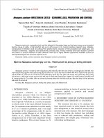
Page from a 1531 Latin translation by Peter Argellata of Al-
Zahrawi’s treatise on surgical and medical instruments.
To Abu al-Qasim Al-Zahrawi (aka Abulcasis), 936–1013 CE
x
CONTRIBUTORS
Christopher Church,
md
Director
Loma Linda Sinus and Allergy Center
Associate Professor
Department of Otolaryngology–Head and Neck Surgery
Loma Linda University School of Medicine
Loma Linda, California
James L. Rutkowski,
dmd, p
h
d
Clinical Instructor
Department of Restorative Dentistry
School of Dental Medicine
University at Buffalo
The State University of New York
Buffalo, New York
Private Practice
Clarion, Pennsylvania
xi
The use of dental implants to restore missing teeth has steadily increased over the past three decades.
It is perhaps not surprising, then, that the number of implant-related complications has grown as well.
Numerous clinical studies involving dental implants have revealed encouraging outcomes; however,
there is an element of risk associated with all clinical procedures, and these encouraging results may
have given rise to unrealistic expectations. Despite careful planning, there is always a potential for surgi-
cal complications. Nevertheless, carrying out routine tasks with care and attention, choosing minimally
invasive techniques when indicated, recognizing evidence of a developing problem, and giving prompt
attention will reduce postoperative complications.
The successful outcome of any surgical procedure requires attention to a series of patient-related and
procedure-dependent parameters. Sound knowledge of surgical anatomy and experience and training
in the fundamentals of internal medicine are important prerequisites for predictable implant surgery.
Also, adequate presurgical planning, appropriate quality and quantity of available bone, a well-executed
surgical technique, good primary stability, a sufficient healing period, and detailed postoperative in-
structions are all factors that play a vital role in the success of dental implant surgery and osseointegra-
tion. Aging, changing health conditions, wear and tear, and inadequate professional maintenance are
important variables influencing prognosis.
This book is designed as a self-instruction guide to the diagnosis and management of surgery-related
complications and to the development of a protocol that allows for the early detection of potential surgi-
cal complications and how to avoid them. It is a well-documented fact that early detection of complica-
tions that are amenable to rescue therapies may reverse the fate of a failing implant or bone grafting
procedure.
The evidence-based methods of complications management described in this book are not meant to
preclude the clinical judgment of experienced clinicians but rather should be applied to either support
or prompt them to rethink their chosen methods of therapy on the basis of existing evidence.
PREFACE
xii
ACKNOWLEDGMENTS
I would like to express sincere gratitude to my parents, Omar Al-Faraje and Nadia Al-Rifai, for their ex-
traordinary sacrifices for too many years. Thank you for your unconditional love and support.
Also to my wife Rana, my lifelong companion and “book widow.” Her support was invaluable as I
was hunched over my computer, sometimes for 12 hours a day. And to our children—Nadia, Omar, and
Tim—who contributed immensely to this book by sacrificing their precious time with “Papa.”
In addition, I would like to thank my teachers at each of the medical institutions I attended. I was
indeed fortunate to have had outstanding anatomical, clinical, and surgical training at the medical
institutes in Russia, the Ukraine, and the United States.
Three special individuals have profoundly influenced my career:
Dr Nizar Al-Tair, my dental mentor, who spent countless hours challenging my knowledge and skills
to deliver excellence and to be my best.
Dr Igor Persidsky, who taught me how to connect patients’ medical problems with their dental needs
and to think like a dental surgeon with internal medicine in mind. I treasure our years of friendship.
Dr Dewhirst Floyd, who gave me a helping hand and believed in me. This book would not have seen
the light without his support during my early years in the dental field.
Finally, I would like to thank all of my students at the California Implant Institute. It is always a pleasure
and an honor to share with you my knowledge and expertise in implant dentistry. For the last few years,
my greatest professional joy has been interacting with my students and colleagues at the California
Implant Institute.
I also would like to express my gratitude to Dr Christopher Church and Dr James Rutkowski for their
contributions to this textbook.
Special thanks to Lisa Bywaters and her editorial team at Quintessence Publishing Company. Their tre-
mendous support throughout the project allowed the creation of this modern, easy-to-consult textbook.
PART 1
Identifying Preoperative
Conditions at Could
Lead to Complications
Complications
1 Inadequate or Excessive Vertical Restorative Space
2 Inadequate Horizontal Restorative Space
3 Limited Jaw Opening and Interarch Distance
4 Inadequate Alveolar Width for Optimal Buccolingual Positioning
5 Maxillary and Mandibular Tori
Buccolingual angulation
Endosseous root-form implants distribute occlusal loads most
effectively when forces are applied in an axial direction. An an-
gulation of 15 degrees or less is considered acceptable. Even
natural teeth are not straight, but rather perpendicular to the
curve of Wilson, the lateral curve of the occlusal table formed
by the inclination of the posterior teeth (Fig 2-1). However, as
implant angulation approaches or exceeds 25 degrees, the
supporting bone is severely compromised through transmis-
sion of occlusal forces (Fig 2-2a). Moreover, if an implant is in-
clined buccolingually and the prosthetic reconstruction is off-
set relative to the implant head for improved occlusion and/or
esthetics, the inclination will introduce a bending moment on
the implant and will lead to a few potential problems.
20
Intraoperative Complications in Implant Placement
PART
2
COMPLICATION 6
Incorrect Implant Angulation
The implant must be angulated correctly in the buccolingual and mesiodistal planes for optimum function and esthetics.
Off-axis loading
Potential biomechanical problems of an excessive lingual tra-
jectory (see Fig 2-2a) include:
• Restoration fracture
• Retaining screw fracture
• Abutment fracture
• Implant body fracture
• Osseous destruction because of unfavorable loading
• Plaque accumulation under ridge lap pontics
Placement of an overly inclined implant is not an accept-
able practice, especially for single-unit restorations. If it is not
possible to place an implant with an angulation of 15 degrees
or less, the treatment plan should be aborted and the implant
placed in a different location, or implant placement should
be delayed and the area grafted using techniques such as
guided bone regeneration (GBR), block grafting (Fig 2-2b),
or ridge splitting, to allow optimum buccolingual angulation
(Fig 2-2c).
Right
maxillary
sinus
Left
maxillary
sinus
Curve of Wilson
Line of
chewing
force
Line of
chewing
force
Implant
long
axis
Implant
long
axis
Fig 2-1 Natural posterior teeth are perpendicular to the curve of Wilson. In
order for posterior implants to be aligned with the direction of chewing forces,
they should also be positioned perpendicular to the curve of Wilson; however,
vertical placement is acceptable because it is a minimal deviation from the
direction of chewing forces.
Fig 2-2 (a) Buccal bone resorption does not justify implant placement with severe lingual angulation (ie, greater than 15
degrees), which potentially leads to
many problems. (b and c) The appropriate solution is ridge augmentation using a bone grafting procedure to allow proper implant placement.
Area of new
grafted bone
Resorbing
bone due
to overload
The direction
of occlusal load
Implant’s long axis
LingualBuccal
a
b c
21
C6
Incorrect Implant Angulation
Mesiodistal angulation
Natural teeth are perpendicular to the curve of Spee, the an-
teroposterior curve formed by the cusp tips of the posterior
teeth (Fig 2-3).
Single implant cases
In single implant cases, excessive mesiodistal angulation
should be avoided. The use of an angled abutment can com-
pensate for slight inclinations (Fig 2-4); however, if the inclina-
tion is too severe, the implant should be removed and rein-
serted in a more upright position, either immediately or after a
period of osseous healing.
To prevent excessive angulation, the surgeon should evalu-
ate the position of the osteotomy after use of the pilot drill by
placing a parallel pin in the pilot hole and taking a radiograph.
If the angulation is not satisfactory, a Lindemann side-cutting
drill can be used to adjust the angulation before continuing
preparation of the implant site (Fig 2-5).
Fig 2-4 (a to i) The implant to replace the miss-
ing right lateral incisor was placed with imperfect
angulation. However, the mesial inclination is mild,
and the use of an angled abutment compensated
for the inclination.
f
ba
d
ihg
Fig 2-3 Curve of Spee.
c
e
32
Intraoperative Complications in Implant Placement
PART
2
Extensive Resorption of the Mandible
As noted in complication 8, the mental foramen may be positioned on the crest of the ridge in a severely resorbed mandible.
Care should be taken to protect the mental nerve by placing the crestal incision lingually; however, if the resorption is extensive
and the mental foramina cannot be clearly identified on the panoramic radiograph or CT scan, a flapless implant insertion proto-
col is recommended to avoid damage to the mental nerve or any of its branches. This technique is shown in Fig 2-23.
COMPLICATION 10
a b c
d e f
g
h i
Fig 2-23 (a) The panoramic radiograph did not reveal the exact location of the mental foramina in this case. A decision was made to place the implants using
a flapless insertion protocol to avoid transecting the mental nerve during incision. (b and c) The alveolar bone within 12 mm on each side of the midline was
established as a low-risk area for implant placement. (d) A disposable tissue punch was used to access the crestal bone. (e) A 2.0-mm pilot drill is used to initi-
ate the implant osteotomies. (f) After the use of each drill, a periodontal probe was used to verify that the osteotomy was completely within the alveolar ridge. (g)
The implant osteotomy was enlarged as needed. (h) The implants were placed. (i) Healing screws are placed for the two-stage insertion protocol. The patient
was treatment planned for a ball-retained overdenture after excessive vertical restorative space was identified. O-ring caps were incorporated into the denture to
disengage the ball attachments before vertical cantilever forces became excessive.
Curved Extraction Socket
33
C11
Curved Extraction Socket
It is challenging to place an immediate implant in an ideal position into a socket after extracting a tooth with significant root cur-
vature (Fig 2-24a). The thick palatal or lingual wall of the socket tends to direct the rotating drill toward the thinner buccal plate,
placing the osteotomy and, subsequently, the implant in an unfavorable and unesthetic location. Perforation of the buccal wall
of the socket may also result.
This difficulty can be overcome using a Lindemann side-cutting drill (Fig 2-24b). The drill should be placed in the socket
first, then the motor activated, and a groove cut in the lingual socket wall (Figs 2-24c), facilitating movement of the subsequent
implant drills in the appropriate direction for correct osteotomy positioning (Fig 2-24d). This technique is often necessary when
placing immediate implants in maxillary anterior and mandibular premolar and anterior sites. Figure 2-25 shows a case of im-
mediate implant surgery in a curved socket.
COMPLICATION 11
a b c
Figs 2-25a to 2-25c (a) Immediate implant surgery in a curved mandibular premolar socket. (b) A Lindemann drill was used to create a groove in the lingual
surface of the alveolus. (c) Subsequent drills are used to further redirect the osteotomy from its natural path down the curved lingual wall of the socket, thus
avoiding perforation of the buccal plate and misalignment of the implant.
Curved extraction
socket
a b
Lindemann
side-cutting drill
d f
Fig 2-24 (a) A curved socket presents a challenge for ideal immediate implant placement because the thick palatal/lingual wall of the socket
tends to redirect the drill toward the thin buccal plate. (b and c) The use of a Lindemann side-cutting drill enables the creation of a depression
or groove in the palatal/lingual side. (d) Cross-sectional view of the redirection of the socket using the Lindemann drill. (e) Clinical view of the
groove created by the Lindemann drill. (f) Placement of the implant in the proper direction in a curved socket.
c
e
144
Complications Associated with Lateral Window Sinus Elevation
PART
4
b
c
a
f
g
d
e
i
h
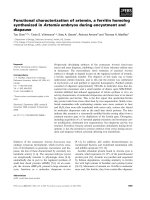
Fig 4-19 Lateral window sinus elevation surgical
protocol. (a) Preoperative view of surgical site. (b)
Anesthesia delivery. (c and d) Adequate flap size is
important for surgical access. The flap is reflected
at least 10 mm beyond the corners of the window.
Lateral Window Sinus Elevation Surgical Protocol
Before discussing the complications that may occur during the lateral window sinus elevation, it is important to present the surgi-
cal protocol that should be followed to minimize the risk of complications.
The lateral window sinus elevation surgical protocol consists of the following eight steps (Fig 4-19):
1. Anesthesia
2. Incision and full-thickness flap reflection
3. Osteotomy and window infracture or removal
4. Sinus membrane elevation
5. Bone graft placement
6. Incision closure
7. Postoperative provisionalization
8. Postoperative instructions and care
The window is outlined (e) and then pushed inward
after being completely separated from the sur-
rounding bone (f to i).
Lateral Window Sinus Elevation Surgical Protocol
145
j
k
n
o
l m
p
q
r
t
s
u
Lateral Window Sinus Elevation Surgical Protocol
Fig 4-19 cont (j and k) Alternatively, the surgeon
may elect to remove the bone flap (eg, when the
buccolingual dimensions of the sinus are narrow).
The bone graft material is placed (l to r), the flap is
sutured (s), and the area is left to heal for a period
of 4 to 9 months (depending on the volume and
type of bone graft materials used) before implant
placement (t and u).