Báo cáo Y học: Functional expression of human liver cytosolic b-glucosidase in Pichia pastoris Insights into its role in the metabolism of dietary glucosides ppt
Bạn đang xem bản rút gọn của tài liệu. Xem và tải ngay bản đầy đủ của tài liệu tại đây (278.85 KB, 10 trang )
Functional expression of human liver cytosolic b-glucosidase
in
Pichia pastoris
Insights into its role in the metabolism of dietary glucosides
Jean-Guy Berrin
1,2
, W. Russell McLauchlan
1
, Paul Needs
1
, Gary Williamson
1
, Antoine Puigserver
2
,
Paul A. Kroon
1
and Nathalie Juge
1,2
1
Nutrition, Health and Consumer Sciences Division, Institute of Food Research, Norwich, UK;
2
Institut Me
Â
diterrane
Â
en de Recherche
en Nutrition, Faculte
Â
des Sciences et Techniques de Saint-Je
Â
ro
Ã
me, Marseilles, France
Human tissues such as liver, small intestine, spleen and
kidney contain a cytosolic b-glucosidase (CBG) that
hydrolyses var ious b-
D
-glycosides, but whose physiological
function is not known. Here, we describe the ®rst hetero-
logous expression of human CBG, a system that facili-
tated a detailed a ssessment of the enzyme speci®city
towards dietary glycosides. A full-length CBG c DNA
(cbg-1) w as cloned from a human liver cDNA library and
expressed in the methylotrophic yeast Pichia pastoris at a
secretion yield of 10 mgáL
)1
. The recombinant CBG
(reCBG) was puri®ed from the supernatant using a single
chromatography step and was shown to be similar to the
native enzyme isolated from human liver in terms of
physical properties and speci®c activity towards 4-nitro-
phenyl-b-
D
-glucoside. Furthermore, the r eCBG displayed a
broad speci®city with respect to the g lycone moiety of
various aryl-glycosides (b-
D
-fucosides, a-
L
-arabinosides,
b-
D
-glucosides, b-
D
-galactosides, b-
L
-xylosides, b-
D
-arabino-
sides), similar to the native enzyme. For the ®rst time, we
show that the human enzyme has signi®cant activity towards
many common dietary xenobiotics including glycosides of
phytoestrogens, ¯avonoids, s imple phenolics and cyanogens
with higher apparent anities (K
m
) and speci®cities (k
cat
/K
m
)
for d ietary xenobiotics than f or other aryl-glycosides. These
data indicate that human CBG hydrolyses a broad range of
dietary glucosides a nd may play a critical role in xenobiotic
metabolism.
Keywords: heterologous expression; xenobiotic metabolism,
¯avonoids; iso¯avones; ®rst-pass metabolism.
b-Glucosidases (b-
D
-glucoside glucohydrolase; EC 3.2.1.21)
are members of glycosyl hydrolase families 1 and 3 [1,2].
b-Glucosidases h ydrolyse O-glycosidic bonds at the t ermi-
nal, nonreducing end of carbohydrates with retention of
anomeric con®guration. They are widely present in nature
where they demonstrate catalytic activity against a broad
range of b-
D
-glycosides.
In humans, sev eral b-glucosidases have been described
and for most of them, the role and physiological substrates
are known. For example, the lysosomal b-glucosidase (Ôacid
b-glucosidaseÕ) hydrolyses glucocerebrosides (glycosphingo-
lipids) present in the lysosomal membranes, a nd a lack of
this enzyme is the cause of the various form s of Gaucher's
disease, one of the hereditary lysosomal storage disorders
[3]. Lactase-phlorizin hyd rolase (LPH) is anchored in the
mucosal membrane in the brush-border of the small
intestine, where it hydrolyses lactose present in milk. A
de®ciency of LPH is the cause of lactose intolerance that is
common except in Northern European adults and a few
small ethnic populations [4]. Another human b-glucosidase
is speci®c for the hydrolysis of pyridoxine 5 ¢-b-
D
-glucopyra-
noside, a common dietary form of vitamin B
6
, and has been
ascribed a role in vitamin B
6
bioavailability [ 5]. A putative
protein, pr edicted from the klotho (kl ) g ene, shows homol-
ogy t o family 1 g lycosyl hydrolase and is also predicted to
occur in the cytosol of certain human cells [6,7] where it
might have a role in human aging [6].
Finally, a b-glucosidase, termed cytosolic b-glucosidase,
is present in the liver, kidney, intestine and spleen of
humans. This c ytosolic b-glucosidase (CBG) h as been
puri®ed from human liver and partially characterized
[8±10]. It is a 53-kDa monomeric protein with a pI of
4.7, a broad and near-neutral pH optimum, and a broad
speci®city w ith respect to the g lycone moiety of substrates.
Human CBG hydrolyses synthetic aryl glycosides (including
4-nitrophenyl and 4-methylumbelliferyl monoglycosides)
[9], but no physiological substrate h as been found and the
function in vivo has yet to be determined. However, during
our research into the mechanisms underlying the absorption
and metabolism of dietary ¯avonoids and iso¯avones, we
demonstrated that crude protein e xtracts derived from
human liver and small intestine tissues ef®ciently hydrolysed
a range of foo d-borne phytochemical (¯avonoid and
iso¯avone) glucosides [11]. The effects of s peci®c enzyme
inhibitors appeared to indicate that the majority of
Correspondence to P. A. Kroon, Nutrition, Health & Consumer Sci-
ences Division, Institute of Food Research, Colney Lane, Norwich,
NR4 7UA, UK. Fax: + 44 1603 255038, Tel.: + 44 1603 255236,
E-mail:
Abbreviations: AOX1, a lcohol oxidase; BMGY, bu ered minimal
glycerol-complex medium; BMMY, buered minimal methanol-
complex medium; ESI, electrospray ionization; CBG, cytosolic
b-glucosidase; cbg-1, cDNA encoding CBG; reCBG, recombinant
CBG; LPH, lactase-phlorizin hydrolase; 4NP, 4-nitrophenol; YNB,
yeast nitrogen base; YPD, yeast extract peptone d extrose.
(Received 12 October 2001, accepted 30 October 2001)
Eur. J. Biochem. 269, 249±258 (2002) Ó FEBS 2002
hydrolytic activity was due to human CBG [11]. CBGs
obtained from o ther mammals have been shown to
hydrolyse some glycosides of plant origin including phen-
olic, pyrimidine, and cyanogenic glycosides [12±14]. We
demonstrated that CBG isolated f rom pig liver hydrolysed
various ¯avonoid glycosides with reasonable turnover
numbers and micromolar K
m
values [14]. Furthermore the
localization of human CBG in metabolic tissues such as the
intestine, liver, k idney and spleen indicate that CBG is
exposed to orally ingested xenobio tic glycosides. The broad
speci®city of the CBG distinguishes this mammalian
b-glucosidase from all o thers and has led to the suggestion
that it is involved in the primary stage of xenobiotic
metabolism [15], but this hypothesis r emained t o b e t ested
using the pure human enzyme. Isolation of CBG from
human tissues is not easy due to dif®culties associated with
obtaining appropriate amounts of suitable tissues, large
variations in activity between tissues obtained from different
individuals [9,16; P. A. Kroon, unpublished d ata], a nd the
need for a multistep fractionation procedure to obtain pure
protein [9,17]. In order to fac ilitate biochemical and
molecular studies on the signi®cance of human CBG in
xenobiotic metabolism, we isolated a human CBG cDNA
(cbg-1) and successfully expressed it heterologously in the
yeast Pichia pastoris. This organism possesses a number of
attributes that renders it an attractive host for the expression
and production of CBG: it can b e grown conveniently to
high den sity levels in a simple and inexpensive medium; it is
able to carry out certain post-translational modi®cation
events such as proteolytic maturation, glycosylation and
disul®de bond formation; under the co ntrol of t he ef®cient
and highly regulated promoter of the alcohol oxidase gene,
AOX1, it c an secrete p roteins to very h igh levels [18±20].
In this report, we show that puri®ed recombinant CBG
possesses similar physical and enzymatic properties to CBG
isolated from human liver. Furthermore, we investigated the
speci®city of the human CBG with r espect to the glycone
and aglycone moieties, and in particular characterized the
ef®ciency of the enzyme in hydrolysing a broad r ange of
dietary xenobiotic glycosides. The potential role for human
CBG in xenobiotic metabolism and uptake is also discussed.
MATERIALS AND METHODS
Materials and strains
The Zero Blunt
TM
TOPO
TM
PCR cloning vector and the
pHIL-S1 shuttle vector [32] were purchased from Invitrogen
(San Diego, CA, USA). Restriction endonucleases and
DNA modifying enzymes were purchased from Promega
(Madison WI, USA) and used according to the manufac-
turer's recommendation. Escherichia c oli DH5 (supE44,
hsdR17, recA1, endA 1, gyrA96, th i-1, relA1) and TOP10
(F
±
mcrA D(mrr-hsdRMS-mcrBC) F80lacZDM15 DlacX74
recA1 deoR araD139 D(ara-leu)7697 gal U galK rpsL(Str
R
)
endA1 nupG) were used for DNA manipulation. Oligonu-
cleotides were s ynthesized by PerkinElmer Applied B iosys-
tems (Warrington, UK). Quercetin-3-xyloside (Q3Xyl;
isolated from apple skins), quercetin-3,4¢-diglucoside
(Q3,4¢Glc) and malonylated quercetin-3-glucoside (Q3Glc-
Mal; both isolated from onions) w ere kind gifts from Keith
Price (IFR, Norwich, UK). Kaempferol-3-glucuronide
(K3GlA; isolated from lettuce) was a kind gift from
S. DuPont (IFR, Norwich, UK). Quercetin-7-glucoside
(Q7Glc) was synthesized as described below. Quercetin
glucuronides (quercetin-3-glucuronide, quercetin-7-glucuro-
nide, quercetin-4¢-glucuronide and quercetin-3¢-glucuro-
nide) were b iosynthesized using pig liver microsomes as a
source of UDP-glucuronosyl transferase (UDP-GT) activ-
ity, UDP-glucuronic acid, UDP-glucosylamine and querce-
tin (all obtained from Sigma Aldrich) as donor, cofactor and
acceptor, respectively, and were puri®ed using s olid-phase
extraction on polyamide followed by preparative HPLC
using a reversed-phase LUNA C-18 column (4.6 ´ 25 mm,
5 lm; Phenomonex, Maccles®eld, UK). Other ¯avonoids
and their conjugates were purchased in the purest form
available from Extrasynthe
Á
se (ZI Lyon Nord, BP 62, 69730
Genay, France) or Apin Chemicals Ltd (Milton Park,
Abingdon, Oxford, UK). Mandelonitrile-b-
D
-glucopyrano-
side (prunasin), mandelonitrile-b-
D
-gentiobioside (amyg-
dalin), 1,4-benzenediol-b-
D
-glucopyranoside (arbutin),
guiacol-b-
D
-glucopyranoside (salicin), 2,4-dinitrophenyl-2-
¯uoro-2-deoxy-b-
D
-glucopyranoside, and the nitrophenyl
glycosyl derivatives were obtained from Sigma Aldrich
(Poole, Dorset, U K).
Synthesis of quercetin-7-
O
-b-
D
-glucopyranoside (Q7Glc)
3¢,4¢,4,5-Tetrabenzoylquercetin [21] (100 mg, 139 lmol),
2,3,4,6-tetra-O-acetyl-a-
D
-glucopyranosyl b romide (170 mg,
3eq.),Ag
2
CO
3
(115 mg,3 eq.),3 A
Ê
sieves (250 mg) and dry
CH
2
Cl
2
(10 mL) and collidine (55 lL, 3 eq.) w ere stirred
under Ar, in the dark, for 3 days. After ® ltration, combined
®ltrate and washings (5% MeOH/CH
2
Cl
2,
100 mL) were
washed with 1
M
HCl (50 mL), H
2
O (50 mL), 0.1
M
Na
2
S
2
O
3
(50 mL), H
2
O (50 mL), saturated NaHCO
3
(50 mL), and H
2
O ( 50 mL), and t hen dried (MgSO
4
). The
evaporated residue was stirred into 1
M
NaOH ( 50 mL)
under Ar (0°, 9 0 m in), warmed to room temperture, heated
at re¯ux (20 min), and cooled. Dowex 50 W resin (H
+
form, 70 mL) was added. Filtrate and washings (50%
aqueous MeOH, 100 mL) were evaporated, dissolved in
10% aqueous MeOH (300 mL), and washed CH
2
Cl
2
(3 ´ 80 mL). The aqueous phase was evaporated, taken
up in MeOH (2.5 m L) and puri®ed by HPLC. Yield 7 m g,
12%.
1
H-NMR (CD
3
OD): d 7.74 (d, 1 H, J
2¢,6¢
2.0 Hz, H-2¢),
7.65 (dd, 1 H, J
6¢,5¢
7.6 Hz, H-6¢), 6.88 (d, 1 H, H-5¢), 6.74 (d,
1H,J
8,6
2.0Hz,H-8),6.44(d,1H,H-6),5.05(d,1H,J
1¢¢,2¢¢
7.2 Hz, H-1¢¢), 3.95 (dd, 1 H, J
6A¢¢,6¢¢B
11.9 Hz, H -6 A ¢¢), 3.73
(dd, 1H, H -6B¢¢), 3.43±3.57 (m, 3 H, H-2¢¢,H-3¢¢,H-4¢¢).
ESMS: m/z 465 [M + H]
+
487 [M + Na]
+
.
Isolation of cytosolic b-glucosidase from human liver
Liver samples were obtained from redundant tissue of
surgical specimens f rom patients undergoing hepatic sur-
gery. The patient c oncerned had given informed consent for
the w ork to be performed. A sample of liver was obtained
fresh, cut into pieces ( 5 g ) and snap-frozen in liquid
nitrogen before use. CBG was isolated from 100 g (fresh
weight) of thawed liver b y a modi®cation of a procedure
described previously [14]. B rie¯y, the isolation involved
homogenization, centrifugation at high speed to remove
membranes and large debris, cation-exchange chromato-
graphy on CM-Sephadex, af®nity chromatography using
octyl-Sepharose, chromatofocussing using a Mono P HR
250 J G. Berrin et al. (Eur. J. Biochem. 269) Ó FEBS 2002
5/20 chromatography column (Amersham Pharmacia Bio-
tech), and gel ®ltration u sing a Superdex 200 HR 10/30 gel
®ltration chromatography column (Amersham Pharmacia
Biotech). Fraction s containing CBG activity were pooled,
mixedwithanequivalentvolumeofethyleneglycoland
stored at )20 °C.
Isolation, sequencing and analysis of
cbg-1
from a human cDNA library
The full length cDNA encoding for human CBG w as
isolated from a human liver kTriplEx
TM
cDNA library
(Clontech, Palo Alto, CA, USA) by h ybridization screening
using a 900-bp o ligonucleotide probe ampli®ed from the
cDNA library by PCR using d egenerate primers designed
against conserved regions in domains II I and IV of human
LPH [22] and guinea pig CBG [23]. The sequence of forward
primer HCG/F2 was 5¢-TAYCGNTTYTCNATHTCN
TGG-3¢. The sequence o f the reverse p rimer HCG/R3 was
5¢-NCCNTTYTCNGTRATRTA-3¢. PCR was p erformed
using 1 lL of library lysate, 20 p mol of primers HCG/F2
and HCG/R3, 0.2 m
M
dNTPs, 2.5 U of Taq polymerase
(Amersham Pharmacia B iotech) 1 0 m
M
Tris/HCl, pH 9 .0,
50 m
M
KCl, 3.5 m
M
MgCl
2
on a PerkinElmer Gene Amp
2400 thermal cycler (PE Biosystems, Foster City, CA, USA)
at 94 °C for 2 min followed by 30 cycles of 94 °Cfor1min,
42.0 °C for 1 min, 72.0 °C for 2 min. The ampli®cation was
completed with a ®nal extension at 72.0 °C for 5 min. T he
probe w as gel-puri®ed using a QIAquick gel extraction k it
(Qiagen Ltd, Crawley, UK) and labelled with horserad-
ish peroxidase using an ECL
TM
direct nucleic acid labelling
and detection k it (Amersham P harmacia Biotech).
The library was plated out on 20 cm ´ 20 cm bioassay
plate ( Nalge Nunc Intern ational, N aperville, USA) for the
primary hybridization screen according to the manufactur-
ers protocol. Plaques were transferred to a nylon membrane
(Hybond N
+
, Amersham Pharmacia Biotech) and cross-
linked using UV irradiation (Stratalinker 2400, Stratagene,
La Jolla, California, USA). Enhanced chemiluminescence
signal generation was carried out using the direct nucleic
acid labelling and detection kit and autoradiography.
Positive colonies from the primary screen were taken
through a secondary screen as described above, except o n
150 mm p lates a t a density of 200±1000 plaques p er pla te.
Single well-isolated positive plaques from the secondary
screen were converted from kTriplEx clones to pTriplEx
clones by in vivo excision and circularization according to
the protocol in the library users manual. The clones were
sequenced on both strands using the ABI Prism BigDye
TM
Terminator Cycle Sequencing kit and an ABI 373 DNA
sequencer. Sequence analysis was carried out using the
Wisconsin
GCG V
10.1 software package (Genetics Computer
Group, Madison, Wisconsin, USA) and sequence align-
ments using
BLAST
v2.0 [24].
Construction of the pHIL-S1/
cbg-1
expression plasmid
The pHIL-S1-derived expression plasmid w ith the cDNA
insert encoding human CBG is shown in Fig. 1. The DNA
manipulations were carried out using standard procedures
[25]. The cDNA fragment (1407 bp) containing the cbg-1
coding region was ampli®ed by PCR from the TriplEx clone
by using Pfu DNA polymerase (Stratagene) and the
upstream primer (5¢-TTTTTT
CTCGAGAAGCTTTCC
CTGCAGGAT-3¢) and downstream primer (5¢-TTTTT
T
GGATCCCTACAGATGTGCTTCAAGGCC-3¢), thus
introducing XhoIandBamHI sites, respectively (underlined)
at each end of the gene. The 5¢ terminus of this construct was
designed to introduce t he Pichia phosphatase sign al
sequence cleavage site (Ala-Arg) in frame with the cbg-1
coding sequence (Fig. 1). As the native PHO1 signal
sequence cleavage site contains a g lutamate re sidue imme-
diately a fter the Ala-Arg residues, a glutamate codon
(GAA) was included in the primer to preserve the
phosphatase's native context. DNA ampli®cation was
carried out through 25 cycles of denaturation (1 min at
94 °C), annealing (0.5 min at 61 °C), and extension
(1.5 min at 7 2 °C) in a DNA thermocycler (PerkinElmer).
The resulting PCR product (1430 bp) was puri®ed using the
Qiaquick PCR puri®cation kit ( Qiagen), subcloned into
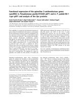
Fig. 1. Nucleotide and amino acid sequences in
the cleavage region between the leader peptide
and mature reCBG. Th e construction of the
vector is detailed under Mate rials and meth-
ods. ss, PHO1 secretion signal seq uence;
5¢AOX1 (Pro), P. pastoris alcohol oxidase
promoter region; 3¢AOX1 (TT), P. pastoris
AOX1 transcriptional terminating sequence.
* i s the N-terminal residue o f the native
human cytosolic be ta-glucosidase.
Ó FEBS 2002 Xenobiotic metabolism by a human b-glucosidase (Eur. J. Biochem. 269) 251
the T OPO vector and subjected to DNA sequencing using
the ABI prism Big Dye
TM
Terminator Cycle Sequencing kit
to con®rm t hat n o e rrors wer e generated during the PCR.
The positive clone was d igested by a c ombination of XhoI
and BamHI, and subsequently the cDNA insert was puri®ed
using the Qiaquick PCR puri®cation kit and ligated into the
XhoIandBamHI sites of the pHIL-S1 vector, i n phase with
the PHO1 signal sequence. E. coli strain DH5 was trans-
formed according to the procedures described in Sambrook
et al. [25]. Transformants were grown in liquid bacterial
cultures, recombinant plasmids isolated using Q iagen col-
umns (Mini-Prep kit), and identity c hecked by restriction
mapping to yield pHIL-S1/cbg-1.
Transformation of
Pichia pastoris
and selection
of a recombinant clone
Transformation of the P. pa storis strain (his4)/GS115 [26]
and screening were achieved using the spheroplast proce-
dure [27], modi®ed as described previously [28]. Brie¯y,
pHIL-S1/cbg-1 ( 1 lg) as well as the pHIL-S1 vector, as
negative control, were digested with BgIII prior to trans-
formation by the spheroplast method. After screening f or
methanol sensitive clones, Mut
s
colonies were used to
inoculate 10 mL BMGY pH 6 . After 2 d ays with shaking at
250 r.p.m., 30 °C, the cells were pelleted a nd resuspended in
2 mL BMMY. Following another 5 days at 30 °C, the
culture w as centrifuged and the amount of reCBG in the
supernatant w as estim ated b y activity measurement ass ays
using 4NPGlc a s substrate.
Expression of
cbg-1
in
P. pastoris
and isolation
of reCBG
Large-scale expression was achieved using 250 mL cultures
in 1 L baf¯ed ¯as ks. Cells grown i n B MGY a t 30 °Ctoa
density of D
600
20±25 were harvested, resuspended in
50 mL of BMMY and incubated with shaking (250 r.p.m.)
in ®ve 5 0 m L loosely cap tubes at 30 °C. The culture was
continued for a total of 5 d ays with aliquots of the
supernatant removed at various time points in order to
monitor p roduction of reCBG. Puri®cation of reCBG
was achieved in a single step using af®nity chromatog-
raphy. Supernatant (50 mL) was loaded onto a column
(1.5 ´ 5 cm) of octyl sepharose previously equilibrated w ith
20 m
M
sodium p hosphate buffer (pH 6.5) containing 1 m
M
EDTA, the column was washed with 20% ethylene glycol in
sodium phosphate buffer and unbound material discar ded.
Bound material was eluted with ethylene glycol (50% v/v) at
a ¯ow rate of 0.5 mLámin
)1
over 1 h . b-Glucosidase-
containing fractions were pooled and checked for purity
by SDS/PAGE.
Enzyme assays
Fractions g enerated during isolation of CBG from human
liver were assayed for CBG a ctivity u sing a spectrophoto-
metric assay where the release of 4-nitrophenol (4NP) from
4-nitrophenyl-b-
D
-glucopyranoside (4NPGlc; 10 m
M
)in
50 m
M
sodium-phosphate buffer (pH 6.5) at 37 °Cis
determined at 400 n m using the molar extinction coef®cient
for 4NP of 18 300
M
)1
ácm
)1
. The p H optimum for r eCBG
was determined b y m easuring the b-glucosidase activity in
50 m
M
sodium phosphate (pH range 2.8±7.6). The thermal
stability of CBG was assessed by measuring the residual
b- glucosidase activity (4NPGlc as substrate) follow ing incu-
bation (30 m in) of CBG samples at various temperatures
(23±70 °C). The activity of puri®ed CBG towards various
nitrophenyl glycosides (a-
D
-glucopyranoside, a-
D
-glucopyr-
anoside, a-
D
-galactopyranoside, a-
L
-arabinopyranoside,
b-
L
-arabinopyranoside, a-
L
-arabino-furanoside, a-
D
-man-
nopyranoside, a-
D
-mannopyranoside, a-
D
-fucopyranoside,
a-
D
-xylopyranoside, a-
L
-rhamnopyranoside) w as deter-
mined u sing the same m ethod. Activities towards phenolic
or mandelonitrile glycosides were determined by measuring
the amount of aglycone released from the substrate (10±
5000 l
M
in 50 m
M
sodium-phosphate buffer), with p artic-
ular care taken to ensure complete solubility of substrates as
described previously [14]. Brie¯y, pure phenolic/mandelo-
nitrile glycosides were dissolved in a small volume of
dimethylsulfoxide prior to dilution with assay buffer
(50 m
M
NaCl/P
i
, pH 6.5; ®nal c oncentration d imethylsulf-
oxide < 2%, v/v), equilibrated at 37 °C , and reactions
started with the addition of enzyme (0.1±1 lgin10lL) in a
®nal volume o f 100 lL. Reactions were terminated by the
addition of acetonitrile/1% aqueous tri¯uoroacetic acid
(50 : 50 , v/v; 100 lL), ®ltered and analysed by reversed-
phase HPLC with online diode-array detection using a
LUNA C-18 co lumn (4.6 ´ 25 mm, 5 lm; Phenomonex,
Maccles®eld, UK) with an injection volume of 20 lL.
Solvents A (water/tetrahydrofuran/tri¯uoroacetic acid,
98 : 2 : 0.1 v/v), B (acetonitrile), C (water/tri¯uoroacetic
acid, 99.9 : 0.1), and D (methanol/tri¯uoroacetic acid,
99.9 : 0.1) were run at a ¯ow rate of 1 mLámin
)1
.The
following gradients were used: incubations containing
arbutin or salicin as substrate; 100% C initial, i ncreasing
D t o 2 0% (10 min), 50% ( 15 min), 100% (5 min), held at
100% (5 min); cyanodin glycosides; 5% B/95% A initial
(5 min), increasing B to 20% (10 m in), 90% (10 min), held
at 90% (5 m in); iso¯avonoid, mandelonitrile and dihydr-
ochalcone glycosides, 17% A/83% B initial (1 min),
increasing B to 90% (10 min), held at 90% (4 min). The
column was re-equilibrated (5 m in) in the appropriate
starting solvent conditions following gradient development.
Standard curves were constructed using HPLC grade
aglycones from which response factors were calculated
and used t o estimate t he amount of product released in test
incubations. For estimations of the apparent af®nity (K
m
)
and k
cat
, steady-state rates were determined over a range of
substrate concentrations (at least 0.2±5.0 ´ K
m
where
possible) and k inetic constants e stimated using a nonlinear
weighted least-squares regression analysis method [29]. The
concentration of phenolic and mandelonitrile glycosides
present in solution at the higher concentrations of substrate
used was con®rmed b y HPLC analysis of t he supernatant
obtained following centrifugation (13 000 g,10min).
Inhibition of reCBG with 2,4-dinitrophenyl-2-¯uoro-
2-deoxy-b-
D
-glucopyranoside
Inhibition studies were performed by incubating reCBG
(100 lL) with 2,4-dinitrophenyl-2-¯uoro-2-deoxy- b-
D
-
glucopyranoside (100 lL) at ®nal inh ibitor concentrations
of 1 and 5 l
M
([E]/[I] ratio s of 1 : 3 and 1 : 15, respectively)
at 37 °C. The b-glucosidase activity remaining after various
incubation periods (see Fig. 3) was determined by adding
252 J G. Berrin et al. (Eur. J. Biochem. 269) Ó FEBS 2002
20 lL of the enzyme/inhibitor mixture to 180 lLof
substrate (4NPGlc), incubating for 30 min at 37 °C, and
measuring the rel ease of 4NP.
Protein assays and protein sequencing
Total protein in crude and semipuri®ed samples was
estimated using the Pierce Protein Assay Reagent with
BSA as standard. For puri®ed reCBG, total protein was
calculated using an e xtinction coef®cient at 2 80 nm
(122 120
M
)1
ácm
)1
) derived fro m the a mino-acid composi-
tion for the primary s tructure for reCBG. Protein sequenc-
ing was performed at the Protein Sequencing & Peptide
Synthesis Facility (John Innes C entre, Norwich, UK) using
an ABI 4 91 Procise sequencer.
Gel electrophoresis
SDS/PAGE was routinely p erformed using 12% homoge-
neous Tris/glycine gels (Novex, Frankfurt, Germany)
according to the manufacturer's instructions, a nd stained
with Coomassie Blue. Molecular m asses were estimated
from plots of log(M
r
) vs. migration for a series of known
standard proteins (LMW Marker Kit; Amersham Pharma-
cia Biotech). Isoelectric focusing w as performed using 5%
homogeneous polyacrylamide gels for the pH range 3±7
(Novex) according to t he manufacturer's i nstructions, and
stained with C oomassie Blue. Values for pI w ere estimated
from plots o f pI vs. distance from the anode for a series of
known protein standards (Low pI Kit; Amersham Phar-
macia Biotech).
RESULTS
Isolation and characterization of cytosolic
b-glucosidase from human liver
Human liver was chosen as a source of CBG a s this o rgan
is potentially a rich source of the enzyme and disease-free
tissue can be obtained fresh during relatively routine
surgical procedures. Isolation of CBG from human liver
has b een described and involves a fairly long series of
fractionation procedures [9,17]. The isolation u sed here
involved cation-exchange chromatography (CBG does not
bind at pH 5.5), hydrophobic i nteraction chromatography
using octyl sepharose (behaves as an af® nity column for
mammalian CBG [17]), chromatofocusing, and removal o f
ampholines by gel ®ltration chromatography. Starting with
100 g fresh liver tissue, this procedure resulted in a small
amount ( 50 lg) of electrophoretically pure protein with
a speci®c activity towards 4NPGlc o f 12.8 lmolámin
)1
ámg
protein
)1
, an apparent molecular m ass (by SDS/PAGE) o f
51.9 kDa and a pI o f 4.7. These values are in good
agreement with previously published values for mamma-
lian liver CBGs [9,12,14,23,30]. We were unable to obtain
an N-terminal sequence for the puri®ed enzyme probably
because, as with guinea-p ig CBG [23], the N-terminus was
blocked.
Human liver
cbg-1
cDNA cloning and sequence analysis
A human liver cDNA library was screened b y a c onven-
tional a pproach using a 900-bp
32
P-labelled DNA fragment
from human CBG. This DNA probe was ampli®ed by PCR
from the c DNA library using two degenerate oligonucleo-
tide primers d esigned against consensus sequences from the
coding regions of domains III and IV of human lactase
phlorizin hydrolase (LPH) [22] and guinea pig cytosolic
b-glucosidase [23]. Five cDNA clones were isolated and
sequenced. The largest clone was found t o contain an ORF
of 1407 nucleotides encoding a p rotein of 496 a mino acids
with a calculated molecular mass of 53.7 kDa. A single
putative glycosylation site was located at N47 of the
deduced amino-acid sequence within the motif KNQT. No
signal sequence was apparent which indicates, as expected,
CBG is located in the cytosol. The nucleotide a nd amin o-
acid sequence has been submitted to the GenBank seq uence
data bank and is available under a ccession number
AF317840.
The primary sequence for human CBG s hared extensive
sequence homology with other mammalian b-glucosidases.
CBG shared 79% nucleotide similarity and 83.6 % ami no-
acid similarity with gu inea pig C BG, and showed homol-
ogy w ith domains III and I V o f mammalian LPH (56 and
57% amino-acid similarity, respectively) and with the
putative cytosolic and membrane-bound forms of human
klotho (42 and 32% amino acid similarity, respectively).
Highly conserved regions were identi®ed including those
surrounding the putative catalytic glutamates, character-
ized by the sequence motifs VKQWITINEA (residues 157±
166) and IYITENG (residues 369±375) found in all
family 1 b-glycosidases [31±34]. Alignment o f the cbg-1
cDNA sequence with the other available sequences for
human CBG [ 35±37] allowed us to identify s everal
nucleotide differences, some of which lead to changes in
the p rotein primary structure. We are con®dent these are
not due to errors in the cbg-1 sequence as it was derived
from a full-length cDNA iso lated using a radiolabelled
cDNA probe. The observed differences may be genuine
and re¯ect genetic polymorphism. It w as therefore impor-
tant to clone, express, and characterize the product arising
from a single gene.
Expression of
cbg-1
in
P. pastoris
The cDNA sequence encoding the entire human liver cbg-1
cDNA was inserted into the expression vector pHIL-S1 i n
frame with the P. pa storis phosphatase signal sequence
(Fig. 1). The resulting expression plasmid was used to
transform P. pastoris and the transformants screened for
the best expression performances. Mut
s
transformants were
grown under noninduced conditions (MGY or BMGY) and
then transferred to medium containing methanol (MMY or
BMMY). Routine activity assays against pNP-b-
D
-gluco-
pyranoside were u sed for the selection of clones with h igh
b-glucosidase productivity. b-Glucosidase activ ity was
found only when rich m edium (BMGY/BMMY) was used
for induction of CBG expression. However, as P. p ast oris
secretes endogenous b-glucosidase activity into the medium,
although at very low level, it was important to discriminate
between the recombinant and endogenous activities. This
was achieved using the ¯avonol glucoside Q4¢Glc, which is a
substrate for human liver CBG (Table 2) but not for
P. pastoris endogenous b-glucosidase, as demonstrated
using media from P. pastoris transformedwithpHIL-S1
lacking the CBG cDNA insertion (data not shown). Hence,
Ó FEBS 2002 Xenobiotic metabolism by a human b-glucosidase (Eur. J. Biochem. 269) 253
although both the Pichia endogenous b-glucosidase and the
human reCBG hydrolysed 4NPGlc, the use of Q4¢Glc
con®rmed that the increased level of b-glucosidase activ ity
was due to the secretion of the human recombinant enzyme.
A representative His
+
Mut
s
transformant was selected for
production of recombinant CBG (reCBG) in shake-¯ask
cultures with secretion yields up to 10 mgáL
)1
after 5 days of
culture. When cells were transformed with the pHIL-S1/
cbg-1 vector and induced with methanol, a single major
protein band of 53 kDa was identi®ed following SDS/
PAGE analysis of the culture supernatant, and only trace
amounts of other proteins were visible a s faint bands (data
not shown). The 53-kDa protein was absent from the
medium of cells transformed with the vector alone.
Puri®cation and characterization of reCBG
A single puri®cation step using octyl-Sepharose se parated
the reCBG from Pichia endogenous b-glucosidase, and
gave an e lectrophoretically pure protein (M
r
53 kDa;
Fig. 2A) with a speci®c activity on 4NPGlc of
10.0 lmolámin
)1
ámg protein
)1
. Eighty-two percent of the
total b-glucosidase activity in the culture supernatant was
recovered in a single peak (chromatogram not shown). No
bands other than the 53-kDa band were visible even
following silver staining, indicating a very h igh level of
purity. The small discrepancy b etween the speci®c activities
for human liver CBG a nd reCBG w as shown to be due to
the different methods used to estimate total p rotein.
Isoelectric focusing of puri®ed reCBG gave t wo bands at
pI 4.7 and 4.8 (Fig. 2B), in good agreement with that
obtained for CBG isolated from human liver. Conventional
Edman sequencing of reCBG indicated a single N-terminal
sequence (REAFP) demonstrating that there had been
correct processing of the PHO1 signal sequence (Fig. 1).
The b-glucosidase inhibitor, 2,4-dinitroph enyl-2-¯uoro-
2-deoxy-b-
D
-glucopyranoside, was a potent inhibitor of
reCBG (Fig. 3). Incubation of reCBG (0.35 l
M
®nal
concentration) in the presence of 1 and 5 l
M
inhibitor
reduced the b-glucosidase activity in a time-dependent
manner; 36 and 70% of the b-glucosidase activity remain ed
following 30 and 50 min incubation with 1 and 5 l
M
inhibitor, respectively. b-Glucosidase activity was not
recovered following extensive dialysis of the inhibited
enzyme, indicating that inhibition was essentially irrevers-
ible. The highest rates for hydrolysis of 4NPGlc over 10 min
were obtained at 50 °C, 2.3-fold faster than at 37 °Cand
4-fold faster than at 58 °C (re¯ecting thermal inactivation).
The enzyme was relatively stable at 37 °C as more than 80%
activity remained after 24 h at this temperature. The pH
optimum for b oth r eCBG and human liver CBG w as 6.5,
with ³ 70% o f optimum activity maintained over the pH
range 5.0±7.5, but < 4% at pH 4.0.
Furthermore, we examined the s peci®city o f reCBG with
respect to the glycone moiety using a series of NP derivatives.
The enzyme catalysed the release of 4NP from six of the 11
4-substituted substrates tested an d the kinetic parameters f or
these are presented in Table 1. We detecte d no measureable
release of NP using 4NP-a-
D
-glucopyranoside, 4NP-a-
L
-
arabinofuranoside, 4NP- a-
L
-rhamnopyranoside, 4NP- a-
D
-
mannopyranoside or 4NP-b-
D
-mannopyrano-side. The
activity towards 2NP-galactopyranoside (10 m
M
)was
10-fold lower than observed for the 4NP-derivative (data
not shown). K inetic analysis under steady-state conditions
indicated that the speci®city (k
cat
/K
m
)ofreCBGfor4NP-
glycosides was b-
D
-fucopyranoside > a-
L
-arabinopyrano-
side > b-
D
-glucopyranoside > b-
D
-galactopyranoside >
b-
D
-xylopyranoside > b-
L
-arabinopyranoside. These data
are in general agreement with those obtained by Daniels
et al. [9] and con®rm that CBG has a broad speci®city that
can accommodate several glycones in the active site,
including b-
D
-linked pentose and hexose s ugars and a-
L
-
or b-
L
-linked arabinopyranosides, although several other
a-linked sugar derivatives (pentose and hexose) are not
hydrolysed by reCBG. Although we detected no me asure-
able release of 4NP from 4NP-b-
D
-mannopyranoside, we
were able to con®rm [16] that this compound was an
Fig. 2. Gel electrophoresis of reCBG. (A) Reducing SDS/PAGE:
Desalted samples (5 lg of puri®ed reCBG) were mixed with 15 lLof
2 ´ SDSsamplebuerandheatedat100°Cfor5minbeforeelec-
trophoresis on a 12% homogeneous Tris/glycine polyacrylamide gel.
The molecular masses of the marker proteins are shown to the right.
Gel was stained with Coomassie blue. (B) Isoelectric focusing: desalted
samples were mixed with 2 ´ sample buer and focused o n a 5%
homogeneous polyacrylamide gel containing ampholines covering the
pH range 3±7. Proteins were stained with Coomassie Blue.
Fig. 3. Time-dependent irreversible inactivation of reCBG by 2 ,4-dini-
trophenyl-2-¯uoro-2-deoxy-b-
D
-glucopyranoside. The enzyme (0.35 l
M
)
was incubated at 37 °C for the indicated period time with 1 l
M
inhibitor ( ,), 5 l
M
inhibitor (h) and withou t inhibitor ( s), an d then
assayed for beta-glucosidase activity at 37 °C for 30 min.
254 J G. Berrin et al. (Eur. J. Biochem. 269) Ó FEBS 2002
effective in hibitor of reCBG [the hydrolysis of 4NP-b-
D
-
glucopyranoside (10 m
M
) was reduced by 98% in the
presence of 4NP-b-
D
-mannopyranoside (10 m
M
)].
Hydrolysis of xenobiotic glycosides by reCBG
The a bility of CBG to hydrolyse a variety of glucosides was
assessed using a wide variety of aglycone structures that
were linked to sugars through various positions on the
aglycone (Tables 2 and 3, Fig. 4 ). The analysis was
performed in order to (a) assess the capacity of CBG to
hydrolyse a variety of plant-derived glycosides which are
commonly ingested by humans, and (b) determine some
relationships between aglycone structure a nd CBG speci-
®city. CBG hydrolysed e f®ciently many of t he compounds
tested, demonstrating lower apparent af®nities (K
m
)and
higher speci®city constants (k
cat
/K
m
) than those obtained
using various nitrophenyl glycosides (compare with data in
Table 1 ). b-
D
-Glucosides of ¯avones, iso¯avones and
¯avonols were hydrolysed p articularly ef®ciently. For
example, the estimates of apparent af®nity and speci®city
constant obtained using the ¯avone glucoside luteolin-4¢-
Glcassubstrate(10l
M
and 117 m
M
)1
ás
)1
, respectively)
were 176-fold l ower and 1 7-fold greater, respect ively, than
those obtained using 4NPGlc as substrate (Table 2 ).
Flavanone glucosides were hydrolysed less ef®ciently (due
to higher K
m
values) compared to glucosides of iso¯avones,
¯avones and ¯avonols. Hydrolysis of cyanogenic glycosides
Table 1. The glycone speci®city of reCBG. The hydrolysis of 4NP-glycosides was determined in 50 m
M
sodium phosphate buer (pH 6.5) at 37 °C
by estimating the release of 4NP spectrophotometric ally at 400 nm. For each substrate, d ata were obtained at various concentrations under steady-
state conditions, and the data ®tted to the Michaelis±Menten eq uation in orde r to obtain estimates for the kinetic constants ( k
cat
, K
m
).
Substrate
k
cat
(s
)1
)
K
m
(m
M
)
k
cat
/ K
m
(m
M
)1
ás
)1
)
4NP-b-
D
-fucopyranoside 10.7 0.0 0.37 0.01 28.9
4NP-a-
L
-arabinopyranoside 5.97 0.45 0.57 0.08 10.4
4NP-b-
D
-glucopyranoside 12.1 0.3 1.76 0.15 6.9
4NP-b-
D
-galactopyranoside 17.6 0.3 3.14 0.15 5.6
4NP-b-
D
-xylopyranoside 0.75 0.02 1.58 0.14 0.48
4NP-b-
L
-arabinopyranoside 0.66 0.09 52.6 8.4 0.013
Table 2. Hydrolysis of xenobiotic glycosides by reCBG. Incubations were performed a t 37 °Cin50m
M
sodium pho sphate bu er ( pH 6.5). T he
release of aglyc one was estimated using re versed-phase HPLC with reference to standard curves constructed using appropriate pure compounds.
Where signi® cant rates of h ydrolysis were observed, steady-state rates were obtained for a r ange of initial substrate concentrations and the data
®tted to the Michaelis±Menten equation in order to obtain estimates for K
m
and k
cat
. Glc, glucoside; diGlc, diglucoside; MalGlc, malonylglucoside;
General, ge ntiobioside; GlA, glucu ronide; GlcRha, rut inoside (1,6-linked rham noglucoside). ND, n ot determined.
Substrate
Speci®c activity
a
(lmolámin
)1
ámg
)1
)
k
cat
(s
)1
)
K
m
(l
M
)
k
cat
/K
m
(m
M
)1
ás
)1
)
Simple Phenolics
Salicyl alcohol-Glc (salicin) 0.171 ND ND ND
Hydroquinone-Glc (arbutin) 0.015 ND ND ND
Iso¯avones (phytoestrogens)
Genistein-7-Glc (genistin) 1.73 1.53 0.04 35 2.9 44
Daidzein-7-Glc (daidzin) 2.75 3.55 0.16 118 11 30
Daidzein-7-MalGlc 0.038 0.24 0.01 3230 130 0.075
Flavonols
Quercetin-4¢-Glc (spiraeoside) 1.19 1.08 0.02 31.8 2.9 34
Quercetin-7-Glc 0.77 0.69 0.02 42.2 3.2 16
Quercetin-3,4¢-diGlc 0.21
b
0.30 0.01 274 21 1.1
Flavones
Apigenin-7-Glc (apigetrin) 1.30 1.53 0.05 21.5 1.6 71
Luteolin-4¢-Glc 1.30 1.17 0.01 10 0.06 117
Luteolin-7-Glc 2.85 3.05 0.07 50 3.2 61
Luteolin-3¢,7-diGlc 1.46
c
ND ND ND
Flavanones
Naringenin-7-Glc 0.93 2.60 0.01 432 33 6.0
Eriodictyol-7-Glc 0.90 1.26 0.03 253 13 5.0
Cyanogenic glycosides
Mandelonitrile-General (amygdalin) 0.100 ND ND ND
Mandelonitrile-Glc (prunasin) 0.184 ND ND ND
a
Speci®c activities are mean data (n ³ 2) and were determined with substrate at a concentration of 500 l
M
, except for apigenin-7-Glc, which
was determined at 200 l
M
.
b,c
Rate calculations were based on appearance of quercetin-3-Glc and luteolin aglycone, respectively.
Ó FEBS 2002 Xenobiotic metabolism by a human b-glucosidase (Eur. J. Biochem. 269) 255
(prunasin, amygdalin) and glucosides of simple phenolics
(salicin, arbutin) occurred at 10% and 1% of the average
rate observed for (iso)¯avonoid monoglucosides, respec-
tively. Malonylation of the glucose in daidzin (malonyl
daidzin) decreased the speci®city 400-fold compared to
daidzin due to increases in K
m
(30-fold) and decreases in k
cat
(15-fold) (Table 2 ). No activity was detected using gluco-
sides of d ihydrochalcones (phlorizin), anthocyanins (e.g.
kuromanin) or secoiridoids (oleuropein). Rutinosides (1,6-
linked rhamnoglucosides) and glucuronides were not
hydrolysed regardless of the conjugation position or
aglycone structure ( Table 3).
CBG demonstrated remarkable speci®city with respect to
the position of glycosylation. For example, although gluco-
sides formed in the 4¢- and 7-position of quercetin were
ef®ciently hydrolysed, the 3-glucoside was not a substrate f or
the enzyme. Indeed, no activity could b e detected on any of
the glucosides c onjugated at the 3-position in the C-ring of
¯avonoids (Table 3). CBG was most active on substrates
conjugated at the 4¢-compared to the 7-position as evidenced
by a lower K
m
and a higher k
cat
/K
m
. It was possible t o
determine the relative effects of aglycone structure o n the
apparent af®nity and speci®city constant using (iso)¯avo-
noids conjugated in the 7 -position. Values for K
m
varied
20-fold, k
cat
5-fold and k
cat
/K
m
14-fold. Some of the
differences could be ascribed to s ingle substitution differ-
ences between otherwise s imilar aglycones, for example t he
presence of a C-5 hydroxyl in genistin reduces the K
m
4-fold and i ncreases k
cat
/K
m
1.5-fold compared to daidzin,
which lacks a C-5 hydroxyl in the aglycone moiety.
However, the major differenc es were observed b etween
aglycones containing variations in the C-ring, wh ich de®ne
the ¯avonoid subclasses. In particular, saturation of the
C-ring to give a ¯avanone (e.g. naringenin, eriodictyol)
rather than ¯avone (e.g. apigenin, luteolin) resulted i n large
increases in K
m
and decreases in k
cat
/K
m
(19- and 12-fold
average, respectively). Quercetin (a ¯avonol) differs from
luteolin (a ¯avone) only i n t hat it i s hydroxylated at the 3-
position, but the effect is to reduce k
cat
/K
m
4-fold, largely
through an increase in k
cat
(Table 2). CBG was tested for
activity against a series of ¯avonol glycosides that differed
only in the glycone moiety [Q3Glu, Q3Gal, Q3Xyl, Q3Ara,
Q3GlA, Q3Rha and Q3GlcMal; K3Glc, K3GlA and
k3(pCA)Glc]. However, we were not able to assess the
effects of the glycone moiety in this way as none of these
compounds were substrates. These data indicate that
¯avonoid-3-glycosides are not substrates for C BG.
DISCUSSION
The m echanism by w hich xenobiotics are metabolized and
absorbed in humans has re ceived much attention due to the
high levels of plant-derived compounds that are ingested
orally and bioactive, or which g enerate potentially t oxic or
bene®cial metabolites [38±41]. The vast majority of t hese
compounds are in the form of b-glycosides (most commonly
b-
D
-glucosides) and hydrolysis to release the relatively more
hydrophobic aglycone is, almost without exception, a
prerequisite to metabolism, conjugation and excretion. It
has been commonly thought that hydrolysis of ingested
glycosides occurs only in the colon, facilitated by microbial
b-glucosidases. However, there is clear evidence to show
that uptake via the c olon is not the only route for dietary
xenobiotics to enter the general circulation. Firstly, phenyl-
glycosides can be actively transported i nto small intestinal
enterocytes by hexose transporters such as the sodium-
dependent glucose transporter (SGLT1 [42±45]). Secondly,
pharmacokinetic data indicate that absorption of many
xenobiotic glycosides occurs very rapidly f ollowing inges-
tion, with uptake clearly occurring before compounds have
reached the colon [46,47]. Furthermore, it has been dem-
onstrated that the bioavailability of some xenobiotics is
dependent mainly on small intestinal uptake [46±49]. Taken
together these ®ndings suggest that the mechanisms by
which xenobiotics are metabolized and absorbed in humans
involve endogenous human enzymes (rather than those
produced by the colon micro¯ora) that able to hydrolyse
glycosides to release the (bioactive) aglycone in the small
intestine. The purpose of this study was to determine
whether the human cytosolic b-glucosidase could function
to deglycosylate dietary ¯avonoid and iso ¯avone glycosides
during ®rst pass metabolism. In order to assess this, cbg-1
Table 3. Xenobiotic glycosides not hydrolysed by r eCBG. In cubation s
were performed for 2 h at 37 °Cin50m
M
sodium phosphate buer
(pH 6.5). Glc, glucoside; diGlc, diglucoside; GlA, glucuronide;
GlcRha, rutinoside (1,6-linked rhamnoglucoside).
Class Compound
Flavonols Quercetin-3-Glc (isoquercitrin)
Quercetin-3-GlcRha (rutin)
Quercetin-4¢-GlA
Kaempferol-3-Glc
Isorhamnetin-3-Glc
Flavanones Naringenin-7-GlcRha (naringin)
Hesperetin-7-GlcRha (hesperidin)
Dihydrochalcones Phloretin-7-Glc (phlorizin)
Secoiridoids Oleuropein
Anthocyanidins Cyanidin-3-Glc (kuromanin)
Cyanidin-3,5-diGlc
Fig. 4. Structure s of the xenobiotic aglycones, potential substrates for
cytosolic b-glucosidase. (A) quercetin (R1, OH; R2, OH), ap igenin (R1,
H, R2, H), luteolin (R1, H, R2, OH); (B) naringenin (R, H ), eriodictyol
(R, OH); (C) daidzein (R, H), genistein (R, OH); (D) h ydroquinone;
(E) salicyl alcohol; (F) mandelonitrile.
256 J G. Berrin et al. (Eur. J. Biochem. 269) Ó FEBS 2002
cDNA was c loned f rom a human liver c DNA library and
expressed heterologously.
The recombinant protein, produced in P. pastoris,was
very similar to CBG isolated from human liver according to
various criteria (electrophoretic mobility, isoelectric point,
speci®c activity towards 4NPGlc). Furthermore, reCBG
hydrolysed various aryl-glycosides ef®ciently and was
inhibited in a time-dependent manner by 2,4-dinitrophe-
nyl-2-¯uoro-2-deoxy-b-
D
-glucopyranoside ( a known mech-
anism-based b-glucosidase inhibitor). This is the ®rst re port
describing expression of a b-glucosidase gene in the
methylotrophic yeast P. pastoris ,anorganismthathas
shown great potential for heterologous protein expression
[19,20,50]. Expression facilitated the puri®cation of CBG
and allowed characterization in s ome detail, especially with
respect to its glycone/aglycone speci®city a nd ability to
catalyse the hydrolysis of dietary xenobiotic glycosides. This
is also the ®rst report describing heterologous expression of
a mammalian CBG, and will facilitate identi®cation of
putative endogenous substrate(s).
CBG f ul®ls many of the criteria required f or an enzyme
involved in xenobiotic metabolism [ 15,51]: (a) af®nity for
amphipathic xenobiotics due to the presence o f polar and
apolar regions involved in su bstrate binding [10,17]; (b) a
broad speci®city with regard t o t he glycone moiety and t o
some extent the aglycone moiety; and (c) it is found in
signi®cant concentrations in the liver and intestine [13]. I n
this report, we show for the ®rst time that human CBG
hydrolyses many xenobiotic glycosides that are c ommonly
ingested as part of the diet, including p hytoestrogens
(abundant in soya products), ¯avonols (onions, endive,
green beans, broccoli, tomatoes, black grapes, berries,
apples skins, tea, leeks, grapefruit), ¯avones (artichokes,
parsley, celery, olive, red pepper, lemon), ¯avanones (citrus
fruits and juices) a nd cyanogens such as mandelonitrile
(cassava) (see Table 2). However, CBG did not hydrolyse
all the xenobiotic s tested, and was inactive on dihydroch-
alcones such as phlorizin (abu ndant in apple skins),
anthocyanodins such as kuromanin (red wine, g rape skins
and seeds, berries, raspberries, strawberries) and secoirid-
oids such as oleuropein (olives). The fourth general property
of enzymes involved in transformation of xenobiotics is
increased enzyme levels in the presence of xenobiotic
substrates, i.e. inducibility. Cloning the human cbg- 1 gene
(i.e. including the 5¢-and3¢-¯anking regions) will facili-
tate future studies co ncerned with controls of expression
for CBG.
In conclusion, a human cDNA encoding CBG has been
cloned and expressed in t he yeast P. pastoris and the
recombinant protein extensively c haracterized. We show
that human CBG hydrolyses a number of xenobiotic
glycosides at appreciable rates and with micromolar af®nity
constants, and have suggested a role for this enzyme in
xenobiotic metabolism.
ACKNOWLEDGEMENTS
The a uthors thank Dr N. Lambe rt for assistance with the puri®cation
of CBG, Dr M. J . Naldrett (Jo hn I nnes C entre, Norwic h, UK) for
protein sequ encing, J . Eagle s for mass spectro scopy, S . D upont and
K. O'Leary for kind gifts of ¯avonoid glycosides and quercetin
glucuronides, respectively, Dr A.J. Day for useful discussions, and the
Anatomic Gift Fo undation (Maryland, USA) for the sample of
human liver. This work was funded b y a Biotechnology and
Biological Sciences Research Council Competitive Strategic Grant
and a Europ ean Union F rame work V G rant ( POLYBIND; QLKI-
1999±00505) to J .G.B.
REFERENCES
1. Henrissat, B. (1991) A classi®cation of glycosyl hydrolases based
on amin o acid se quence similarities. Biochem. J. 280, 309±316.
2. Henrissat, B. & Davies, G. (1997) Structural and sequence-based
classi®cation of glycoside hydrolases. Curr. Opin. Struct. Biol. 7,
637±644.
3. Neufeld, E.F. (1991) Lysosomal storage diseases. Annu. Rev.
Biochem. 60, 257±280.
4. Hollox, E.J., Poulter, M., Zvarik, M., Ferak, V., Krause, A.,
Jenkins, T., Saha, N., Kozlov, A.I. & Swallow, D.M. (2001)
Lactase haplotype diversity in the old world. Am.J.Hum.Genet.
68, 160±172.
5. McMahon, L.G., Nakano, H., Levy, M.D. & Gregory III, J.F.
(1997) Cytosolic pyridoxine-beta-
D
-glucoside hydro lase f rom
porcine jejunal mucosa. Puri®cation, properties, and comparison
with broad speci®city beta-glucosidase. J. Biol. Chem. 272,
32025±32033.
6. Kuro, O.M., Matsumura, Y., Aizawa, H., Kawaguchi, H., Suga,
T., Utsugi, T., Ohyama, Y., Kurabayashi, M., Kaname, T.,
Kume, E., et al. (1997) Mutation of the mouse klotho gene leads to
a syndrome resembling ageing. Nature 390, 45±51.
7. Matsumura, Y., Aizawa, H., Shiraki-Iida, T., Nagai, R., Kuro, O.,
M. & Nabeshima, Y. (1998) Identi®cation o f the human klotho
gene and its two transcripts encoding membrane and secreted
klotho protein. Biochem. Biophys. Res. Com. 242, 626±630.
8. Ben Yoseph, Y. Shapira, E. Edelman, D. Burton, B.K. & Nadler,
H.L. (1977) Puri®cation and properties of neutral beta-
galactosidases from human liver. Arch. Biochem. Biophys. 184,
373±380.
9. Daniels, L.B. Coyle, P.J. Ch iao, Y.B. Glew, R.H. & Labow, R.S.
(1981) Puri®cation and characterization of a cytosolic broad
speci®city beta-glucosidase fro m human liver. J. Biol. Chem. 256,
13004±13013.
10. Gopalan, V. Daniels, L.B. Glew, R.H. & Claeyssens, M. ( 1989)
Kinetic analysis of the interaction of alkyl glycosides with two
human beta-glucosidases. Biochem. J. 262, 541±548.
11. Day, A.J. DuPont, M.S. & R idley, S. (1998) Deglycosylation of
¯avonoid and iso¯avonoid glycosides by human small intestine
and liver beta-glucosidase activity. FEBS Le tt. 436, 71±75.
12. LaMarco, K.L. & Glew, R.H. (1986) Hydrolysi s of a n aturally
occurring beta-glucoside by a broad-speci®city beta-glucosidase
from liver. Biochem. J. 237, 469±476.
13. Gopalan, V. Pastuszyn, A. Galey, W.R. & Glew, R.H. (1992)
Exolytic hydrolysis of toxic plant glucosides by guinea pig liver
cytosolic beta-glucosidase. J. Biol. Chem. 267, 14027±14032.
14. Lambert, N. Kroon, P.A. Faulds, C.B. P lumb, G .W. McL auch-
lan, W.R. Day, A.J. & Williamson, G. (1999) Puri®cation of
cytosolic beta-glucosidase from p ig liver and its reactivity
towards ¯avonoid glycosides. Bioc him. Biophys. Acta 1435,
110±116.
15.Glew,R.H.Gopalan,V.Forsyth,G.W.&VanderJagt,D.J.
(1993) The m ammalian cytosolic broad-speci®city b-glucosidase.
In b-Glucosidases: Biochemistry and Molecular Biology (Esen, A.,
ed.). ACS, Washington, DC.
16. Forsyth,G.W.Romero,K.M.Alverson,J.VanderJagt,D.J.&
Glew, R.H. (1993) Variable expression of leukocyte cytosolic
broad-speci®city beta-glucosidase activity. Clin. Chim. Acta. 216,
11±21.
17. LaMarco, K.L. & Glew, R.H. (1985) Galactosylsphingosine
inhibition of the b road-speci®city c ytosolic beta-gluc osidase of
human liver. Arch. Biochem. Biophys. 236, 669±676.
Ó FEBS 2002 Xenobiotic metabolism by a human b-glucosidase (Eur. J. Biochem. 269) 257
18. Hollenberg, C. P. & Gellissen, G. (1 997) Production of recombi-
nant proteins by methylotrophic yeasts. Curr. Opin. Biotechnol. 8,
554±560.
19. Cereghino, J.L. & Cregg, J.M. (2000) Heterologous protein
expression in the methylotrophic yeast Pichia pastoris. FEMS
Microbiol. Rev. 24, 45±66.
20. Sreekrishna, K. Potenz, R.H.B. Cruze, J.A. McCombie, W.R.
Parker, K.A. Nelles, L. Mazzaferro, P.K. Holden, K.A. Harrison,
R.G., Wood, P.J. et al. (1988) High level expression of heterolo-
gous proteins in methylotrophic yeast Pichia pastoris. J. Basic.
Microbiol. 28, 265±278.
21. Farkas, L., No
Â
gra
Â
di, M., Vermes, B., Wolfner, A., Wagner, H.,
Ho
È
rhammer, L. & Kra
È
mer, H. (1969) Transacylation reactions in
the ¯avonoid series. V. S ynthesis of q uercimeritrins an d querce tin-
3,7-diglucosides. Chem. Ber. 10 2, 2583±2587.
22. Mantei, N., Villa, M., Enzler, T., Wacker, H., Boll, W., James, P.,
Hunziker, W. & S emenza, G. (1988) Complete primary structure
of human a nd rabbit lactase-phlorizin h ydrolase: implications for
biosynthes is, memb rane anc horin g and e volution of t he enz yme.
EMBO J. 7, 2705±2713.
23. Hays, W.S., Jenison, S.A., Yamada, T., Pastuszyn, A. & Glew,
R.H. (1996) Primary structure of the cytosolic beta-glucosidase of
guinea pig liver. Biochem. J. 319, 829±837.
24. Altschul, S.F., Gish, W., Miller, W., Myers, E.W. & Lipman, D.J.
(1990) Basic local alignment search tool. J. Mol. Biol. 215,
403±410.
25. Sambrook, J ., Fritsch, E.F. & Maniatis, T. (1989) Molecular
Cloning: a Laboratory Manual, 2nd edn. Cold Spring Harbor
Laboratory, Cold Spring Harbor, New York, USA.
26. Cregg, J.M., Barringer, K.J., Hessler, A.Y. & Madden, K.R.
(1985) Pichia pastoris as a host system for transformations. Mol.
Cell. Biol. 5, 3376±3385.
27. Hinnen, A., Hicks, J.B. & Fink, G.R. ( 1978) Transformation of
yeast. Proc. Natl Acad. Sci. USA 75, 1929±1933.
28. Juge, N., Williamson, G., Puigserver, A., Cummings, N.J.,
Connerton,I.,F.&Faulds,C.,B.(2001)High-levelproductionof
recombinant Aspergillus niger cinnamoyl esterase (FAEA) in the
methylotrophic yeast Pichia pastoris. FEMS Yeast Res. 1,
127±132.
29. Leatherbarrow, R.J. (1998) Gra®t, Version 4.0. Erithacus Soft-
ware Ltd , Staines, Midd lesex, UK.
30. Legler, G. & Bieberich, E. (1988) Active site directed inhibition of
a cytosolic beta-glucosidase from calf liver by bromoconduritol B
epoxide and bromoconduritol F. Arch. Biochem. Biophys. 260,
437±442.
31. Wiesmann, C., Beste, G., Hengstenberg, W. & Schulz, G.E. (1995)
The three-dimensional structure of 6-phospho-bet a-galactosidase
from Lactococcus lactis. Structure 3, 961±968.
32. Barrett, T., Suresh, C.G., T olley, S.P., Dodson, E.J. & Hughes,
M.A. (1995) The crystal structure of a cyanogenic beta-glucosidase
from white clover, a family 1 glycosyl hydrolase. Structure 3,
951±960.
33. Burmeister, W.P., C ottaz, S., Driguez, H., Iori, R., Palmieri, S . &
Henrissat, B . (1997) The crystal structures of Sinapis alba myro-
sinase and a covalent glycosyl±enzyme intermediate provide
insights into the substrate recogn ition and active-site machinery of
an S-glycosidase. Structure 5, 663±675.
34. Sanz-Aparicio, J ., Hermoso, J.A., M artinez-Ripoll, M.,
Lequerica, J.L. & Polaina, J. (1998) Crystal structure of beta-
glucosidase A from Bacillus polymyxa: insights into the catalytic
activity in family 1 glycosyl hydrolases. J. Mol. Biol. 275, 491±502.
35. Hays, W.S., VanderJagt, D.J. & Glew, R .H. (1999) cDNA and
amino acid sequences of th e human liver cytosolic beta-glucosi-
dase. Patent WO 99/03874.
36. Yahata, K., Mori, K., Arai, H., Koide, S., Ogawa, Y., Mukoyama,
M.,Sugawara,A.,Ozaki,S.,Tanaka,I.,Nabeshima,Y.&Nakao,
K. (2000) Molecular c loning and e xpression of a novel klotho-
related protein. J. Mol. Med. 78, 389±394.
37. deGraaf, M., vanVeen, I., vanderMeulen-Muileman, I.H.,
Gerritsen, W.R., Pinedo, H.M. & Haisma, H.J. (2000) Cloning
and characterization of human liver cytosolic beta-glucosidase. In
GenBank sequence database, accession no. AJ278964.1. NCBI,
Bethesda, MD, US A.
38. Kuo, S .M. (1997) Dietary ¯avonoid and cancer prevention: evi-
dence and potential mechanism. Crit. Rev. Oncogenesis 8, 47±69.
39. Rice-Evans, C.A., Miller, N.J. & Paganga, G. (1996) Structure-
antioxidant activity relationships of ¯avonoids and phenolic acids.
Free Rad. Biol. M ed. 20, 933±956.
40. Bravo, L. (1998) Polyphenols: Chemistry, dietary sources,
metabolism and nutrit ional signi®cance. Nutr. Rev. 56, 317±333.
41. Scalbert, A. & Williamson, G. (2000) Dietary intake and bio-
availability o f polyphenols. J. Nutr. 130, 2073±2085.
42. Mizuma, T., O hta, K., Hayashi, M. & Awazu, S. ( 1993) Com -
parative study of active absorption by the intestine and disposition
of anomers of sugar-conjugated compounds. Biochem. Pharmacol.
45, 1520±1523.
43. Lostao, M.P., Hirayama, B.A., Loo, D.D. & Wright, E.M. (1994)
Phenylglucosides and the Na
+
/glucose cotransporter ( SGLT1) :
analysis of interactions. J. Membr. Biol. 142, 161±170.
44. Gee, J.M., DuPont, M.S., Day, A.J., Plumb, G.W., Williamson,
G. & Johnson, I.T. ( 2000) I ntestinal tra n sport o f q uercetin gly -
cosides in rats involves b oth deglycosylation and interaction with
the h exose transport pathway. J. Nutr. 130, 2765±2771.
45. Walgren, R.A., Lin, J.T., Kinne, R.K. & Walle, T. (2000) Cellular
uptake of dietary ¯avonoid quercetin 4¢-beta-glucoside by sodium-
dependent glucose transporter SGLT1. J. Ph arma col. Exp. Ther.
294, 837±843.
46. Hollman, P.C., van der Gaag, M.S., Mengelers, M.J., van Trijp,
J.M., d e Vries, J .H. & Ka tan, M.B. (1996) Absorption and dis-
position kinetics of the dietary antioxidant quercetin in man. Free
Radic. Biol. Med. 21, 703±707.
47. Hollman, P.C., van Trijp, J.M., Buysman, M.N., van der Gaag,
M.S., Mengelers, M.J., de Vries, J.H. & Katan, M.B. (1997)
Relative bioavailability of the antioxidant ¯avonoid quercetin
from various foods in man. FEBS Lett. 418, 152±156.
48. Hollman, P.C., de Vries, J.H., van Leeuwen, S.D., Mengelers,
M.J. & Katan, M.B. (1995) Absorption of dietary quercetin gly-
cosides and querce tin in healthy ileostomy volunteers. Am.J.Clin.
Nutr. 62, 1276±1282.
49. Morand, C., Manach, C., Crespy, V. & Remesy, C. (2000)
Respective bioavailability of quercetin aglycone and its glycosides
in a rat model. Biofactors 12, 169±174.
50. Huecas, S., Villalba, M., Gonzalez, E., Martõ
Â
nez-Ruiz,A.&
Rodrõ
Â
guez, R. (1999) Production and detailed characterization of
biologically active olive pollen allergen Ole e1 secreted by the yeast
Pichia pastoris. Eur. J. Biochem. 261, 539±546.
51. Jakoby, W.B. & Z eigler, D.M. ( 1990) The enzymes of detoxi®-
cation. J. Biol. Chem. 265, 20715±20718.
258 J G. Berrin et al. (Eur. J. Biochem. 269) Ó FEBS 2002