Báo cáo Y học: Functional epitope of common c chain for interleukin-4 binding ppt
Bạn đang xem bản rút gọn của tài liệu. Xem và tải ngay bản đầy đủ của tài liệu tại đây (343.26 KB, 10 trang )
Functional epitope of common c chain for interleukin-4 binding
Jin-Li Zhang, Manfred Buehner and Walter Sebald
Theodor-Boveri-Institut fu
¨
r Biowissenschaften (Biozentrum), Physiologische Chemie II, Universita
¨
tWu
¨
rzburg, Germany
Interleukin 4 (IL-4) can a ct on target cells through an IL-4
receptor c omplex consisting of the IL-4 receptor a chain and
the c ommon c chain (c
c
). A n IL-4 epitope for c
c
binding has
previously been identified. In this study, the c
c
residues
involved in IL-4 binding were defi ned by alanine-scanning
mutational analysis. The epitope comprises c
c
residues I100,
L102, and Y103 on loop EF1 together with L208 on loop
FG2 as the major binding determinants. These predomin-
antly h ydrophobic determinants i nteract with t he hydro-
phobic IL-4 epitope composed of residues I11, N 15, a nd
Y124. Double-mutant cycle a nalysis revealed co-operative
interaction between c
c
and IL-4 side chains. Seve ral c
c
residues involved in IL-4 binding have b een previo usly
shown to be mutated in X-linked severe combined
immunodeficiency. The importance of these binding residues
for c
c
function is discussed. These r esults provide a basis f or
elucidating the molecular recognition mechanism in the IL-4
receptor system and a paradigm for other c
c
-dependent
cytokine receptor systems.
Keywords:commonc chain; interleukin 4; mutagenesis;
protein–protein interaction; structure/function.
Interleukin-4 ( IL-4) is a multifunctional cytokine that plays
a critical role in t he regulation of immune responses [1,2]. It
induces the generation o f Th2-dominated early immune
response [3] and determines the immunoglobulin class
switching t o I gE [4]. Dysregulation of IL-4 function is
strongly correlated w ith type I hyper sensitivity reactions,
such as allergies and asthma [5]. The IL-4 receptor complex
is therefore a potential target f or the development o f
antiallergic drugs. The central role of IL-4 in the develop -
ment of Th2 cells sugge sts that it may be of benefit in the
treatment of autoimmune disease characterized by an
imbalance of Th cells [6]. Its ability to induce growth arrest
and apoptosis in leukemic lymphoblasts in vitro [7] suggests
that IL-4 is also a p romising cytokine for the treatment o f
high-risk acute lymphoblastic leukemia. Understanding the
molecular recognition m echanism in the IL-4 receptor
system is a p rerequisite for the rational design of IL-4-like
drugs.
IL-4 is one of the s hort-chain four-helix bundle cytokines.
Its effects depend on binding to and s ignaling t hrough a
receptor complex consisting of a p rimary high-affinity
binding subunit, the IL-4Ra, a nd a l ow-affinity receptor,
depending on the cell type, the common c chain (c
c
;typeI
IL-4 receptor [8]) or IL-13Ra1 chain (type II IL-4 receptor
[9]). All three receptors are members of the type I cytokine
receptor superfamily, w hich is charac terized by the presence
of at least one cytokine-binding homology r egion (CHR)
composed of two fibronectin type III domains. The
membrane distal domain contains a set of four conserved
cysteines, and the membrane proximal domain contains a
WSXWS motif [10]. T he fibronectin type III domain is
comprised of s even b strands, t he sequences of which are
conserved b etween members o f the family, while loop
sequences connecting the b strands vary between family
members and putatively contain r esidues that mediate
distinct intermolecular c ontacts. These loop regions were
therefore selected for this mutational analysis.
A comprehensive mutational analysis of I L-4 i n w hich
single residues were replaced by alanine or charged residues
yielded high-resolution data on the binding epitopes f or the
receptor chains. The IL-4 site 1 binding epitope for IL-4Ra
consists of a mixed charge pair (E9, R88) as major
determinants and five minor determinants l ocated on helices
A, B, and C [11]. The importance of site 1-binding
determinants and their partner residues o n I L-4Ra (D72,
Y183 as key binding determinants) was subsequently
confirmed and further d efined by d etermining the c rystal
structure of the 1 : 1 IL-4/IL-4Ra ectodomain (IL-4-
binding protein, I L-4BP) complex [12] and by mutational
analysis of the IL-4BP binding epitope [13]. The results have
already be en used for the rational design of IL-4 minipro-
teins [14]. The IL-4 s ite 2 epitope for c
c
comprises residues
I11 and N15 on helix A together with Y 124 on helix D as
major b inding determinants and three minor determinants
K12, R121, an d S125 o n helices A a nd D [ 15]. A double
mutant of IL-4 that completely inhibits responses induced
by IL-4 and IL-13 by disrupting the binding of the IL-4 site
2 epitope to c
c
or IL-13Ra1provedtobeaverypromising
anti-asthma drug [ 16–18]. T wo f urther I L-4 m utants that
selectively inhibit IL-4-induced activity on endothelial cells
appeared to b e good candidate drugs for the treatment of
certain autoimmune diseases [6] and high-risk acute
lymphoblastic l eukemia [7]. However, t he residues on c
c
that contribute to IL-4 site 2 binding remain uncertain.
Correspondence to W. Sebald, Theodor-Boveri-Institut fu
¨
rBiowis-
senschaften (Biozentrum), Physiologische Chemie II, Universita
¨
t
Wu
¨
rzburg, Am Hubland, D -97074 Wu
¨
rzburg, Germany.
Fax: + 49 931 888 4113, Tel.: + 49 931 888 4111,
E-mail:
Abbreviations: IL-4, interleukin-4; IL-4Ra, interleukin-4 receptor a
chain; IL-4BP, IL-4 binding protein; c
c
, common c chain; IL-13Ra1,
IL-13 receptor a1 chain; CHR, cytokine-binding homology region;
Jak, Janus kinase; XSCID, X-linked severe combined immunodefi-
ciency; hGHR, human growth hormone receptor; hEPOR, hum an
erythropoietin receptor; b
c
,commonb chain.
(Received 14 November 2001, revised 16 January 2002, accepted
21 January 2002)
Eur. J. Biochem. 269, 1490–1499 (2002) Ó FEBS 2002
c
c
is shared by several important cytokine receptor
complexes, including those for IL-2, IL-4, IL-7, IL-9, IL-15
[8] and also for the recently described new member of the
cytokine family, I L-21 [19]. c
c
alone binds ligands with very
low affinity (K
d
% 150 l
M
for IL-4) [15]. Recruitment of c
c
into receptor complexes for the above cytokines increases
receptor af finity for binding [20–22]. c
c
participates in
cytokine signaling in several receptor complexes via JAK3
[23]. Mutations of either c
c
or JAK3 result in X-linked
severe combined immunodeficiency (XSCID) which is
characterized by a failure in T and NK cell d evelopment
[24]. c
c
-knockout mice have been generated a nd their
immune system successfully reconstituted by gene therapy
[25,26]. Initial attempts a t gene t herapy for patients w ith
XSCID had been successful for more than 10 m onths
[27,28]. Thus, defining the IL-4-binding determinants on c
c
is important not only for elucidating molecular recognition
and activation mechanisms in the IL-4 receptor system and
possibly providing a paradigm for other c
c
-dependent
cytokine receptor systems, but also for delineating the
molecular pathology of XSCID.
So far, the binding epitopes o f human and m urine c
c
for some c
c
-dependent cytokines have been studied.
A molecular mapping study using the antagonistic
monoclonal a ntibody PC.B8, which reacts with a discon-
tinuous site on human c
c
, localized c
c
binding residues to
four loops, but did not identify single specific residues for
ligand binding [29]. Mutational analysis of murine c
c
employing heterodimeric IL-2R and IL-7R on whole cells
suggests that c
c
epitopes for IL-2 and IL-7 binding
overlap a nd comprise at least t hree distinct putative loop
segments of the c
c
protein [ 30]. Here we report the effect
of single amino-acid substitutions in the human c
c
ectodomain on IL-4 binding. Biosensor techniques
employing s oluble r ecombinant I L-4, IL-4-BP and the
wild type or mutant forms of human c
c
ectodomain
revealed the c ontributions of c
c
residues to IL-4 binding.
The possible co-operativity between some residues on t he
c
c
epitope and t he IL-4 site 2 epitope was a nalyzed b y
double-mutant cycle analysis.
EXPERIMENTAL PROCEDURES
Protein expression and purification
The ectodomain of human c
c
comprising amino-acid
residues 1–232 [20] was expressed with a C-terminal
thrombin cleavage site (LVPRGS) plus a His
6
tag in SF9
insect cells according t o the manufacturer’s instructions
(PharMingen). The protein was isolated from the culture
medium of infected SF9 cells by standard procedures
involving Ni
2+
/nitriloacetate/agarose (Qiagen), digested
with thrombin (Sigma), and purified by ge l-filtration
chromatography through a Superdex 200 HR 10/30 c ol-
umn (Pharmacia). After exhaustive dialysis against water,
the purified protein was freeze-dried and stored at )80 °C.
ThecDNAforthemurinec
c
ectodomain comprising
residues 1–233 [31] was cloned into the temperature-
regulated e xpression vector pRpr9 fd [32], expressed i n
Escherichia coli strain KS 474, and refolded as desc ribed
[33]. The refolded protein was purified to homogeneity by
gel-filtration chromatography through a Superdex 200 HR
10/30 column, and stored at )80 °C.
The A 182, C207 IL-4BP variant was produced in SF9
cells, purified, and biotinylated at C207 as described [32].
IL-4 and IL-4 variants were expressed in E. coli, refolded,
and pu rified to homogeneity as described [11,34]. Protein
concentrations were determined by measuring A
280
,using
an absorption coefficient (e
280
) ¼ 8860
M
)1
Æcm
)1
for
IL-4, e
280
¼ 7370
M
)1
Æcm
)1
for A124 IL-4, e
280
=
66 930
M
)1
Æcm
)1
for IL-4BP, e
280
¼ 61 450
M
)1
Æcm
)1
for
human c
c
, e
280
¼ 60 170
M
)1
Æcm
)1
for A103 human c
c
,
and e
280
¼ 45 660
M
)1
Æcm
)1
for murine c
c
.
Mutagenesis of the c
c
ectodomain
cDNA for human c
c
ectodomain was submitted to in vitro
cassette mutagenesis employing synthetic double-stranded
oligonucleotides. The c
c
variants were expressed and
purified as the wild-type human c
c
ectodomain.
Biosensor interaction analysis
The binding of c
c
variants to I L-4/IL-4BP was recorded on a
BIAcore 2000 system ( Pharmacia B iosensor) as described
[15]. Briefly, a CM5 biosensor chip was first loaded with
streptavidin in flow cells 1 and 2. Subsequently biotinylated
A182,C207 IL-4BP was immobilized at the streptavidin
matrix of flow cell 2 at a density of % 200 resonance units.
The following reaction cycle was applied using the c om-
mand COINJECT: ( a) IL-4 at 0.1 l
M
in HBS buff er (1 0 m
M
Hepes, pH 7.4, 150 m
M
NaCl, 3 .4 m
M
EDTA, 0.005%
surfactant P20) was perfused over flow cells 1 and 2 at a flow
rate of 10 lLÆmin
)1
at 25 °C f or 2 min; (b) 0.1 l
M
IL-4 plus
c
c
ectodomain o r c
c
variants at 1–10 l
M
inthesamebuffer
were perfused in the same way for 2 min; (c) H BS buffer
alone was perfused f or 5 min; ( d) free receptors were
regenerated by perfusion with 0.1
M
acetic acid/1
M
NaCl
for 30 s. Sensograms were recorded at a data-sampling r ate
of 2.5 Hz and evaluated as described [15]. Equilibrium
binding of c
c
variants at 1, 2, 3, 5, 10 l
M
was measured for at
least three times in duplicate. The mean standard deviation
(mean r) was 13.8% ± 6 .5% for the K
d
values calculated
from the fi ve variant concentrations. For the double mutant
cycle analysis [35], the same procedure as a bove was used
except that IL-4 variants [15,36] at 0.1 l
M
and c
c
variants at
2, 4, 6, 10, 20 l
M
were perfused (the mean r was
16.4% ± 7.4% for the K
d
values). The loss of binding free
energy on mutatio n for IL-4 and c
c
wascalculatedasddG
(kJÆmol
)1
) ¼ 5.69 log K
d
(mutant)/K
d
(wild-type). The
interaction energy between two residues was calculated by
the double-mutant cycle method as in Eqn. (1):
ddG
int
¼ ddG
X-A
þ ddG
Y-B
À ddG
X-A;Y-B
ð1Þ
where ddG
X-A
and ddG
Y-B
are t he changes in binding
energy on mutation of X to A and Y to B (mutation of IL-4
and c
c
in this experiment), respectively, and ddG
X-A, Y-B
the
change on the simultaneous mutation of X to A and Y to B.
ddG
int
is a measure of the co-operativity of the interaction
of the two components mutated. ddG
int
¼ 0indicatesthat
the pair o f residues analyzed do n ot interact. A positive
value of ddG
int
means that two residues interact favorably,
and a negative value means that the two residues repel each
other [37]. The individual errors (2 r, a ¼ 0.95) calculated
from the mean for ddG
int
areshowninTable3.
Ó FEBS 2002 Mutagenesis of human c
c
ectodomain (Eur. J. Biochem. 269) 1491
Molecular modeling of the IL-4–IL-4BP–c
c
ternary
complex
The present model i s based on the crystal structure of the
complexofIL-4andIL-4BP(PDBentry1IAR[12]),
augmented by the model of c
c
derived f rom human growth
hormone receptor (hGHR), as obtained from an older
model (T. Mueller, & W. Kammer, personal communica-
tion, Universita
¨
tWu
¨
rzburg, Germany)
1
of the ternary
complex of IL-4–IL-4BP–c
c
. This old model was based on
the struc ture of f ree IL-4 (PDB entry 1 HIK [ 38]) a nd of
models of the e xtracellular domains of IL-4Ra and c
c
obtained by analogy modeling following the structure of the
hGHR complex (PDB entry 3HHR [39]). The 3 HHR data
were o btained from the protein databa nk (PDB [40]). The
old model was built in such a way that all cysteine residues
formed proper d isulfide bonds, and all evidence from
mutation experiments a vailable a t the time was used to
adjust the binding epitope s of the recep tor chains. The
resulting alignment r equired some nontr ivial rebuilding with
insertions an d deletions, and, consequently, t he resulting
model of the IL-4 receptor complex had to be extensively
energy refined. The p rogram
O
[41] was used f or model
building, and the program
X
-
PLOR
[42] for energy refinement.
The differences between the experimentally determined
binary complex and the corresponding components of the
old model were significant in detail, but the gross changes
were small en ough that t he binding topology of c
c
could be
transferred to the new m odel without major problems.
The local program
DISDM
2 was used (H. J . Hecht, &
M. Buehner, unpublished r esults) t o build and adjust the
present model using the data of mutational a nalysis of IL-4
and c
c
. The program runs under Open-VMS and uses
Datagraph VTC 8002 and VTC 8003 terminals for display.
All model building was performed manually. For online
refinement of conformational energy, the p rogram
EREF
was used [43], which is called from within
DISDM
2.
RESULTS
Site-specific mutagenesis of amino acids
in the c
c
ectodomain
Alanine substitutions were targeted to residues in four
putative i nterconnecting loops and the interdomain segment
of the human c
c
ectodomain based on the published models
[44–46], and sequence alignment performed between c
c
and
several cytokine receptors, the major ligand-binding deter-
minants of w hich were identified. These include the hGHR
[39,47], the human erythropoietin receptor (hEPOR
[48,49]), IL-4BP [12], and the human gp130 (hgp130
[50,51]). E ighteen c
c
variants were generated with amino-
acid substitutions in the AB1, EF1, BC2, FG2 loops and t he
interdomain segment (Fig. 1). A deletion m utant lacking
residues 1–33 of the N-terminus of c
c
,namedc
c
CHR, was
also generated to find o ut whether this N-terminal region of
c
c
is required for ligand bindin g.
All human c
c
wild-type or variant proteins could b e
purified to apparent homogeneity by Ni
2+
/nitrilotriacetate/
agarose and gel fi ltration. The wild-type human c
c
ectodo-
main expressed in SF9 cells was recovered as monomeric
and dimeric species [52,53]. The murine c
c
ectodomain
expressed i n E. coli occurred as a monomer (Fig. 2). Initial
biosensor studies showed that the different forms of human
and m urine p roteins exhibited similar b inding affinity for
the IL-4–IL-4BP complex. The mixture of monomeric and
dimeric human c
c
interacts with the complex with a K
d
of
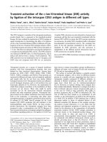
Fig. 1. Amino-acid substitutions in the ectodomain of the human com-
mon c chain (c
c
). The amino-acid sequence of c
c
is shown with boxed
portions i ndicating predicted b-strands which are designated by the
letter below the box. R esidues substituted in this study are indicated by
asterisks.
Fig. 2. Gel-filtration analysis of the human c
c
ectodomain expr essed in
SF9 cells and the m urine c
c
ectodomain expressed in E. coli. The samples
were applied to a Superdex 200 HR 10/30 c olumn and eluted w ith the
same buffer. The two peaks of human c
c
represent dimer (A) and
monomer (B).
1492 J L. Zhang et al.(Eur. J. Biochem. 269) Ó FEBS 2002
4 l
M
, a nd murine c
c
with a K
d
of 1.6 l
M
(Fig. 3 and
Table 1). In addition, different preparations of wild-type
human c
c
ectodomain consistently showed a K
d
of 4 l
M
irrespective of the monomer t o d imer ratio (data not
shown). Therefore, the mixtures o f dimeric and monomeric
human c
c
protein w ere u sed for all b iosensor measurements.
The c
c
epitope for IL-4 binding
The method of measuring the binding of c
c
to IL-4–IL-4BP
by biosensor was established previously [15]. The d issocia-
tion constant K
d
evaluated from the concentration
dependence of e quilibrium binding proved to be very
reliable for measuring the interaction of the c
c
ectodomain
with IL-4–IL-4BP. The measured K
d
for interaction of c
c
ectodomain variants with IL-4–IL-4BP are compiled in
Table 1. Eight c
c
variants including c
c
CHR exhibited
unchanged binding characteristics. Changes in binding
affinity were observed in 11 c
c
variants. The K
d
of six
variants was too high to be r eliably determined. A r ough
estimate yields K
d
values of about 200–300 l
M
for I100A,
L102A, Y103A and L208A, and K
d
values of about 500–
1000 l
M
for C160A and C209A. The K
d
values of five
variants, N128A, H15 9A, L161A, E162A, and G210A,
were found to be increased threefold to fourfold compared
with the K
d
of wild-type c
c
, suggesting that these residues are
part of the c
c
binding interface, but do not play a key role in
binding. The loss of binding affinity of the four variants
I100A, L102A, Y103A a nd L208A i s not likely to be c aused
by extended structural alterations, a s I 100A, L102A, and
L208AwerereportedtobindtoIL-2andIL-7withthe
same affinity a s wild-type c
c
, and the Y 103A mutation
resulted in only twofold to thre efold reduced IL-2 and IL-7
binding [30]. Thus, the four residues I100, L 102, Y103 and
L208 are hot spots on c
c
, contributing > 9 kJÆmol
)1
each.
The five minor residues investigated contribute only 2.9–
3.5 kJÆmol
)1
. The two cysteine variants C160A and C209A
exhibited a largely r educed binding affinity ( K
d
> 500 l
M
).
Thismaybecausedbystructuralperturbationofthe
protein. A direct role in binding, however, cannot be
excluded for these residues.
Double-mutant cycle analysis of the IL-4–c
c
interface
The c o-operativity of the interaction of some residues on the
IL-4 site 2 epitope and the c
c
ectodomain was determined in
this experiment. Double-mutant cycles were con structed
only for the mutants with minimal effects on b inding
(Tables 2 and 3), because the K
d
values for the interaction
between variants o f the main binding residues I100, L102,
Y103, a nd L2 08 of c
c
andIL-4variantsaswellasthe
interaction betwee n variant I 11A o f I L-4 a nd c
c
variants
were too high to b e reliably d etermined. Interaction be tween
residues on IL-4 and c
c
can be grouped according t o their
coupling energies (Table 3). The main binding determinant
of IL-4 Y124 failed to e xhibit positive coupling energies with
any of the c
c
residues analyzed. IL-4 Y124 probably
interacts w ith the main functional side chains of the receptor
located on loop EF1 (I100, L102, Y103), the binding of the
alanine variants of which was too weak to be analyzed by
this approach. Remarkably, IL-4 S125 neighboring Y124
does show coupling to receptor N128 in addition to that to
Fig. 3. Sensograms rec ording the binding of h uman and m urine c
c
ectodomains to the IL-4–IL-4BP complex. IL-4BP was i mmobiliz ed on
the biosensor matrix. At time zero, perfusion with 100 n
M
IL-4 was
initiated. The saturation binding of IL-4 was arbitrarily set as zero.
After 120 s, perfusion was continued with 1 00 n
M
IL-4 plus c
c
ecto-
domain. In different cycles, 5 l
M
human (a) or murine (b ) c
c
ecto-
domains w e re applied. Perfusion with buff er alone starte d at time
240 s. The ruler indicates resonance units (RU) corresponding to
1–10 l
M
murine c
c
ectodomain. Th e resonance u nit f or 5 l
M
human c
c
corresponded to that for 1.6–2 l
M
murine c
c
.
Table 1. Equilibrium binding between c
c
ectodomain mutants and
IL-4–IL-4BP. The dissociation constants K
d
were evaluated from
equilibrium binding between wild-type (wt) or mutants (mut) of the c
c
ectodomain and immobilized IL-4BP saturated with IL-4. The l oss of
free energy of binding on mutation was calculated as ddG
(kJÆmol
)1
) ¼ 5.69 log K
d
(mut)/K
d
(wt).
Alanine
variant
Equilibrium binding
ddG
(kJÆmol
)1
)
K
d
(l
M
) K
d
(mut)/K
d
(wt)
Murine c
c
(wt) 1.6
Human c
c
(wt) 4.0 1.0 0.0
Human c
c
CHR 4.5 1.1 0.2
Loop 1 (AB1)
N44A 2.8 0.7 )0.9
V45A 3.3 0.8 )0.5
Loop 3 (EF1)
E99A 5.5 1.4 0.8
I100A >320 >80 >11
L102A >240 >60 >10
Y103A >300 >80 >11
Q104A 5.6 1.4 0.8
Loop 4 (ID)
Q127A 2.4 0.6 )1.3
N128A 15 3.7 3.2
Loop 5 (BC2)
N158A 1.4 0.4 )2.3
H159A 17 4.1 3.5
C160A >900 >230 >13
L161A 13 3.2 2.9
E162A 13 3.2 2.9
Loop 6 (FG2)
P207A 3.4 0.9 )0.4
L208A >166 >40 >9
C209A >490 >120 >12
G210A 15 3.7 3.2
Ó FEBS 2002 Mutagenesis of human c
c
ectodomain (Eur. J. Biochem. 269) 1493
G210. The IL-4 side c hain of N15 functionally interacts with
the central receptor s ide chain N128, a nd also with H159
located at the periphery of the functional c
c
epitope. The
relative positions of the c oupling side c hains as proposed by
our theoretical model of the ternary complex (see below) are
presented i n t he open-book view in Fig. 4A,B. T he two
receptor side chains N1128 and H159 are 12 A
˚
apart in t he
c
c
model. This could indicate that our c
c
model is
inaccurate, because this model does not completely fit the
results of t he double-mutant cycle analysis. Alternatively,
the interaction of IL-4 side chain N15 with H159 (coupling
energy only 0.8 k JÆmol
)1
) m ay be indirect. Of p articular
interest is the I L-4 side chain of R121, which, after being
substituted with D or E, leads t o a selective IL-4 agonist
specifically impaired in IL-13R a1 binding [6,7,16,17,34].
The IL-4 R 121 was distinct in showing positive coupling
during interaction with the c
c
side chain L161.
Model of the structure of the IL-4–IL-4BP–c
c
ternary
complex
In a series of steps, c
c
was adapted to achieve a good fit to
the core structure (the binary complex; Fig. 5). The
procedure s tarted with moving the whole chain (Ôrigid
bodyÕ). Then domains and subdomains were moved indi-
vidually. The binary core complex was changed as little as
possible, being an experimentally determined structure and
thus the most reliable p art of the model, but some minor
changes in side chain o rientation could not be avoided for
proper a daptation. An important point was to keep t he
C-terminal domains of the receptor c hains c lose together, as
this was expected to be essential for dimer formation and
thereby signaling through the membrane. The structures of
c
c
and the ternary complex were modeled so that residues
that exhibit positive coupling energies during double-
mutant cycle analysis w ere placed c lose to each other.
Occasionally, however, t here was a Ôconflict of interestÕ
between the requirements of interaction and those of
dimerization.
DISCUSSION
This mutational analysis defines human c
c
residues involved
in IL-4 binding. T he residues are located in the EF1, BC2,
and FG2 loops and the interdomain segment of c
c
.The
functional b inding epitope of c
c
includes r esidues I100,
L102, Y103, and L208 as major binding dete rminants and
five residues, N128, H159, L161, E162, and G210, as minor
determinants. O ur results a lso show that the truncated c
c
CHR has the same binding affinity as the complete c
c
ectodomain, indicating that the short N-terminal region of
c
c
is not required for ligand binding. This is true for most
type I cytokine receptors, except for hgp130 [51] and
granulocyte colony-stimulating factor receptor [54]. There-
fore, c
c
CHR, the short form of the c
c
ectodomain, may be
Table 2. Double mutant cycle analysis of interaction between c
c
and
IL-4. The d issoc iation constants K
d
were evaluated from equilibrium
binding between wild-type (wt) o r mutants (mut) o f the c
c
ectodomain
and im mobilized IL-4BP saturated w ith w ild-t ype o r mutants of IL-4.
The loss of free e nergy o f binding on mutation was calculated a s
ddG ¼ 5.69 log K
d
(mut)/K
d
(wt). ddG
sum
is the sum of th e l osses of f ree
energy of binding upon m ut ation for IL-4 and c
c
separately. ND,
Sensogram could not be evaluated because of weak binding.
IL-4
variants
c
c
chain
variants
K
d
(l
M
)
ddG
(kJÆmol
)1
)
ddG
sum
(kJÆmol
)1
)
wt wt 4.0
N15A wt 20 4.0
R121A wt 12 2.8
Y124F wt 6.9 1.4
S125A wt 8.5 1.9
wt N128A 15 3.2
wt H159A 17 3.5
wt L161A 13 2.9
wt E162A 13 2.9
wt G210A 15 3.2
N15A N128A 34 5.3 7.2
N15A H159A 61 6.7 7.5
N15A L161A 75 7.2 6.9
N15A E162A ND – 6.9
N15A G210A 59 6.7 7.2
R121A N128A 50 6.2 6.0
R121A H159A 75 7.2 6.3
R121A L161A 27 4.7 5.7
R121A E162A 127 8.6 5.7
R121A G210A 81 7.4 6.0
Y124F N128A 22 4.2 4.6
Y124F H159A 33 5.2 4.9
Y124F L161A 24 4.5 4.3
Y124F E162A 83 7.5 4.3
Y124F G210A 29 4.9 4.6
S125A N128A 19 3.9 5.1
S125A H159A 57 6.6 5.4
S125A L161A 53 6.4 4.8
S125A E162A 37 5.5 4.8
S125A G210A 22 4.2 5.1
Table 3. Co-operativity between r esidue pairs in the i nteraction interface o f c
c
and IL -4. The couplin g e nergy between a p a ir of residues was c alc ulated
as d dG
int
¼ ddG
sum
) ddG (data from Table 2) ac cording to eqn ( 1). T he underlined values indicate f avorable i nteraction. The numbers in
parentheses are the calculated errors (2 r, a ¼ 0.95). ND, Sensogram could not be evaluated because of weak binding.
c
c
chain
variants
ddG of IL-4 variants (kJÆmol
)1
)
N15A R121A Y124F S125A
N128A
1.9 (0.62) )0.2 (0.66) 0.4 (0.80) 1.2 (0.90)
H159A
0.8 (0.64) )0.9 (1.10) )0.3 (0.83) )1.2 (0.86)
L161A )0.3 (0.99)
1.0 (0.80) )0.2 (0.64) )1.6 (0.72)
E162A ND )2.9 (0.90) )3.2 (0.76) )0.7 (0.68)
G210A 0.4 (0.66) )1.4 (0.87) )0.3 (0.48)
0.9 (0.67)
1494 J L. Zhang et al.(Eur. J. Biochem. 269) Ó FEBS 2002
Fig. 5. M odel of I L-4–IL-4BP–c
c
ternary complex. The s tructures o f I L-4, IL-4BP, and CHR of c
c
are depicted as ribbons and colored blue, red,
and green, respectively. The major binding residues on c
c
and IL-4 site II epitopes are represented by s ticks. The figure was g enerated using
MOLSCRIPT
and
RASTER
3
D.
Fig. 4. O pen-book view o f complementary functional IL -4 (site 2 ) (A) and c
c
(B) binding epitopes, and missense mutations in the putative loops of c
c
implicated in patients with XSCID (62) (C) . The structures of IL-4 and c
c
from our model are depicted as ribbons. The mutated residues are
represented b y space-filling m odels. The colors o f residues i n IL-4 and c
c
binding s ites indicate the loss o f binding free energy [ddG
(kcalÆmo l
)1
) ¼ 1.36 log (K
d
variant/K
d
wild-type)] d ue to alanine substitutio n (see Tab les 1 and 2; 1 kcalÆmol
)1
¼ 4.18 kJÆmol
)1
). The data for
I11, K12, an d Y124 wer e taken from Letzeler
2
et a l. [15]. The letters in parentheses in (C) indic ate the other mutations found in the s ame p osition .
The figure was produced with
MOLSCRIPT
and
RASTER
3
D
.
Ó FEBS 2002 Mutagenesis of human c
c
ectodomain (Eur. J. Biochem. 269) 1495
better suited to form crystals of IL-4–IL-4BP–c
c
than the
complete c
c
ectodomain for solving the structure of t he low-
affinity complex by X-ray diffraction.
It appears that binding of c
c
to IL-4 is sustained
predominantly by h ydrophobic i nteractions. O f t he nine
residues involved in IL-4 binding, five, in particular all four
major d eterminants, are hydrophobic. We propose that
residues I100, L102, and Y103 of loop EF1, a nd L208 of
FG2 form a hydrophobic c luster to interact with the
hydrophobic epitope composed of residues I11, N15, a nd
Y124 on helices A and D of IL-4 ( [12,15]; Figs 4 and 5).
Similar hydrophobic determinants have b een found in
several type I cytokine receptors, including hGHR [39],
hEPOR [48], hgp130 [50], and the human common b chain
(hb
c
[55–57]). Two of the three loops EF1, BC2 a nd FG2 of
these receptors appear to establish two major f unctional
interfaces with the ligands, and the binding is dominated by
one or two h ydrophobic aromatic r esidues. For example,
W104 and W 169 in loops EF1 and BC2 o f hGHR, F 93 and
F205 in loops EF1 and FG2 of h EPOR, F169 in loop EF1
of hgp130, and Y365 and Y421 in loops BC2 and FG2 of b
c
are all key residues in binding interactions (Fig. 6). In terms
of c
c
, Y103 is homologous to W104 of hGHR, to F93 of
hEPOR and to F169 of hgp130, and the FG2 loop
containing L208 may have a similar function to the loop
containing W169 in hGHR. In this regard, Y103 and L208
may h ave t he most im portant r ole in the hydrophobic
cluster for binding to IL-4.
The two c
c
variants, C160A and C209A, exhibited very
high K
d
values (> 490 l
M
and > 90 0 l
M
, respectively).
The two cysteines may form a disulfide bond between loops
BC2 and FG2. This prediction is consistent with our model
and one of the published models [46] of c
c
. The contribution
of the two residues to binding could not be directly
determined. The disulfide bond may be only important for
maintaining the structural integrity of c
c
. However, i t
cannot be ruled out that the disulfide group participates
directly in binding. These questions may be answered when
the structures of both free c
c
and the IL-4–IL-4BP–c
c
ternary complex are solved.
Double-mutant cycle analysis could identify co-operativ-
ity between two side chains [ 35], and predict a more d etailed
map of interacting residues without knowledge of t he
structures of the two proteins analyzed. Unfortunately, the
coupling e nergies between the m ajor determinants on c
c
and
IL-4 site 2 cannot be measured because of the low b inding
affinity of the alanine variants. Nevertheless, our experiment
revealed favorable interactions between several pairs of c
c
and IL-4 side chains. The results support our prediction of
hydrophobic interaction between the functional c
c
epitope
and I L-4 site 2 reasonably w ell. Accordingly, the b inding
epitope of c
c
can be divided into two functional interfaces
(Figs 4A,B and 5): ( a) I100, L 102, and Y 103 on t he EF1
loop interact mainly with IL-4 Y124 and S125; (b) L208 and
other residues on the BC2 a nd FG2 loops interact mainly
with IL-4 N15, and probably I11 (coupling w ith c
c
residues
could not be determined). The most i mportant is the
interface on the EF1 loop of c
c
, because the partner residue
IL-4 Y124 is a k ey determinant f or binding ( contributing
10.9 kJÆmol
)1
) [ 15], a nd the Y124D mutant exhibits a
complete antagonist activity [36]. The IL-4 R121 which is
more important for IL-13Ra1 binding [6,7,16,17] was found
to interact with L161 on the BC2 and FG2 interface of c
c
.
Its interaction with the binding residues on t he EF1 loop of
c
c
could not be exclud ed. Therefore, it would be interesting
to determine t he IL-1 3Ra1 epitope for IL-4 binding and
compare it with the c
c
epitope defined in this experiment.
It is unfortunate for our modeling process that the most
effective mutations did not yield interaction data. Therefore,
we had to rely on the residues of the weaker (but
measurable) interaction which, although they are expected
to work over larger distances and t hus provide less stringent
constraints than d esirable, nevertheless l ead to a quite
reasonable model as far as the gross features are concerned.
For all the details on a t ruly atomic scale, howeve r, we have
to await the crystal structure of the ternary complex.
This study focu ses o n t he molecular description of the
mechanism of recognition between human IL-4 and c
c
.
Nevertheless, it will be important to understand how the c
c
mutations and the associated changes in IL-4 binding affect
the biological a ctivity of c
c
during IL-4 signaling or t he
signaling of the cytok ines that d epend on c
c
. Previous
experiments with IL-4 mutant proteins [15] revealed that
substitutions in the c
c
binding e pitope l ead to partial
agonists and IL-4 antagonists. The binding affinity of such
mutants t o t he receptor on whole cells was a t m ost threefold
reduced compared with wild-type IL-4 (see, e.g [6]),
indicating that c
c
binding contributes only m arginally to
IL-4 binding affinity with the whole receptor complex (see
also [21]). Remarkably, only a l owering of t he c
c
binding
affinity of more than 100-fold, measured b y Biosensor in
certain IL-4 mutants, produced partial agonist activities of
less than 20%. It could be p redicted that the c
c
mutant
proteins with reduced IL-4 bindin g affinity will exhibit the
same alterations in biological activity as the c omplementary
IL-4 proteins. Furthermore, some point mutations
have been reported t o a brogate or diminish t he high
affinity of IL-4 (and also IL- 2 and IL-7) for Epstein-Barr
virus-transformed B cell lines or cells transfected with
Fig. 6. Alignment of loops of different cytokine receptors involved in
ligand binding. The struct ure-based sequence alignment of hIL-4Ra,
hGHR, hEPOR, and hgp130 was taken from Hage et al.[12].The
sequence s o f h c
c
and hb
c
were aligned manually. The c entral part of the
EF1, BC2, and FG2 l oops of receptors are selectively shown. Key
residues fo r b inding are underlined. The d eletions are marked Ô–Õ.The
interaction of the receptor with the respective ligand is classified as
follows:
a
AD or AC helix interface involved in receptor binding;
b
polarity of the interface;
c
affinity of ligand–receptor interaction.
1496 J L. Zhang et al.(Eur. J. Biochem. 269) Ó FEBS 2002
mutant sequences derived from patients with XSCID
[58–61]. Two c
c
mutants (A134V and R202C) were found
to produce t wofold and fourfold reduced IL-4 and IL-2
binding, and to be less effective in modulating Jak3
activation stimulated by IL-4 and IL-2, respectively
[60,61]. A134 is located at the periphery of the c
c
epitope
identified in this study and has not been included in the
present experiment.
Some of the residues in the c
c
epitope for IL-4 binding as
identified in this study (Y103, L161, L208 and G210) have
been found to be mutated in patients with XSCID
(Fig 4B,C [62]). The XSCID phenotype seems to be caused
predominantly by the dis ruption of IL-7 and/or IL-15
signaling [28,63]. Thus, the c
c
epitopes for binding of IL-4
and of IL-7 and/or IL-15 most likely share Y103, L161,
L208 and G 210 as binding det erminants. Remarkably,
L161 and G210 of c
c
are only minor determinants for IL-4
binding. T he severe deficiency produced in XSCID m ay
result from the particular substitutions (G210R and L161S;
Fig. 4C [62]); this could be more disruptive than an alanine
substitution. Alternatively, L161 and G210 may be major
determinants for IL-7 and/or IL-15 binding. More d etailed
molecular information is needed on how far the c
c
epitopes
for binding of IL-4, IL-2, IL-7, I L-9, IL-15, and IL-21 differ
or coincide. Then t he severity of the clinical manifestation in
patients with XSCID c an possibly be correlated w ith c
c
mutations in major or minor binding determinants.
Thecommonnatureofc
c
raises the possibility that
common residues for binding different ligands may exist in
this receptor. Indeed, some common residues contributing
to binding of different ligands have been found in hb
c
[55,56]
and hgp130 [51]. Our result and the mutagenesis analysis of
the b inding of the m urine c
c
chain t o I L-2 a nd IL-7 [30]
show that Y103 of c
c
is a key ligand-interacting residue for
IL-2, IL-4, and IL-7. Y103 is probably a common critical
residue for all c
c
-dependent receptor systems. In a ddition, in
that study [30], the counterpart of three dominated residues
I100, L 102 a nd L208 of human c
c
for I L-4 binding were
reported not to be important for IL-2 a nd IL-7 binding.
These residues are probably unique to IL-4 binding, a s
suggested by the fact that c
c
binding sites for different
cytokines overlap but are not identical [29,64]. However, it
cannot be ruled out that some of the binding residues of c
c
defined in our study also participate in IL-2 and IL-7
binding, as, in the aforementioned s tudy, only one re sidue
(Y103) was shown to be directly involved in IL-2 and IL-7
binding. Y 103A or Y103R mutations resulted in only
slightly (twofold to threefold) reduced IL-2 and IL-7
binding [30]. The difference between these results and our
own may partly originate f rom the different methods
applied. Therefore, further studies will be required to
determine w hether Y103 and other residues i dentified in
the present study are also involved in binding of other
c
c
-dependent cytokines.
The co-ordinate of the model of the IL-4–IL-4BP–c
c
ternary complex is available from the authors.
ACKNOWLEDGEMENTS
The authors are grateful to Dr J . Nickel for h elpful discussion,
Dr Siddiqi for drawing the figures, and W. Ha
¨
delt and C. So
¨
der for
excellent technical assistance. This work was supported by the S FB 487,
TP B2, and by the Fonds der Chemischen I ndustrie.
REFERENCES
1. Paul, W.E. (1991) Interleukin-4: a prototypic immunoregulato ry
lymphokine. Blood 77, 1859–1870.
2. Nelms, K ., K eegan, A.D ., Z amorano, J., Ryan, J.J. & Paul, W.E.
(1999) The IL-4 r eceptor: s ignaling mechanisms and biologic
functions. Annu. Rev. Immunol. 17, 701–738.
3. Paul, W.E. & Seder, R .A. (1994) Lymphocyte responses and
cytokines. Cell 76, 241–251.
4. Coffman, R.L., Lebman, D.A. & Rothman, P. (1993) Mechanism
and regulation of immunoglobulin isotype switching. Adv.
Immunol. 54, 229–270.
5. Romagnani, S. (1994) Mechanism and regulation of immuno-
globulin isotype switching. Curr. Opin. Immunol. 6, 838–846.
6. Shanafelt, A.B., Forte, C.P., Kasper, J.J., Sanchez-Pescador, L.,
Wetzel, M., Gundel, R. & Greve, J.M. (1998) An imm une cell-
selective interleukin 4 agonist. Proc. Natl Acad. Sci. USA 95,
9454–9458.
7. Srivannaboon, K., Shanafelt, A.B., T odisco, E., F orte, C.P.,
Behm, F.G., Raimondi, S.C., Pui, C.H. & Campana, D. (2001)
Interleukin-4 variant (BAY 36–1677) selectively induces apoptosis
in acute lymphoblastic leukemia cells. Blood 97, 752–758.
8. Sugamura, K., Asao, H., Kondo, M., T anaka, N., Ishii, N.,
Nakamura, M. & Takeshita, T. (1995) The common gam ma-chain
for multiple cytokine receptors. Adv. Immunol. 59, 225–277.
9. Gauchat, J.F., Schlagenhauf, E., Feng, N.P., Moser, R., Yamage,
M., Jeannin, P., Alouani, S., Elson, G ., Notarangelo, L.D., Wells,
T., Eugster, H.P. & Bonnefoy, J.Y. (1997) A novel 4-kb inter-
leukin-13 receptor alpha mRNA e xpressed in human B, T, and
endothelial cells encoding an alternate type-II interleukin-4/inter-
leukin-13 receptor. Eur. J . Immu nol . 27, 971–978.
10. Bazan, J.F. (1990) Structural design & molecular evolution of a
cytokine receptor superfamily. Proc. Natl A cad. Sc i. U SA 87 ,
6934–6938.
11. Wang, Y., Shen, B.J. & Sebald, W. (1997) A mixed-charge pair
in human interleukin 4 dominates high–affinity interaction
with the receptor alpha chain. Proc. Natl Acad. Sci. USA 94,
1657–1662.
12. Hage, T., Sebald, W. & Reinemer, P. (1999) Crystal structure of
the interleukin-4/receptor alpha chain com plex reveals a mo saic
binding interface. Cell 97, 271–281.
13. Zhang, J.L., Simeonowa, I., W ang, Y. & Sebald, W. (2002) The
high-affinity interaction of human IL-4 and the receptor a chain is
constituted by two independent binding clusters. J. M ol. Biol. 315 ,
399–407.
14. Domingues, H., Cregut, D., Sebald, W., Oschkinat, H. & Serrano,
L. (1999) Rational design of a GCN4-derived mimetic of inter-
leukin-4. Nat. Struct. Biol. 6, 652–656.
15. Letzelter,F.,Wang,Y.&Sebald,W.(1998)Theinterleukin-4site-
2 epitope determining binding of the common receptor gamma
chain. Eur. J. Biochem. 257, 11–20.
16. Tony, H.P., Shen, B.J., Reusch, P. & S ebald, W. (1994) De sign of
human interleukin-4 antagonists inhibiting interleukin-4-depend-
ent and interleu kin-13-dependen t responses in T-cells and B-cells
with high efficiency. Eur. J. Biochem. 225, 659–665.
17. Schnarr, B., Ezernieks, J., Sebald, W. & Duschl, A. (1997) IL-4
receptor complexes containing or lacking the gamma C chain are
inhibited by an overlapping set of antagonistic I L-4 mutant pro-
teins. Int. Immunol. 9, 861–868.
18. G runewald , S .M., Werthmann, A., S ch narr, B ., Klein, C.E.,
Brocker, E.B., Mohrs, M., Brombacher, F., Sebald, W. &
Duschl, A. (1998) An antagonistic IL-4 mutant prevents type I
allergy in the m ouse: inhibition of the IL-4/IL-13 receptor system
completely abrogates humoral immune response to allergen and
development of allergic symptoms in vivo. J. Immunol. 160 ,
4004–4009.
19. Asao, H., Okuyama, C., Kumaki, S., Ishii, N ., Tsuchiya, S.,
Foster, D. & Sugamura, K. (2001) The common gamma-chain is
Ó FEBS 2002 Mutagenesis of human c
c
ectodomain (Eur. J. Biochem. 269) 1497
an indispensable subunit of the IL-21 receptor complex.
J. Immunol. 167, 1–5.
20. Takeshita, T., A sao, H., O htani, K., Ishii, N., K umaki, S.,
Tanaka, N., Munakata, H., Nakamura, M. & Sugamura, K.
(1992) Cloning of the gamma chain of the human IL-2 receptor.
Science 257, 379–382.
21. Russell, S.M., Keegan, A.D., Harada, N ., Nakamura, Y.,
Noguchi, M., Leland, P., Friedmann, M.C., Miyajima, A., Puri,
R.K., Paul, W.E. & Leonard, W.J. (1993) Interleukin-2 receptor
gamma chain: a functional co mponent of the in terleukin-4
receptor. Science 262 , 1880–1883.
22. Kondo, M., Takeshita, T., Hig uchi, M., Nakamura, M., S udo, T.,
Nishikawa, S. & Sugamura, K. (1994) Functional participation
of the IL-2 receptor g amma ch ain in IL -7 recep tor comp lexes.
Science 263, 1453–1454.
23. Leonard, W.J. & O’Shea, J.J. (1998) Jaks and STATs: biological
implications. Annu. Rev. Immunol. 16, 293–322.
24. Leonard, W.J. (1996) The molecular basis of X-linked severe
combined immunodeficiency: d efective cytokine receptor signal-
ing. Annu.Rev.Med.47, 229–239.
25. Lo, M., Bloom, M.L., Imada, K., Berg, M., Bollenbacher, J.M.,
Bloom, E.T., Kelsall, B.L. & Leonard, W.J. ( 1999) Restoration of
lymphoid populations in a murine m od el of X -lin ked severe
combined immun odeficien cy by a gene-therapy approac h. Blood
94, 3027–3036.
26. Soudais, C., Sh iho, T., Sharara, L.I., Guy-Grand, D., Taniguchi,
T., F ischer, A . & Di Santo, J.P. (200 0) Stable and f unctional
lymphoid re co nstitution of common cytokine receptor gamma
chain d eficient mice by r etrov iral-media ted gene transfe r. Blo od 95,
3071–3077.
27. Cavazzana-Calvo, M., Hacein-Bey, S ., de Saint Basile, G ., Gross,
F.,Yvon,E.,Nusbaum,P.,Selz,F.,Hue,C.,Certain,S.,Casa-
nova,J.L.,Bousso,P.,Deist,F.L.&Fischer,A.(2000)Gene
therapy of hu man severe c ombined immunodeficiency (SCID)-X1
disease. Science 288, 669–672.
28. Leonard, W.J. (2000 ) X-linked s evere c ombined i mmunodefi-
ciency: from molecular cause to gene therapy within seven years.
Mol. Med. Today 6, 403–407.
29. Raskin, N., Jakubowski, A., Sizing, I.D., Olson, D.L., Kalled,
S.L., Hession, C.A., Benjamin, C.D., Baker, D.P. & B urkly, L.C.
(1998) Molecular mapping with functional antibodies localizes
critical sites on the human I L receptor common gamma (gam ma c)
chain. J. Immunol. 161, 3474–3483.
30. Olosz, F. & Malek, T.R. (2000) Thre e loops of the common
gamma chain ectodomain req uired f or the bind ing of i nterleuk in-2
and interleukin-7. J. Biol. Chem. 275, 30100–30105.
31. Kumaki,S.,Kondo,M.,Takeshita,T.,Asao,H.,Nakamura,M.
& Sugamura, K. ( 1993) Clon ing o f t he mo use interleukin 2
receptor gamma chain: demonstration of fu nc tional differences
between the mo use a nd hu man r eceptor s. Biochem. Biophys. Res.
Commun. 193, 356–363.
32. Shen, B.J., Hage, T. & S ebald, W . (1996) Global a nd local
determinants for the kinetics o f interl eukin- 4/interleuk in-4
receptor alpha chain i nteraction . A biosensor s tudy employing
recombinant interleukin-4-binding protein. Eur. J. Biochem. 240,
252–261.
33. Sondermann, P. & Jacob, U. (1999) Human Fcgamma receptor
IIb expresse d in E sch erichia coli reveals IgG binding capability.
Biol. Chem. 380, 717–721.
34. Kruse, N., Shen, B.J., Arnold, S., Tony, H.P., Muller, T. &
Sebald, W. (1993) Two distinct functional sites of human inter-
leukin 4 are identified by variants impaired in either r ecept or
binding or receptor activation. EMBO J. 12, 5121–5129.
35. Schreiber, G. & Fersht, A.R. ( 1995) E nergetics of pr otein–protein
interactions: analysis of the barnase–barstar interface by
single mutations and do uble mutant cycles. J. Mol. Biol. 248,
478–486.
36. Kruse, N., Tony, H.P. & Sebald, W. (1992) Conversion of human
interleukin-4 into a high affin ity antagonist by a s ingle amino acid
replacement. EMB O J. 11, 3237–3244.
37. Sch reiber, G ., Frisch, C. & Fersht, A.R. (1997) The role of Glu73
of barnase in catalysis and t he binding o f b arstar. J. M ol. Biol. 270 ,
111–122.
38. Mueller, T., Oehlenschlaeger, F. & Buehner, M. (1995) Human
interleukin-4 and variant R88Q: phasing X-ray diffraction data
by molecular r eplacement using X-ray and nuclear magnetic
resonance models. J. Mol. Biol. 24 7, 360–372.
39. de Vos, A.M., U ltsch, M. & K ossiakoff, A .A. ( 1992) Human
growth hormone and extracellular domain of its recep tor: crystal
structure of the complex. Science 255, 306–312.
40. Bernstein, F.C., Koetzle, T.F., Williams, G.J.B., Meyer, E.F. Jr,,
Brice, M.D., Rodgers, J.R., K ennard, O., Shimanouchi, T. &
Tasumi, M. ( 1977) The P rotein Data Bank: a compute r-based
archival file for m a cromolecular structures. J. Mol. Bio l. 112,535–
542.
41. Jones, T.A., Zou, J.Y., Cowan, S.W. & Kjelgaard, M. (1991)
Improved meth ods f or b indin g prote in m od els i n e lectron density
maps an d the location of errors in these models. Act a Crystallogr.
A47, 110–119.
42. Bruenger, A.T. (1992) X-Plor, Version 3.1. A System for X-Ray
Crystallography and NMR. Yale University Press, New Haven,
CT, USA.
43. Jack, A. & Levitt, M. (1978) Refinement of large structures by
simultaneous minimization of energy and R. factor. Acta Crys-
tallogr. A34, 931–935.
44. Bamborough, P., Hedgecock, C.J. & Richards, W.G. (1994) The
interleukin-2 and interleukin-4 receptors studied by molecular
modelling. Structure 2, 839–851.
45. Gustchina, A., Zdanov, A ., Schalk-Hihi, C. & Wlodawer, A.
(1995) A model of the complex between interleukin-4 and its
receptors. Proteins 21, 140–148.
46. Kroemer, R.T. & Richards, W.G. (1996) Homology modeling
study of t he human interleukin -7 rec eptor complex. Protein Eng. 9,
1135–1142.
47. Clackson, T. & W ells, J.A. ( 1995) A hot spot of binding energy in
a hormone–receptor inter face. Science 267, 383–386.
48. Livnah, O ., Stura, E.A., J oh nson, D.L., M iddleton, S.A.,
Mulcahy, L.S., Wrighton, N.C., Dower, W.J., Jolliffe, L.K. &
Wilson, I.A. (1996) Functional mimicry of a protein hormone by a
peptide agonist: the EPO rece ptor complex at 2.8 A
˚
. Science 27 3,
464–471.
49. Middlet on, S.A., Barbone, F.P., J ohnson, D.L., Thurmond, R .L.,
You, Y., McMahon, F.J., Jin, R., Livnah, O., Tullai, J., Farrell,
F.X., Goldsmith, M .A., Wilson, I.A. & Jolliffe, L.K. (1999)
Shared and unique determinants of th e erythropoietin (EPO)
receptor are i mportant for binding EPO and EPO mimetic pep-
tide. J. Biol. Chem. 274, 14163–14169.
50. Horsten, U., Muller-Newen, G., G erhartz, C., Wollmer, A.,
Wijdenes, J., Heinrich, P .C. & Grotzi nger, J. (19 97) Molecular
modeling-guided mutagenesis of the extracellular part of gp130
leads to the identification of c ontact s ites in th e i nter leukin-6
(IL-6). J. Biol. Chem. 272, 23748–23757.
51.Kurth,I.,Horsten,U.,Pflanz,S.,Dahmen,H.,Kuster,A.,
Grotzinger, J., Heinrich, P.C. & Muller-Newen, G. (1999) Acti-
vation of t he s ignal transducer glycoprotein 130 by both IL-6 and
IL-11 requires two dis tinct bind ing epitopes. J. Immunol. 162,
1480–1487.
52. Hoff man, R.C., Castner, B. J., Gerhart, M ., Gibson, M.G.,
Rasmussen,B.D.,March,C.J.,Weatherbee,J.,Tsang,M.,
Gustchina, A., Schalk-Hihi, C., Reshetnikova, L. & Wlodawer, A.
(1995) Direct evidence of a heterotrimeric complex of human
interleukin-4 with its receptors. Protein Sci. 4, 382–386.
53. Baker, D.P., Whitty, A., Zafari, M.R., Olson, D.L., Hession,
C.A., Miatkowski, K., Avedissian, L.S., Foley, S.F., McKay,
1498 J L. Zhang et al.(Eur. J. Biochem. 269) Ó FEBS 2002
M.L., Benjamin, C.D. & Burkly, L.C. (1998) The m urine a nti-
human co mmon gamma chain m onoclo nal a ntibody CP.B8
blocks the second step in the formation of the intermediate affinity
IL-2 receptor. Biochemistry 37, 14337–14349.
54. Hiraoka, O., Anaguchi, H., Asakura, A. & Ota, Y. (1995)
Requirement for the immunoglobulin-like domain of granulocyte
colony-stimulating factor receptor in formation of a 2: 1 receptor–
ligand complex. J. Biol. Chem. 270, 25928–25934.
55. Woodcock, J.M., Z acharakis, B., Plaetinck, G., Bagl ey, C.J.,
Qiyu, S., Hercus, T.R., Tavernier, J. & Lopez, A.F. (1994) Three
residues in the common beta c hain of the human GM -CSF, IL-3
and IL-5 receptors are essential for GM-CSF and IL-5 but not
IL-3 high affinity binding and in teract with Glu21 of GM-CSF.
EMBO J. 13, 5176–5185.
56. Wo odcock, J .M., B agley, C.J., Zacharakis, B. & L opez, A .F.
(1996) A single tyrosine residue in the membrane-proximal
domain of the granulocyte-macrophage colony-stimulating factor,
interleukin (IL) - 3, and IL-5 receptor c ommon beta-chain is
necessary and sufficien t for high affinity bin ding and signalin g by
all three ligands. J. Biol. Chem. 271, 25999–26006.
57. Haman,A.,Cadieux,C.,Wilkes,B.,Hercus,T.,Lopez,A.,Clark,
S. & Hoang, T. (1999) A s ingle tyrosine residue in the membrane-
proximal domain of the granulocyte-macrophage colony-stimu-
lating facto r, interleukin (IL)-3, and IL-5 re ceptor common
beta-chain is necessary and sufficient for high a ffin ity b inding and
signaling by all three ligands. J. Biol. Chem. 274, 34155–34163.
58. DiSanto, J.P., Dautry-Varsat, A., Certain, S., Fischer, A. & de
Saint Basile, G . (1994) Interleukin-2 (IL-2) r eceptor gamma chain
mutations i n X-linked severe combined immuno deficiency disease
result in the l oss of h igh-affinity IL-2 receptor binding. Eur.
J. Immunol. 24, 475–479.
59. Ishii,N.,Asao,H.,Kimura,Y.,Takeshita,T.,Nakamura,M.,
Tsuchiya, S., Konno, T., Maeda, M., Uchiyama, T. & Sugamura,
K. (1994) Impairment of ligand binding and growth s ignaling of
mutant IL-2 receptor gamma-chains i n p atients with X-linked
severe combined im mu nodeficie ncy. J. Immunol. 153, 1310–1317.
60. K umaki, S., Ishii, N., Mineg ishi, M ., Tsu chiya, S., Cosman, D.,
Sugamura, K. & Konno, T. ( 1999) Functional role o f interleukin-4
(IL-4) and IL-7 in the development of X -linked severe combined
immunodeficiency. Blood 93, 607–612.
61. Sharfe, N., Shahar, M. & Roifman, C.M. (1997) An interleukin-2
receptor gamma chain mutation with normal thymus morpho-
logy. J. Clin. Invest. 100, 3036–3043.
62. Puck, J.M. (1996) A database of gamma c-chain defects causing
human X-SCID. Immunol. Today 17, 507–511.
63. Malek, T.R., Porter, B.O. & He, Y.W. (1999) Multiple gamma
c-dependent cytokines r egulate T-cell develo pment. Immunol.
Today 20, 71–76.
64. He, Y.W., Adkins, B., Furse, R.K. & Malek, T.R. (1995)
Expression and function of the gamma c s ubunit of the IL-2, IL-4,
and IL-7 receptors. Distinct interaction of gamma c in the IL-4
receptor. J. Immunol. 154, 1596–1605.
Ó FEBS 2002 Mutagenesis of human c
c
ectodomain (Eur. J. Biochem. 269) 1499