Báo cáo Y học: The SK-N-MC cell line expresses an orexin binding site different from recombinant orexin 1-type receptor pptx
Bạn đang xem bản rút gọn của tài liệu. Xem và tải ngay bản đầy đủ của tài liệu tại đây (377.6 KB, 8 trang )
The SK-N-MC cell line expresses an orexin binding site
different from recombinant orexin 1-type receptor
Heike A. Wieland
1,
*, Richard M. So¨ll
2,3
, Henri N. Doods
1
, Dirk Stenkamp
1
, Rudolf Hurnaus
1
,
Ba¨ rbel La¨ mmle
1
and Annette G. Beck-Sickinger
2
1
Division of Preclinical Research, Boehringer Ingelheim Pharma KG, Biberach, Germany;
2
Institute of Biochemistry,
University of Leipzig, Germany;
3
Department of Applied Biosciences, Swiss Federal Institute of Technology, Zurich, Switzerland
Orexin A and B (also known as hypocretins), two r ecently
discovered neuropeptides, play an important role in food
intake, sleep/wake cycle and neuroendocrine functions.
Orexins are endogenous ligands of two G-protein-coupled
receptors, termed OX
1
and OX
2
. This work presents the first
short orexin A and B analogues, orexin A 23–33 and orexin
B 18–28, with high affinity (119 ± 49 and 49 ± 23 n
M
)for
OX
1
receptors expressed o n SK-N-MC cells and indicates
the importance of the C-terminal part of the o rexin peptides
for this ligand–receptor interaction. However, t hese C-ter-
minal fragments of orexin did n ot displace the
125
I-labelled orexin B from the recombinant orexin 1
receptor stably expressed in Chinese hamster ovary cells. To
examine the role of the shortened orexin A 23–33 in feeding,
its e ffects in m imicking or a ntagonizing the effects of o rexin
A were studied in rats after administration via the lateral
hypothalamus. In contrast with orexin A, which potently
induced feeding up to 4 h after administration, orexin A
23–33 neither induced feeding nor inhibited orexin
A-induced feeding. Modafinil (VigilÒ), which was shown
earlier to activate o rexin neurons, d isplayed binding ne ither
to the orexin receptor e xpressed on SK-N-MC cells nor to
the r ecombinant orexin 1 r eceptor, which indicates that
modafinil d isplays i ts antinarcoleptic action via another yet
unknown mechanism. PCR and subsequent sequencing
revealed expression of the full-length o rexin 1 receptor
mRNA in SK-N-MC and NT-2 cells. Interestingly,
sequencing of several cDNA clones derived from RNA of
both SK-N-MC and NT-2 cells differed from t he published
nucleotide sequence at position 1375. Amino acid prediction
of this AfiG change results in an isoleucinefivaline sub-
stitution at the protein level, which may provide evidence for
an ed iting process.
Keywords: food intake; h ypocretin; ligand–receptor inter-
action; obesity; orexin.
Two novel neuropeptides, orexin A and B, were recently
discovered independently by two groups and identified as
potent stimulators of food intake after i ntracerebroventric-
ular administration [1–4]. Further investigations revealed a
broad involvement of these peptides in the regulation of
many physiological and behavioural activities that are
associated with f eeding behaviour [4–7], in the modulation
of neuroendocrine function and the sleep/wake cycle
[8–12]. Both peptide amides derive from prepro-orexin, a
precursor p rotein produced in defined r egions of the lateral
and perifornical hypothalamus, whose mRNA i s up-regu-
lated upon fasting. O rexin immunoreactive n eurons are,
however, distributed widely in the brain, including regions
of the cerebral cortex, the medial groups of the thalamus,
the circumventricular organs, the limbic s ystem and the
brain s tem [13–15]. A key r ole for orexins in n arcolepsy has
been described [10,16–18]. It was shown recently that the
anti-narcoleptic drug Modafinil (VigilÒ), the mechanism of
action of which is unknown, might act through the orexin
pathway [10].
Orexin A consists of 33 amino acids, is C -terminally
amidated and contains t wo intramolecular disulfide bonds,
that connect cysteine residues from positions 6–12 and 7–14,
respectively. Orexin B consists of 28 residues and sh ares
46% i dentity w ith o rexin A, mainly a t the C t ermin us. The
three-dimensional solution structure of orexin B was
recently determined b y two-dimensional NMR and shows
two a helices, connected by a short linker sequence at
position 20–23 [19]. The structure of orexin A is conserved
among human, rat, mouse and cow, whereas r odent
orexin B contains two amino acid substitutions compared
with the human sequence: proline i nstead of serine in
position two and asparagine instead of s erine in position 18.
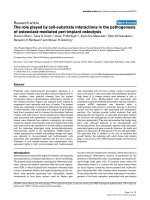
Xenopus laevis has orexins that differ slightly from the
human sequence, but the C-terminal decapeptide of
orexin A and B and the positions next to the disulfide
bonds in orexin A remain conserved (Fig. 1), which
suggests some importance in biological activity of these
peptide regions [20].
Orexin A and B are endogenous ligands of two closely
related (64% amino acid identity [1]) heptahelical
G-protein-coupled receptors, termed OX
1
and OX
2
. They
induce an intracellular i ncrease in free Ca
2+
concentration
after activation of the receptors [1,21]. Orexin A shows
Correspondence to A. G. Beck-Sickinger, Institute of Biochemistry,
University of Le i pzig, Talstr. 33, 04103 L e ipzig, Germany.
Fax: +49 341 9736 998, Tel.:+ 49 341 9735 901,
E-mail:
Abbreviations:OX
1
receptor, orexin 1 receptor; OX
2
receptor, orexin 2
receptor;NPY,neuropeptideY;HOBt,N-h ydroxybenzotriazole;
CHO, Chinese hamster ovary; IC
50
, 50% inhibitory concentration.
*Present address: Aventis Phar ma Deutschland G mbH, DG
Thrombotic Diseases/Degenerative J o int Diseases, H811, D-65926
Frankfurt, Germany.
(Received 2 4 September 200 1, revised 7 December 2001, a ccepted
12 December 2 001)
Eur. J. Biochem. 269, 1128–1135 (2002) Ó FEBS 2002
higher affinity to the OX
1
receptor, whereas the binding
affinity of the two peptides to OX
2
receptor is in the same
order of magnitude [10].
Up to now, little is known about the structure–activity
relationship, except for the relevanc e of the C-terminal
segment of o rexin A [22]. O nly r ecently, a subtype selective
nonpeptide antagonist was described in vitro [23]. We
describe here the shortest orexin A and B analogues that
bind to OX-receptors. We have also determined that the
orexin type 1 receptor is expressed by SK-N-MC cells, a
human neuroblastoma cell line, although with a pharma-
cological profile different from that of the recombinantly
expressed OX
1
receptor. In addition, we describe an amino
acid position that differs in clones derived from RNA that
has been isolated from SK-N-MC cells.
MATERIALS AND METHODS
Materials
N
a
-Fmoc-protected amino a cids w ere f rom A lexis
(La
¨
ufelfingen, Switzerland). The side-chain protecting
groups were tert-butyl for serine and threonine, and trityl
for a sparagine and histidine. The 4-(2¢,4¢-dimethoxyphenyl-
Fmoc-aminomethyl)-phenoxy (Rink Amide) resin was from
Novabiochem (La
¨
ufelfingen). N-hydroxybenzotriazole
(HOBt), trifluoroacetic a cid, thioanisole, p -thiocresol, tri-
methylsilylbromide, 1,2-ethanedithiol, p iperidine, tert -buta-
nol, 1,1,1-trifluoroethanol and dimethylformamide were
from Fluka. N,N ¢-diisopropylcarbodiimide w as from
Aldrich. Dimethylformamide (pure) and diethylether were
from Scharlau (La Jota, Barcelona, Spain). Acetonitrile was
from Romil (Cambridge, England).
Dulbecco’s modified E agle’s medium was from B ioWhit-
taker; OPTI-MEM and Lipofectamine were from G ibco
BRL; fetal bovine serum was from BioWhittaker; Hepes
was from Fluka; geneticin was from Gibco BRL; Pefabloc
SC was from Merck;
125
I-labelled Tyr-human orexin B
(specific activity 2130 CiÆmmol
)1
) was from Anawa (Zu
¨
rich,
Switzerland);
125
I-labelled Tyr-human orexin A (spec ific
activity 2130 CiÆmmol
)1
was from NEN; orexin B was from
Bachem (Heidelberg, Germany).
Modafinil (Vigil
Ò
) was from Laboratoire L. Lafon,
Merckle, Blaubeuren (Germany), NT-2 cells were from
Stratagene.
Peptide synthesis
The C-terminal undecapeptides of the o rexins, orexin A
23–33 and orexin B 18–28, and the analogues of orexin B
and orexin A 23–33 were synthesized by automated
multiple solid-phase peptide s ynthesis on a peptide synthe-
sizer (Syro, MultiSynTech, Bochum, Germany) using Rink
Amide resin (30 mg, resin loading 0.6 mmol Æg
)1
). Amino
acids were attached by the Fmoc-strategy in a double
coupling procedure, using a 10-fold excess of Fmoc-amino
acid, H OBt and N,N ¢-diisopropylcarbodiimide in d imethyl-
formamide a nd a reaction t ime o f 4 0 min per coupling.
Fmoc-deprotection was accomplished w ith 40% piperidine
in dimethylform amide f or 3 min, 2 0% piperidine for 7 min
and finally 40% piperidine for a further 5 m in. The
orexin A fragment was cleave d from the resin with a
mixture o f trifluoroacetic a cid/thioanisole/p-thiocresol
(90 : 5 : 5, v/v), precipitated from ice-cold diethylether,
collected by ce ntrifugation and washe d four times with
diethylether. The methionine-containing orexin B fragment
was cleaved from the resin using a mixture of trifluoroacetic
acid/thioanisol/ethanedithiol ( 90 : 7 : 3, v/v), precipitated
and washed as described. Partial oxidation of the methio-
nine residue was reduced by dissolving the p eptide (15 mg,
0.014 mmol) in 1 mL trifluoroacetic acid, followed by the
addition of ethanedithiol (15.7 lL, 0.2 mol ÆL
)1
)and
trimethylsilylbromide (13 lL, 0.1 molÆL
)1
) [24]. The solu-
tion was shaken for 40 min at room temperature and the
peptide w as precipitated and washed as described. P urifica-
tion of the peptide was achieved by preparative H PLC on a
C18-column (Waters, 5 lm, 25 · 300 mm) with a linear
gradient of 10–30% A in B; A ¼ 0.08% trifluoroacetic
acid in acetonitrile, B ¼ 0.1% trifluoroacetic acid in
water) and a flow rate of 15 mLÆmin
)1
. The peptides were
dissolved in tert-butanol/water (1 : 3) and lyophilized.
Analytical characterization of the peptides w as achieved
by electrospray ionization MS (SSQ 710, Finnigan MAT,
Bremen, Germany) and by analytical reversed-phase HPLC
on a LiChrospher RP18-column (5 lm, 3 · 125 mm,
Merck, Darmstadt, Germany) using linear gradients of
5–50% over 30 min (I), 10–60% over 30 min (II), 10–40%
over 30 min (III) or 20–40% over 30 min (IV). Analyt ical
data were as expected [orexin A, 23–33: molecular mass (m),
m
expected
1036 Da; m
found
, 1036.1 ± 0.5 Da; HPLC reten-
tion time (I), 17.3 min; [G23] orexin A 23–33: m
expected
1022 Da; m
found
1021.5 ± 0.6 Da; HPLC retention time
(II), 11.4 min. Orexin B 18–28: m
expected
1070 Da; m
found
1069.9 ± 0.1 Da; HPLC retention t ime (III), 12.9 min.
[L28] orexin B: m
expected
,2881Da;m, 2881.3 ± 0.4 Da;
HPLC retention time (IV) 16.6 min.
cDNA subcloning and nucleotide sequence
determination
PCR w as used to amplify the full-length o rexin 1 receptor
according t o a ccession number AF041243 [1]. Oligonucleo-
tides f rom MWG Biotech (Ebersberg, Germany) were used
as primers: OX
1
-f, 5¢-GTAGAGCCTAGGATGCCCCT-
3¢;OX
1
-r: 5¢-AGGAAGTGACTTATCCAGAGT-3¢.
Total RNA from SK-N-MC cells and NT-2 cells were
used as templates. Isolation of total RNA was performed
with an RNeasy Total RNA Kit (Qiagen). RT-PCR was
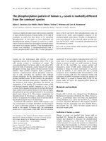
Fig. 1. Sequence o f (A) matu re orexin A
peptides of human, bovine and rat origin and (B)
mature orexin-B peptides. Deviations from the
human s equences are underlined.
U ¼ pyr oglutamic acid.
Ó FEBS 2002 SK-N-MC cell line expresses different orexin binding sites (Eur. J. Biochem. 269) 1129
performed using the Superscript Preamp lification System
(Gibco/BRL). After 3 min at 94 °C, the reactions were
subjected t o 3 5 cycles of: denaturation, 1 min at 94 °C;
annealing, 2 min at 60 °C; elongation 2 min at 72 °Cina
primus plus cycler (MWG Biotech). PCR products of the
expected size were cloned in pCR2.1TOPO using the TOPO
TA Cloning Kit from Invitrogen. The sequence was
confirmed using the BigDye Terminator Cycle Sequencing
with an ABI 377 Sequencer using the M13 Forward ()20)
and Reverse primers (Invitrogen).
cDNA from orexin type 1 receptor was from Receptor
Biology (Beltsville, MD, USA), sequenced and OX
1
R-
cDNA was subcloned into pcDNA3.1/HisA vector from
Invitrogen.
Cell culture
Transfection into Chinese hamster ovary (CHO) cells was
performed using the lipofectamine PLUS method according
to the m anufacturer’s protocol (Gibco/BRL) using expres-
sion plasmids encoding the orexin 1 receptor.
Binding assays with transfected cells
CHO cells were grown in nutrient m ixture Ham’s F12
medium with 10% fetal bovine serum from BioWhittaker
(Boehringer Ingelheim Bioproducts Partnership, Verviers,
Belgium), nonessential amino acids, hygromycin B, 2 m
M
L
-glutamine and 1% geneticin (Gibco/BRL) at 37 °Cand
5% CO
2
until they were confluent in a 24-well plate. The
medium was aspirated. The cells were washed twice with
0.25 mL NaCl/P
i
. Incubation buffer [0.2 mL; 84.7 m
M
NaCl, 3 0 m
M
KCl, 1.2 m
M
MgSO
4
Æ7H
2
0, 11.2 m
M
NaH
2
PO
4
,bufferedwithHepes,15 m
M
(4-(2-hydroxyethyl)-
1-piperazine e thanesulfonic acid, pH 7.5; from SERVA,
Heidelberg, Germany)] and at the day of the experiment
5.5 m
M
glucose, 0.1% BSA, 0.05 mgÆmL
)1
bacitracin was
added. The total v olume (0.25 mL) contained 100 p
M
final
concentration of
125
I-labelled Tyr-human (h) orexin B or
125
I-labelled Tyr-human orexin or
125
I-labelled h nueropep-
tide Y (NPY)-Tyr36 (specific activity: 2000 CiÆmmol
)1
;
Amersham) and increasing concentrations of the cold
ligand orexin B or increasing concentrations of test
compounds. After 120 min of gentle shaking a t room
temperature the supernatant was removed followed by t wo
washes with 0.25 mL NaCl/P
i
. Lysis buffer was a dded
(NaCl/P
i
containing 2% Triton · 100) and after one wash
with 0.5 mL radioactivity was counted.
Membrane preparation and binding assay
on SK-N-MC cells
For m embrane p reparation of SK-N-MC ce lls, t he cells
were grown in MEM (MEM with Earl’s salt, 10% fetal
bovine serum, 1 m
M
sodium pyruvate, 1 % nonessential
amino acids, 4 m
M
glutamine). Confluent cells were
removed with 0.02% EDTA/ NaCl/P
i
and resu spended in
10 mL incubation buffer (MEM/25 m
M
Hepes containing
0.5% BSA, 50 l
M
phenylmethanesulfonyl fluoride, 0.1%
bacitracin, 3 .75 m
M
CaCl
2
), then washed twice with 10 mL
NaCl/P
i
. After addition of 5 mL preparation buffer ( 5 m
M
Hepes, 0.32
M
sucrose, 50 l
M
pefabloc, pH 7 .0) the cells
were removed with a rubber policeman. After centrifugation
at 4 °C, 10 min, 48 200 g the supernatant was decanted and
centrifuged at 4 °C, 30 min, 48 200 g. The pellet was
resuspended in 15 mL NaCl/P
i
. The sample was recentri-
fuged at 4 °C, 50 min, 150 g and the pellet was resuspended
in incubation buffer (84.7 m
M
NaCl, 3 0 m
M
KCl, 1.2 m
M
MgSO
4
Æ7H
2
O, 11.2 m
M
NaH
2
PO
4
bufferedwithHepes).
After counting, the cells were diluted to a final concentration
of 1.0 · 10
6
cellsÆmL
)1
and homogenized using an Ultra-
Thurrax. After the addition of 5.5 m
M
glucose, 0.1% BSA
and 50 lg bacitracin, 200 ll of t his cell suspension was
incubated for 2 h at room temperature with 100 p
M
125
I-labelled orexin B and increasing concentrations of
orexin, orexin a nalogues or NPY (Neosyste
`
me, Strasbourg,
France) in a total volume of 0.25 mL. Unbound radio-
activity was separated by filtration through Whatman GF/
C filters presoaked in 0.5% po lyethylenimine. The filters
were washed three times with ice-cold 0.9% NaCl. A ll tips
and vials were siliconized.
Competition binding experiments were analysed by a
nonlinear least-squares fitting method with a one- or two-
binding site model, respectively (
RS/1
software package,
BBN Research Systems, Cambridge, MA, USA). The
maximum specific r adioligand binding was s et to 100%. All
data (n ¼ 3) are e xpressed a s mean ± SEM.
Circular dichromism
Conformational properties of the peptides were investigated
by CD spectroscopy using a JASCO model J720 spectro-
polarimeter ov er 190 –250 nm at 20 °CinaN
2
atmosphere.
The peptides were dissolved in 20 m
M
NaCl/P
i
at neutral
pH containing 0%, 30%, 50% or 70% trifluoroethanol and
in pure trifluoroethan ol, in a concentration range of
200–300 l
M
. Each measurement was repeated three times
using a thermostatable sample cell with a path of 0.02 cm
and the following parameters: r esponse time, 2 s; scan
speed, 2 0 nmÆmin
)1
; s ensitivity of 10 mdeg; s tep resolution,
0.2 nm; band width, 2 nm. The CD spectrum of the solvent
was subtracted from the CD spectra of the peptide solutions
to eliminate the interference from c ell, solvent and optical
equipment. High f requency noise was reduced by means of
a low-path Fourier-transform filter. The ellipticity was
expressed as the mean-residue molar ellipticity [Q]
R
in
degÆcm
2
Ædmol
)1
.
Rodent model of food intake
Adult male Chbb:Thom rats weighing between 300 a nd
340 g were individually housed and maintained on a
12 light: 12 h dark cycle beginning at 06.00 hours. Tap
water and standard laboratory chow were available
throughout. After 1 week of habituation to their new
housing conditions, t he animals w ere a naesthetized with
sodium pentobarbital (60 mgÆkg
)1
, intraperitoneally) for
the placement of stainless steel guide cannulae. Cannulae
(26 gauge) were placed 1 mm above the lateral hypothal-
amus according to the stereotaxic coordinates: AP : 2.1,
L : 2.0, V : 7.2 (+1 mm injection tip: 8.2). Guide cannulae
were maintained in place on the skull w ith s mall metal
screws and d ental a crylic cement. Cannulae were closed
with a stainless steel stylet when not in use. Rats were
allowed to recover for a t l east 1 week and were adapted t o
the injection procedure. On the day of the experiments drugs
1130 H. A. Wieland et al. (Eur. J. Biochem. 269) Ó FEBS 2002
were injected between 01.00 and 02.00 p.m. Injection
cannulae (33 gauge) were inserted 1 mm beyond the tips
of the guide cannulae. The injection c annulae were attached
by polyethylene t ubing to a Hamilton microsyringe mount-
ed in an infusion pump. Injection volume was 0.4 lLgiven
slowly over 40 s.
Groups of six to eight rats received either saline ( c ontrol),
1.0 nmol Ærat
)1
orexin A unilaterally, o r 1 nmolÆrat
)1
orexin
A and 3 nmol rat
)1
orexin A 23–33 into the lateral
hypothalamus and food intake was monitored for 4 h. In
the s econd set o f experiments 1.0 nmol orexin A 23–33 was
given with the injection of 1.0 nmol orexin A in order to
antagonize the effects of o rexin A.
RESULTS
The peptides were synthesized by automated multiple
peptide synthesis on a R ink Amide resin to directly obtain
the peptide amides after cleavage of the peptides from the
resin [25]. In addition to the native sequences h-orexin A
and B, w e u sed t wo C-terminal segments h-orexin A 23–33
and h-orexin B 18–28. B ecause of the differ ent length o f the
natural orexins, these two C-terminal segments are homo-
logous and correspond to the C-terminal undecapeptide of
orexin A and orexin B, respectively. Nine amino acids are
identical, whereas orexin B contains a C-terminal methio-
nine in contrast with leucine in orexin A (Fig. 1). This led to
the orexin B analogue [L28] orexin B to make s ure that a ny
identified differences are not owing to the different
sequences. The second variable position is residue 23
(orexin A , alanine)/18 (orexin B, glycine). To investigate
the role of this exchange we investigated [G23] orexin A
23–33 (Table 1) .
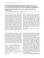
The binding affinity of the peptides was tested on SK-N-
MC cells.
125
I-labelled orexin B binding was inhibited in a
dose-dependent fashion with a K
i
of 118 ± 57 n
M
(Table 1,
Fig. 2) and to a similar order of magnitude on NT-2 cells,
another human cell-line (data not shown). All curves
displayed a monophasic shape with slopes close to unity.
125
I-labelled orexin B could be displaced by the orexin A and
orexin B fragments in the range of human orexin B itself or
with slightly improved affinity (Table 1). Sub stitution of
orexin A at position 23 did not improve affinity significantly.
Several atte mpts to detect specific
125
I-labelled orexin A
binding was unsuccessful with SK-N-MC cells whereas
recombinant CHO cells expressing the OX
1
receptor
revealed a 5 0% inhibitory concentration (IC
50
)of
10 ± 6 n
M
for inhibition of
125
I-labelled orexin A binding
by orexin A. The C-terminal fragments orexin A 23–33
and orexin B 18–28 do not displace
125
I-labelled orexin B
from the recombinant receptor; neither does [G23]
h-orexin A 23–33 displace
125
I-labelled orexin A. The first
selective orexin 1 receptor antagonist (SB-334867-A) has
been described recently, with a pK
i
value of 7.17 n
M
[23,26]. We tested a compound related to SB-334867,
published earlier by G. Chan et al. [27], 1-(4-
N,N-dimethylaminophenyl)-3-chinolin-4yl-urea), named
EXBN8016BS. This compound displayed an IC
50
of
149 ± 3 n
M
for the inhibition of
125
I-labelled orexin A at
the r ecombinant OX
1
receptor whereas it c annot inhibit
the
125
I-labelled orexin B binding to both the recombinant
OX
1
receptor or the orexin 1 receptor expressed on
SK-N-MC cells. M odafinil was shown earlier to activate
orexin-responsive n eurons. Therefore, w e e xamined
whether Modafinil a cts indirectly via inhibitory orexin
autoreceptors. Modafin il displayed no s ignificant a ffinity
for the orexin B binding site of SK-N-MC cells or of
recombinantly expressed OX
1
receptors. Sensitivity of
orexin B binding to NPY has been observed (Table 1)
with an IC
50
of % 450 n
M
.
Table 1. Binding affinity of h-orexin A and B, C-terminal orexin A and B fragments and reported antagonists on SK-N-MC cells and CHO cells stably
transfected w ith the huma n OX
1
receptor (1 00 p
M
radioligand).
Ox1 receptor
125
I-labelled orexin B
IC
50
[n
M
]
SK-N-MC cells
125
I-labelled orexin B
K
i
[n
M
]
Ox1 receptor
125
I-labelled orexin A
IC
50
[n
M
]
h-orexin A > 1000 (n ¼ 3) 882 ± 286 10 ± 6
h-orexin B 138 ± 28 118 ± 57 16 ± 15
[L28]h-orexin B 370 ± 200 – 180 ± 126
h-orexin A 23–33 > 10 000 (n ¼ 3) 119 ± 46 –
[G23]h-orexin A 23–33 9400 ± 450 93 ± 80 > 10 000 (n ¼ 2)
h-orexin B 18–28 > 10 000 (n ¼ 3) 49 ± 23 –
Modafinil > 10 000 (n ¼ 3) > 10 000 (n ¼ 3) –
EXBN8016BS
a
> 10 000 (n ¼ 3) > 10 000 (n ¼ 3) 149 ± 3
NPY 454 ± 241 (n ¼ 2) 450 ± 49 (n ¼ 2) –
a
1-(4-N,N-dimethyl-aminophenyl)-3-chinolin-4yl-urea) [27].
Fig. 2. Receptor binding studies with
125
I-labelled orexin B and orexin B
(m) using SK-N-MC cells.
Ó FEBS 2002 SK-N-MC cell line expresses different orexin binding sites (Eur. J. Biochem. 269) 1131
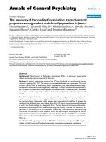
Structure
The structure of the peptides w as investigated by CD
spectroscopy in aqueous solutions at neutral pH,
containing increasing amounts of trifluoroethanol. All
peptides adopted mainly random structure. Fi g. 3 s hows
the CD spectra of orexin A 2 3–33 dissolved in water (A),
50% t rifluoroethanol in water ( B), 70% trifluoroethanol
in water (C) and pure trifluoroethanol (D) in order to
see any stabilizing effects of the solvent. All other
peptides showed comparable CD spectra (data not
shown). The negative band at 198 nm in aqueous
solution, an indication of randomly structured pep tides,
was shifted to 202 nm in all trifluoroethanol-containing
samples. The negative CD value of the water solution at
190 n m was raised to positive values in trifluoroethanol-
containing solutions. These shifts indicate partial forma-
tion o f an a helix in trifluoroethanol-contain ing samples.
Analysis of the spectra by a secondary structure
estimation program (
JASCO
, J-700 for Windows) based
on the method of Yang et al. [ 28] revealed a slightly
increasing amount of a helix with increasing amount of
trifluoroethanol, although the maximum amount of helix
was only % 11% (Fig. 3).
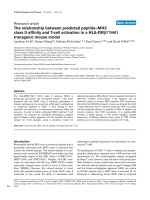
Food intake
Administration of 1 nmol orexin A into the third ventricle
of rats significantly increased food intake after 2 and 4 h
whereas a trend was seen after 6 and 8 h and no effec t
was seen after 24 h. One nanomole of orexin A also
significantly increased food intake after administration
into the lateral hypothalamus (Fig. 4A). Administration
of orexin A 23–33 together with orexin A in order to
evaluate a potential antagonistic property of orexin A 23–
33 did not reveal any effect (Fig. 4A). Orexin A 23–33 in
a dose range of 1 and 3 nmol per rat did not induce
feeding (Fig. 4B).
Sequencing
To study orexin binding we searched for a suitable cellu lar
system and screened neuronal cell lines of human origin
(e.g. SK-N-MC and NT-2) for orexin receptor binding
sites.
Analysis of the cDNA derived from t otal RNA r evealed
that these cell lines co ntain intronless o rexin 1 receptor
transcripts that s eem t o b e partially edited in the c odon for
the isoleucine/valine site at position 1375 (amino acid 408)
beyond transmembrane region 7 (Fig. 5, Table 2). Fig. 5
shows that all other amino acids are 100% identical to the
published sequence [1].
Analysis of the human genomic DNA revealed that
adenosine is present at this position, which leads to an
isoleucine at this position in the protein (personal commu-
nication, Receptor Biology Inc).
DISCUSSION
The sequence o f the C-terminal decapeptide of o rexin A
and B is conserved throughout all s pecies examined. Here
we show that the C -terminal orexin fragments, orexin A
23–33 and orexin B 18–28, bind to the orexin receptor
expressed on SK-N-MC cells with an affinity in the same
range or with four- t o eightfold improved affinity c ompared
Fig. 3. CD spectra and secondary structure according to the calculation
of Yang et al. [28] of orexin A 23–33. Solvent: (A) water, pH 7.0;
(B) 50% trifluoroethanol in water; (C) 70% trifluoroethanol in water;
(D) pure t rifluoroethano l.
Fig. 4. Food intake studies. Food intake after administration
of orexin A via the lateral hypothalamus and after administration of
orexin A 23–33 and orexin A (A). Food intake after administration of
orexin A 23–33 (B).
1132 H. A. Wieland et al. (Eur. J. Biochem. 269) Ó FEBS 2002
with orexin B and orexin A, respective ly. On the contrary,
initial data with N-terminal fragments, e.g. orexin B 1–10 or
orexin B 1 –13, showed no significant affinity to th is orexin
receptor (data not shown). This indicates an important
sequence motif in the C-terminus of the peptides, that might
play an essential role in binding of the peptides to t his
receptor. This is in accordance with the recently published
three-dimensional structure of orexin B, solved by two-
dimentional NMR. I t s howed the peptide to consist of two
a-helices, connec ted by a short linker [19]. The C-terminal
helix of the mature human orexin B extended from r esidue
22 to residue 28. Although these seven residues constitute
the major part of the ore xin A and B fragments, a helical
structure, as postulated f or the matu re orexin B, could be
found neither in orexin A 23–33 nor in orexin B 18–28.
Even dissolving the peptides in solutions containing high
amounts of trifluoroethanol, an a helix-inducing s olvent,
resulted in peptides with maximal 11% a helix and still 89%
random structure. This indicates that besides the confor-
mational properties, t he amino acid side chains might also
play an important role in binding of the orexin fragments to
the receptors, in particular the trifunctional residues aspar-
agine, histidine and threonine and additionally for orexin B
18–28 serine and the C-terminal methionine. The confor-
mational influence of the C-terminal part of the o rexins
remains unclear. The introduction of a helix inducers into
C-terminal peptide fragments might increase binding affin-
ity, because the native peptide contains a stable a helix at the
C-terminus.
Binding studies on SK-N-MC cells, a human n euroblas-
toma cell line that is known to express NPY Y
1
receptors
[29], revealed that this cell line also expresses receptors of the
orexin family, which, after sequencing, turned out to
correlate with the cloned OX
1
receptor. Both neuropeptides,
NPY and orexins, are involved in the regulation of food
intake. Sensitivity of orexin A binding to NPY has been
described earlier by studying
125
I-labelled orexin A binding
[30]. Interestingly some affinity of NPY could be identified
fororexinreceptorsaswell.Assomecross-reactivityhas
been reported [31], and orexin-induced food intake seems to
involve the NPY pathways [22], this m ight b e one mode of
regulation.
Our results indicate the importance of the C-terminal part
of th e orexins for binding interaction o f the ligand a nd its
receptor expressed on SK-N-MC cells. The lack of affinity
of the fragments for the recombinant OX
1
receptor suggests
a different pharmacological profile a lthough a sequence
almost i dentical t o the rec ombinant receptor is expressed in
the neuroblastoma cell line. This discrepancy might be
explained b y p ost-translational modifications, heterodimer-
ization (re viewed in [33]) or different ac cessory proteins of
the orexin 1 receptor e xpressed in SK-N-MC cells, similar
to those found for the CGRP receptor [34] possibly resulting
in different binding profiles. The binding to an orexin 2
receptor is possib le, but unlikely, because the dual e xpres-
sion of both receptors with a different pharmacological
profile should r esult in a biphasic inhibition curve. This w as
not observed.
The different IC
50
values of ligands, e.g. EXBN 8016BS
or orexin A, to inhibit either
125
I-labelled ore xin A or
Fig. 5. Sequence similarity of the o rexin 1
receptor cloned both from SK-N-MC and NT-2
cells. Posi tion X means G o r C at nucleotide
1375 coding for amino acid 408 which is either
translated into isoleucine [1] or valine
(Table 2).
Table 2. Position 137 5 is nuc leotide 1375 within the O X
1
receptor
cDNA which i s either t ranslated to isoleucine or valine.
Cells/clone Pos 1375 Nucleotide at
NT-2-cells
1 G Val
2G
3 A Ile
4A
5A
6A
7A
8A
SK-N-MC
2G
3G
Database
(Acc.no AF 041243) [1]
A Ile
Ó FEBS 2002 SK-N-MC cell line expresses different orexin binding sites (Eur. J. Biochem. 269) 1133
125
I-labelled orexin B binding at the recombinan t OX
1
receptor might be due to different binding epitopes of the
receptors as found for other agonist/antagonist systems of
neuropeptides, such as NPY o r substance P [35,36]. It was
hypothesized earlier [10] that modafinil may promote
wakefulness t hrough orexin neurons. Sin ce, however, mod-
afinil has low a ffinity fo r orexin 1 receptors, this activation
might i nvolve other orexin receptors or could have another
mechanism.
The feeding effects with orexin A are a t variance w ith the
strong and persistent feeding response observed by S akurai
et al . [1], but similar to those d escribed by others in rats [3]
and mice [37]. O rexin B showed no effects (data not shown)
which is in agreement with earlier reports [3,4,37]. This is
why we chose the shortened o rexin A fragment and not
orexin B 18–28 for f eeding studies. The lack of feeding
effects of orexin A 23–33 indicates that the orexin B binding
site expressed on SK-N-MC cells is not represented in the
lateral hypothalamus and therefore might not be involved in
the regulation of food intake.
However, this study has revealed a single nucleotide
mismatch between corresponding cDNAs encoding orexin 1
receptors. Human genomic DNA analysis (personal com-
munication, Receptor Biology) indic ated that alternative
exons c ould be excluded as a potential source for this
nucleotide exchange. Hence, editing of the RNA transcribed
from these genes best explains our observation w hich is
similartotheAfiG editing described in glutamate-gated
channels [38,39] and with the G p rotein-c oupled seroton in-
2C receptor [40]. Single nucleotide polymorphism cannot be
excluded at this point but the G was not found on a genomic
level. Subtle kinds of regulation of G p rotein-coupled
receptors coupling to G proteins have been described earlier
[33]. For example, transcripts encoding the 5-HT
2C
recep-
tor, a phospholipase C-coupled receptor, un dergo R NA
editing events in which the g enomically encoded adenosine
residues are converted to i nosines by a double-stranded
RNA adenosine deaminase(s). Seven major 5-HT
2C
recep-
tor isoforms are predicted, e ncoded by 11 distinct R NA
species and differing in their second intracellular loops [40].
This post-transcriptional modification leads to a 10- to
15-fold reduction in efficacy of the coupling of 5-HT
2C
to
the G protein.
We conclude that the s tructural s tudies o f C-terminal
orexin A and B fragments and their binding affinity to the
orexin receptors as presented in this w ork, provide a nother
step in characterization o f the r eceptor binding mode of
orexin A and B and provide important information for
pharmacological a nd biochemical investigations. The
observed A/G exchange in SK-N-MC and NT-2, two
human cell lines, might indicate an editing process which
will be further investigated.
ACKNOWLEDGEMENTS
The financial support of the Deutsche Forschungsgemeinschaft
(Be 1264-3/1) is kindly acknowledged. We further acknowledge the
skilled technical e xpertise of E. Liebhardt a nd S. Schacherl-Schmid.
REFERENCES
1. Sakurai, T., Amemiya, A., Ishii, M., Matsuzaki, I., Chemelli,
R.M., T anaka, H., Williams, S.C., Richardson, J.A., Kozlowski,
G.P., Wilson, S., Arch., J.R.S., Buckingham, R.E., Haynes, A.C.,
Carr, S.A., Annan, R.S., McNulty, D.E., Liu, W S., Terrett, J.A.,
Elshourbagy, N.A., Bergsma, D.J. & Yanagisawa, M. ( 1998)
Orexins and orexin receptors: a family of hypothalamic neuro-
peptides and G protein-coupled receptors that regulate feeding
behavior. Cel l 92, 573 –585.
2. De Lecea, L., Kilduff, T.S., Peyron, C., Gao, X B., Foye, P.E.,
Danielson, P.E., Fukuhara, C., Battenberg, E.L.F., Gautvik, V.T.,
Bartlett,F.S., I.I.Frankel, W.N., Van den Pol, A.N., Bloom, F.E.,
Gautvik, K.M. & Sut cliffe, J.G. (1998) The hypocretins: hypo-
thalamus-specific peptides with neuro excitatory activity. Proc.
Natl Acad. S ci. USA 95 , 322–327.
3. Haynes, A.C., Jackson, B., Overend, P., Buckingham, R.E.,
Wilson, S., T adayyon, M. & Arch., J.R.S. ( 1999) Effects of single
and chronic intracerebroventricular administration o f the or exins
on feeding in the rat. Peptides 20 , 1099–1105.
4. Ida, T., Nakahara, K., Katayama, T., Murakami, N. &
Nakazato, M . (1999) Effect of lateral cerebroventricular injection
of the appetite-stimulating neuropeptide, orexin and neuropeptide
Y, on the various behavioral activities of rats. Brain Res. 821,
526–529.
5. Takahashi, N., Okumura, T., Yamad a, H. & Kohgo, Y. (1999)
Stimulation of gastric acid secretion by centrally administered
orexin-A in conscious r ats. Biochem. Biophys. Res. Commun. 254,
623–627.
6. Griffond, B., Risold, P.Y., Jacquemard, C ., Colard, C. &
Fellmann, D. (1999) Insulin-induced hypoglycemia increases
preprohypocretin (orexin) mRNA in the r at lateral hypothalamic
area. Neurosci. L ett. 262, 77–80.
7. Dyer, C.J., Touchette, K.J., Carroll, J.A., Allee, G.L. &
Matteri, R.L. (1999) Cloning of porcine prepro-orexin cDNA
and effects of a n intramuscular injection of synthetic porcine
orexin-B on food intake in young pigs. Domestic Anim Endocrinol.
16, 145–148.
8. Van den Pol, A.N., Gao, X.B., Obrietan, K., Kilduff, T.S. &
Belousov, A.B. (1998) Presynaptic and postsynaptic actions and
modulation of neuroendocrine neurons by a new hypothalamic
peptide, hypocretin/orexin. J. Neurosci. 18, 7 962–7971.
9. Date, Y., Mondal, M.S., Matsukura, S., Ueta, Y., Yamashita, H.,
Kaiya, H., Kangawa, K . & Nakazato, M. ( 2000) Distribution of
orexin/hypocretin in the rat median eminence and pituitary. Brain
Res. Mol. Brain Res. 76, 1–6.
10. Chemelli, R.M., Willie, J.T., Sinton, C.M., Elmquist, J.K.,
Scammell, T., Lee, C., Richardson, J.A., Williams, S.C., Xiong,
Y., Kisanuki, Y., Fitch, T.E., Nakazato, M., Hammer, R.E.,
Saper, C.B. & Yanagisawa, M . ( 1999) Narcolepsy in or exin
knockout mice: molecular genetics of sleep regulation. Cell 98,
437–451.
11. Singer, C.M. & Lewy, A.J. (1999) Does our DNA determine when
we sleep? Nat. Med. 5 , 983.
12. Hagan, J.J., Leslie, R.A., Patel, S., E vans, M.L., W attam, T.A.,
Holmes, S., Benham, C.D., Taylor, S.G., Routledge, C.,
Hemmati, P., Munton, R.P., Ashmeade, T.E., Shah, A.S.,
Hatcher, J.P., Hatcher, P.D., Jones, D.N.C., Smith, M.I., Piper,
D.C., Hunter, A .J., Porter, R.A. & Upton, N. (1999) Orexin A
activates locus coeruleus cell firing and increases arousal in the rat.
Proc. N atl Acad. Sci. USA 96 , 10911–10916.
13. Nambu, T., Sakurai, T., Mizukami, K., Hosoya, Y., Yanagisawa,
M. & Goto, K. (1999) Distribution of orexin neurons in the adult
rat brain. Brain R es. 827, 2 43–260.
14. Dube, M.G., Kalra, S.P. & Kalra, P.S. (1999) Food intake elicited
by central administration of orexins/hypocretins: identification of
hypothalamic sites o f action. Brain Res. 842, 4 73–477.
15. Peyron, C., Tighe, D.K., van den Pol, A.N., de Lecea, L., H eller,
H.C., Sutcliffe, J.G. & Kilduff, T.S. (1998) Neurons containing
hypocretin (orexin) project to multiple neuronal systems.
J. Ne urosci. 18, 9996–10015.
1134 H. A. Wieland et al. (Eur. J. Biochem. 269) Ó FEBS 2002
16. N ishino, S., Ripley, B., Overeem, S., Lammers, G.J. & Mignot, E.
(2000) Hypocretin (orexin) deficiency in human narcolepsy.
Lancet 35 5, 39–40.
17. Lin,L.,Faraco,J.,Li,R.,Kadotani,H.,Rogers,W.,Lin,X.,Qiu,
X., de J on g, P.J., Nishino, S . & Mignot, E. ( 1999) the sleep d is-
order canine narcolepsy is caused by a mutation in the hypocretin
(orexin) receptor 2 gene. Cell 98, 365–376.
18. Siegel, J.M. (1999) Narcolepsy: a key role for hypocretins
(orexins). Cell 98, 4 09–412.
19. Lee,J.H.,Bang,E.,Chae,K.J.,Kim,J.Y.,Lee,D.W.&Lee,W.
(1999) Solution structure o f a n ew hypothalamic neuropeptide,
human h ypocretin-2/orexin-B. Eu r. J. Bioc hem. 266 , 831–839.
20. S hibahara, M., Sakurai, T., N amb u, T., Takeno uchi, T., Iwaasa,
H., Egashira, S.I., Ihara, M. & Goto, K. (1999) Structure, tissue
distribution, a nd pharmacological characterization of Xenopus
orexins. Peptides 20, 1 169–1176.
21. Smart, D ., Jerman, J.C., Brough, S.J., Rus hton, S.L., Murdoch,
P.R., Jewitt, F., Elshourbagy, N.A., Ellis, C.E., Middlemiss, D.N.
& B rown, F. (1999) Characterization of recombinant h uman
orexin receptor pharmacology in a Chinese hamster ovary cell-line
using F LIPR. Br. J. Pharmaco l. 128,1–3.
22. Darker, J.G., Porter, R.A., Eggleston, D.S., Smart, D., Brough,
S.J., Sabido-David, C. & Jerman, J.C. (2001) Structure–activity
analysis of truncated orexin-A analogues at the orexin-1 receptor.
Bioorg. Med. Chem. L et t. 11, 737– 740.
23. S mart, D ., Sabido-David, C., Br ough, S.J., Jewitt, F., Johns, A.,
Porter, R.A. & Jerman, J.C. (2001) SB-334867-A: the first
selective o rex in-1 receptor anta gonist. Br.J.Pharmacol.132,
1179–1182.
24. Beck,W.&Jung,G.(1994)ConvenientreductionofS-oxidesin
synthetic peptides, lipopeptides and peptide libraries. Lett. Pept.
Sci. 1, 3 1–37.
25. So
¨
ll, R. & Beck-Sickinger, A.G. (2000) On the synthesis of orexin
A: a novel protocol to obtain peptides with two intramolecular
disulfide bonds. J. Pept. Sci. 6,387.
26. Rodgers, R.J., Halford, J.C., Nunes de Souza, R.L., Canto de
Souza, A.L., Piper, D.C., Arch., J.R., Upton, N., Porter, R.A.,
Johns, A. & B lundell, J .E. (2001) SB-334867, a selective orexin-1
receptor an tago nist, e nh ances behavioural satiety a n d b locks the
hyperphagic e ffect of orexin-A in r ats. Eur. J. Neurosci. 13, 1444–
1452.
27. Chan,G.,Johns,A.,Jurewicz,A.,Porter,R.&Widdowson,K.
Phenyl urea and phenyl thiourea derivatives as HFGAN72
antagonists. WO 99/09024.
28. Yang, J.T., Wu, C.S. & Martinez, H.M., (1986) Calculation of
protein conformation from circular dichromism. Methods Enzy-
mol. 130 ,208.
29. B eck-Sickinger, A.G., Wieland, H.A., Wittneben, H., Willim,
K.D.,Rudolf,K.&Jung,G.(1994)Complete
L
-alanine scan of
neuropeptide Y reveals ligands binding to Y1 and Y2 recep-
tors with distinguished conformations. Eur. J. Biochem. 225 ,
947–958.
30. Kane, J.K., Tanaka, H., Parker, S.L., Yanagisawa, M. & Li, M.D.
(2000) Sensitivity of orexin-A binding to phospholipase C in hibi-
tors, neuropeptide Y, and secretin. Biochem. Biophys. Res.
Commun. 271, 959– 965.
31. Jaszberenyi, M ., Bujdoso, E. & T elegdy, G. (2001) The role of
neuropeptide Y in orexin-induced hypothalamic-pituitary-adrenal
activation. J. Neuroendocrinol. 13, 438–441.
32. Yamanaka, A., Kunii, K., Nambu, T., Tsujino, N., Sakai, A.,
Matsuzaki, I., Miwa, Y., Goto, K. & Sakurai, T. (2000) Orexin-
induced food intake involves neuropeptide Y pathway. Brain Res.
859, 404–409.
33. Bockaert, J. & Pin, J .P. (1999) Molec ular tinkering of G protein-
coupled receptors: an evolu tionary process. EMBO J. 18, 1723–
1729.
34. McLatchie, L., Fraser, N., Main, M., Wise, A., Brown, J.,
Thompson, N., S olari, R., Lee, M. & F o ord, S. ( 1998) RAMPs
regulate the transport and ligand specificity of the calcitonin-
receptor-like r eceptor. Nature 393, 333– 339.
35. S autel, M., Rudolf, K., Wittneben, H., Herzog, H., Martinez, R.,
Munoz, M., Eberlein, W., Engel, W., Walker, Ph & Beck-Sick-
inger, A.G. (1996) Neuropeptide Y and the non peptide antagonist
BIBP 3226 share a large overlapping binding site at the human Y
1
receptor. Mol . Pharmacol. 50 , 285–292.
36.Rosenkilde,M.M.,Cahir,M.,Gether,U.,Hjorth,S.A.&
Schwartz, T .W. (1994) Mutations along transmembrane se gment
II of the NK-1 r eceptor affect substance P competition with
non-peptide antagonists but not substance P binding. J. Biol.
Chem. 269, 2 8160–28164.
37. Lubkin, M. & Stricker-Krongrad, A. (1998) I ndependent feeding
and metabolic a ctions of Orexins i n mice. Biochem. Biophys. Res.
Commun. 253, 241– 245.
38. Sommer, B., Ko
¨
hler, M., Sprengel, R. & Seeburg, P.H. (1991)
RNA editing in brain controls a d eterminant of io n flow in
glutamate gated channels. Cell 67, 11– 19.
39. Higushi, M ., Single, F., Ko
¨
hler, M., Som mer, B., Sprengel, R. &
Seeburg, P.H. (1993) RNA editing of AMPA receptor subunit
GluR-B: a base-paired intron-exon structure determines position
and efficiency. Cell 75, 1 361–1370.
40. B urns, C.M., Chu, H., R ueter, S.M., H utchinson, L.K., C an ton,
H., Sanders- Bush, E . & Emeson, R.B. (1997) Regu lation of
serotonin-2C receptor G-protein coupling by RNA editing.
Nature 387, 303–308.
Ó FEBS 2002 SK-N-MC cell line expresses different orexin binding sites (Eur. J. Biochem. 269) 1135