Báo cáo Y học: Interaction of bovine coagulation factor X and its glutamic-acid-containing fragments with phospholipid membranes A surface plasmon resonance study pdf
Bạn đang xem bản rút gọn của tài liệu. Xem và tải ngay bản đầy đủ của tài liệu tại đây (208.42 KB, 6 trang )
Interaction of bovine coagulation factor X and its
glutamic-acid-containing fragments with phospholipid membranes
A surface plasmon resonance study
Eva-Maria Erb
1
, Johan Stenflo
1
and Torbjo¨ rn Drakenberg
2
1
Department of Clinical Chemistry, University Hospital Malmo
¨
, Lund University, Malmo
¨
, Sweden;
2
Department of Biophysical
Chemistry, Lund University, Lund, Sweden
The interaction of blood coagulation factor X and its
Gla-containing fragments with negatively charged phos-
pholipid membranes composed of 25 mol% phosphatidyl-
serine (PtdSer) and 75 mol% phosphatidylcholine (PtdCho)
was studied by surface plasmon resonance. The binding to
100 mol% PtdCho membranes was negligible. The calcium
dependence in the membrane binding was evaluated for
intact bovine factor X (factor X) and the fragment con-
taining the Gla-domain and the N-terminal EGF (epidermal
growth factor)-like domain, Gla–EGF
N
,fromfactorX.
Both proteins show the same calcium dependence in the
membrane binding. Calcium binding is cooperative and half-
maximum binding was observed at 1.5 m
M
and 1.4 m
M
,
with the best fit to the experimental data with three
cooperatively bound calcium ions for both the intact protein
and the fragment. The dissociation constant (K
d
) for binding
to membranes containing 25 mol% PtdSer decreased from
4.6 l
M
for the isolated Gla-domain to 1 l
M
for the frag-
ments Gla–EGF
N
and Gla–EGF
NC
(the Gla-domain and
both EGF-like domains) fragments and to 40 n
M
for the
entire protein as zymogen, activated enzyme or in the active-
site inhibited form. Analysis of the kinetics of adsorption and
desorption confirmed the equilibrium binding data.
Keywords: blood coagulation; membrane binding; calcium
dependence; factor X; Gla-domain.
Blood coagulation factor X belongs to the family of vitamin
K-dependent proteins. It consists of an NH
2
-terminal
c-carboxyglutamic acid (Gla)-containing domain, followed
by two epidermal growth factor (EGF)-like domains and a
serine protease (SP) domain [1]. The Gla-domain mediates
Ca
2+
-dependent binding to biological membranes, for
example the platelet membrane [2]. Binding of factor X
and other Gla domain-containing coagulation factors is
greatly enhanced after platelet activation, due to the
exposure of negatively charged phosphatidylserine (PtdSer)
on the cell surface. The crystal structure of the Ca
2+
-loaded
form of prothrombin fragment 1 showed that six or seven of
the Gla residues ligate four to five Ca
2+
in the interior of the
protein and that three conserved residues with hydrophobic
side-chains, Phe4, Leu5 and Val8 in bovine factor X, form a
hydrophobic patch on the surfase of the domain [3–5].
These residues are thought to mediate membrane-binding
by inserting their side-chains into the membrane. This
hypothesis gained support from site directed mutagenesis
studies. In protein C the Leu5 fi Gln mutation reduces
membrane affinity and biological activity [5,6]. NMR
studies have illustrated how Ca
2+
induces a drastic
conformational transition in the Gla domain [7]. The Gla-
residues at positions 6, 7, 16, 20, and 29 (bovine factor X
numbering), solvent exposed in the absence of Ca
2+
,turnto
the inside of the domain where they coordinate Ca
2+
,
whereas the three hydrophobic residues, Phe4, Leu5 and
Val8, located in the interior of the domain in the absence of
Ca
2+
, become solvent exposed and form the hydrophobic
patch [7]. These results, as well as studies utilizing a synthetic
Gla domain with Leu6 and Phe9 (factor IX, residues 5 and 8
in factor X) substituted for a hydrophobic photoactivable
crosslinking agent, suggested that there is an important
hydrophobic component in the interaction of Gla-contain-
ing proteins with biological membranes [8].
Although the Gla domain sequence is highly conserved
among the various hemostatic Gla-containing proteins, the
dissociation constant (K
d
) for binding to model membranes
varies by as much as three orders of magnitude [9].
Presumably, this is caused by still poorly understood
electrostatic interactions between the Ca
2+
-bound Gla
domain and phosphate head groups in the phospholipid
membrane. This notion also gains support from numerous
studies where site-directed mutagenesis was employed to
establish the functional role of individual amino acids in Gla
domains [9–11].
Membrane binding of vitamin K-dependent coagulation
factors has previously been studied by ellipsometry [12,13],
light scattering [9,14–16] and fluorescence polarization [17].
The K
d
values determined for the same coagulation factor
Correspondence to T. Drakenberg, Department of Biophysical
Chemistry, Lund University, P.O. Box 124, SE-221 00 Lund, Sweden.
Fax: + 46 46 222 45 43, Tel.: + 46 46 222 44 70,
E-mail:
Abbreviations: PtdSer, phosphatidylserine; PtdCho,
phosphatidtylcholine; Gla, c-carboxy glutamic acid; EGF-like,
epidermal growth factor-like; Gla–EGF
N
, a fragment comprising the
Gla domain and the first EGF domain of factor X; Gla-EGF
NC
,a
fragment comprising the Gla domain, the first and the second EGF
domain of factor X; RU, response units.
Note: this work was funded in part by the EU Biotechnology program
(contract no BIO4-CT96-0662).
(Received 20 December 2001, revised 23 April 2002,
accepted 7 May 2002)
Eur. J. Biochem. 269, 3041–3046 (2002) Ó FEBS 2002 doi:10.1046/j.1432-1033.2002.02981.x
under similar conditions by different methods varied by as
much as two orders of magnitude [12,13,17]. We therefore
decided to investigate membrane binding by surface
plasmon resonance. With this method the kinetics of
membrane interaction is measured in real time. Also, the
proteins do not have to be labeled with fluorescent
compounds as in, for instance, fluorescence energy transfer
studies. We have previously characterized the surfaces
generated by liposome binding to the Biacore L1 sensor
chip [18]. This sensor chip consists of a dextran matrix to
which hydrophobic residues are covalently bound. Our
results indicate that the liposomes were captured on the
modified dextran matrix and subsequently fuse to generate
a homogeneous lipid membrane. Moreover, a flat mem-
brane is favorable as compared to the curvature of the
liposomes [19–21]. To elucidate the impact of domains
other than the Gla domain on membrane binding, we have
now investigated the membrane-binding properties of
coagulation factor X and Gla domain-containing frag-
ments of this protein.
MATERIALS AND METHODS
Materials
The lipids 1-palmitoyl 2-oleoyl-sn-glycero-3-phosphocho-
line and 1,2-dioleoyl-sn-glycero-3-[phospho-
L
-serine] were
obtained from Avanti Polar Lipids (Alabaster, AL, USA),
polycarbonate filters were from SPI suppllies (West Chester,
PA, USA). All other reagents were obtained from Merck
(Darmstadt, Germany) or Sigma (St Louis, MO, USA). The
peptide corresponding to the Gla domain (residues 1–46) of
factor X, was chemically synthesized using standard Fmoc
chemistry. The fragments Gla–EGF
N
(residues 1–86) Gla–
EGF
NC
(residues 1–140, 154–183) were generated by
digestion of bovine factor X with trypsin [22]. Bovine
factor X, factor Xa and DEGR-factor Xa were purchased
from Haematologic Technologies Inc. (Burlington, VT,
USA). All surface plasmon resonance experiments were
performed on either a BIAcore X or a BIA2000 together
with L1 pioneer sensor chips (Biacore AB, Uppsala,
Sweden).
Membrane generation
Liposomes were prepared by the extruder technique and
bound to the L1 sensor chip as described previously [18].
In brief, liposomes containing either 100 mol% PtdCho,
10 mol% PtdSer/90 mol% PtdCho, 25% mol% PtdSer/
75 mol% PtdCho or 40 mol% PtdSer/60 mol% PtdCho
were injected into a Biacore instrument equipped with a L1
sensor chip. The flow rate was 10 lLÆmin
)1
. Liposomes
were captured on the sensor chip and spontaneously fused
to generate a flat lipid membrane surface. Excess liposomes
were removed by two 60 s pulses with 5 m
M
EDTA,
pH 8.0 at a flow rate of 5 lLÆmin
)1
. The running buffer
was then changed to 10 m
M
Tris/HCl, 150 m
M
NaCl,
pH 7.4 (Tris buffer) containing 0.1% (w/v) bovine serum
albumin (BSA). For titration experiments the buffer was
made 0–10 m
M
in CaCl
2
. For binding experiments, the
Ca
2+
concentration was 10 m
M
. All solutions used in the
Biacore experiments were degassed and filtered through
0.22 lmfilters.
Ca
2+
-dependence of membrane binding
Factor X and Gla–EGF
N
were diluted in the Tris buffer
containing 0.1% (w/v) BSA, 0–10 m
M
CaCl
2
to a final
concentration of 39 n
M
and 2 l
M
CaCl
2
, respectively. The
running buffer always had the same Ca
2+
concentration as
the protein containing buffer. Association was followed for
180 s at a flow rate of 10 lLÆmin
)1
, followed by a 600-s
dissociation phase using the same flow rate. The membrane
was regenerated by two 60 s pulses with 5 m
M
EDTA
pH 8.0 at a flow rate of 5 lLÆmin
)1
. The binding data were
fittedtoEqn(1).
Y ¼ R ½Ca
2þ
n
=ð½Ca
2þ
n
þ K
n
0:5
Þð1Þ
where R is the maximum response signal, n is the number of
cooperatively bound Ca
2+
ions needed for membrane
binding and K
0.5
is the Ca
2+
concentration at which half-
maximum binding occurs.
Kinetics of membrane binding
Membrane binding experiments on factor X, factor Xa,
DEGR-factor Xa and the Gla-containing fragments of
factor X were performed with membranes containing
either 25 mol% PtdSer and 75 mol% PtdCho or
100 mol% PtdCho in the presence of 10 m
M
Ca
2+
.The
Ca
2+
concentration used here would be expected to
almost completely saturate the Ca
2+
binding sites in the
Gla domain. The response signal, when using membranes
containing 25 mol% PtdSer, was corrected for the back-
ground binding to membranes composed of 100%
PtdCho. Data were evaluated with the program
BIAEVAL-
UATION
3.0 using either the simple bimolecular interaction
model or a two-step binding model as described by the
following equations. The rate equation for the bivalent
analyte model:
A þ B )
*
k
on;1
k
off;1
AB ð2Þ
AB þ B )
*
k
on;2
k
off;2
AB
2
ð3Þ
where
d½B=dt ¼À2k
on;1
½A½Bþk
off;1
½ABÀk
on;2
½AB[B]
þ 2k
off;2
½AB
2
ð4Þ
d½AB=dt ¼ 2k
on;1
½A½BÀk
off;1
½AB
À k
on;2
½AB[B] þ 2k
off;2
½AB
2
ð5Þ
d½AB
2
=dt ¼ k
on;2
½AB½BÀ2k
off;2
½AB
2
ð6Þ
The rate equations for the conformational change model:
A þ B )
*
k
on;1
k
off;1
AB ð7Þ
AB )
*
k
on;2
k
off;2
AB
Ã
ð8Þ
where
d½B=dt ¼Àk
on;1
½A½Bþk
off;1
½ABð9Þ
3042 E M. Erb et al.(Eur. J. Biochem. 269) Ó FEBS 2002
d½AB=dt ¼ k
on;1
½A½BÀk
off;1
½ABÀk
on;2
½AB
þ k
off;2
½AB
Ã
ð10Þ
d½AB
Ã
=dt ¼ k
on;2
½ABÀk
off;2
½AB
Ã
ð11Þ
The concentrations at t ¼ 0are[B]
0
¼ R
max
, R
max
¼
response at full saturation, [AB]
0
¼ 0and[AB
2
]
0
¼ 0.
The total response signal is the sum of the initial response
signal R
i
plus the signals from the complexes AB and AB
2
or
AB* for the bivalent model or for the conformational
change model, respectively.
Equilibrium response signals
Equilibrium response signals were plotted vs. the protein
concentration. The K
d
values were determined by fitting the
data to Eqn (2) assuming a single class of binding sites:
saturation ¼½protein=ð½proteinþK
d
Þ: ð12Þ
The equilibrium response signal is the sum of the signals
from the intermediate complex AB and the final complex
AB
2
. However, the contribution of the second binding step
to the total response is about 15%, and therefore the
evaluation of the equilibrium response signals by Eqn (2)
gives a good approximation for the K
d
values of the first
binding step. The uncertainties given in Table 1 are
therefore set to 15%.
RESULTS
Ca
2+
-dependence of membrane binding
The Ca
2+
concentration dependence of membrane binding
was determined by measuring the equilibrium response
signal at different Ca
2+
concentrations. Factor X and the
fragment Gla–EGF
N
were bound to membranes containing
25 mol% PtdSer/75 mol% PtdCho at a concentration of
39 n
M
and 2 l
M
, respectively. Binding of both species to
membranes composed of 100 mol% PtdCho was less then
5% of the binding to membrane containing 25 mol%
PtdSer. The Ca
2+
titration curves of factor X and Gla–
EGF
N
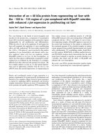
binding indicate cooperative binding (Fig. 1). Half-
maximal binding occurred at a calcium concentration of 1.5
and 1.4 m
M
for factor X and Gla–EGF
N
, respectively,
which is close to the concentration of free calcium in blood of
1.2 m
M
. The best fit to the data in Fig. 1 was obtained
assuming three cooperatively bound Ca
2+
ions. As shown in
Fig. 1 the membrane binding of intact factor X and the Gla–
EGF
N
fragment, showed very similar Ca
2+
-dependencies,
indicating that neither the second EGF domain nor the
serine protease domain alter those Ca
2+
-binding properties
of factor X that are relevant to membrane binding. Experi-
ments using membranes containing either 10 mol% PtdSer/
90 mol% PtdCho or 40 mol% PtdSer/60 mol% PtdCho
showedthesameCa
2+
-dependence as 25 mol% PtdSer/
75 mol% PtdCho for binding intact factor X and Gla–
EGF
N
(data not shown).
Kinetics of membrane binding
The kinetics of binding to PL membranes of the zymogen
factor X, activated factor X (factor Xa) and the active site
inhibited form DEGR-factor Xa as well as the the factor X
peptides were studied with surface plasmon resonance. The
Ca
2+
concentration was 10 m
M
to ascertain that the Ca
2+
binding sites of the Gla domain were completely satur-
ated. Figure 2 presents the binding of factor X to the
Table 1. Kinetic constants for binding of factor X and its Gla-containing fragments to membranes containing 25 mol% PtdSer in the presence of
10 m
M
Ca
2+
obtained by evaluation of association and dissociation phases (I) and equilibrium binding data (II) as described in Materials and methods.
k
on
(MÆs)
)1
k
off
(s
)1
) K
d
(
M
) (I) K
d
(
M
) (II)
Gla (8.0 ± 2.2) · 10
3
(3.7 ± 0.2) · 10
)2
(4.6 ± 1.3) · 10
)6
(9.4 ± 1.4) · 10
)6
Gla–EGF
N
(4.5 ± 1.1) · 10
4
(3.8 ± 0.2) · 10
)2
(8.4 ± 2.1) · 10
)7
(1.7 ± 0.3) · 10
)6
Gla–EGF
N,C
(6.7 ± 2.1) · 10
4
(4.3 ± 0.2) · 10
)2
(6.4 ± 2.0) · 10
)7
(2.0 ± 0.3) · 10
)6
Factor X (8.3 ± 1.9) · 10
5
(3.2 ± 0.2) · 10
)2
(3.9 ± 0.9) · 10
)8
(3.7 ± 0.6) · 10
)8
Factor Xa (4.5 ± 0.8) · 10
5
(3.6 ± 0.2) · 10
)2
(8.0 ± 1.5) · 10
)8
(5.2 ± 0.8) · 10
)8
DEGR-factor Xa (5.3 ± 1.3) · 10
5
(3.7 ± 0.2) · 10
)2
(8.0 ± 1.5) · 10
)8
(6.2 ± 0.9) · 10
)8
Fig. 1. Ca
2+
-dependence in the membrane binding of factor X (A) and
the fragment Gla–EGF
N
(B) as determined by surface plasmon reson-
ance. Binding experiments were performed on 25 mol% PtdSer-con-
taining membranes (solid symbols) and 100 mol% PtdCho-containing
membranes (open symbols). The solid curve is the best fit to the
experimental data points obtained by Eqn (1), assuming n ¼ 3
(c
2
¼ 359.2); the dotted line assuming n ¼ 4(c
2
¼ 595.7); the dashed
line assuming n ¼ 2(c
2
¼ 715.3).
Ó FEBS 2002 Membrane binding of coagulation factor X (Eur. J. Biochem. 269) 3043
phospholipid membrane at various protein concentrations.
Similar sensorgrams were obtained for the other forms of
factor X and fragments, although with different concentra-
tions for half maximum binding (data not shown). In a first
attempt the association and dissociation processes were
treated as simple one step processes. However, with this
approach it was not possible to obtain a reasonable agree-
ment between observed and calculated sensorgrams. Mod-
els with two on-rates and two off-rates improved the fit
significantly. Moreover, a model including a conformation-
al change and a model including a bivalent analyte both
gave good fits to the experimental data. The results obtained
with the bivalent analyte model is shown in Fig. 2. In all cases
there is a dominating fast process with an almost constant
off-rate for all the proteins (3.2–4.8 10
)2
Æs
)1
). The difference
in binding affinity is therefore the result of different on-rates
(Table 1). The isolated Gla domain (the fragment with the
lowest molecular mass, about 5 kDa) shows the lowest on
rate, even though from thermodynamic aspects it would be
expected to show a higher on rate. This may be explained by
assuming that only a small fraction of the fragment has a
conformation that is commensurate with membrane-bind-
ing. The on-rates for Gla–EGF
N
and Gla–EGF
NC
are about
a factor of five higher than for the Gla-domain. This can
presumably be attributed to a stabilizing effect of the
N-terminal EGF domain on the Gla domain [7]. The entire
protein has an on-rate that is two orders of magnitude faster
than for the Gla-domain presumably due to a further
stabilization of the structure of the Gla-domain, indicating
that less than 1% of the free isolated Gla-domain has a
conformation that is appropriate for membrane binding.
Equilibrium binding isotherms
The concentration dependence of factor X binding is shown
in Fig. 2. It is apparent that the adsorption is rapid and that
a plateau is reached within 100–200 s. Figure 3 shows the
binding isotherms of factor X and its peptides. Their mem-
brane binding affinities increase in the order Gla < Gla–
EGF
N
¼ Gla–EGF
NC
<factor X ¼ factor Xa ¼ DEGR-
factor Xa (Table 1). Although both the first and second
binding step contribute to the equilibrium response signal,
the first binding step is the dominating process and the
influence from the second one, whether a conformational
change or a bifunctional ligand, has been neglected. The
consistency of the K
d
values resulting from the evaluation of
the equilibrium response signals and those obtained by
evaluating the first step in the association phase of the
sensorgrams justifies this assumption.
DISCUSSION
Calcium binding to the Gla domain is known to be crucial
for the induction of a conformation in the domain that
mediates membrane binding. Early studies employing
equilibrium dialysis established the existence of about 10
Ca
2+
-binding sites, at least three of which mediate cooper-
ative binding [23–26]. By studies of the binding of divalent
cations other than Ca
2+
, for example Mg
2+
,Mn
2+
and
Ba
2+
, it became evident that there is one class of binding
sites that is cation nonspecific and binds all four metal ions
in a cooperative manner [26–29]. Moreover, metal ion-
binding to the cation nonspecific sites induces quenching of
the intrinsic protein fluorescence [26,28,30]. The Ca
2+
concentration necessary to induce half-maximal fluores-
cence quenching in factor X and in the fragment that
consists of the Gla domain linked to the first EGF domain
was determined to about 0.5 m
M
[31]. The conformation
induced by cation binding to the nonspecific sites does not
support membrane-binding [27,29]. The second class of
binding sites is Ca
2+
-specific, and metal ion-binding to these
sites induces a membrane binding conformation. From
NMR studies of the Mg
2+
form of a Gla-domain it became
evident that unlike Ca
2+
-binding, Mg
2+
-binding to the
Fig. 3. Equilibrium isotherms of factor X and its Gla-containing frag-
ments binding to membranes containing 25 mol% PtdSer in the presence
of 10 m
M
Ca
2+
. The measured equilibrium binding signal is plotted
against the solution phase concentration of factor X (d), factor Xa
(m), DEGR-factor Xa (n), Gla–EGF
NC
(e), Gla–EGF
N
(r)andGla
(.). Solid lines indicate the least-square fit of the Langmuir model to
this data as described in Materials and methods. The estimated binding
parameters are listed in Table 1.
Fig. 2. Adsorption and desorption kinetics of factor X to 25 mol%
PtdSer containing membranes. Experiments were performed using
10 m
M
Tris/HCl,pH7.5,150m
M
NaCl, 10 m
M
CaCl
2
,0.1%(w/v)
BSA as running buffer at a flow rate of 10 lLÆmin
)1
.FactorXwas
diluted in the same buffer to the final concentration of 44 n
M
(h),
22 n
M
(j), 11 n
M
(n), 5.5 n
M
(m), 2.8 n
M
(s)and1.4n
M
(d). The
protein was injected at t ¼ 0 and binding to the membrane is apparent
during the association phase (180 s). The protein-containing buffer
was then replaced by running buffer, resulting in dissociation of the
protein from the membrane. The solid curves were calculated using
equations 4–6.
3044 E M. Erb et al.(Eur. J. Biochem. 269) Ó FEBS 2002
Gla-domain did not induce the native conformation in
residues 1–11 of the Gla-domain [8]. Moreover, NMR
studies of the Ca
2+
-free form of the Gla-domain established
that the metal ion binding translocated the residues that
constitute the hydrophobic patch from the interior of the
domain to the surface, allowing them to interact with the
phospholipid membrane [7]. Furthermore, these results
support the notion that the nature of this drastic conform-
ational transition must be highly cooperative with respect
to Ca
2+
due to noncompensated electrostatic repulsion
between carboxylate groups with, for instance, only one
Ca
2+
bound in this region.
We have now found that the Ca
2+
concentration that
induces half-maximal membrane binding of factor X and
the fragment Gla–EGF
N
to PtdSer-containing membranes
is about 1.5 m
M
. This is consistent with results from light
scattering experiments with other Gla domain-containing
proteins. Thus the Ca
2+
-concentration necessary to
induce half-maximal binding has been determined to be
0.55 m
M
,0.9m
M
and 1.2 m
M
for factor IX [32], factor
VII [33] and protein C [5], respectively. We have found
that the membrane-binding of intact factor X and Gla–
EGF
N
show about the same Ca
2+
dependence, indicating
that Ca
2+
-binding to domains other than the Gla domain
and the N-terminal EGF-like domain does not influence
the membrane-binding properties of factor X. Our results
also demonstrate that the membrane binding is cooper-
ative with respect to Ca
2+
, presumably reflecting the
cooperative Ca
2+
-binding to sites in the Gla domain.
Interestingly, the Ca
2+
concentration necessary to induce
membrane-binding corresponds rather closely to the
concentration of free Ca
2+
in blood (1.2 m
M
). It is thus
possible that binding of at least some Gla domain-
containing proteins to biological membranes will be
sensitive to local variations in the Ca
2+
concentration in
the immediate vicinity of the membrane.
We found that the isolated factor X Gla domain exhibits
low affinity binding to PtdSer-containing membranes with a
K
d
of 4.6 l
M
. This agrees well with the value of 2.4 l
M
for
factor IX (1–47) [8] and 3.7 l
M
for human protein C (1–48)
[34] measured under similar conditions (1 l
M
Ca
2+
,40%
PtdSer) by resonance energy transfer and circular dichro-
ism, respectively. The C-terminal helix of the factor X Gla
domain of Gla–EGF
N
(residues 33–41) interacts with the
adjacent EGF
N
domain [8]. Presumably, this interaction
stabilizes the Gla domain and contributes to the five-fold
higher affinity of Gla–EGF
N
(K
d
¼ 1 l
M
) for phospholipid
membranes as compared to the isolated Gla domain. The
second EGF domain does not appear to provide any further
stabilization. The membrane affinity of the intact protein is
about 10-fold higher than the affinity for Gla–EGF
N
and
Gla–EGF
NC
and about 100-fold higher than the affinity to
the isolated Gla domain. No significant difference in
membrane affinity could be detected between the zymogen,
the activated protein and the active site-inhibited form. It
should be pointed out that the results from equilibrium
binding studies are consistent with the data resulting form
the evaluation of association and dissociation phases. The
differences in the K
d
values resulting from the different
evaluations of the experiments are in the same range as
observed previously [13,35]. The K
d
determined for factor X
is consistent with the value determined by McDonald
et al.[9].
The effect of the serine protease domain upon the
membrane affinity of the intact protein is enigmatic. It could
be due to a long distance conformational change in the
protein mediated through the two EGF-domains. In this
context it should be noted that mutation of Ca
2+
ligating
amino acids in the N-terminal part of the first EGF-like
domain of factor X influences the amidolytic activity of the
intact protein [36]. However, direct interactions between the
Gla and serine protease domains, intra or intermolecular,
might also explain the difference in binding affinities.
Another factor contributing to the higher on-rate for the
intact protein is the net charge. The Gla–EGF
NC
fragment is
highly negatively charge, especially when not saturated
with Ca
2+
()29 without Ca
2+
and )15 with 7Ca
2+
). The
C-terminal serineprotease domain, however, has anet charge
of +8, making the whole protein less negatively charged.
Therefore the equilibrium concentration of the intact protein
near the negatively charged surface will be higher than for the
fragments resulting in a higher apparent on-rate. Using the
same argument the on-rate of the Gla–EGF
NC
fragment
should be lower than for the Gla-domain as it is more
negatively charged. The stabilizing effect of EGF
N
on the
structure of the Gla-domain is therefore even more than what
is reflected by the fivefold increase in the on-rate.
ACKNOWLEDGEMENTS
This work was supported by grants from the Swedish Medical Research
Council and EU Project BIO-CT-96-0662.
REFERENCES
1. Furie, B. & Furie, B.C. (1988) The molecular basis of blood
coagulation. Cell 53, 505–518.
2. Mann, K.G., Krishnaswamy, S. & Lawson, J.H. (1992) Surface-
dependent hemostasis Semin-Hematol. 29, 213–226.
3. Soriano Garcia, M., Padmanabhan, K., deVos, A.M. & Tulinsky,
A. (1992) The Ca
2+
ion and membrane binding structure of the
Gla-domain of Ca-prothrombin fragment 1. Biochemistry 31,
2554–2566.
4. Arni,R.K.,Padmanabhan,K.,Padmanabhan,K.P.,Wu,T.P.&
Tulinsky, A. (1994) Structure of the non-covalent complex of
prothrombin kringle 2 with PPACK-thrombin. Chem. Phys.
Lipids 68, 59–66.
5. Zhang, L. & Castellino, F.J. (1994) The binding energy of human
coagulation protein C to acidic phospholipid vesicles contains a
major contribution from leucine 5 in the gamma-carboxyglutamic
acid domain. J. Biol. Chem. 269, 3590–3595.
6. Christiansen, W.T., Jalbert, L.R., Robertson, R.M., Jhingan, A.,
Prorok, M. & Castellino, F.J. (1995) Hydrophobic amino acid
residues of human anticoagulation protein C that contribute to its
functional binding to phospholipid vesicles. Biochemistry 34,
10374–10382.
7. Sunnerhagen, M., Forse
´
n, S., Hoffre
´
n, A.M., Drakenberg, T.,
Teleman, O. & Stenflo, J. (1995) Structure of the Ca(2+)-free Gla
domain sheds light on membrane binding of the blood coagulation
proteins. Nat. Struct. Biol. 2, 504–509.
8. Freedman, S.J., Blostein, M.D., Baleja, J.D., Jacobs, M., Furie,
B.C. & Furie, B. (1996) Identification of the phospholipid binding
site in the vitamin K-dependent blood coagulation protein factor
IX. J. Biol. Chem 271, 16227–16236.
9. McDonald, J.F., Shah, A.M., Schwalbe, R.A., Kisiel, W.,
Dahlba
¨
ck, B. & Nelsestuen, G.L. (1997) Comparison of naturally
occurring vitamin K-dependent proteins: correlation of amino
Ó FEBS 2002 Membrane binding of coagulation factor X (Eur. J. Biochem. 269) 3045
acid sequences and membrane binding properties suggests a
membrane contact site. Biochemistry 36, 5120–5127.
10. Stenflo, J. & Dahlba
¨
ck, B. (1994) Vitamin K-dependent proteins.
In The Molecular Basis of Blood Diseases (Stamatoyannopoulos,
G.,Nienhuis,A.W.,Majerus,P.W.&Varmus,H.,eds),pp.565–
598. Saunders, Philadelphia, PA, USA.
11. Thariath, A. & Castellino, F.J. (1997) Highly conserved residue
arginine-15 is required for the Ca
2+
-dependent properties of the
c-carboxyglutamic acid domain of human anticoagulant Protein
C and activated protein C. Biochem. J. 322, 309–315.
12. Giesen, P.L., Willems, G.M., Hemker, H.C. & Hermens, W.T.
(1991) Membrane-mediated assembly of the prothrombinase
complex. J. Biol. Chem 266, 18720–18725.
13. Willems, G.M., Janssen, M.P., Salemink, I., Wun, T.C. &
Lindhout, T. (1998) Transient high affinity of tissue factor path-
way inhibitor-Factor Xa complex to negatively charged phos-
pholipid membranes. Biochemistry 37, 3321–3328.
14.Cutsforth,G.A.,Whitaker,R.N.,Hermans,J.&Lentz,B.R.
(1989) A new model to describe extrinsic protein binding to
phospholipid membranes of varying composition: application to
human coagulation proteins. Biochemistry 28, 7453–7461.
15. Krishnaswamy, S., Jones, K.C. & Mann, K.G. (1988) Pro-
thrombinase complex assembly. Kinetic mechanism of enzyme
assembly on phospholipid vesicles. J. Biol. Chem 263, 3823–3834.
16. Nelsestuen, G.L., Kisiel, W. & Di Scipio, R.G. (1978) Interaction
of vitamin K dependent proteins with membranes. Biochemistry
17, 2134–2138.
17. Nesheim, M.E., Kettner, C., Shaw, E. & Mann, K.G. (1981)
Cofactor dependence of Factor Xa incorporation into the pro-
thrombinase complex. J. Biol. Chem 256, 6537–6540.
18. Erb, E M., Chen, X., Allen, S., Roberts, C.J., Tendler, S.J.B.,
Davies, M.C. & Forse
´
n, S., (2000) Characterization of the
surface generated by liposome binding to the modified dextran
matrix of a surface plasmon resonance sensor chip. An. Biochem.
280, 29–35.
19. Abbott, A.J. & Nelsestuen, G.L. (1987) Association of a protein
with membrane vesicles at the collisional limit: studies with blood
coagulation Factor Va light chain also suggest major differences
between small and large unilamellar vesicles. Biochemistry 26,
7994–8003.
20. Greenhut, S.F., Bourgeois, V.R. & Roseman, M.A. (1986) Dis-
tribution of cytochrome b
5
between small and large unilamellar
phospholipid vesicles. J. Biol. Chem. 261, 3670–3675.
21. Silversmith, R.E. & Nelsestuen, G.L. (1986) Interaction of com-
plement proteins C5b-6 and C5b-7 with phospholipid vesicles:
effects of phospholipid structural features. Biochemistry 25, 7717–
7725.
22. Persson, E., Bjo
¨
rk, I. & Stenflo, J. (1991) Protein structural
requirements for Ca
2+
binding to the light chain of factor X.
Studies using isolated intact fragments containing the c-carbo-
xyglutamic acid region and/or the epidermal growth factor-like
domains. J. Biol. Chem. 266, 2444–2452.
23. Nelsestuen, G.L. & Suttie, J.W. (1972) Mode of action of vitamin
K and calcium binding properties of bovine prothrombin. Bio-
chemistry 11, 4961–4964.
24. Stenflo, J. & Ganot, P. (1973) Binding of Ca
2+
to normal and
dicoumarol-induced prothrombin. Biochem. Biophys. Res.
Commun. 50, 98–104.
25. Henriksen, R.A. & Jackson, C.M. (1975) Cooperative calcium
binding by the phospholipid binding region of bovine pro-
thrombin: a requirement for intact disulfide bridges. Arch. Bio-
chem. Biophys. 170, 149–159.
26. Prendergast, F.G. & Mann, K.G. (1977) Differentiation of metal
ion-induced transitions of prothrombin fragment 1. J. Biol. Chem.
252, 840–850.
27. Borowski, M., Furie, B.C., Bauminger, S. & Furie, B. (1986)
Prothrombin requires two sequential metal-dependent conforma-
tional transitions to bind phospholipid. J. Biol. Chem. 261, 14969–
14975.
28. Nelsestuen, G.L., Broderius, M. & Martin, G. (1976) Role of
c-carboxyglutamic acid. Cation specificity of prothrombin and
factor X-phospholipid binding. J. Biol. Chem. 251, 6886–6893.
29. Liebman, H.A., Furie, B.C. & Furie, B. (1987) The factor IX
phospholipid-binding site is required for calcium-dependent acti-
vation of factor IX by factor XIa. J. Biol. Chem. 262, 7605–7612.
30. Nelsestuen, G.L. (1876) Role of gamma-carboxyglutamic acid. An
unusual protein transition required for the calcium-dependent
binding of prothrombin to phospholipid. J. Biol. Chem. 25, 5649–
5656.
31. Persson, E., Valcarce, C. & Stenflo, J. (1991) The c-carboxyglut-
amic acid and epidermal growth factor-like domains of Factor X.
J.Biol. Chem. 266, 2453–2458.
32. Christiansen, W.T. & Castellino, F.J. (1994) Properties of
recombinant chimeric human protein C and activated protein C
containing the c-carboxyglutamic acid and trailing helical stack
domains of protein C replaced by those of human coagulation
factor IX. Biochemistry 33, 5901–5911.
33. Geng, J.P. & Castellino, F.J. (1997) The properties of human
protein C, factor VII, and factor IX are exchangeable with respect
to directing gamma-carboxylation of these proteins. Thromb.
Haemost. 77, 926–933.
34. Colpitts, T.L. & Castellino, F.J. (1994) Calcium and phospholipid
binding properties of synthetic c-carboxyglutamic acid-containing
peptides with sequence counterparts in human protein C. Bio-
chemistry 33, 3501–3508.
35. Haseley, S.R., Talaga, P., Kamerling, J.P. & Vliegenthart, J.F.
(1999) Characterization of the carbohydrate binding specificity
and kinetic parameters of lectins by using surface plasmon
resonance. Anal. Biochem. 274, 203–210.
36. Lentig, P.J., Christophe, O.D., Maat, H., Rees, D.J.G. & Mertens,
K. (1996) Ca
2+
binding to the first epidermal growth factor-like
domain of human blood coagulation factor IX promotes enzyme
activity and factor VIII light chain binding. J. Biol. Chem. 271,
25332–25337.
3046 E M. Erb et al.(Eur. J. Biochem. 269) Ó FEBS 2002