Báo cáo y học: "Molecular action of methotrexate in inflammatory diseases" ppt
Bạn đang xem bản rút gọn của tài liệu. Xem và tải ngay bản đầy đủ của tài liệu tại đây (143.78 KB, 8 trang )
266
AICAR = aminoimidazolecarboxamidoribonucleotide; Fc = crystallizable fragment (of antibody); IFN = interferon; IL = interleukin; RA = rheumatoid
arthritis; Th = T helper (cells); TNF = tumor necrosis factor.
Arthritis Research Vol 4 No 4 Chan and Cronstein
Introduction
The demonstration in 1985 that low-dose, intermittent
methotrexate is a potent and effective therapy for rheuma-
toid arthritis (RA) [1] led to a dramatic change in the way
that patients with RA are treated. Indeed, methotrexate is
no less efficacious than specific anti-tumor-necrosis-factor
(anti-TNF) therapy for the relief of symptomatic joint inflam-
mation in early RA, and the difference between methotrex-
ate and etanercept with respect to protection from
structural injury in RA is probably not biologically signifi-
cant [2]. Thus, methotrexate remains the cornerstone of
therapy for RA, and understanding the mechanism(s)
responsible for the therapeutic efficacy of this agent may
lead to the development of new therapies.
History and clinical pharmacology
Methotrexate was first developed in the 1940s as a spe-
cific antagonist of folic acid. This drug inhibits the prolifera-
tion of malignant cells, primarily by inhibiting the de novo
synthesis of purines and pyrimidines. Because administra-
tion of high doses of reduced folic acid (folinic acid) or
even folic acid itself can reverse the antiproliferative effects
of methotrexate, it is clear that methotrexate does act as an
antifolate agent. Interestingly, although not originally
designed as such, methotrexate appears to be a ‘pro-drug’,
i.e. a compound that is converted to the active agent after
uptake. Methotrexate is taken up by cells via the reduced
folate carrier and then is converted within the cells to
polyglutamates [3]. Methotrexate polyglutamates are long-
lived metabolites that retain some of the antifolate activities
of the parent compound, although the potency for inhibition
of various folate-dependent enzymes is shifted [3–6].
Proposed mechanisms of action of
methotrexate
Low-dose methotrexate was introduced for the treatment
of RA because of its presumed antiproliferative properties,
although it was unclear how inhibiting proliferation of the
lymphocytes thought to be responsible for synovial inflam-
mation in RA for one day a week might lead to effective
suppression of disease activity. However, it soon became
clear that inhibition of folic acid metabolism could not be
completely responsible for the anti-inflammatory effect of
methotrexate. During the past 15 years, it has become
clear that administration of folic acid in doses of 1–5 mg
per day helps to prevent much of the toxicity of methotrex-
ate without interfering with the anti-inflammatory efficacy
of the drug, whereas very high doses of folinic acid also
prevent methotrexate toxicity but may interfere with its effi-
cacy [7–20]. There are two potential explanations for the
Review
Molecular action of methotrexate in inflammatory diseases
Edwin S L Chan and Bruce N Cronstein
Division of Clinical Pharmacology, NYU School of Medicine, New York, NY, USA
Corresponding author: Bruce N Cronstein (e-mail: )
Received: 1 November 2001 Revisions received: 27 November 2001 Accepted: 12 December 2001 Published: 19 March 2002
Arthritis Res 2002, 4:266-273
© 2002 BioMed Central Ltd (
Print ISSN 1465-9905; Online ISSN
1465-9913)
Abstract
Despite the recent introduction of biological response modifiers and potent new small-molecule
antirheumatic drugs, the efficacy of methotrexate is nearly unsurpassed in the treatment of inflammatory
arthritis. Although methotrexate was first introduced as an antiproliferative agent that inhibits the
synthesis of purines and pyrimidines for the therapy of malignancies, it is now clear that many of the anti-
inflammatory effects of methotrexate are mediated by adenosine. This nucleoside, acting at one or more
of its receptors, is a potent endogenous anti-inflammatory mediator. In confirmation of this mechanism of
action, recent studies in both animals and patients suggest that adenosine-receptor antagonists, among
which is caffeine, reverse or prevent the anti-inflammatory effects of methotrexate.
Keywords: adenosine receptor, inflammation, methotrexate, rheumatoid arthritis
267
Available online />capacity of high doses of folinic acid to reverse the thera-
peutic effects: first, folinic acid may bypass the effects of
methotrexate on reduction of folic acid and thereby
bypass the therapeutic effects of the drug; alternatively,
folinic acid but not folic acid may compete with methotrex-
ate for a single transport site into the cell (Fig. 1) and may
thus interfere with cellular uptake of methotrexate [21].
Moreover, the expected inhibition of cellular proliferation is
manifested as bone marrow suppression, and oral and gas-
trointestinal ulcers, and may require lowering the dose of
the drug and, usually, the efficacy of the therapy, suggesting
that inhibition of cellular proliferation alone is not responsi-
ble for the anti-inflammatory effects of methotrexate. Thus,
folate antagonism appears to play, at most, a minimal role in
the anti-inflammatory mechanism of methotrexate.
Another potential mechanism by which methotrexate may
diminish inflammation in the joint is by diminishing cytokine
production. Numerous studies have demonstrated dimin-
ished levels of inflammatory cytokines in the serum of
patients. The adenosine A
2A
receptor agonist CGS-21680
is a potent inhibitor of neutrophil leukotriene synthesis in
vitro, and, similarly, methotrexate therapy leads to dimin-
ished production of leukotriene B
4
by neutrophils stimulated
ex vivo [22,23]. The mechanism by which methotrexate
diminishes these cytokine levels remains unexplained and it
is difficult to determine from these studies whether the
effects of methotrexate therapy on production of inflamma-
tory mediators results in diminished inflammation or is sec-
ondary to other anti-inflammatory events.
Similarly, methotrexate-mediated effects on T-cell function,
either in vivo or in vitro, have been demonstrated. Indeed,
Genestier and colleagues have reported that methotrexate
diminishes antigen-stimulated T-cell proliferation both in
vitro and in T cells taken from patients taking methotrexate
[24]. That the effects of methotrexate on T-cell function
are completely reversed by folic acid and that the effects
of therapy on T cells studied ex vivo are present for only
48 hours a week would strongly suggest that this cannot
be responsible for the bulk of the anti-inflammatory effects
of the drug.
A third proposed mechanism of action is based upon the
observation that polyamines accumulate in the synovium
of patients with RA and that metabolism of these
polyamines by macrophages leads to the production of
toxic oxygen products that diminish stimulated T-cell func-
tion [25–27]. Indeed, methotrexate therapy does diminish
polyamine levels in the joints of patients with RA [28–30],
but this effect, like that of methotrexate on T-cell prolifera-
tion, is reversed by folic acid. Moreover, there are more
than enough toxic oxygen metabolites being generated in
the rheumatoid synovium to mediate the tissue damage
present in this disease; another source of toxic agents
would add relatively little.
Methotrexate induces adenosine release
Our laboratory originally proposed the hypothesis that the
beneficial effects of methotrexate result from the intracellu-
lar accumulation of intermediates in purine biosynthesis
that, by a mechanism that has not been completely
worked out, leads to increased concentrations of adeno-
sine in the extracellular space [31]. This hypothesis
sprang from the prior demonstration that intracellular
accumulation of specific intermediates in the de novo syn-
thesis of purines leads to adenosine release [32] and from
our interest in the anti-inflammatory effects of adenosine,
which are mediated by specific receptors on inflammatory
cells. Prior work had demonstrated that methotrexate
polyglutamates inhibit the enzyme aminoimidazolecarbox-
amidoadenosineribonucleotide (AICAR) transformylase
more potently than the other enzymes involved in purine
biosynthesis [4,5,33]. This inhibition occurred at pharma-
cologically relevant concentrations of methotrexate and
might be expected to occur more readily with infrequent
loading with methotrexate, since methotrexate polygluta-
mates are long-lived metabolites (persisting for weeks).
The presence of increased concentrations of AICAR
metabolites in the urine of RA patients treated with
methotrexate supports these findings [34,35]. The accu-
mulation of AICAR and its metabolites has a direct
inhibitory effect on at least two key enzymes, adenosine
deaminase and AMP deaminase, with the end result of
increased concentrations of adenosine and adenine
nucleotides intracellularly [4]. Methotrexate in doses
similar to that used in the treatment of RA has been known
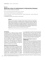
Figure 1
Methotrexate-induced metabolic changes lead to increased extra-
cellular adenosine. ADA = adenosine deaminase; AICAR = amino-
imidazolecarboxamidoribonucleotide; AICAside = aminoimidazole-
carboxamidoribonucleoside; AK = adenosine kinase; AMPDA = AMP
deaminase; DHF = dihydrofolate; DHF
glu
= dihydrofolate
polyglutamate; ecto-5′NT = ecto-5′nucleotidase; FAICAR = formyl-
AICAR; IMP = inosine monophosphate; MTX = methotrexate; MTX
glu
=
methotrexate polyglutamate; RFC1 = reduced folate carrier 1.
268
Arthritis Research Vol 4 No 4 Chan and Cronstein
to cause the accumulation of AICAR in animal models of
RA, and this accumulation is associated with an elevation
in adenosine concentration in the extracellular space
[32,36]. The exact mechanisms by which the elevation of
extracellular adenosine arises are not fully understood, but
dephosphorylation of adenine nucleotides is likely to be a
major contributor, partly because of the ubiquitous nature
of ATP in tissues and partly because of the widespread
existence of ecto-5′-nucleotidase, an enzyme that cat-
alyzes the dephosphorylation of AMP to adenosine [37].
All this evidence points to adenosine as a key mediator in
the anti-inflammatory actions of methotrexate. In vivo exper-
iments support this contention. The nonselective adeno-
sine receptor antagonist 8-phenyl theophylline potentiated
inflammatory responses in a hamster-cheek-pouch model
[38]. Infusion of adenosine directly into the knee in rats
inhibited the development of adjuvant-induced arthritis, and
an adenosine receptor antagonist effectively reduced the
severity of joint inflammation in a collagen-induced arthritis
model in mice [39,40]. We have previously shown that the
anti-inflammatory effects of methotrexate in carrageenan-
induced mouse air pouch inflammation is reversed by an
antagonist to the adenosine A
2A
receptor, or by the addi-
tion of adenosine deaminase, an adenosine-metabolizing
enzyme, suggesting that adenosine is indeed responsible
for the anti-inflammatory effects of methotrexate in vivo
[36]. An interesting study by Silke et al. showed that inges-
tion of caffeine, a nonselective antagonist of adenosine
receptors, in coffee correlates with poor clinical response
to methotrexate, and patients with a high caffeine intake are
more likely to discontinue methotrexate than those with a
low caffeine intake [41].
To better appreciate how adenosine influences biological
responses in the network of events taking place in an
inflammatory milieu, something must be said about this
autocoid and the cellular receptors with which it interacts
to produce these physiological responses. Adenosine
receptors, or P1 receptors, fall into four known subclasses:
A
1
, A
2A
, A
2B
, and A
3
. These are members of the large,
seven-transmembrane-receptor family of receptors that
influence cell signaling mechanisms by coupling to G pro-
teins. The receptor sequences have been characterized
and, with the exception of the A
3
receptor, they are highly
conserved during evolution. Adenosine receptors modulate
a vast array of physiological functions, from heart rate to
the state of wakefulness. Adenosine, acting on P1 recep-
tors, exerts a number of actions on a variety of cell types
relevant to the anti-inflammatory effect of methotrexate.
Cellular effects
Neutrophils
Neutrophils, a hallmark of acute inflammation, are among
the first cells recruited into the inflammatory site. The limi-
tation of neutrophilic-mediated damage relies in part on
the modification of the adhesive capacity and ability to
generate chemical damage, properties under purinergic
influence. The resting neutrophil has a number of mecha-
nisms that, once activated, can damage tissues. One of
these is latent nicotinamide adenine dinucleotide phos-
phate (NADPH) oxidase, a multimolecular complex that is
assembled at the plasma membrane upon activation of the
neutrophil and that generates oxygen radicals [42]. The
first in the chain of these oxygen radicals is superoxide
anion, and it was the discovery in 1983 that superoxide
generation, as stimulated by a variety of agents including
the chemoattractant N-formyl-leucyl-phenylalanine (f MLP),
the complement component C5a, and the calcium
ionophore A23187, was inhibited by adenosine that
sparked an interest in the anti-inflammatory properties of
adenosine [43,44]. This physiological action of adenosine
has subsequently been ascribed to its action on the
adenosine A
2A
receptor, which is present on the neu-
trophilic surface membrane [45]. An important second
messenger to adenosine-A
2A
-receptor signaling in this
respect appears to be 3′,5′-cyclic adenosine monophos-
phate (cAMP), the intracellular concentration of which
increases with neutrophilic adenosine A
2A
receptor stimu-
lation. cAMP further activates protein kinase A down-
stream and inhibition of protein kinase A reverses the
effects of cAMP analogues but not of adenosine receptor
agonists on stimulated neutrophilic superoxide anion gen-
eration [46]. The cAMP–protein-kinase-A-dependent
adenosine inhibition of neutrophil oxidative activity is medi-
ated via the adenosine A
2A
receptor [47]. One direct con-
sequence of the interruption of superoxide anion formation
and respiratory burst reactions is the protection of vascu-
lar endothelial cells from neutrophil-mediated injury [48].
The adenosine-A
2A
-receptor-mediated effects on neutro-
phil function are dose-related. At concentrations similar to
those required to inhibit the release of superoxide anions,
adenosine, acting through A
2A
receptors, inhibits adher-
ence to endothelial cells by stimulated neutrophils [49].
This may be related in part to dose-related preferential
recruitment of receptor subtype, since the adenosine A
1
receptor exhibits many opposing physiological functions
to those mediated by the A
2A
receptor, including stimula-
tion of neutrophil adherence to endothelial cells. Adeno-
sine also inhibits the release of vascular endothelial
growth factor from neutrophils, thereby enhancing vascu-
lar permeability [50]. The dose-dependent response in
adenosine action is also seen with Fc-gamma-receptor-
mediated neutrophil phagocytosis, which is enhanced by
A
1
receptor stimulation but inhibited via A
2
receptors [51].
In addition, adenosine also inhibits the TNF-induced gen-
eration of elastase by neutrophils [52].
Expression of adhesive molecules is an important event
that guides neutrophil recruitment into an inflammatory
site through adhesion to the vascular endothelium.
269
Adenosine has been known to be a modulator of the
expression or function of adhesive molecules including
β
2
-integrin, L-selectin, and CD11b/CD18 [49,53,54]. The
activity of adenosine in the modulation of neutrophil adhe-
sion again demonstrates the opposing roles of A
1
and A
2
receptors [49].
Macrophages
Cells of the monocyte–macrophage series are abundant
in the rheumatoid synovium and pannus and contribute
significantly to the tissue damage seen in both acute and
chronic disease, as recently reviewed by Kinne and col-
leagues [55]. Macrophages, the differentiated tissue form,
are also critical producers of cytokines that play a promi-
nent role in promoting proinflammatory responses that cul-
minate in tissue damage. Like neutrophils, their capacity to
phagocytose opsonized particles and to generate super-
oxide anions plays a major role in eliciting tissue damage.
Inhibition of Fc-gamma-receptor phagocytic activity in cul-
tured monocytes is exhibited by adenosine at high con-
centrations such as that seen with tissue damage and is a
function mediated via adenosine A
2
receptors, while low
concentrations of adenosine have the opposite effect on
Fc-gamma-receptor phagocytic activity mediated via
adenosine A
1
receptors [56]. Similarly, adenosine inhibits
the generation of superoxide anions by monocytes stimu-
lated with N-formyl-leucyl phenylalanine [57].
One of the well known though uncommon side effects of
methotrexate treatment is the formation of subcutaneous
nodules, often similar in histological appearance though
not in distribution to those found in rheumatoid disease. A
hallmark of these subcutaneous nodules is the existence
of the multinucleated giant cell, formed by fusion of
macrophages. The fusion of macrophages into multinucle-
ated giant cells is enhanced by stimulation of the adeno-
sine A
1
receptor and is inhibited by activation of the A
2
receptor [58,59].
The recent success of anti-TNF therapy highlights the role
of cytokines as important mediators of inflammatory activ-
ity. Not surprisingly, methotrexate, still one of the most
effective disease-modifying antirheumatic drugs for the
treatment of RA, acting through the release of adenosine,
also inhibits the production of TNF-α, although the adeno-
sine receptor involved in this action remains controversial
[60–63]. Modulation of cytokine production by adenosine
extends far beyond TNF-α and includes observable effects
on IL-6, IL-8, IL-10, IL-12, and macrophage inflammatory
protein-1α (MIP-1α) [40,64,65]. Cytokines themselves
can regulate the expression of adenosine receptors on
monocytic cells and thereby modulate adenosine-medi-
ated responses, as we and others have recently shown
[66,67]. Macrophage production of nitric oxide and nitric
oxide synthase is also inhibited by adenosine, probably via
A
2B
receptors [65,67].
Endothelial cells
Endothelial cells are effective transit barriers between
vessels and tissue and as such are notable in inflammation
not only because of their expression of adhesive mole-
cules, which allow leukocytes their access to inflammatory
sites. The effectiveness of this barrier function relies in
part on the preservation of impermeability to circulating
cells homing in to take part in inflammatory reactions in the
tissues. Adenosine enhances this barrier function by
decreasing enthothelial permeability via A
2B
receptor and
helps limit potential tissue damage [68,69]. Production of
inflammatory cytokines such as IL-6 and IL-8 and expres-
sion of adhesive molecules such as intercellular adhesion
molecule-1 (ICAM-1) and E-selectin by endothelial cells
are also suppressed by adenosine [70]. Another important
aspect of inflammation lies in the proliferation and migra-
tion of endothelial cells in the process of angiogenesis,
which is enhanced by the presence of adenosine, proba-
bly acting through A
2
receptors [71–73]. Adenosine may
also induce apoptosis of endothelial cells, thus potentially
enhancing the extravasation of inflammatory fluids [74].
Humoral and cellular immune responses
Rheumatoid factor, or autoantibodies directed against the
Fc portion of IgG, is a hallmark of RA, although its exact
role in the pathogenesis of the disease has been debated.
The effect of methotrexate on the levels of circulating IgM
rheumatoid factors has also been controversial. While
some workers have reported no suppression of serum
rheumatoid factor levels with methotrexate treatment,
Alarcon et al. observed significant drops in the levels of
both IgM and IgA rheumatoid factors in methotrexate-
treated patients, and particularly of the concentration of
IgM rheumatoid factor in those who showed clinical
improvement [75]. These findings were confirmed by other
groups in studies done both in vivo and ex vivo [76–80],
although it is unclear whether this is a primary or sec-
ondary effect of adenosine.
T lymphocytes have received much attention in relation to
the pathogenesis of RA and opinions differ in their contri-
bution to the causation of the disease. The presence of
these cells in the affected synovium and the strong
ethnicity-dependent HLA–DR associations implicate T
lymphocytes as key players in the disease process. One
possible explanation of the beneficial actions of methotrex-
ate in RA is the diminution of both the size and reactivity of
the T-lymphocyte population. There are suggestions that
this may be accomplished by the induction of apoptosis in
activated T cells [24]. This suggestion is consistent with
the observations of reductions in peripheral blood T and B
lymphocyte populations after short-term methotrexate
treatment [81], and methotrexate induction of apoptosis in
inflammatory cells may be relevant to its antirheumatic
actions in vivo [82]. In contrast, significant increases in
the CD3- and CD4-positive peripheral blood cells and
Available online />270
enhancement of stimulated lymphocyte proliferation have
been observed after long-term treatment with methotrex-
ate [83], and adenosine, acting through A
2A
and A
2B
receptors, may play a role in T-cell deactivation [84,85].
Nonetheless, the role of these shifts in T-cell function and
trafficking in the pathogenesis of RA is unclear.
Phlogistic responses
Cytokines are messengers with major roles in inflammatory
and immune responses and have been targets of interest in
recent therapeutic developments in chronic arthritis, with
TNF-α and IL-1 as the focus of interest [86]. In animal
models of chronic arthritis, methotrexate was thought to be
useful in reducing the production of IL-1 [87,88]. In
support of these findings, clinical studies of RA patients
receiving methotrexate treatment have documented reduc-
tions in monocytic IL-1 production but not serum concen-
trations of IL-1 [89]. Others have disputed this view and
suggested that alterations in IL-1 responses were related
to diminutions in the ability of cells to respond to IL-1 rather
than to direct inhibition of its production, perhaps through
dose-dependent ligand binding [90–92].
Methotrexate is also known to suppress TNF activity by
suppressing TNF-induced nuclear factor-κB activation in
vitro, in part related to a reduction in the degradation and
inactivation of an inhibitor of this factor, IκBα, and proba-
bly related to the release of adenosine [93]. The genera-
tion of TNF-α by peripheral blood mononuclear cells is
suppressed by an adenosine kinase inhibitor, by virtue of
its ability to limit adenosine uptake and metabolism and
thereby enhance extracellular adenosine concentration
[94]. TNF-α synthesis in T cells and macrophages is sup-
pressed [95]. In the murine collagen-induced arthritis
model, in vivo intraperitoneal methotrexate treatment
reduced TNF serum levels and diminished TNF production
by splenic T cells and macrophages [96]. Methotrexate
suppresses the production of both TNF and IFN-γ by T-
cell-receptor-primed T lymphocytes from both healthy
human donors and RA patients [97]. In early RA, in which
the disease duration is less than 6 months, methotrexate
treatment is associated with a significant decrease of
TNF-α-positive CD4
+
T cells, while the number of T cells
expressing the anti-inflammatory cytokine IL-10 increased
[98]. Methotrexate is also known to suppress the IL-6-
induced generation of reactive oxygen species in the syn-
oviocytes of RA patients [99]. Serum IL-6 levels have also
declined after methotrexate treatment in RA patients in
some studies [100]. Constantin et al. reported that ex vivo
treatment of peripheral blood monocytes with methotrex-
ate increased expression of IL-4 and IL-10 while IL-2 and
interferon-γ expression were decreased, suggesting that
the immunoregulatory role of methotrexate is also targeted
at adjusting the balance between Th1 proinflammatory
and Th2 anti-inflammatory cytokines [101]. Again, the mol-
ecular mechanism of these changes is unclear.
Conclusion
Our search for mechanisms governing the inflammatory
response has uncovered many facets relevant to the patho-
genesis of arthitic diseases. The success of methotrexate
as an antirheumatic agent rests on its many actions that
affect a wide variety of pathogenic mechanisms, many of
which are mediated by the release of adenosine. The mole-
cular mechanism for many of these phenomena is related
to the enhanced release of adenosine into the extracellular
space, where it can activate its receptors on relevant cell
types. In this respect, methotrexate is an excellent example
of how knowledge and continuing research in molecular
biology and pharmacology can be employed in the refine-
ment of existing medications originally used on an observa-
tional basis. Such understanding will form the basis for the
development of new and more effective therapy for the
treatment of rheumatic diseases.
Acknowledgements
This work was supported by grants from the National Institutes of Health
(AR41911, GM56268), Medco Research, Inc., and the General Clinical
Research Center (M01RR00096) and by the Kaplan Cancer Center.
References
1. Weinblatt ME, Coblyn JS, Fox DA, Fraser PA, Holdsworth DE,
Glass DN, Trentham DE: Efficacy of low-dose methotrexate in
rheumatoid arthritis. N Engl J Med 1985, 312:818-822.
2. Bathon JM, Martin RW, Fleischmann RM, Tesser JR, Schiff MH,
Keystone EC, Genovese MC, Wasko MC, Moreland LW, Weaver
AL, Markenson J, Finck BK: A comparison of etanercept and
methotrexate in patients with early rheumatoid arthritis. N
Engl J Med 2000, 343:1586 - 1593.
3. Chabner BA, Allegra CJ, Curt GA, Clendeninn NJ, Baram J,
Koizumi S, Drake JC, Jolivet J: Polyglutamation of methotrexate.
Is methotrexate a prodrug? J Clin Invest 1985, 76:907-912.
4. Baggott JE, Vaughn WH, Hudson BB: Inhibition of 5-aminoimi-
dazole-4-carboxamide ribotide transformylase, adenosine
deaminase and 5-adenylate deaminase by polyglutamates of
methotrexate and oxidized folates and by 5-aminoimidazole-
4-carboxamide riboside and ribotide. Biochem J 1986, 236:
193-200.
5. Allegra, CJ, Drake JC, Jolivet J, Chabner BA: Inhibition of phos-
phoribosylaminoimidazolecarboxamide transformylase by
methotrexate and dihydrofolic acid polyglutamates. Proc Natl
Acad Sci U S A 1985, 82:4881-4885.
6. Chabner BA, Myers CE: Clinical Pharmacology of Cancer
Chemotherapy. In Cancer: Principles and Practice of Oncology.
Edited by DeVita VT, Hellman S, Rosenberg SA. Philadelphia: JB
Lippincott, 1989: 349-395.
7. Ortiz Z, Shea B, Suarez-Almazor M, Moher D, Wells G, Tugwell P:
Folic acid and folinic acid for reducing side effects in patients
receiving methotrexate for rheumatoid arthritis. Cochrane
Database Syst Rev 2, 2000.
8. Suarez-Almazor ME, Belseck E Shea B, Wells G, Tugwell P:
Methotrexate for rheumatoid arthritis. Cochrane Database Syst
Rev 2, 2000.
9. Ravelli A, Migliavacca D, Viola S, Ruperto N, Pistorio A, Martini A:
Efficacy of folinic acid in reducing methotrexate toxicity in juve-
nile idiopathic arthritis. Clin Exp Rheumatol 1999, 17:625-627.
10. Pincus T: Folic and folinic acid supplementation reduces
methotrexate gastrointestinal side effects in rheumatoid
arthritis. Clin Exp Rheumatol 1998, 16:667-668.
11. Morgan SL, Baggott JE, Lee JY, Alarcon GS: Folic acid supple-
mentation prevents deficient blood folate levels and hyperho-
mocysteinemia during longterm, low dose methotrexate
therapy for rheumatoid arthritis: implications for cardiovascu-
lar disease prevention. J Rheumatol 1998, 25:441-446.
12. Ortiz Z, Shea B, Suarez-Almazor ME, Moher D, Wells GA, Tugwell
P: The efficacy of folic acid and folinic acid in reducing
methotrexate gastrointestinal toxicity in rheumatoid arthritis.
Arthritis Research Vol 4 No 4 Chan and Cronstein
271
Available online />A metaanalysis of randomized controlled trials. J Rheumatol
1998, 25:36-43.
13. Hunt PG, Rose CD, McIlvain-Simpson G, Tejani S: The effects of
daily intake of folic acid on the efficacy of methotrexate
therapy in children with juvenile rheumatoid arthritis. A con-
trolled study. J Rheumatol 1997, 24:2230-2232.
14. Shiroky JB: The use of folates concomitantly with low-dose
pulse methotrexate. Rheum Dis Clin North Am 1997, 23:969-
980.
15. Shiroky JB Folic acid and methotrexate in rheumatoid arthritis.
Ann Intern Med 1996, 124:73-74.
16. Kavanaugh A, Kavanaugh D: Folic acid and methotrexate in
rheumatoid arthritis. Ann Intern Med 1996, 124:73; discussion 74.
17. Cooper BA: Folic acid and methotrexate in rheumatoid arthri-
tis. Ann Intern Med 1996, 124:73; discussion 74.
18. Dijkmans BA: Folate supplementation and methotrexate. Br J
Rheumatol 1995, 34:1172-1174.
19. van Ede AE, Laan RF, Rood MJ, Huizinga TW, van de Laar MA,
van Denderen CJ, Westgeest TA, Romme TC, de Rooij DJ,
Jacobs MJ, X de Boo TM, van der Wilt GJ, Severens JL, Hartman
M, Krabbe PF, Dijkmans BA, Breedveld FC, van de Putte LB:
Effect of folic or folinic acid supplementation on the toxicity
and efficacy of methotrexate in rheumatoid arthritis: a forty-
eight week, multicenter, randomized, double-blind, placebo-
controlled study. Arthritis Rheum 2001, 44:1515-1524.
20. Endresen GK, Husby G: Folate supplementation during
methotrexate treatment of patients with rheumatoid arthritis.
An update and proposals for guidelines. Scand J Rheumatol
2001, 30:129-134.
21. Matherly LH, Czajkowski CA, Angeles SM: Identification of a
highly glycosylated methotrexate membrane carrier in K562
human erythroleukemia cells up-regulated for tetrahydrofo-
late cofactor and methotrexate transport. Cancer Res 1991,
51:3420-3426.
22. Sperling RL, Benincaso AI, Anderson RJ, Coblyn JS, Austen KF,
and Weinblatt ME: Acute and chronic suppression of
leukotriene B
4
synthesis ex vivo in neutrophils from patients
with rheumatoid arthritis beginning treatment with methotrex-
ate. Arth.Rheum. 1992, 35:376-384.
23. Surette ME, Krump E, Picard S, Borgeat P: Activation of
leukotriene synthesis in human neutrophils by exogenous
arachidonic acid: inhibition by adenosine A[2a] receptor ago-
nists and crucial role of autocrine activation by leukotriene
B[4]. Mol Pharmacol 1999, 56:1055-1062.
24. Genestier L, Paillot R, Fournel S, Ferraro C, Miossec P, Revillard
JP: Immunosuppressive properties of methotrexate: apopto-
sis and clonal deletion of activated peripheral T cells. J Clin
Invest 1998, 102:322-328.
25. Flescher E, Bowlin TL, Ballester A, Houk R, Talal N: Increased
polyamines may downregulate interleukin 2 production in
rheumatoid arthritis. J Clin Invest 1989, 83:1356-1362.
26. Flescher E, Bowlin TL, Talal N: Regulation of IL-2 production by
mononuclear cells from rheumatoid arthritis synovial fluids.
Clin Exp Immunol 1992, 87:435-437.
27. Yukioka K, Wakitani S, Yukioka M, Furumitsu Y, Shichikawa K,
Ochi T, Goto H, Matsui-Yuasa I, Otani S, Nishizawa Y: Polyamine
levels in synovial tissues and synovial fluids of patients with
rheumatoid arthritis. J Rheumatol 1992 19:689-692.
28. Furumitsu Y, Yukioka K, Kojima A, Yukioka M, Shichikawa K, Ochi
T, Matsui-Yuasa I, Otani S, Nishizawa Y, Morii H: Levels of
urinary polyamines in patients with rheumatoid arthritis. J
Rheumatol 1993, 20:1661-1665.
29. Nesher G, Osborn TG, Moore TL: In vitro effects of methotrex-
ate on polyamine levels in lymphocytes from rheumatoid
arthritis patients. Clin Exp Rheumatol 1996, 14:395-399.
30. Nesher G, Moore TL: The in vitro effects of methotrexate on
peripheral blood mononuclear cells. Modulation by methyl
donors and spermidine. Arthritis Rheum 1990, 33:954-959.
31. Cronstein BN, Eberle MA, Gruber HE, Levin RI: Methotrexate
inhibits neutrophil function by stimulating adenosine release
from connective tissue cells. Proc Natl Acad Sci U S A 1991,
88:2441-2445.
32. Gruber HE, Hoffer ME, McAllister DR, Laikind PK, Lane TA,
Schmid-Schoenbein GW, Engler RL: Increased adenosine con-
centration in blood from ischemic myocardium by AICA ribo-
side: effects on flow, granulocytes and injury. Circulation
1989, 80:1400-1411.
33. Allegra CJ, Hoang K, Yeh GC, Drake JC, Baram J: Evidence for
direct inhibition of de novo purine synthesis in human MCF-7
breast cells as a principal mode of metabolic inhibition by
methotrexate. J Biol Chem 1987, 262:13520-13526.
34. Baggott JE, Morgan SL, Koopman WJ: The effect of methotrex-
ate and 7-hydroxymethotrexate on rat adjuvant arthritis and
on urinary aminoimidazole carboxamide excretion. Arthritis
Rheum 1998, 41:1407-1410.
35. Luhby AL, Cooperman JH: Aminoimidazole carboxamide excre-
tion in vitamin B12 and folic acid deficiencies. Lancet 1962,
2:1381-1382.
36. Cronstein BN, Naime D, Ostad E: The antiinflammatory mecha-
nism of methotrexate: increased adenosine release at
inflamed sites diminishes leukocyte accumulation in an in
vivo model of inflammation. J Clin Invest 1993, 92:2675-2682.
37. Morabito L, Montesinos MC, Schreibman DM, Balter L, Thompson
LF, Resta R, Carlin G, Huie MA, Cronstein BN: Methotrexate and
sulfasalazine promote adenosine release by a mechanism
that requires ecto-5
′′
-nucleotidase-mediated conversion of
adenine nucleotides. J Clin Invest 1998, 101:295-300.
38. Rosengren S, Arfors KE, Proctor KG, Potentiation of leukotriene
B4-mediated inflammatory response by the adenosine antag-
onist, 8-phenyl theophylline. Int J Microcirc: Clin Exp 1991, 10:
345-357.
39. Green PG, Basbaum AI, Helms C, Levine JD: Purinergic regula-
tion of bradykinin-induced plasma extravasation and adju-
vant-induced arthritis in the rat. Proc Natl Acad Sci U S A
1991: 88:4162-4165.
40. Szabo C, Scott GS, Virag L, Egnaczyk G, Salzman AL, Shanley
TP, Hasko G: Suppression of macrophage inflammatory
protein [MIP]-1alpha production and collagen-induced arthri-
tis by adenosine receptor agonists. Br J Pharmacol 1998, 125:
379-387.
41. Silke C, Murphy MS, Buckley T, Busteed S, Molloy MG, Phelan M:
The effects of caffeine ingestion on the efficacy of methotrex-
ate. Rheumatology [Oxford] 2001, 40(suppl1):S34.
42. Halliwell B, Hoult JR, Blake DR: Oxidants, inflammation, and
anti-inflammatory drugs. FASEB J 1988, 2:2867-2873.
43. Cronstein BN, Kramer SB, Weissmann G, Hirschhorn R: Adeno-
sine: a physiological modulator of superoxide anion genera-
tion by human neutrophils. J Exp Med 1983, 158:1160-1177.
44. Cronstein BN, Kramer SB, Weissmann G, Hirschhorn R: A new
physiological function for adenosine: regulation of superoxide
anion production. Trans Assoc Am Physicians 1983, 96:384-391.
45. Cronstein BN, Rosenstein ED, Kramer SB, Weissmann G,
Hirschhorn R: Adenosine; a physiologic modulator of superox-
ide anion generation by human neutrophils. Adenosine acts
via an A2 receptor on human neutrophils. J Immunol 1985,
135:1366-1371.
46. Cronstein BN, Haines KA, Kolasinski SL, Reibman J: Occupancy
of G alpha s-linked receptors uncouples chemoattractant
receptors from their stimulus-transduction mechanisms in
the neutrophil. Blood 1992, 80:1052-1057.
47. Sullivan GW, Rieger JM, Scheld WM, Macdonald TL, Linden J:
Cyclic AMP-dependent inhibition of human neutrophil oxida-
tive activity by substituted 2-propynylcyclohexyl adenosine
A[2A] receptor agonists. Br J Pharmacol 132:1017-1026.
48. Cronstein BN, Levin RI, Belanoff J, Weissmann G, Hirschhorn R.
Adenosine: an endogenous inhibitor of neutrophil-mediated
injury to endothelial cells. J Clin Invest 2001, 78:760-770.
49. Cronstein BN, Levin RI, Philips MR, Hirschhorn R, Abramson SB,
Weissmann G: Neutrophil adherence to endothelium is
enhanced via adenosine A1 receptors and inhibited via
adenosine A2 receptors. J Immunol 1992, 148:2201-2206.
50. Wakai A, Wang JH, Winter DC, Street JT, O’Sullivan RG,
Redmond HP: Adenosine inhibits neutrophil vascular endothe-
lial growth factor release and transendothelial migration via
A2B receptor activation. Shock 2001, 15:297-301.
51. Salmon JE, Cronstein BN: Fcgamma receptor-mediated func-
tions in neutrophils are modulated by adenosine receptor
occupancy: A1 receptors are stimulatory and A2 receptors are
inhibitory. J Immunol 1990, 145:2235-2240.
52. Ottonello L, Amelotti M, Barbera P, Dapino P, Mancini M, Tortolina
G, Dallegri F: Chemoattractant-induced release of elastase by
tumor necrosis factor- primed human neutrophils: auto-regu-
lation by endogenous adenosine. Inflamm Res 1999, 48:637-
642.
272
Arthritis Research Vol 4 No 4 Chan and Cronstein
53. Firestein GS, Bullough DA, Erion MD, Jimenez R, Ramirez-Wein-
house M, Barankiewicz J, Smith CW, Gruber E, Mullane KM: Inhi-
bition of neutrophil adhesion by adenosine and an adenosine
kinase inhibitor: the role of selectins. J Immunol 1995, 154:
326-334.
54. Wollner A, Wollner S, Smith JB: Acting via A2 receptors, adeno-
sine inhibits the upregulation of Mac-1 [CD11b/CD18]
expression on FMLP-stimulated neutrophils. Am J Resp Cell
Mol Biol 1993, 9:179-185.
55. Kinne RW, Brauer R, Stuhlmuller B, Palombo-Kinne E, Burmester
GR: Macrophages in rheumatoid arthritis. Arthritis Res 2000,
2:189-202.
56. Salmon JE, Brogle N, Brownlie C, Edberg JC, Kimberly RP, Chen
BX, Erlanger BF: Human mononuclear phagocytes express
adenosine A1 receptors. A novel mechanism for differential
regulation of Fc gamma receptor function. J Immunol 1993,
151:2775-2785.
57. Leonard EJ, Shenai A, Skeel A: Dynamics of chemotactic
peptide-induced superoxide generation by human mono-
cytes. Inflammation 1987, 11:229-240.
58. Merrill, JT, Shen C, Schreibman D, Coffey D, Zakharenko O,
Fisher R, Lahita, J. Salmon RG, Cronstein BN: Adenosine A
1
receptor promotion of multinucleated giant cell formation by
human monocytes: a mechanism for methotrexate-induced
nodulosis in rheumatoid arthritis. Arth Rheum 1997, 40:1308-
1315.
59. Merrill TJ, Shen C, Schreibman D, Coffey D, Zakharenko O, Fisher
R, Lahita RG, Salmon J, Cronstein BN. Adenosine A
1
receptor
promotion of multinucleated giant cell formation by human
monocytes, a mechanism for methotrexate-induced nodulo-
sis in rheumatoid arthritis. Arthritis Rheum. 1995, 38
(Suppl):S157.
60. Eigler A, Greten TF, Sinha B, Haslberger C, Sullivan GW, Endres
S: Endogenous adenosine curtails lipopolysaccharide-stimu-
lated tumour necrosis factor synthesis. Scand J Immunol
1997, 45:132-139.
61. Prabhakar U, Brooks DP, Lipshlitz D, Esser KM: Inhibition of
LPS-induced TNF alpha production in human monocytes by
adenosine [A2] receptor selective agonists. Int J Pharmacol
1995, 17:221-224.
62. Sajjadi FG, Takabayashi K, Foster AC, Domingo RC, Firestein GS:
Inhibition of TNF-alpha expression by adenosine: role of A3
adenosine receptors. J Immunol 1996, 156:3435-3442.
63. McWhinney CD, Dudley MW, Bowlin TL, Peet NP, Schook L,
Bradshaw M, De M, Borcherding DR, Edwards CK 3rd: Activa-
tion of adenosine A3 receptors on macrophages inhibits
tumor necrosis factor-alpha. Eur J Pharmacol 1996, 310:209-
216.
64. Bouma MG, Stad RK, van den Wildenberg FA, Buurman WA: Dif-
ferential regulatory effects of adenosine on cytokine release by
activated human monocytes. J Immunol 1994: 153:4159-4168.
65. Hasko G, Szabo C, Nemeth ZH, Kvetan V, Pastores SM, Vizi ES:
Adenosine receptor agonists differentially regulate IL-10, TNF-
alpha, and nitric oxide production in RAW 264.7 macrophages
and in endotoxemic mice. J Immunol 1996, 157:4634-4640.
66. Khoa ND, Montesinos MC, Reiss AB, Delano D, Awadallah N,
Cronstein BN: Inflammatory cytokines regulate function and
expression of adenosine A
2A
receptors in human monocytoid
THP-1 cells. J Immunol 2001, 167:4026-4032.
67. Xaus J, Mirabet M, Lloberas J, Soler C, Lluis C, Franco R, Celada
A: IFN-gamma up-regulates the A2B adenosine receptor
expression in macrophages: a mechanism of macrophage
deactivation. J Immunol 1999, 162:3607-3614.
68. Lennon PF, Taylor CT, Stahl GL, Colgan SP: Neutrophil-derived
5
′′
-adenosine monophosphate promotes endothelial barrier
function via CD73-mediated conversion to adenosine and
endothelial A2B receptor activation. J Exp Med 1998, 188:
1433-1443.
69. Richard LF, Dahms TE, Webster RO: Adenosine prevents per-
meability increase in oxidant-injured endothelial monolayers.
Am J Physiol 1998, 274:H35-H42.
70. Bouma MG, van den Wildenberg FAJM, Buurman WA: Adeno-
sine inhibits cytokine release and expression of adhesion
molecules by activated human endothelial cells. Am J Physiol
1996, 39:C522-C529.
71. Grant MB, Tarnuzzer RW, Caballero S, Ozeck MJ, Davis MI,
Spoerri PE, Feoktistov I, Biaggioni I, Shryock JC, Belardinelli L:
Adenosine receptor activation induces vascular endothelial
growth factor in human retinal endothelial cells. Circ Res
1999, 85:699-706.
72. Ethier MF, Chander V, Dobson JG, Jr: Adenosine stimulates
proliferation of human endothelial cells in culture. Am J
Physiol 1993, 265:H131-H138.
73. Sexl V, Mancusi G, Baumgartner-Parzer S, Schutz W, Freissmuth
M: Stimulation of human umbilical vein endothelial cell prolif-
eration by A2-adenosine and beta 2-adrenoceptors. Br J Phar-
macol 1995: 114:1577-1586.
74. Harrington EO, Smeglin A, Newton J, Ballard G, Rounds S:
Protein tyrosine phosphatase-dependent proteolysis of focal
adhesion complexes in endothelial cell apoptosis. Am J
Physiol Lung Cell Mol Physiol 2001, 280:L342-L353.
75. Alarcon GS, Schrohenloher RE, Bartolucci AA, Ward JR, Williams
HJ, Koopman WJ: Suppression of rheumatoid factor produc-
tion by methotrexate in patients with rheumatoid arthritis. Arth
Rheum 1990, 33:1156-1161.
76. Spadaro A, Riccieri V, Sili Scavalli A, Taccari E, Zoppini A: One
year treatment with low dose methotrexate in rheumatoid
arthritis: effect on class specific rheumatoid factors. Clin
Rheumatol 1993, 12:357-360.
77. Olsen NJ, Teal GP, Brooks RH: IgM-rheumatoid factor and
responses to second-line drugs in rheumatoid arthritis.
Agents Actions 1991, 34:169-171.
78. Moore S, Ruska K, Peters L, Olsen NJ: Associations of IgA and
IgA-rheumatoid factor with disease features in patients with
rheumatoid arthritis. Immunol Invest 1994: 23:355-365.
79. Olsen NJ, Callahan LF, Pincus T: Immunologic studies of
rheumatoid arthritis patients treated with methotrexate. Arthri-
tis Rheum 1987, 30:481-488.
80. Olsen NJ, Murray LM: Antiproliferative effects of methotrexate
on peripheral blood mononuclear cells. Arthritis Rheum 1989,
32:378-385.
81. Wascher TC, Hermann J, Brezinschek HP, Brezinschek R,
Wilders-Truschnig M, Rainer F, Krejs GJ: Cell-type specific
response of peripheral blood lymphocytes to methotrexate in
the treatment of rheumatoid arthritis. Clin Invest 1994, 72:
535-540.
82. Nakazawa F, Matsuno H, Yudoh K, Katayama R, Sawai T, Uzuki M,
Kimura T: Methotrexate inhibits rheumatoid synovitis by induc-
ing apoptosis. J Rheumatol 2001, 28:1800-1808.
83. Weinblatt ME, Trentham DE, Fraser PA, Holdsworth DE, Falchuk
KR, Weissman BN, Coblyn JS: Long-term prospective trial of
low-dose methotrexate in rheumatoid arthritis. Arth Rheum
1988, 31:167-175.
84. Mirabet M, Herrera C, Cordero OJ, Mallol J, Lluis C, Franco R:
Expression of A2B adenosine receptors in human lymphocytes:
their role in T cell activation. J Cell Sci 1999, 112:491-502.
85. Dong RP, Kameoka J, Hegen M, Tanaka T, Xu Y, Schlossman SF,
Morimoto C: Characterization of adenosine deaminase
binding to human CD26 on T cells and its biologic role in
immune response. J Immunol 1996, 156:1349-1355.
86. van den Berg WB: Anti-cytokine therapy in chronic destructive
arthritis. Arthritis Res 2001, 3:18-26
87. DiMartino MJ, Johnson WJ, Votta B,. Hanna N: Effect of
antiarthritic drugs on the enhanced interleukin-1 [IL- 1] pro-
duction by macrophages from adjuvant-induced arthritic [AA]
rats. Agents Actions 1987, 21:348-350.
88. Novaes GS, Mello SB, Laurindo IM, Cossermelli W: Low dose
methotrexate decreases intraarticular prostaglandin and
interleukin 1 levels in antigen induced arthritis in rabbits. J
Rheumatol 1996, 23:2092-2097.
89. Chang DM, Weinblatt ME, Schur PH: The effects of methotrex-
ate on interleukin 1 in patients with rheumatoid arthritis. J
Rheumatol 1992, 19:1678-1682.
90. Segal R, Mozes E, Yaron M, Tartakovsky B: The effects of
methotrexate on the production and activity of Il-1. Arth
Rheum 1989, 32:370-377.
91. Chang DM, Baptiste P, Schur PH: The effect of antirheumatic
drugs on interleukin 1 [IL-1] activity and IL-1 and IL-1 inhibitor
production by human monocytes. J Rheumatol 1990, 17:1148-
1157.
92. Brody M, Bohm I, Bauer R: Mechanism of action of methotrex-
ate: experimental evidence that methotrexate blocks the
binding of interleukin 1 beta to the interleukin 1 receptor on
target cells. Eur J Clin Chem Clin Biochem 1993, 31:667-674.
273
Available online />93. Majumdar S, Aggarwal BB: Methotrexate suppresses Nf-
kappaB activation through inhibition of IkappaBalpha phos-
phorylation and degradation. J Immunol 2001, 167:2911-2920.
94. Eigler A, Matschke V, Hartmann G, Erhardt S, Boyle D, Firestein
GS, Endres S: Suppression of TNF-alpha production in human
mononuclear cells by an adenosine kinase inhibitor. J Leukoc
Biol 2000, 68:97-103.
95. Becker C, Barbulescu K, Hildner K, Meyer zum Buschenfelde KH,
Neurath MF: Activation and methotrexate-mediated suppres-
sion of the TNF alpha promoter in T cells and macrophages.
Ann N Y Acad Sci 1998, 859:311-314.
96. Neurath MF, Hildner K, Becker C, Schlaak JF, Barbulescu K,
Germann T, Schmitt E, Schirmacher P, Haralambous S, Pas-
parakis M, Meyer Zum Buschenfelde KH, Kollias G, Marker-
Hermann E: Methotrexate specifically modulates cytokine
production by T cells and macrophages in murine collagen-
induced arthritis [CIA]: a mechanism for methotrexate-medi-
ated immunosuppression. Clin Exp Immunol 1999, 115:42-55.
97. Hildner K, Finotto S, Becker C, Schlaak J, Schirmacher P, Galle
PR, Marker-Hermann E, Neurath MF: Tumour necrosis factor
[TNF] production by T cell receptor-primed T lymphocytes is a
target for low dose methotrexate in rheumatoid arthritis. Clin
Exp Immunol 1999, 118:137-146.
98. Rudwaleit M, Yin Z, Siegert S, Grolms M, Radbruch A, Braun J,
Sieper J: Response to methotrexate in early rheumatoid arthri-
tis is associated with a decrease of T cell derived tumour
necrosis factor alpha, increase of interleukin 10, and pre-
dicted by the initial concentration of interleukin 4. Ann Rheum
Dis 2000, 59:311-314.
99. Sung JY, Hong JH, Kang HS, Choi I, Lim SD, Lee JK, Seok JH,
Lee JH, Hur GM: Methotrexate suppresses the interleukin-6
induced generation of reactive oxygen species in the synovio-
cytes of rheumatoid arthritis. Immunopharmacology 2000,
47:35-44.
100. Spadaro A, Taccari E, Riccieri V, Sensi F, Sili Scavalli A, Zoppini
A: Relationship of soluble interleukin-2-receptor and inter-
leukin-6 with class-specific rheumatoid factors during low-
dose methotrexate treatment in rheumatoid arthritis. Rev
Rhum Engl Ed 1997, 64:89-94.
101. Constantin A, Loubet-Lescoulie P, Lambert N, Yassine-Diab B,
Abbal M, Mazieres B, de Preval C, Cantagrel A: Antiinflammatory
and immunoregulatory action of methotrexate in the treat-
ment of rheumatoid arthritis: evidence of increased inter-
leukin-4 and interleukin-10 gene expression demonstrated in
vitro by competitive reverse transcriptase-polymerase chain
reaction. Arthritis Rheum 1998, 41:48-57.
Correspondence
Bruce N Cronstein MD, Division of Clinical Pharmacology, NYU School
of Medicine, 550 First Avenue, New York, NY 10016, USA.
Tel: +1 212 263 6404; fax: +1 212 263 8804; e-mail: