Frontotemporal branch of the facial nerve and fascial layers in the temporal region a cadaveric study to define a safe dissection plane sihag RK, gupta SK, sahni d, aggarwal a neurol india
Bạn đang xem bản rút gọn của tài liệu. Xem và tải ngay bản đầy đủ của tài liệu tại đây (3.81 MB, 13 trang )
14/5/2021
Frontotemporal Branch of the Facial Nerve and Fascial Layers in the Temporal Region: A Cadaveric Study to Define a Safe Dissection Pl…
Open access journal indexed with Index
Medicus
Users online: 43
Home | Login
About
Editorial
board
Articles
NSI
Publications
Search
Instructions
Online
Subscribe
Submission
Videos
Etcetera
Contact
Impact Factor for 2019 is 2.128 Click here to view op
Navigate Here
Search
GO
Resource Links
» Similar in PUBMED
» Search Pubmed for
Sihag RK
Gupta SK
Sahni D
Aggarwal A
ORIGINAL ARTICLE
Year : 2020 | Volume : 68 | Issue : 6 | Page : 1313-1320
Frontotemporal Branch of the Facial Nerve and Fascial Layers in the Temporal
Region: A Cadaveric Study to Define a Safe Dissection Plane
Rakesh K Sihag1, Sunil K Gupta1, Daisy Sahni2, Ashish Aggarwal1
1 Department of Neurosurgery, Postgraduate Institute of Medical Education and
Chandigarh, India
» Search in Google Scholar Research,
2 Department of Anatomy, Postgraduate Institute of Medical Education and
for
Research, Chandigarh, India
Sihag RK
Gupta SK
Date of Web
19-DecSahni D
Publication
2020
Aggarwal A
»Related articles
Correspondence Address:
Dr. Sunil K Gupta
Department of Neurosurgery, PGIMER, Chandigarh 160012
India
Temporal fascia
facial nerve
dissection plane
Key Messages: This
article
Source of Support: None, Conflict of Interest: None
through cadaver studies
defines the disposition
of the frontotemporal
Check
branch of the nerve in
relation to the different
temporal fascia and the DOI: 10.4103/0028-3886.304113
fat pads is necessary t
/>
1/13
14/5/2021
Frontotemporal Branch of the Facial Nerve and Fascial Layers in the Temporal Region: A Cadaveric Study to Define a Safe Dissection Pl…
» Article in PDF (2,461
KB)
» Citation Manager
» Access Statistics
» Reader Comments
» Email Alert *
» Add to My List *
* Registration required
(free)
In this Article
» Abstract
» Material and Methods
» Observations and...
» Discussion
» Conclusions
» References
» Article Figures
» Article Tables
Article Access Statistics
Viewed
1650
Printed
36
Emailed
0
PDF Downloaded
66
Comments
[Add]
» Abstract
Background: Anatomy of the temporal region is complex with controversy over
the relationship of fascial planes with the upper division of the facial nerve.
Objective: This study aimed to identify the safe surgical landmarks to preserve the
frontotemporal branch of the facial nerve during surgery and define the safest
approach for surgical procedures in this region.
Material and Methods: The anatomical relationship of the frontal branch of the
facial nerve, superficial temporal artery (STA), fascial planes, and fat pads was
determined after dissection on 10 cadaveric heads, that is (20 sides) Dissection
was performed layer by layer from skin to bone.
Results: The temporoparietal fascia was made up of multiple (3–4) layers above
the zygomatic arch and these layers were integrated with thin fibrous septa. The
frontotemporal branch of the facial nerve (FTFN) was observed in a deeper part of
temporoparietal fascia and superficial fat pad. The frontotemporal branch of the
facial nerve (FTFN) crossed the zygomatic arch as two branches in 25%, as three
branches in 65% and as four branches in 10% of specimens.
Conclusions: Interfascial dissection between two layers of deep temporal fascia
through the intermediate fat pad is superior to other approaches because of the lack
of facial nerve branches in this plane. The Intermediate fat could be easily
separated from deep layer of deep temporal fascia.
Keywords: Temporal fascia, facial nerve, dissection plane
Key Messages: This article, through cadaver studies, defines the disposition of the
frontotemporal branch of the nerve in relation to the different temporal fascia and
the fat pads is necessary to stay in the correct dissection plane.
How to cite this article:
Sihag RK, Gupta SK, Sahni D, Aggarwal A. Frontotemporal Branch of the Facial
Nerve and Fascial Layers in the Temporal Region: A Cadaveric Study to Define a
Safe Dissection Plane. Neurol India 2020;68:1313-20
How to cite this URL:
Sihag RK, Gupta SK, Sahni D, Aggarwal A. Frontotemporal Branch of the Facial
Nerve and Fascial Layers in the Temporal Region: A Cadaveric Study to Define a
Safe Dissection Plane. Neurol India [serial online] 2020 [cited 2021 May
14];68:1313-20. Available from: />2020/68/6/1313/304113
Knowledge of the anatomical landmarks is an essential prerequisite for any
surgical procedure. The relationship of the temporal fascial planes with the upper
division of the facial nerve remains controversial.[1],[2],[3],[4],[5],[6],[7],[8],[9],[10],
[11],[12],[13],[14],[15],[16]
/>
2/13
14/5/2021
Frontotemporal Branch of the Facial Nerve and Fascial Layers in the Temporal Region: A Cadaveric Study to Define a Safe Dissection Pl…
The frontotemporal division of the facial nerve (FTFN) supplies muscles of the
forehead and face through temporal and zygomatic branches. Small branches of
the frontotemporal nerve supply the orbicularis oculi, frontalis, and auricularis
muscles. The anatomy of FTFN in tissue layers has been debated extensively in
the literature. Many of these studies lack in exact dimension and measurements
from fixed landmarks to identify the nerve intra-operatively.[1],[2],[3],[7],[9],[10],[11],
[12],[13],[14],[16],[17]
FTFN is prone to injury in surgical procedures like pterional, frontotemporal
orbitozygomatic (FTOZ) craniotomy either directly or indirectly due to the
stretching of a nerve. This cadaveric study aimed to identify the safe surgical
landmarks to preserve the FTFN during surgery.
» Material and Methods
The anatomical relationships of the FTFN, superficial temporal artery (STA),
fascial planes, and fat pads were determined on 10 cadaveric heads, that is (20
sides). Layer by layer dissection was done from skin to bone. Different names
were described in the literature for fascia, fat pads, and nerve; therefore, preferred
names are taken to avoid confusion [Table 1].
Table 1: Nomenclature of fascia, fat pads, and
nerve in literature
Click here to view
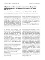
Surgical technique
The skin incision started 3 cm below the base of the zygomatic arch, 1 cm anterior
to the tragus, curving forward toward the frontal region ending in the midline at
the hairline [Figure 1]. Incision involved skin, subcutaneous tissue superficial to
the temporoparietal fascia. The temporoparietal fascia was dissected layer by layer
inferiorly up to parotidomasseteric fascia and reflected anterolaterally with the
scalp flap. The STA was identified and separated from the skin by dissection in the
subcutaneous plane.
Figure 1: Surface marking of the skin Incision.
Incision started 3 cm below the base of the
zygomatic arch and 1 cm anterior to the tragus,
curving upward and backward above the pinna
and then forward toward the frontal region and
ended in the midline at the hairline (left side)
Click here to view
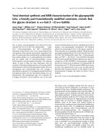
Two standard lines were drawn based on the skeletal landmark, the L1 line
connecting the lateral bony canthus (LBC) to the upper level of the tragus,
/>
3/13
14/5/2021
Frontotemporal Branch of the Facial Nerve and Fascial Layers in the Temporal Region: A Cadaveric Study to Define a Safe Dissection Pl…
corresponding to the upper border of the zygomatic arch (Z) and line L2
perpendicular to L1 at lateral bony canthus [Figure 2]a. Distances were measured
at points, where the branches of nerve and vessels crossed L1 and L2. The
frontotemporal branch of the facial nerve usually had three rami; the point where
anterior rami of a frontotemporal branch of facial nerve crossed L1 is the N1 point,
middle rami N2, and posterior rami N3 point. If multiple branches were crossing
then thickest was selected for an objective. If only two branches crossed, then
anterior and posterior were considered. The point where the uppermost nerve
crossed the L2 was designated as F, and distance from lateral bony canthus to
where the frontal branch of superficial temporal artery (STA) cross the L2 was A1
[Figure 2]b.
Figure 2: (a) Surface marking showing line L1
and L2 and (b) the measuring points on the two
standard lines. LBC- Lateral bony canthus, L1
line connecting the lateral bony canthus to the
upper level of the tragus, L2 line perpendicular
to L1 at lateral bony canthus, N1 point where
anterior rami of FTFN crossed L1, middle rami
N2 and posterior rami N3 point, F- distance from
LBC to where the upper most nerve crossed L2,
A1 distance between lateral bony canthus to
where the frontal branch of STA crosses the L2.
(Left side)
Click here to view
» Observations and Results
Following fascial layers and fat pads were observed: temporoparietal fascia,
superficial fat pad, the superficial layer of deep temporal fascia, intermediate fat
pad, deep layer of deep temporal fascia, deep fat pad and temporal muscle [Figure
3].
Figure 3: Coronal dissection of temporal fossa
showing fascial planes and fat pads. S -skin, TPF
-Temporoparietal fascia, DTF -Deep temporal
fascia, S-DTF- superficial layer of deep temporal
fascia, D-DTF-deep layer of deep temporal
fascia, SFP- superficial fat pad, IFPintermediate fat pad, DFP- deep fat pad, STVsuperficial temporal vein, STA- superficial
temporal artery, FTFN- frontotemporal branch of
facial nerve, Z- zygomatic arch, PG- parotid
gland, TM- temporal muscle)
Click here to view
The temporoparietal fascia was made up of multiple (3–4) layers above the
zygomatic arch and these layers integrated with thin fibrous septa deep to the
subcutaneous tissue. These layers were well defined just above the middle part of
/>
4/13
14/5/2021
Frontotemporal Branch of the Facial Nerve and Fascial Layers in the Temporal Region: A Cadaveric Study to Define a Safe Dissection Pl…
the zygomatic archand condensed with each other near the superior temporal line
to become a thin single layer. This thin layer merged with deep temporal fascia
and was attached to the superior temporal line. Anteriorly, this fascia inserted into
the lateral orbital rim [Figure 4]. The temporoparietal fascia which descended
below the zygomatic arch continued as a superficial musculoaponeurotic system
(SMAS) and had no attachment to the zygomatic arch. The temporoparietal fascial
layers above the zygomatic arch were easier to dissect from the superficial layer of
the deep temporal fascia because of the fatty layer present between them, mainly
in the middle part.
Figure 4: Temporoparietal fascia covering STA,
STV and FTFN. TPF- Temporoparietal fascial
layer, Fr-STA- Frontal branch of STA, STVSuperficial temporal vein. (Right side)
Click here to view
The superficial fat pad was present between the temporoparietal fascia and
superficial layer of deep temporal fascia. The thickness of this fat pad was
variable, depending on the habitu of the cadaver. Maximum thickness was found
in the middle third. The superficial temporal vessels were found in the outer part
of temporoparietal fascia whereas the frontotemporal branch of the facial nerve
(FTFN) was observed in the deeper part of temporoparietal fascia and superficial
fat pad [Figure 4], [Figure 5], [Figure 6] and [Figure 7].
Figure 5: Dissection of the layers of temporal
fascia. STL-Superior temporal line, S-DTFsuperficial layer of deep temporal fascia, DDTF-Deep layer of deep temporal fascia, SFPsuperficial fat pad, IFP- intermediate fat pad. 5aRight side; 5b-Left side
Click here to view
Figure 6: Intermediate fat pad (a) and deep fat
pad (b). D-DTF- deep layer of deep temporal
fascia, TM- Temporal muscle, DFP- deep fat pad
(Left side)
Click here to view
Figure 7: Frontotemporal branches of facial
nerve in planer dissection of temporal region. 1,
2,3-branches of FTFN, S-DTF- superficial layer
of DTF, TPF- Temporoparietal fascia, STAsuperficial temporal artery, SFP- superficial fat
pad. 7a-Left, 7b-Right, 7c-Left
Click here to view
There was tight adhesion observed over the zygomatic arch between the
temporoparietal fascia and superficial layer of the deep temporal fascia, which
covered the zygomatic arch. The deep temporal fascia which covers the temporal
muscle is attached to the superior temporal line, and inferiorly this fascia splits
/>
5/13
14/5/2021
Frontotemporal Branch of the Facial Nerve and Fascial Layers in the Temporal Region: A Cadaveric Study to Define a Safe Dissection Pl…
into the superficial and deep layer [Figure 5]a. The superficial layers continued
inferiorly through the anterior surface of the zygomatic arch to form
parotidomasseteric fascia, and deep layer inferiorly attached to the posterosuperior
margin of the zygomatic arch. These two facial layers were well separated in the
middle third by the fat pad and fused in their anterior and posterior third. The
superficial and deep layers become thicker as it descends towards the zygomatic
arch from the superior temporal line [Figure 5].
In between both layers of deep temporal fascia, the fat pad present termed as an
intermediate fat pad [Figure 6]a. Multiple fibrous septa extend from superficial
layer of the deep temporal fascia to this fat pad and this fat layer was more densely
adherent to superficial layer by multiple fibrous septa and was loosely adherent to
the deep layer. This fat layer was observed near the middle third of the zygomatic
arch and disappeared below the arch. Sometimes deep fat pad was present below
the deep layer of deep temporal fascia [Figure 6]b.
Frontotemporal branch of facial nerve
Facial nerve gave frontotemporal branch within the parotid gland, courses within
parotid, emerged from the anterosuperior surface and then crossed over zygomatic
arch. Itusually consisted of more than two branches in each cadaver half [Figure
7]. The FTFN crossed the zygomatic arch as a single twig in 0.0% cases, as 2
branches in 25%, as 3 branches in 65% and as 4 branches in 10% of specimens. It
crossed the zygomatic arch area and curved forward inside the deeper layers of
temporoparietal fascia and in the superficial fat pad. As it coursed upward, it
became superficial and near superior orbital rim, pierced temporoparietal fascia to
supply frontalis and orbicularis muscles [Figure 8]a. The FTFN usually had 3
branches at the upper border of the zygomatic arch, called the anterior, middle, and
posterior rami. The distance of the most anterior branch of the FTFN from lateral
bony canthus (LBC) to 3.2 cm and posterior was 5.1 cm.
Figure 8: (a) Frontotemporal branches of facial
nerve on right side pierced temporoparietal
fascia and supply frontalis and orbicularis
muscles; (b) Superficial temporal artery and its
course in temporoparietal fascia
Click here to view
Superficial temporal artery
It traveled in a superficial plane of the temporoparietal fascia. The branches of the
FTFN were anteroinferior to superficial temporal artery [Figure 4], [Figure 8]a and
[Figure 8]b. The average distance of superficial temporal artery bifurcation from
the upper border of the zygomatic arch was 24 mm. In 90% of cases, the
bifurcation took place above the upper border of the zygomatic arch, and in the
rest of 10% cases over the arch.
Measurements
The distance from lateral bony canthus (LBC) to the points N1, N2, N3 where the
FTFN branches (anterior, middle, posterior) crossed L1 measured and A1, F
measured on L2.
/>
6/13
14/5/2021
Frontotemporal Branch of the Facial Nerve and Fascial Layers in the Temporal Region: A Cadaveric Study to Define a Safe Dissection Pl…
(N1 point where anterior rami of FTFN crossed L1, middle rami N2 and posterior
rami N3 point, F- distance from LBC to where the uppermost nerve crossed L2,
A1 distance between lateral bony canthus to where the frontal branch of STA
crosses the L2) [Table 2] and [Table 3].
Table 2: Measurements (in cms) of different
crossing points of branches of frontotemporal
brances of frontal nerve and of superficial
temporal artery
Click here to view
Table 3: Average, standard deviation and range
of both sides (in cm)
Click here to view
The average distance between LBC and N1 was 3.2 (±0.55) cm, between LBC and
N2 4.2 (±0.59) cm, between LBC and N3 5.1 (±0.49) cm, between LBC and F, 2.4
(±0.54) cm, and between LBC and A1, 3.4 (±0.42) cm The average width of FTFN
across the L1 (upper border of the zygomatic arch) was about 1.9 cm and the
distance between each ramus was about 1.2 cm [Table 4]. The average distance
between a frontal branch of superficial temporal artery (STA) and the FTFN was
1.1 cm (range: 0.5–2.3). No significant difference was observed between the two
sides in the t test.
Table 4: Distance between each ramus on L1
(N1-N2-N3) in cm
Click here to view
» Discussion
Anatomical texts of fascial planes in the temporoparietal region are confusing and
controversial because of dense adhesions and there is inconsistent use of
nomenclature of fascial layers.
Fascial layers and fat pads
Mitz and Peyronie[18] described that temporoparietal fascia presents as a
fibromuscular sheet between the facial muscle and the dermis and forms part of
the superficial muscular aponeurotic system (SMAS). Hing et al.[19] observed that
temporoparietal fascia was attached to the zygomatic arch. We observed that
temporoparietal fascia was made up of multiples layers (3–4) above the zygomatic
arch and these layers were integrated by fibrous septa [Figure 4]. Our results are
similar that observed by Babakurban et al.[11] and Tellioglu et al.[20]
Campiglio et al.[12] and Beheiry et al.[21] observed that above the level of the
zygomatic arch, the temporal muscle fascia separated into two sheets, the
superficial sheet abutting the zygoma and continuing as parotidomasseteric fascia
and the deep sheet abutting the posterior surface of the arch. The above two fascial
/>
7/13
14/5/2021
Frontotemporal Branch of the Facial Nerve and Fascial Layers in the Temporal Region: A Cadaveric Study to Define a Safe Dissection Pl…
sheets were well separated in the middle third by a fat pad and fused in the anterior
and posterior third.
Yasargil et al.[22] observed that at the orbital level the deep temporal fascia split
into two layers. The superficial layer was attached to the lateral border of the
zygoma and deep layer to the medial border of the zygomatic arch and both layers
were separated by a fat layer.
Our observations are consistent with most of the studies[13],[16],[23],[24], inthat the
temporoparietal fascia was made up of multiple (3–4) layers above the zygomatic
arch which were integrated with thin fibrous septa deep to the subcutaneous tissue.
These layers were well defined just above the middle part of the zygomatic arch.
These multiple layers condensed with each other near the superior temporal line
and became a thin single layer. This thin layer merged with deep temporal fascia
and was attached to the superior temporal line. Anteriorly, this fascia inserted into
the lateral orbital rim.
The deep temporal fascia was a thick, dense tough fibrous layer covering the
temporal muscle; superiorly it attached to the superior temporal line, anteriorly to
the orbital rim as a single layer, and split into two layers 2 cm below the superior
temporal line [Figure 5] and [Figure 6]. The superficial layer of deep temporal
fascia crosses the zygomatic arch and continues as the parotidomasseteric fascia
and deep layer inserted to the posterosuperior edge of the zygomatic arch.
Frontotemporal branch of the facial nerve
We observed that FTFN was not a single nerve, but multiple branches crossing the
zygomatic arch and anastomosing with each other [Figure 7]. The finding of the
multiplicity of branches was consistent with the finding of Gosain et al.[15], Sabini
et al.[10] but different from Pitanguy et al.[2] who observed a single branch.
The FTFN crossed the zygomatic arch as a single branch in 0.0% cases, as two
branches in 25%, as three branches in 65% and as four branches in 10% of
specimens. This is at variance with the findings of some other studies[11],[25]
[Table 5].
Table 5: Number of Branches of FTFN across the
zygomatic arch
Click here to view
In our study, we observed that the frontal branch of superficial temporal artery
(STA) was located on average 3.4 cm from lateral bony canthus (LBC) on L2 and
distance between artery and nerve was on an average, 1.1 cm. The mean distance
between lateral bony canthus (LBC) and anterior rami of the frontotemporal
branch of the facial nerve (N1) was 3.2 cm, between LBC and middle rami (N2)
4.2 cm, and between LBC and posterior rami (N3) 5.1 cm [Table 3]. Our results
are consistent with that observed by Ishikawa.[26]
Beheiry[21] reported that frontotemporal branches of the facial nerve are found
within layers of temporoparietal fascia and fat pad. Ammirati et al. 16 observed
that the terminal branches of the temporal branch penetrated the temporoparietal
/>
8/13
14/5/2021
Frontotemporal Branch of the Facial Nerve and Fascial Layers in the Temporal Region: A Cadaveric Study to Define a Safe Dissection Pl…
fascia at different levels. In our study, we showed that the FTFN traversed in
deeper planes of temporoparietal fascia above the zygomatic arch and then pierced
the fascia near the superior orbital rim to supply frontalis and orbicularis muscle.
Dissection plane for the preservation of frontotemporal branch of the facial
nerve (FTFN)
In literature, different approaches have been described for safe surgical dissection
to preserve the FTFN.
Ammirati et al.[16] observed that terminal branches of the frontotemporal branch
of facial nerve penetrated the temporoparietal fascia at different levels and due to
wrong identification of intermediate fat pad as the superficial fat pad,
frontotemporal branches were injured during interfacial dissection, so he described
submuscular dissection to preserve nerve branches. Stuzin et al.9 describes that
dissection should start in the superficial fat pad and 2 cm above the zygomatic
arch dissection deepen into an intermediate fat pad to preserve nerve. Coscarella et
al. 13 proposed submuscular dissection (deep to the temporal muscle) or subfascial
dissection (deep to the deep temporal fascia layer) based on the observation that
the frontotemporal branch courses within the superficial fat pad. Beheiry and
Hamid[21] observed that the frontotemporal branch of facial nerve coursed within
layers of temporoparietal fasciaand recommended interfascial dissection.
Yasargil[22] described interfascial dissection which comprises splitting of two
layers of deep temporal fascia through an intermediate fat pad, thus protecting the
FTFN. Splitting of two layers directly exposes the zygomatic arch and
subperiosteal dissection can be done. In another recent article based on cadaver
dissections, authors favored the subfascial method, below both the layers of deep
fascia, for preservation of facial nerve.[27] Facial nerve danger zones during
various plastic surgery procedures have been the object of study for preservation
of frontal, cervical and marginal branches.[28] A safety zone for incisions used for
supraorbital key hole surgery in relation to the frontozygomatic junction has been
described.[29]
In our study, we reviewed the results of various approaches that may be used for
dissection near the zygomatic arch [Figure 9]. In addition, the advantages and
disadvantages of these various approached were compared [Table 6].
Figure 9: Various surgical approaches shown in
figure [1, 2, 3-interfascial (red color line), 4subfascial (blue line), 5-sub muscular approach
(black line)]
Click here to view
Table 6: Comparison of various dissection
approaches in temporal region [Figure 9]
Click here to view
/>
9/13
14/5/2021
Frontotemporal Branch of the Facial Nerve and Fascial Layers in the Temporal Region: A Cadaveric Study to Define a Safe Dissection Pl…
1. The first approach between temporoparietal fascia and the superficial layer
of deep temporal fascia through superficial fat pad [Figure 9] shown by red
line 1].
2. The second approach between two layers of deep temporal fascia through an
intermediate fat pad [Figure 9] shown by red line 2].
3. The third approach is the combination of the above two approaches which
started between temporoparietal fascia and the superficial layer of deep
temporal fascia and near the zygomatic arch (around 2 cm above the arch)
deepens into intermediate fat pad [Figure 9] shown by red line 3].
4. The fourth approach below the deep layer of deep temporal fascia and above
the temporal muscle [Figure 9] shown by blue line 4].
5. Fifth approach the skin, fascia and temporal muscle raised as a single flap,
temporal muscle separated from temporal bone [Figure 9] shown by black
line 5].
In the first approach, we observed that temporoparietal fascia and the superficial
layer of deep temporoparietal fascia are separated by the superficial fat pad and
these two fascial layers are adherent to each other by fibrous septa over and below
the zygomatic arch. The fat pad disappears below the zygomatic arch so dissection
between these two layers increases the risk of injury to the frontotemporal nerve
(FTFN).
In the second approach, both the layers of deep temporal fascia are separated by an
intermediate fat pad and this fat pad loosely adheres to the deep layer of deep
temporal fascia and contains fewer blood vessels, so dissection done in this
relatively avascular plane and no risk of injury to FTFN. As the superficial layer of
deep temporal fascia crosses over the zygomatic arch, so dissection can be
continued in a subperiosteal plane over zygoma.
In the third approach, it is sometimes difficult to differentiate between superficial
fat pads and deep fat pad, so the chance of injury to the facial nerve is more
compared to the second approach.
In the fourth approach, the deep layer of deep temporal fascia is densely adherent
to the temporal muscle and contains multiple small blood vessels, so dissection in
this plane can lead to more blood loss, but risk of injury to facial nerve branches is
minimal.
The fifth approach combines skin, fascia, and muscle flap, which minimizes the
risk of injury to nerve, but due to the large bulk of temporal muscle there is
decreased visualization along with the sphenoid ridge and due to excessive
retraction, an increased chance of post-operative atrophy of temporal muscle.
» Conclusions
The temporoparietal fascia was made up of multiple layers (3–4) above the
zygomatic arch, the superficial temporal artery, and vein traversed in superficial
layers and frontotemporal branches of the facial nerve in deeper layers of
temporoparietal fascia. The frontotemporal branch of the facial nerve usually
contained three rami over the upper border of the zygomatic arch and its distance
from LBC was nearly constant, and the branches of FTFN were found inferior to
/>
10/13
14/5/2021
Frontotemporal Branch of the Facial Nerve and Fascial Layers in the Temporal Region: A Cadaveric Study to Define a Safe Dissection Pl…
the frontal branch of STA. Interfascial dissection between two layers of deep
temporal fascia through intermediate fat pad was superior to other approaches
because of the lack of facial nerve fibers in this plane, with negligible chances of
injury to FTFN.
Financial support and sponsorship
Nil.
Conflicts of interest
There are no conflicts of interest.
» References
1. Furnas DW. Landmarks for the trunk and the temporofacial division of the
facial nerve. Br J Surg 1965;52:694-6.
2. Pitanguy I, Ramos AS. The frontal branch of the facial nerve: The importance
of its variations in face lifting. Plast Reconstr Surg 1966;38:352-6.
3. Gosain AK. Surgical anatomy of the facial nerve. Clin Plast Surg
1995;22:241-51.
4. Chen TH, Chen CH, Shyu JF, Wu CW, Lui WY, Liu JC. Distribution of the
superficial temporal artery in the Chinese adult. Plast Reconstr Surg
1999;104:1276-9.
5. Marano SR, Fischer DW, Gaines C, Sonntag VK. Anatomical study of the
superficial temporal artery. Neurosurgery 1985;16:786-90.
6. Mwachaka P, Sinkeet S, Ogeng'o J. Superficial temporal artery among
Kenyans: Pattern of branching and its relation to pericranial structures. Folia
Morphol 2010;69:51-53.
7. Pinar YA, Govsa F. Anatomy of the superficial temporal artery and its
branches: Its importance for surgery. Surg Radiol Anat 2006;28:248-53.
8. Abul-Hassan HS, von Drasek Ascher G, Acland RD. Surgical anatomy and
blood supply of the fascial layers of the temporal region. Plast Reconstr Surg
1986;77:17-28.
9. Stuzin JM, Wagstrom L, Kawamoto HK, Wolfe SA. Anatomy of the frontal
branch of the facial nerve: The significance of the temporal fat pad. Plast
Reconstr Surg 1989;83:265-71.
10. Sabini P, Wayne I, Quatela VC. Anatomical guides to precisely localize the
frontal branch of the facial nerve. Arch Facial Plast Surg 2003;5:150-2.
11. Babakurban ST, Cakmak O, Kendir S, Elhan A, Quatela VC. Temporal branch
of the facial nerve and its relationship to fascial layers. Arch Facial Plast Surg
/>
11/13
14/5/2021
Frontotemporal Branch of the Facial Nerve and Fascial Layers in the Temporal Region: A Cadaveric Study to Define a Safe Dissection Pl…
2010;12:16-23.
12. Campiglio GL, Candiani P. Anatomical study on the temporal fascial layers
and their relationships with the facial nerve. Aesthetic Plast Surg 1997;21:6974.
13. Coscarella E, Vishteh AG, Spetzler RF, Seoane E, Zabramski JM. Subfascial
and submuscular methods of temporal muscle dissection and their relationship
to the frontal branch of the facial nerve. Technical note. J Neurosurg
2000;92:877-80.
14. Lettieri S. Frontal branch of the facial nerve: Galeal temporal relationship.
Aesthetic Surg J 2008;28:143-6.
15. Gosain AK, Sewall SR, Yousif NJ. The temporal branch of the facial nerve:
How reliably can we predict its path? Plast Reconstr Surg. 1997;99:1224-33.
16. Ammirati M, Spallone A, Ma J, Cheatham M, Becker D. An
anatomicosurgical study of the temporal branch of the facial nerve.
Neurosurgery 1993;33:1038-43.
17. Vasconez LO, Core GB, Gamboa-Bobadilla M, Guzman G, Askren C,
Yamamoto Y. Endoscopic techniques in coronal brow lifting. Plast Reconstr
Surg 1994;94:788-93.
18. Mitz V, Peyronie M. The superficial musculo-aponeurotic system (SMAS) in
the parotid and cheek area. Plast Reconstr Surg 1976;58:80-8.
19. Hing DN, Buncke HJ, Alpert BS. Use of the temporoparietal free fascial flap
in the upper extremity. Plast Reconstr Surg 1988;81:534-44.
20. Tellioglu AT, Tekdemir I, Erdemli EA, Tuccar E, Ulusoy G. Temporoparietal
fascia: An anatomic and histologic reinvestigation with new potential clinical
applications. Plast Reconstr Surg 2000;105:40-5.
21. Beheiry EE, Abdel-Hamid FA. An anatomical study of the temporal fascia and
related temporal pads of fat. Plast Reconstr Surg 2007;119:136-44.
22. Yasargil MG, Reichman MV, Kubik S. Preservation of the frontotemporal
branch of the facial nerve using the interfascial temporalis flap for pterional
craniotomy. Technical article. J Neurosurg 1987;67:463-6.
23. Ramirez OM. Endoscopic techniques in facial rejuvenation: An overview. Part
I. Aesthetic Plast Surg 1994;18:141-7.
24. Hwang K, Kim DJ. Attachment of the deep temporal fascia to the zygomatic
arch: An anatomic study. J Craniofac Surg 1999;10:342-5.
25. Zani R, Fadul R, Jr., Da Rocha MA, Santos RA, Alves MC, Ferreira LM.
Facial nerve in rhytidoplasty: Anatomic study of its trajectory in the overlying
skin and the most common sites of injury. Ann Plast Surg 2003;51:236-42.
26. Ishikawa Y. An anatomical study on the distribution of the temporal branch of
the facial nerve. J Craniomaxillofac Surg 1990;18:287-92.
/>
12/13
14/5/2021
Frontotemporal Branch of the Facial Nerve and Fascial Layers in the Temporal Region: A Cadaveric Study to Define a Safe Dissection Pl…
27. Tayebi Meybodi A, Lawton MT, Yousef S, Sánchez JJG, Benet A. Preserving
the facial nerve during orbitozygomatic craniotomy: Surgical anatomy
assessment and stepwise illustration. World Neurosurg 2017;105:359-68.
28. Stuzin JM, Rohrich RJ. Facial Nerve Danger Zones. Plast Reconstr Surg
2020;145:99-102.
29. García-García S, González-Sánchez JJ, Kakaizada S, Lawton MT, Benet A.
Facial nerve preservation for supraorbital approaches: Anatomical mapping
based on consistent landmarks. Oper Neurosurg (Hagerstown) 2020;18:52-9.
Figures
[Figure 1], [Figure 2], [Figure 3], [Figure 4], [Figure 5], [Figure 6], [Figure 7],
[Figure 8], [Figure 9]
Tables
[Table 1], [Table 2], [Table 3], [Table 4], [Table 5], [Table 6]
Site Map | Home | Contact Us | Advertise With Us | Feedback | Copyright and Disclaimer
Online since 20th March '04
Published by Wolters Kluwer - Medknow
Editorial and Ethics Policies
/>
13/13