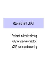
Recombinant DNA I - Basics of molecular cloning Polymerase chain reaction cDNA clones and screening
Bạn đang xem bản rút gọn của tài liệu. Xem và tải ngay bản đầy đủ của tài liệu tại đây (240.07 KB, 23 trang )
Recombinant DNA I
Basics of molecular cloning
Polymerase chain reaction
cDNA clones and screening
Recombinant DNA Technology
•
Utilizes microbiological selection and screening
procedures to isolate a gene that represents as
little as 1 part in a million of the genetic material in
an organism.
•
DNA from the organism of interest is divided into
small pieces that are then placed into individual
cells (usually bacterial).
•
These can then be separated as individual
colonies on plates, and they can be screened to
find the gene of interest.
•
This process is also called molecular cloning.
DNA pieces are joined in vitro to form
recombinant molecules
•
Generate sticky ends on the DNA, e.g. with
restriction endonucleases
•
Tie DNA molecules from different sources
together with DNA ligase
Restriction endonucleases generate ends
that facilitate mixing and matching
GAATTC
CTTAAG
GAATTC
CTTAAG
G
CTTAA
AATTC
G
G
CTTAA
AATTC
G
EcoRI cut
Mix and ligate
G
CTTAA
AATTC
G
G
CTTAA
AATTC
G
Recombinant
molecules
GAATTC
CTTAAG
GAATTC
CTTAAG
Parental
molecules
DNA ligase covalently joins two DNA molecules
•
Uses ATP or NADH to provide energy to seal
nicks
P
P
P
P
P
P
P
P
P
P
P
P
P
P
P
P
P
P
P
P
P
P
A
G
G
A
A
T
T
C
G
T
A
T
C
C
T
T
A
A
G
C
A
T
OH
OH
nick
nick
P
P
P
P
P
P
P
P
P
P
P
P
P
P
P
P
P
P
P
P
P
P
A
G
G
A
A
T
T
C
G
T
A
T
C
C
T
T
A
A
G
C
A
T
T4 DNA ligase + ATP
DNA ligase will seal the nicks that remain after annealing two fragments together
Alternate method to join DNA:
homopolymer tails
Alternate
method to
join DNA:
linkers
Introduction of recombinant DNA into
living cells via vectors
•
Autonomously replicating DNA molecules
–
(have an origin of replication)
•
Selectable marker, such as drug resistance
•
Insertion site for foreign DNA
–
(often a genetically engineered multiple cloning
region with sites for several restriction enzymes)
Plasmid vectors
•
Circular, extrachromosomal, autonomously
replicating DNA molecules
•
Frequently carry drug resistance genes
•
Can be present in MANY copies in the cell
A common plasmid cloning vector: pUC
ColE1 origin
of replication
lacZ
mulitple
cloning
sites
Ap
R
pUC
ColE1 ori
lacZ
Ap
R
pUC recombinant
Lac+, or blue colonies
on X-gal in
appropriate
strains of E. coli
Lac-, or white colonies
on X-gal in
appropriate
strains of E. coli
foreign DNA
High copy
number
Transformation of E. coli
•
E. coli does NOT have a natural system to
take up DNA
•
Treat with inorganic salts to destabilize cell
wall and cell membrane
•
During a brief heat shock, some of the
bacteria takes up a plasmid molecule
•
Can also use electroporation
Phage vectors
•
More efficient introduction of DNA into
bacteria
•
Lambda phage and P1 phage can carry
large fragments of DNA
–
20 kb for lambda
–
70 to 300 kb for P1
•
M13 phage vectors can be used to generate
single-stranded DNA
YAC vectors for cloning large DNA inserts
CEN4ori
URA3
TEL TEL
TRP1
CEN4
ori
SUP4
TRP1
URA3TEL TEL
pYAC3
BB
S
Cut with restriction
Enzymes S + B
Ligate to very large
Fragments of genomic
DNA
Large insert, 400 to
as much as 1400 kb
11.4 kb
Yeast artificial chromosome = YAC
Not to scale.
Bacterial artificial chromosomes
•
Are derived from the fertility factor, or F-
factor, of E. coli
•
Can carry large inserts of foreign DNA, up to
300 kb
•
Are low-copy number plasmids
•
Are less prone to insert instability than
YACs
•
Have fewer chimeric inserts (more than one
DNA fragment) than YACs
•
Extensively used in genome projects
BAC vectors for large DNA inserts
Cut with restriction enzyme E, remove “stuffer”
Ligate to very large fragments of genomic
DNA
SacBII
promoter
oriF
Cm(R)
pBACe3.6
E
E
S
11.5 kb
SacB+: SacBII encodes levansucrase,
which converts sucrose to levan,
a compound toxic to the bacteria.
Large insert, 300kb
oriF
Cm(R)
promoter
S
SacBII
SacB-: No toxic levan produced on sucrose
media: positive selection for recombinants.
Not to scale.
PCR provides access to specific DNA segments
•
Polymerase Chain Reaction
•
Requires knowledge of the DNA sequence
in the region of interest.
•
As more sequence information becomes
available, the uses of PCR expand.
•
With appropriate primers, one can amplify
the desired region from even miniscule
amounts of DNA.
•
Not limited by the distribution of restriction
endonuclease cleavage sites.
Polymerase chain reaction, cycle 1
Primer 1
Primer 2
Template
1. Denature
2. Anneal primers
3. Synthesize new DNA with polymerase
Cycle 1
Polymerase chain reaction, cycle 2
1. Denature
2. Anneal primers
3. Synthesize new DNA with polymerase
Cycle 2
PCR, cycle 3
1. Denature
2. Anneal primers
3. Synthesize new DNA with polymerase
Cycle 3 (focus on DNA segments bounded by primers)
2 duplex
molecules
of desired
product
PCR, cycle 4: exponential increase in
product
Cycle 4: Denature, anneal primers, and synthesize new DNA:
6 duplex
molecules
of desired
product
PCR, cycle 5: exponential increase in
product
Cycle 5: Denature, anneal primers, and synthesize new DNA:
14 duplex
molecules
of desired
product
PCR: make large amounts of a
particular sequence
•
The number of molecules of the DNA
fragment between the primers increases
about 2-fold with each cycle.
•
For n = number of cycles, the amplification
is approximately [2exp(n-1)]-2.
•
After 21 cycles, the fragment has been
amplified about a million-fold.
•
E.g. a sample with 0.1 pg of the target
fragment can be amplified to 0.1 microgram
PCR is one of the most widely used
molecular tools in biology
•
Molecular genetics - obtain a specific DNA
fragment
–
Test for function, expression, structure, etc.
•
Enzymology - place fragment encoding a
particular region of a protein in an expression
vector
•
Population genetics - examine polymorphisms in a
population
•
Forensics - test whether suspect’s DNA matches
DNA extracted from evidence at crime scene
•
Etc, etc