Biology 11 pptx
Bạn đang xem bản rút gọn của tài liệu. Xem và tải ngay bản đầy đủ của tài liệu tại đây (42.86 MB, 687 trang )
Ripped by Jack Truong, if you bought this, you got ripped off.
UNIT
1
Cellular Functions
It has been said that we are made of the stuff of
stars. What do you think this means? The pine
wood cells pictured on the right and all other
organisms on Earth are made mostly of only six
common chemical elements. These elements
originated under the conditions of massive
gravity and heat found in stars. Evidence that the
molecules of life — compounds containing carbon,
hydrogen, and oxygen — exist throughout the
universe is found in comets like Hale-Bopp, shown
below. Scientists have recently found that such
rocks, travelling through space, transport compounds
and molecules that form the basis of life on Earth.
Within cells, these molecules are transformed
into living organisms with a multitude of complex
strategies for survival. The same few molecules are
used over and over in different combinations to
make literally millions of different structures and to
carry out all the different functions needed by
living things. The processes involved in sustaining
life all begin at the molecular level within the
microscopic spaces of the cell. This includes the
storage and release of the energy needed to power
cellular process — which ultimately comes from
the Sun.
2
Unit
Contents
Chapter 1
Exploring the Micro-
universe of the Cell .
Chapter 2
Organizing Life . . . .
Chapter 3
Cells, Energy, and
Technology . . . . . . .
Unit
Investigation . . . . .
110
78
42
4
Overall
Expectations
In this Unit, you will
discover
What molecules make up
cells
How the cell membrane
separates cells from their
external environment but
allows substances into
and out of the cell
What special functions cell
structures have and how
these contribute to
keeping an organism alive
What processes in cells
capture and release the
energy needed for survival
and how we harness these
processes
The image below may look like a single-
celled organism, but it is actually a comet
called Hale-Bopp. What does a comet have
to do with the pine wood cells on the right?
Look ahead to pages
110–111.
You can start planning
your investigation well in
advance by knowing what
you will need.
As you work through the
unit, watch for ideas and
materials that will help you
prepare your experimental
design.
UNIT INVESTIGATION
3
CHAPTER
1
Reflecting
Questions
Exploring the
Micro-universe of the Cell
4
The micro-universe of the cell is a
world of stunning beauty, high
drama, and battles to the death. All
of it relies on and revolves around
the molecules of life. Why does the
didinium in the photograph on the
right hunt the paramecium — a larger
micro-organism? The didinium
cannot make all the molecules it
needs, such as proteins, from the
substances dissolved in its watery
environment. So the didinium must
acquire these molecules from its prey.
It then uses the molecules to build
and repair cellular structures and as a
source of energy for cellular processes.
The didinium and paramecium, as
well as the vorticella pictured below,
separate themselves from the outside
world with a cell membrane. How
then does the didinium “eat” the
paramecium? If the didinium opened
a hole in its cell membrane large
enough to take in the paramecium,
the didinium’s own cell contents
would leak out into the water
surrounding it. Indeed, how do any
of these cells take in molecules they
need or excrete wastes? Clearly, the
cell membrane must do much more
than separate the cell contents from
the external environment. How does
this living edge of the cell function?
Cellular dramas are also taking
place in the human body. For example,
cells that line your stomach live no
longer than four days because the
acid produced there eventually
destroys them. As the old cells die,
replacement cells emerge to face the
acidic battleground. If this did not
happen, you would not get the
nutrients you need to feed the cells
of your body.
Earlier courses introduced you to
cells and cell reproduction. In this
chapter, you will discover the
molecules of life. In particular, you
will investigate the large molecules
— carbohydrates, lipids, proteins,
and nucleic acids — that nourish,
build, and direct the living cell. You
will also examine the role that the
cell membrane plays in transporting
substances into and out of the cell.
Beautiful but deadly, the single-
celled vorticella pictured below use
the coiled spring in their cilia to leap
out to grab their prey (bacteria).
What are the key
molecules of life?
How does the cell
membrane define the
living cell and separate it
from its environment?
How does a cell control
the movement of materials
that enter and leave it?
5
Chapter
Contents
1.1 The Molecular Basis of Life 6
Thinking Lab:
Life: A Winning Experiment 6
MiniLab: The Resolving
Power of Skin 7
MiniLab: Modelling Sugars 11
Investigation 1-A: What’s Here?
Testing for Macromolecules 18
MiniLab: Manipulating
Macromolecules 20
1.2 Cell Membrane Structure 21
1.3 Through the Cell
Membrane 25
MiniLab: Random Walking 26
Design Your Own Investigation
1-B: Osmosis in a Model Cell 28
Thinking Lab: Relative
Concentration Challenge 32
1.4 Bulk Membrane Transport 35
MiniLab: Freezing Cells 37
THINKING LAB
1.1
SECTION
The Molecular Basis of Life
6
MHR • Cellular Functions
EXPECTATIONS
Describe the structure and
function of important
biochemical compounds.
Test for macromolecules
found in living organisms.
Use three-dimensional models
of important compounds.
Figure 1.1 These bacteria
remained dormant in a salt
crystal, probably from before
the time of the dinosaurs. In
2000, scientists revived them
by giving them water and
carbon-containing compounds.
When you think about cells, what first comes to
mind? How small they are? How such tiny living
things can do so much work? How a single
fertilized egg cell can produce all the many
specialized cells of a large organism, such as a
human being? This chapter, and the other chapters
in this unit, will help you answer these questions
— and perhaps also help you find new ones to ask.
Less than two hundred years ago, people did
not know of the existence of cells. The development
of the first microscopes finally gave scientists
access to the miniature world of the cell. Early
investigators discovered what you now take for
granted: that all living things are made up of one or
more cells. Other scientists determined that cells
are also the fundamental functional units of life.
What does this mean?
Life: A Winning Experiment
Background
Where do cells come from? Prior to the development and
acceptance of the cell theory in 1864, at least one early
investigator thought that mice could be generated
spontaneously by leaving a dirty shirt in a bucket. In 1860,
the Paris Academy of Sciences offered a prize to anyone
who could prove or disprove the spontaneous generation of
life. The biologist Louis Pasteur took up the challenge. The
two Erlenmeyer flasks shown here reproduce the results of
Pasteur’s winning experiment. Each flask and the stopper
were sterilized. Each contains 100 mL of vegetable broth
that was boiled for 10 min. Then, the sterilized stopper
was placed in one flask, while the second was left
unstoppered. This is what the flasks looked like five days
after they were filled.
Analyze
1. Describe any differences you observe in the broth of the
two flasks.
2. If you see any evidence of life generating life in these
photographs, where did the living organisms come from?
MINI LAB
The Resolving Power of Skin
You may not think of your skin as an exploratory tool that
has resolving power, but that is one of its functions. The
network of nerves in your skin gives you greater resolving
power in some places than in others. What does this
mean? Tape two pencils together, and ask a classmate to
touch both pencil points gently on the following spots while
you keep your eyes closed: a fingertip, the palm of your
hand, the back of your hand, and the back of your neck.
Ask your classmate to record what you felt each time, two
points or one.
Analyze
1. Which part of your skin has the greatest resolving
power (lets you clearly distinguish the two pencil
points)? Which has the least resolving power?
2. Suggest how differences in sensitivity to touch are
related to differences in the number and closeness of
nerve endings in your skin.
7
Exploring the Micro-universe of the Cell • MHR
What must the cells pictured in Figure 1.1 do to
stay alive? Like you, they have to obtain and ingest
food and water, get rid of wastes, grow, and respond
to changes in their environment. At some point,
they will reproduce, creating more cells. Each one
of these cells has to perform key life processes.
How does one cell do all that? Each cell uses
energy to fashion the structures it needs out of
materials available in its external environment —
atoms and molecules. Each cell also maintains a
sophisticated barrier between itself and the outside
world: the cell membrane. For example, the parasites
pictured in Figure 1.2 have cell membranes that
help them evade the human immune system.
How have scientists learned so many of the cell’s
secrets? Technology and scientific inquiry have
provided many answers. The technology for
examining cells you probably know best is the
compound light microscope. However, its glass
lenses can only magnify the cell enough to allow
you to see some of the larger cell features. Light
microscopes cannot resolve — or form distinct
images of — objects as close together as are most
structures in the cell.
The nature of visible light itself limits the
resolving power of a light microscope. When a light
wave passes through a specimen with structures
less than 0.2 µm apart, the wave bounces back from
Figure 1.2 The flat, undulating cells (trypanosomes) you see
among the red blood cells enter the bloodstream when a
tsetse fly bites and cause a disease called African sleeping
sickness. The structure of their cell membranes can make
them difficult for the human immune system to destroy.
the two features as if they consisted of a single
point. The features are too close together to block
the light wave separately, which would reveal them
as two points.
Before the invention of the electron microscope,
how did biologists gather information about the
inner workings of the cell? Living things depend
on chemical reactions, which take place at the
level of the molecule. So scientists used chemical
knowledge and procedures to learn about the world
of the cell: the molecules that living cells use,
form, excrete, and interact with. This section will
introduce you to that world.
Explain why biologists describe the cell as the unifying
structure that links all life.
PAUSE
RECORD
Look at the ingredients list on a milk package. You will see the
word pasteurized connected with the ingredients. Find out what
this word means. Why does it appear on a milk carton? Explain
in your own words where this term came from and how it
relates to cells.
LINK
Word
To review the cell theory, turn to Appendix 1.
FAST FORWARD
8
MHR • Cellular Functions
Living Organisms Rely on Small
Molecules
You may not think of your body in terms of
chemical reactions, yet you rely on your cells to
perform trillions of chemical reactions every second.
Without these, you could not remain alive. The
study of these reactions and the molecules and
processes involved in them is called biochemistry.
Some of the smallest molecules involved in
biochemical reactions are the most important.
Your breath contains three kinds of small
molecules critical to life. When you “see your
breath” on winter days, what you are seeing? Like
clouds, your visible breath consists of condensed
water vapour molecules (
H
2
O
) released through
your lungs. Your exhaled breath also contains two
other kinds of small molecules important to your
cells: oxygen (
O
2
) and carbon dioxide (
CO
2
). The
oxygen is left over from the previous inhalation
(your body absorbs only a small fraction of the
oxygen you take in with each breath).
Your cells use the oxygen molecules that do
pass in through your lungs to help release energy
from simple food molecules. This process,
called cellular respiration, can be summarized in
an equation:
C
6
H
12
O
6
+ 6O
2
6CO
2
+ 6H
2
O + energy
glucose oxygen carbon water
dioxide
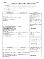
Figure 1.3
Life as we know it would not exist without these
small molecules.
Thus, the carbon dioxide and water you exhale are
waste products of this reaction, which occurs in
your cells. The compounds produced by the
process of converting food into energy are small
molecules. Figure 1.3 uses models to illustrate how
atoms in molecules of water, oxygen, and carbon
dioxide are arranged.
Water: The Primary Molecule of Life
Water is the most abundant molecule in any cell.
The unique chemical properties of water enable it
to act as a carrier for dissolved molecules inside and
outside the cell, and as a raw material in essential
cell reactions. It also functions as a lubricant
between organs, tissues, and individual cells.
These properties of water make possible life as
we know it.
remains liquid over a wide temperature range,
including temperatures at which most small
molecules are gases (such as room temperature)
dissolves most substances involved in living
processes, such as oxygen, carbon dioxide,
glucose, amino acids (components of proteins),
and sodium chloride (salt)
changes temperature gradually when heated or
cooled, so it protects cells from rapid temperature
changes and provides a stable environment for
cell reactions
is the only pure substance that expands when it
becomes a solid, which means that it floats when
it freezes (see Figure 1.5)
Figure 1.4 Water molecules cling together, which helps
water to creep up thin tubes, such as those running from
the roots to the tops of plants.
O
2
H
2
O
CO
2
9
Exploring the Micro-universe of the Cell • MHR
The special properties of water are determined
by its chemical structure. The uneven distribution
of electrical charges on a water molecule allows
one water molecule to attract another water
molecule at room temperature enough to form a
liquid. (Larger molecules such as oxygen and
carbon dioxide remain gases at room temperature.)
Figure 1.4 shows how this property is important to
plants. Molecules with uneven charge distribution
are said to be polar (because they have oppositely
charged “poles”). Although carbon dioxide
contains oxygen, it has an even distribution of
electrical charge. This means that it is nonpolar.
Organic Compounds
The term organic compound refers to molecules
that contain both carbon and hydrogen, which
means that molecules such as oxygen, water, and
carbon dioxide are inorganic. Although living
things require water to perform their life functions,
and most also require oxygen, these molecules
can be generated without the involvement of
living things.
The molecules that form a more permanent part
of living cells all have a carbon “backbone.” This
abundance of carbon in organic compounds is why
scientists call life on Earth carbon based. Each
carbon atom can form up to four bonds with other
atoms. Hydrocarbon molecules (organic molecules
containing only carbon and hydrogen) come in an
enormous range of sizes and shapes, including
open-ended chains and closed, loop-like “rings,”
such as those shown in Figure 1.6. From previous
studies, you may recognize the lines joining the
atoms in this figure as covalent bonds.
Figure 1.6 These hydrocarbon molecules have relatively
simple structures.
H
H
CH
C
H
H
C
H
H
C
H
H
C
H
H
C
H
H
C
H
H
C H
H
H
C
C
CC
C
H
C
H
H
H
H
H
To review chemical bonding, turn to Appendix 2.
FAST FORWARD
ice 0˚
Ice acts as an insulator and prevents the water below it
from freezing, which protects aquatic organisms in the winter.
Water provides an external
environment for many organisms
both single-celled and
multicellular.
water 4˚
pond
micro-organisms
frog
hibernating
in mud
Figure 1.5 All organisms require water to live.
10
MHR • Cellular Functions
In addition to carbon and hydrogen, many
organic molecules contain other elements, the
most important of which are oxygen, nitrogen,
phosphorous, and sulfur. You may recall from
earlier studies that normal air is about 20% oxygen
and 78% nitrogen, so it is not surprising that many
organic molecules contain these two elements.
Living cells make and use a variety of organic
molecules, such as glucose (a sugar). The cells of
plants and some other organisms manufacture
glucose through the process of photosynthesis
summarized in this equation:
6CO
2
+ 6H
2
O6O
2
+ C
6
H
12
O
6
carbon water oxygen glucose
dioxide
Both plants and animals use glucose as a food from
which they obtain energy.
In this chapter, you will explore only the
principal organic molecules contained in
carbohydrates, lipids, and proteins, as well as
the nucleic acids. that make up the DNA in
chromosomes. Figure 1.7 illustrates foods
containing these molecules. All of these organic
compounds are very large molecules, or
macromolecules (macro means large), composed
of smaller subunits.
The Structure and Biological
Function of Carbohydrates
Very interested in the food produced by plants,
early scientists chemically analyzed sugars and
starches. They discovered that these compounds
always contain carbon, hydrogen, and oxygen —
almost always in the same proportion: two atoms of
hydrogen and one atom of oxygen for every atom of
carbon, or
CH
2
O
. Since the formula for water is
H
2
O
, the scientists concluded that sugars and
starches consist of carbons with water attached to
them, or carbohydrates (hydro means water).
Carbohydrates provide short- or longer-term energy
storage for living organisms.
Molecule
Water
Oxygen
Carbon dioxide
Glucose
Chemical formula Atomic mass units
18
32
44
180
H
2
O
O
2
CO
2
C
6
H
12
O
6
This chart gives you the chemical formulas for a number of
important biological molecules and the mass of each in atomic
mass units. Use this information to determine the mass of a
molecule of table sugar (sucrose), which has the chemical
formula
C
12
H
22
O
11
.
LINK
Math
To view the periodic table, turn to Appendix 11.
FAST FORWARD
l
ig
h
t
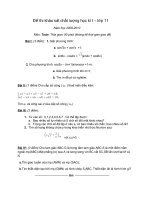
Figure 1.7 Foods rich in carbohydrates, lipids, and proteins.
MINI LAB
Modelling Sugars
In this lab, you will construct models of two glucose
molecules and join them together to make a disaccharide
molecule. To do this, you will need the following molecular
model materials: 12 carbon atoms, 24 hydrogen atoms,
12 oxygen atoms, and 50 bonds.
Use the diagram of glucose in Figure 1.8 above as a
guide to building your glucose molecule models. Note that
glucose has a ring structure consisting of five carbon atoms
and an oxygen atom. A sixth carbon is attached to one of
the ring carbons. Keep in mind that each carbon has four
bonding sites. Set both of your glucose models side by side
so that they match the orientation of the glucose molecules
on the left side of Figure 1.9. Break and re-make bonds to
build a model of the disaccharide maltose.
Analyze
1. Which atoms are involved in the breaking and making
of bonds when a disaccharide is formed? Suggest a
reason for this.
2. Describe how you think the glucose and fructose
subunits in sucrose are linked. Build a molecular model
to show this.
3. How might a cell use the three-dimensional shape of a
glucose molecule to orient the connecting atoms
between two glucose “rings”?
11
Exploring the Micro-universe of the Cell • MHR
A carbohydrate molecule with three to seven
carbon atoms (and the corresponding number
of hydrogen and oxygen atoms) is called a
monosaccharide, or simple sugar (mono means one;
sakkharon means sugar). Figure 1.8 shows the
single, closed ring-like structures of three common
monosaccharides: glucose, fructose, and galactose.
A disaccharide, or double sugar, is made up of
two simple sugars (di means two). Figure 1.9 shows
how two glucose units link together to form one
molecule of the disaccharide maltose. Malted
products such as beer contain maltose. You may be
more familiar with another disaccharide, sucrose,
which is made by joining glucose with fructose.
Sucrose is in many food products, from brownies
to barbeque sauce.
Figure 1.8 Living cells use molecules of glucose or other
simple sugars, such as fructose and galactose, as a quick
source of energy. Although all three simple sugars have the
same composition of atoms, the arrangement of these
atoms differs slightly in each molecule.
C
OH
OH
C
H
H
C
OH
H
C
HO
H
CO
H
CH
2
OH
glucose
C
OH
HO
C
H
H
C
H
C
HO
O
CH
2
OH
fructose
C
OH
OH
C
H
H
C
OH
H
C
H
HO
CO
CH
2
HO
CH
2
HO
galactose
Figure 1.9 Note the role played by water when glucose units are linked to form
maltose and when maltose is broken apart to form individual glucose molecules.
++
H
O
C
6
H
12
O
6
glucose glucose
HO
2
HO
2
HO
H
O
C
6
H
12
O
6
C
12
H
22
O
11
O
O
maltose
water
O
OH
12
MHR • Cellular Functions
A polysaccharide is a complex carbohydrate
consisting of many simple sugars linked together
(poly means many). Figure 1.10 shows the structure
of the polysaccharides starch, glycogen, and
cellulose. Starch performs the important function
of energy storage in plants. Glycogen performs the
same function in animals. Compare the structures
of the starch and glycogen molecules, and note the
many “branches” on the glycogen molecule. The
larger amount of branching in glycogen means that
glycogen molecules pack more glucose units into a
single cell than do starch molecules.
Plants produce an even larger polysaccharide
macromolecule called cellulose, out of which they
build their cell walls. Cellulose is considered a
structural molecule because it protects individual
cells and provides support for the whole plant. As
a polysaccharide made up of glucose units,
cellulose also stores a great deal of energy.
However, only a few bacterial species produce the
digestive chemicals needed to break cellulose
down into glucose units and release energy. So —
to obtain nourishment from grass, leaves, wood,
and other cellulose-rich plant materials — animals
such as cattle, rabbits, and termites must host these
bacteria in their guts. The human gut does not host
these bacteria, so the food energy in cellulose is not
directly accessible to us.
Figure 1.10 Look at the structural differences among the
polysaccharides starch, glycogen, and cellulose. Notice that
all three consist of glucose subunits.
The Structure and Biological
Function of Lipids
Lipids are a diverse group of macromolecules that
have one important feature in common: they do not
dissolve in water. Living organisms use lipids for
many purposes: long-term nutrient and energy
storage, insulation, cushioning of internal organs,
and hormones to send messages around the body.
Lipids are also the primary structural component of
the cell membrane of every cell.
The lipid with which you are likely most
familiar is fat. Fats include not only substances
such as butter but also oils such as canola oil.
Whether in solid or liquid form, one gram of lipid
contains 2.25 times as much energy as one gram
of carbohydrate.
Figure 1.11 The white walrus has just returned from an
extended swim in extremely cold water. You can see its
blubber right through its skin because the blood vessels in
its skin have constricted (narrowed) to conserve heat in the
cold water. Without normal blood flow, the skin becomes
nearly transparent, making the walrus appear white.
In terms of mass, most of the world’s carbohydrate exists in
the form of cellulose.
BIO FACT
Glycogen
glucose
subunits
glucose
subunits
crosslink
bonds
Cellulose
Starch
glucose
subunits
potato
liver
cotton
13
Exploring the Micro-universe of the Cell • MHR
All fat molecules have the same basic three-
branched structure. Figure 1.13 shows how this
structure forms in a chemical reaction involving
one molecule of an alcohol called glycerol and
three molecules of fatty acid. Another name for
this structure is a triglyceride.
Figure 1.13 On this simple model of a triglyceride (fat)
macromolecule, the triangles represent glycerol’s three
reaction sites.
A fatty acid is a hydrocarbon chain with a
difference: at one end, the carbon has an acidic
— COOH group instead of hydrogen attached to it.
It is this acidic group of a fatty acid that attaches to
one of the three main reaction sites on a glycerol
molecule, as shown in Figures 1.13 and 1.14. The
triglyceride produced is nonpolar. This means that
it will not be attracted to (polar) water molecules,
which is why fats are insoluble in water.
Glycerol always has the same composition;
not so for the three fatty acids, which may be
identical or nonidentical, short or long, saturated
or unsaturated. In the hydrocarbon chain of a
saturated fatty acid, each of the carbon atoms
beyond the one bonded to oxygen is bonded to four
other atoms. An unsaturated fatty acid has bonding
sites (double bonds) where additional hydrogen
atoms could be attached. Figure 1.14 shows the
difference between a saturated and an unsaturated
fatty acid.
If unsaturated fatty acids dominate, the resulting
fat will likely be liquid at room temperature. If
saturated fatty acids dominate, the resulting fat will
likely be solid at room temperature.
Figure 1.14 (A) This fatty acid is saturated with hydrogen.
(B) This fatty acid has room for two more hydrogen atoms,
one on each of the highlighted carbon atoms. Such a fatty
acid is called unsaturated.
C
H
H
H H
C
H
C
H
C
H
H
C
O
HO
C
H
H
C
H
H
H
C
H
H
C
H
C
H
C
H
H
C
O
C
H
H
H
HO
A
B
To learn about double bonds, turn to Appendix 2.
FAST FORWARD
Compare Figure 1.12 (lipid formation) with Figure 1.9
(carbohydrate formation). What do the two reactions have in
common? (Hint: Look at the blue highlighting on each
figure.) How do they differ?
PAUSE
RECORD
glycerol
Fatty acid 1
Fatty acid 2
Fatty acid 3
Figure 1.12 Notice that in each fatty acid chain of a triglyceride molecule only the
carbon atom at the glycerol end has oxygen attached to it. All the rest of the
carbon atoms on the fatty acid have only hydrogen atoms attached to them.
+
COHH
H
COHH
C
C
OHH
H
H
H
COH
H
COH
COH
H
C
H
C
H
C
H
H
C
O
C
H
H
H C
HO
H
C
H
C
H
C
H
H
C
C
C
H
H
H
O
C
H
H
H H
C
H
C
H
C
H
H
C
O
HO
C
H
H
C
H
H
HC
H
H
H H
C
H
C
H
C
H
H
C
O
C
H
H
C
H
H
H
C
H
H
H H
C
H
C
H
C
H
H
C
O
H
C
H
H
H H
C
H
C
H
C
H
H
H
glycerol 3 fatty acids fat 3 water
molecules
HO
HO
+
HO
2
3
14
MHR • Cellular Functions
The Structure and Biological
Function of Proteins
Most cellular structures are made of various
types of protein. Proteins also serve many other
functions in cells. In fact, they display greater
structural complexity and functional diversity than
either lipids or carbohydrates.
Your hair and fingernails are both made of the
same type of protein, keratin, yet each has its own
distinctive properties. The bones and muscles
inside your hand and the ligaments and tendons
connecting them also contain distinctly different
proteins. Without these proteins, you would not be
able to move your hand.
In addition to their structural functions,
proteins also
function as enzymes to facilitate chemical
reactions (the enzyme amylase in your saliva
begins the breakdown of starches into simple
sugars while you chew)
help transport substances across cell membranes
or to different parts of an organism (the
hemoglobin in your blood transports oxygen
from your lungs to each cell in your body)
Figure 1.15 Feathers, spider webs, wool, and silk are made
up of proteins. In fact, feathers consist mostly of the same
protein, keratin, that makes up human nails and hair.
act as chemical messengers (some hormones are
proteins rather than lipids, such as the insulin
that helps to regulate the amount of glucose
available to cells)
Like other macromolecules, proteins are
assembled from small units. In proteins, the
building blocks are amino acid molecules.
Figure 1.16 shows the chemical structure of five
representative amino acids. Note the unhighlighted
part of each amino acid. It contains two carbon
atoms, two oxygen atoms, four hydrogen atoms,
and one nitrogen atom per molecule. The number
and arrangement of these atoms is identical for
all but one amino acid (proline). What differs
substantially from one amino acid to another is the
highlighted remainder group (or R group).
Steroids (and cholesterol) are lipids too, although the
structure of these molecules differs markedly from the
structure of fats. News reports from the sports world may
have led you to think of steroids as harmful to health. In
fact, your body makes several different kinds of steroids
from the fats you eat. You need all of these steroids for
normal health and development. Your body manufactures
all the steroids it needs, so injecting or ingesting steroids
can lead to abnormal development of sex organs and even
early death.
BIO FACT
Figure 1.16 Note that these five representative amino acids differ from one
another by their R groups.
H
H
N
HH
H
H
C
O
OH
C
C
alanine
Remainder group,
or R group
valine cysteine
H
HH
H
H
H
C
CH H
H
C
N
O
OH
C
CH
H
H
H
H
H
H
H
C
SH
N
O
OH
C
C
C
C
C
C
H
H
H
H
H
H
C
C
C
N
O
OH
C
C
H
H
phenylalanine
15
Exploring the Micro-universe of the Cell • MHR
A chemical linkage called a peptide bond joins
individual amino acids together. Figure 1.17 shows
how a peptide bond between two amino acids is
formed or broken. Regardless of which R group is
present, amino acids always bond to each other in
the manner shown in Figure 1.17. However, a
chain of amino acids is not yet a protein, only a
polypeptide.
Figure 1.19 on the next page shows the steps
between a peptide bond and a finished protein
molecule. The final shape of the protein’s three-
dimensional structure determines what properties
it will have and therefore what functions it can
perform.
If a protein molecule is exposed to extreme
temperatures, extreme pH conditions (very acidic
or very basic), or harsh chemicals, it will unfold or
change shape. When this happens, the protein is
said to have been denatured. The protein loses its
ability to perform its normal function.
Why can some proteins such as enzymes or
hemoglobin function in a water solution while
others (such as the keratin in your fingernails) are
usually insoluble in water? This depends on how
the polypeptide(s) making up a protein are twisted
and folded. When the parts of the R groups that can
interact with water end up on the outside of the
final protein structure, the protein is soluble in
water. When the parts of the R groups that do not
interact with water or react only slightly with it
end up on the outside, the protein will not dissolve
in water.
Figure 1.18 This computer-generated image of a protein
molecule makes the protein’s complex, three-dimensional
structure easier to visualize.
Humans need 20 amino acids — known as
the common amino acids — to make the protein
macromolecules required for healthy body
structures and functions. Your body can
manufacture 12 of these amino acids from non-
protein food sources. The other eight must be
present in your food because your body cannot
manufacture them for itself. These eight are
referred to as essential amino acids.
With 20 different amino acids to combine,
proteins exist in thousands of distinctly different
forms. Each kind of organism manufactures its
own characteristic proteins or variations on
proteins common to a number of species, such as
hemoglobin. Indeed, it is our proteins that make us
different from ants, amoebas, or ash trees.
To enhance your learning about macromolecules, go to your
Electronic Learning Partner.
PLAY
Figure 1.17 In the first stage of the formation of a polypeptide, two amino acids
are linked together. The R groups appear only as “R” because they do not take
part in the reaction that produces or breaks a peptide bond.
water
C
H
R
H H
C
O
OH H
NH
H
C
H
R
CNH
H
C
R
C
O
OH
NN
H
H
C
R
C
O
OH
O
peptide
bond
amino acid amino acid dipeptide
++
HO
2
16
MHR • Cellular Functions
C
O
H
N
R
C
N
O
C
R
C
H
O
C
N
H
H
N
O
C
C
O
H
N
R
C
O
C
NH
C
O
H
N
R
C
N
O
C
R
C
H
O
C
N
H
CR
H
N
CR
O
C
C
O
H
N
R
C
O
C
NH
H
H
H
H
C
R
C
C
C
O
C
N
H
CH
R
O
O
O
O
H
H
H
H
H
O
O
O
O
C
CH
R
O
C
N
H
O
C
CH
R
C
N
H
O
C
CH
R
N
H
O
C
CH
R
N
CH
R
N
H
O
C
N
CH
R
peptide bond
CH
pleated sheet
α (alpha) helix
amino acid
+
H
3
N
COO
−
Many amino acids are
joined together to form
a polypeptide chain.
Figure 1.19 The formation of a protein molecule from a polypeptide
When a polypeptide grows
beyond 30 amino acids, it
begins to either coil up into a
helix or bend into a pleated
sheet. The dotted lines
represent the weak attraction
between the O and H
“sidearms” that holds the
molecule in a helical or
pleated shape.
The helix then folds into a three-dimensional
structure, the exact shape of which depends
on which R groups are present and in
what order.
Many proteins contain two or more
folded polypeptides joined together.
Figure 1.20 Generalized nucleotide. Nucleotides consist
of a five-carbon simple sugar (ribose in the case of RNA and
deoxyribose in DNA), a nitrogen base, and a phosphate
group, symbolized here by .
Nucleic Acids
Nucleic acids direct the growth and development
of every living thing by means of a chemical code.
They determine how the cell functions and what
characteristics it has.
The cell contains two types of nucleic acid: RNA
(ribonucleic acid) and DNA (deoxyribonucleic
acid). You may already have learned that DNA is
the main component of the genes, or hereditary
material, in all cells. Each gene contains
instructions for making RNA. RNA, contains the
instructions for making proteins. These proteins
make up much of the structure of a cell and control
how it functions.
Like proteins and carbohydrates, nucleic acids
consist of long chains of linked subunits. These
subunits are called nucleotides, which are depicted
in Figure 1.20. DNA is made up of just four
different nucleotides. So is RNA. Each DNA
nucleotide has an RNA nucleotide counterpart.
RNA consists of a single, long chain of
nucleotides. In DNA, two enormous nucleotide
chains are attached in a ladder-like structure,
which then coils into a double helix shape.
Figure 1.22 illustrates this DNA structure.
Figure 1.21 This image shows the shape of individual
atoms on a section of a DNA molecule. It was mapped using
a probe through which a tiny electric current flows.
P
P
S
O
NN
pentose sugar
nitrogen-
containing
base
phosphate
17
Exploring the Micro-universe of the Cell • MHR
A
A
A
G
T
T
C
G
T
C
P
S
P
S
P
S
P
S
P
S
P
S
P
S
P
S
P
S
S
A
T
A
T
C
G
G
C
one
nucleotide
P
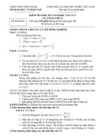
Figure 1.22 DNA’s structure. Each DNA strand contains carbon rings (sugar) and
phosphate molecules, while the ladder “rungs” between the strands consist of
nitrogen bases.
SKILL FOCUS
Conducting research
Performing and recording
Analyzing and interpreting
Communicating results
Investigation
1•A
18
MHR • Cellular Functions
Pre-lab Questions
Glucose is a monosaccharide. What does this
mean?
Proteins are made of amino acids. What atom
is present in an amino acid that is not present
in a sugar molecule?
Identify two health hazards related to using a
copper sulfate solution.
Problem
How can you determine the presence of glucose,
starch, lipid, and protein in various samples?
CAUTION: Be careful when handling iodine,
Benedict’s solution, Sudan IV, and Biuret reagent
as they are toxic. Avoid allowing the hot water
bath to boil vigorously because this can cause
test tubes to break. Clean up spills immediately,
and notify your teacher if a spill occurs.
Materials
safety goggles 40 mL sucrose solution
disposable gloves 40 mL starch solution
apron 40 mL distilled water
marker Benedict’s solution in a
6 graduated cylinders dropper bottle
12 test tubes iodine solution in a
test tube rack dropper bottle
hot water bath Sudan IV solution
test tube clamp (0.5% alcohol solution)
test tube brush in a dropper bottle
40 mL protein solution Biuret reagent in a
(2% gelatin solution) dropper bottle
40 mL vegetable oil glassware soap
40 mL glucose solution
Procedure
Follow your teacher’s instructions for the
disposal of the test solutions and samples.
Use the same graduated cylinder to measure
samples of the same substance for all four
parts of this investigation. For example, use
the same graduated cylinder to measure out
vegetable oil each time.
Perform parts B, C, and D of this investigation
while you heat samples for part A.
Carefully clean your work area after you finish
each test.
Wash glassware throughly with soap and water.
Part A
1. Set up the hot water bath as shown below.
Use a medium setting for the hot plate.
2. Mark the six graduated cylinders with the
numbers 1 to 6.
3. Mark six test tubes with the numbers 1 to 6.
4. Measure out 10 mL of protein solution into
graduated cylinder 1, 10 mL of vegetable oil
into graduated cylinder 2, 10 mL of glucose
solution into graduated cylinder 3, 10 mL of
sucrose solution into graduated cylinder 4,
10 mL of starch solution into graduated
cylinder 5, and 10 mL of distilled water into
graduated cylinder 6.
What’s Here? Testing for Macromolecules
Biochemists have developed standard tests to determine the presence
of the most abundant macromolecules made by cells: carbohydrates,
lipids, and proteins. In this investigation, you will conduct standard
tests to determine the presence of glucose, starch, lipid, and protein in
known samples. Each test involves an indicator, which is a chemical
that changes colour when it reacts with a specific substance.
19
Exploring the Micro-universe of the Cell • MHR
5. Add 10 mL of each sample to the test tube
with the same number.
6. Add 5 drops of Benedict’s solution to each
test tube. Safely mix the contents of each test
tube by swirling the test tube as shown below.
7. Heat each test tube in the hot water bath
for 5 min. If your hot water bath is large
enough, heat two test tubes at a time. After
5 min, use a test tube clamp to move each
test tube to the test tube rack.
8. When all the test tubes have been heated
and removed, turn off the source of heat and
let the water bath cool.
9. Record your observations for each test tube.
10. When the test tubes have cooled, wash
them. When the hot water bath has cooled,
pour out the water and wash the glassware.
Part B
1. Repeat steps 3, 4, and 5 from Part A.
2. Add 5 drops of iodine solution to each test
tube. Carefully mix the contents of each test
tube.
3. Record your observations for each test tube.
Then wash the test tubes.
Part C
1. Repeat steps 3, 4, and 5 from Part A.
2. Add 5 drops of Sudan IV solution to each
test tube. Safely mix the contents of each
test tube.
3. Record your observations for each test tube.
Then wash the test tubes thoroughly.
Part D
1. Repeat steps 3, 4, and 5 from Part A.
2. Add 5 drops of Biuret reagent to each test
tube. Safely mix the contents of each test
tube.
3. Record your observations for each test tube.
Then wash the test tubes and graduated
cylinders.
Post-lab Questions
1. Describe a positive test for starch. Explain
how you know.
2. Describe a positive test for glucose. Explain
how you know.
3. Describe a positive test for lipids. Explain
how you know.
4. Describe a positive test for protein. Explain
how you know.
Conclude and Apply
5. What was the purpose of testing distilled
water for each part of the investigation?
6. Suppose you have a sample of breakfast
cereal that may contain one, two, three, or
all four of the macromolecules you tested
for in this investigation. Write a procedure
describing how you would test the sample
to determine which macromolecules
it contains.
Exploring Further
7. Physicians often want to know the glucose
and lipid levels in a patient’s blood and
whether proteins are present in a patient’s
urine. Research to find out what this
information might show about an
individual’s health.
A.
Benedict’s
solution +
heat
B.
Iodine
solution
C.
Sudan IV
solution
D.
Biuret
reagent
Sample
1. protein
solution
2. vegetable
oil
20
MHR • Cellular Functions
SECTION REVIEW
1. List the key life processes of cells.
2. Identify three inorganic molecules important
for cells.
3. Describe the unique properties of water. Explain
how each property is important to cells.
4. Copy and complete this chart:
5. What is a peptide bond?
6. Why are some amino acids described as
essential amino acids?
7. Some people add cold milk to hot coffee. Others
heat milk so that it is hot and steamy. Does heating
milk change its chemical make-up? Predict any
changes, and design a lab that would test your
prediction.
8. Some oils, such as olive oil, are liquid at room
temperature. How can the structure of the oil
molecules be changed so that they are almost solid
at room temperature?
9. Find a Materials Safety Data Sheet, and identify
health hazards related to Biuret’s reagent.
10. Explain how computer molecular-model
simulations could benefit biomedical research.
How is whole milk different from skim milk?
• Design a series of tests to identify the macromolecules
in whole milk. Which indicators would you use?
• Predict which macromolecules you would find if you
performed the tests you designed above. Would you
expect different results with skim milk? Explain.
UNIT INVESTIGATION PREP
MC
MC
K/U
I
K/U
K/U
Macromolecule
type
Diagram
Sample
Molecule
monosaccharide
carbohydrate
lipid (2 examples)
protein (2 examples)
nucleic acid
Function in
the cell
K/U
K/U
K/U
K/U
MINI LAB
Manipulating Macromolecules
The study of biological molecules has been revolutionized
by the use of computers. Today, sophisticated software
programs allow biochemists to explore, build, and
manipulate three-dimensional models of macromolecules.
In this lab, you will use the Internet to view and manipulate
similar models. (You may need to download free software
to run the simulations, such as Chime. Check with your
teacher before you download anything onto a school
computer.) Your teacher will give you a list of sites that
contain three-dimensional models of proteins and other
macromolecules. Go to each site, and use the simulations
to view and manipulate the molecular models.
Analyze
1. Describe each type of model the site(s) allowed you to
view, for example, a bail-and-stick model, space-filling
model, and so on.
2. How does rotating a molecule change what you can
see about it?
3. Draw structural formulas for two of the three-
dimensional models that you viewed.
4. What did the computer simulations of molecules show
you that would be more difficult to see using molecular
model kits?
1.2
SECTION
Cell Membrane Structure
21
Exploring the Micro-universe of the Cell • MHR
EXPECTATIONS
Identify the structure and
function of phospholipids.
Describe the fluid-mosaic
structure of the cell
membrane.
Figure 1.23 From an altitude of
10 000 m, a city may look quiet
and still. From 1000 m, it
becomes clear that buses,
trucks, and cars are moving.
Airplanes fly into its airport.
Ships and boats come and go
from its harbour. How does this
city resemble a cell?
When viewed with even the most powerful
light microscope, the cell membrane looks like
nothing more than a thin, dark line. Yet if the cell
membrane functioned only as a barrier separating
the inside of the cell from its external environment,
how could the cell survive? How would the cell get
the raw materials it needs to build macromolecules?
The cell membrane must also regulate the movement
of materials from one environment to the other.
Figure 1.24 What was the original purpose of this wall
around the old part of Québec City? How did its original
function resemble that of a cell membrane?
The efficient operation of a city such as the one
pictured in Figure 1.23 would soon grind to a halt
without adequate routes for the flow of people and
things in and out. Similarly, the activities of a
living cell depend on the ability of its membrane to
transport raw materials into the cell
transport manufactured products and wastes out
of the cell
prevent the entry of unwanted matter into the cell
prevent the escape of the matter needed to
perform the cellular functions
Getting the Cell Membrane in Focus
The development of the electron microscope gave
scientists the information they needed to begin
exploring how the cell membrane performs its
regulatory functions. An electron microscope uses
beams of electrons instead of light to produce
images. Electron microscopes and other devices
separate electrons from their atoms and focus them
into a beam. For example, the image on a TV set is
formed by electron beams that cause the inner
coating on the screen to glow.
Compared to light, an electron beam has a very
short wavelength — so short that it can pass
between two cell features less than 0.2 µm apart
and form an image of them that shows two distinct
and separate points.
22
MHR • Cellular Functions
Figure 1.25 James Hillier was in his early twenties when
his professor asked him to help build a practical electron
microscope. The microscope that Hiller and Albert Prebus
built is now on display at the Ontario Science Centre in
Toronto. It has 7000x magnification.
The first really usable electron microscope was
built in 1938 at the University of Toronto by two
graduate students, James Hillier (1915–) and Albert
Prebus (1913–1997). Their microscope revealed
that what look like “grains” under the light
microscope are complex cellular structures. In
Chapter 2, you will learn more about these
structures. This section continues the story of
research into the cell membrane.
When electron microscopy finally yielded a
more detailed view, microscopists saw that the
cell membrane is in fact a bilayer, or a structure
consisting of two layers of molecules. Chemical
analysis revealed that this bilayer is composed
mainly of phospholipid molecules, a type of lipid.
Phospholipids have two fatty acids bonded to a
glycerol “backbone.” The third glycerol reaction
site is bonded to a chain containing phosphorus,
and in some cases nitrogen as well.
This makes the shape and properties of a
phospholipid quite different from those of a
triglyceride. The phosphate chain forms a “head,”
while the two fatty acids form two “tails.” The
electric charge in the molecule is unevenly
distributed, as shown in Figure 1.27: the molecule
has a polar head and nonpolar tails.
The polar head of a phospholipid molecule is
attracted to water molecules, which are also polar.
This makes the phosphorus end of a phospholipid
water soluble. The hydrocarbon chains in the fatty-
acid tails of the phospholipid are not attracted to
water molecules. They are, however, compatible
with other lipids.
Figure 1.28 shows what can happen when a film
of phospholipid molecules is spread in a water
sample. Through a combination of attraction and
repulsion, the phospholipids spontaneously
arrange themselves into a spherical, cage-like
bilayer. Their water-attracting polar heads face both
the inside and the outside of the sphere, while
Earlier in this chapter, you learned that hydro means water.
Many textbooks use the terms hydrophobic and hydrophilic to
describe the way that molecules interact with water. Write a
definition for each of these words, including the word soluble
in one definition and insoluble in the other. Which end of a
phospholipid is hydrophobic and which is hydrophilic?
LINK
Word
Figure 1.26 Electron microscopy showed that the cell membranes of both plant
and animal cells have a two-layered structure. This gave scientists the clue they
needed to begin unravelling the mystery of how the cell membrane works.
plant cell
membrane
animal cell membrane
23
Exploring the Micro-universe of the Cell • MHR
Figure 1.27 Constructed much like a triglyceride (fat),
phospholipids contain a phosphate group and sometimes
also a nitrogen group.
their water-averse, nonpolar lipid tails face each
other. This sandwich-like phospholipid structure,
called a phospholipid bilayer, forms the basis of
the cell membrane.
The Fluid-Mosaic Membrane Model
The fact that lipids do not dissolve in water creates
a border around the cell. The phosphate edges of
this border help to define and contain the more
fluid lipid centre. However, there is much more to
a cell membrane than its phospholipid bilayer.
Figure 1.28 The molecular structure of a phospholipid
bilayer. Unlike the cell membrane of a living cell, this bilayer
contains only water inside it.
Based on intensive research by biochemists
and electron microscopists, biologists have inferred
that the cell membrane also contains a mosaic of
different components scattered throughout it, much
like raisins in a slice of raisin bread. For example,
numerous protein molecules stud the phospholipid
bilayer. The phospholipid molecules and some of
these proteins can drift sideways in the bilayer, a
phenomenon which supports the idea that the
phospholipid bilayer has a fluid consistency. Thus,
this description of the cell membrane is called the
fluid-mosaic membrane model.
Figure 1.29 on the next page shows how proteins
and phospholipids fit together in the continuous
mosaic of an animal cell membrane. Note that this
cell membrane also contains another type of lipid:
cholesterol molecules. Cholesterol allows animal
cell membranes to function in a wide range of
temperatures. At high temperatures, it helps
maintain rigidity in the oily membrane bilayer. At
low temperatures, its keeps the membrane fluid,
flexible, and functional — preventing cell death
from a frozen membrane. Cholesterol also makes
the membrane less permeable to most biological
molecules. Plants have a different lipid that serves
a similar function in their cell membranes.
The shapes of the membrane proteins vary
according to their function, and each type of cell
has a characteristic arrangement of proteins in its
membrane. For example, the membrane of a human
red blood cell includes 50 different protein types
arranged in a pattern that only other cells from
humans with the same blood type can “recognize.”
water
The ability of phospholipids to spontaneously form a
spherical bilayer in water likely played a key role in the
formation of the first cells about 3.8 billion years ago.
BIO FACT
CH
2
CH
2
CH
2
CH
2
CH
2
CH
2
CH
2
CH
2
CH
2
CH
2
CH
2
CH
2
CH
2
CH
2
CH
2
CH
2
CH
2
CH
2
CH
2
CH
2
CH
2
CH
2
CH
2
CH
2
CH
2
CH
2
CH
3
CH
2
CH
2
CH
2
CH
2
CH
2
CH
2
CH
2
CH
2
CH
3
CH
3
CH
3
CH
3
head
tail
polar head
group
nitrogen
group
glycerol
phosphate
group
nonpolar
tail group
fatty acids
P
CH
O
O
OCOC
O
O
CH
CH
O
A
B
+
N
O
−
24
MHR • Cellular Functions
Outside cell
Inside cell
filaments of
the cytoskeleton
peripheral
protein
cholesterol
integral
protein
phospholipid
bilayer
glycolipid
glycoprotein
carbohydrate
chain
Figure 1.29 Fluid-mosaic model of membrane structure.
Notice that many lipids and proteins facing the exterior of
the cell have carbohydrate chains attached to them, while
on the interior of the cell, parts of the cell’s skeleton (called
its cytoskeleton) support the membrane. Each type of cell
has its own unique “fingerprint” of carbohydrate chains that
distinguish it from other kinds of cells.
SECTION REVIEW
1. List the functions of the cell membrane.
2. Compare the structures of a phospholipid and a
fatty acid using a simple diagram of each type of
molecule. Label any differences in polarity.
3. Make a model cell membrane that shows the
different components. Include a legend that makes
your model easy to understand.
4. Cells are organized differently from the world
outside the cell membrane. Draw a diagram of a
predator cell, showing how this organization inside
the cell is different from the material outside the cell.
Then make a second diagram to show the impact
that opening a hole in the cell membrane would have
on the cell.
5. Identify the component(s) of the cell membrane
that give it a fluid consistency.
6. Why does the cell membrane require a fluid
consistency?
7. Why does your body manufacture cholesterol
even if you do not eat any foods that contain
cholesterol?
8. Explain why the electron microscope is better
than the light microscope for looking at the cell
membrane.
9. What other cellular structures might the electron
microscope provide useful information about that a
light microscope could not?
10. Oil acts as an organic solvent. What kinds of
problems would organisms coming into contact with
an oil spill have?
MC
K/U
K/U
K/U
K/U
K/U
C
C
C
K/U
1.3
SECTION
Through the Cell Membrane
25
Exploring the Micro-universe of the Cell • MHR
EXPECTATIONS
Explain the dynamics of the
transport of substances
through the cell membrane,
including facilitated diffusion.
Design and carry out an
investigation to examine the
movement of substances
across a membrane.
Figure 1.30 Two different water
environments meet when the
Thompson River joins the
Fraser River, much like the
internal environment of a cell
and the extracellular fluid
meeting at the cell membrane.
The conditions inside every cell must remain
nearly constant for it to continue performing its
life functions. The steady state that results from
maintaining near-constant conditions in the
internal environment of a living thing is called
homeostasis. The structure chiefly responsible for
maintaining homeostasis inside a living cell is the
cell membrane.
You have seen that the cell membrane’s structure
is remarkably complex. The cell membrane uses
several methods to transport molecules of different
sizes and with different properties in and out of the
cell. The primary methods it uses rely on the fact
that the cell membrane is semi-permeable, allowing
some molecules to pass through it while preventing
others from doing so. This section will examine
those transport methods that involve substances
moving through the cell membrane.
On both sides of the cell membrane, water is
the solvent, the meeting place for all of the other
chemicals. As you learned in Section 1.1, water
has special properties that make it a functional
medium for living reactions. For example, the
external environment of a single-celled life form,
such as the amoeba shown in Figure 1.31, consists
primarily of water. This external environment also
contains other microscopic aquatic organisms,
decaying organic matter, and dissolved gases (such
as oxygen) and other inorganic substances.
Figure 1.31 This amoeba has little sensory equipment,
limited locomotion, and a seemingly fragile membranous
covering. Yet it copes with an external environment as
complex as yours.
In the case of a multicellular organism, every
cell is bathed in a thin layer of extracellular fluid.
The extracellular fluid consists of a variable
mixture of water and dissolved materials. Some are
substances that a particular cell type requires; some
are substances needed by all cells. Other materials
are wastes that the cell has already discarded —
and that the organism will eventually get rid of.
Diffusion and the Cell Membrane
One passive method by which small molecules
move through the cell membrane is diffusion.
Diffusion is the movement of molecules from a
region where they are more concentrated to one