Báo cáo khoa học: Recent insights into cerebral cavernous malformations: animal models of CCM and the human phenotype pptx
Bạn đang xem bản rút gọn của tài liệu. Xem và tải ngay bản đầy đủ của tài liệu tại đây (473.68 KB, 8 trang )
MINIREVIEW
Recent insights into cerebral cavernous malformations:
animal models of CCM and the human phenotype
Aubrey C. Chan
1
, Dean Y. Li
1,2
, Michel J. Berg
3
and Kevin J. Whitehead
1,2
1 Molecular Medicine Program, University of Utah, Salt Lake City, UT, USA
2 Division of Cardiology, University of Utah, Salt Lake City, UT, USA
3 Department of Neurology, University of Rochester Medical Center, NY, USA
Introduction
Cerebral cavernous malformation (CCM) is a common
vascular disease consisting of clusters of dilated, thin-
walled vessels lacking smooth muscle support and
prone to hemorrhage. They are found in 1 in 200–250
individuals in the general population [1,2]. Although
named for their predilection for the central nervous
system (CNS), CCMs are also found in the retina, skin
and other organs [3]. CCMs can be sporadic or famil-
ial, with the familial form manifesting with earlier
onset and a higher number of malformations. The
familial form is linked to three genes, KRIT1
(KREV1 ⁄ RAP1A interaction trapped-1 also known as
CCM1) [4,5], CCM2 (also known as OSM or Osmo-
sensing scaffold for MEKK3) [6–8] and PDCD10 (Pro-
grammed cell death 10, also known as CCM3) [9]. The
genetics of cavernous malformations is reviewed by
Riant et al. [10].
The proteins encoded by these three genes are struc-
turally unrelated and lack catalytic domains. Consider-
able progress has been made characterizing the
interaction partners and the signaling pathways of the
CCM proteins. The biochemistry of these pathways is
reviewed by Faurobert & Albiges-Rizo [11]. Although
such basic mechanistic studies are necessary to come
to a more complete understanding of the underlying
cellular processes that lead to disease, these studies are
difficult to interpret without the context of in vivo cor-
relation. Furthermore, these studies have been per-
formed in a variety of cell types, both primary cultures
and established laboratory cell lines. An understanding
Keywords
animal model; cavernous angioma; CCM;
CCM2; cerebral cavernous malformation;
Krit1; mouse model; OSM; PDCD10;
zebrafish
Correspondence
K. J. Whitehead, Molecular Medicine
Program, University of Utah, 15 N. 2030
East, Salt Lake City, UT 84112, USA
Fax: +1 801 585 0701
Tel: +1 801 585 1694
E-mail:
(Received 8 September 2009, revised 5
November 2009, accepted 6 November
2009)
doi:10.1111/j.1742-4658.2009.07536.x
Cerebral cavernous malformations are common vascular lesions of the cen-
tral nervous system that predispose to seizures, focal neurologic deficits
and potentially fatal hemorrhagic stroke. Human genetic studies have iden-
tified three genes associated with the disease and biochemical studies of
these proteins have identified interaction partners and possible signaling
pathways. A variety of animal models of CCM have been described to help
translate the cellular and biochemical insights into a better understanding
of disease mechanism. In this minireview, we discuss the contributions of
animal models to our growing understanding of the biology of cavernous
malformations, including the elucidation of the cellular context of CCM
protein actions and the in vivo confirmation of abnormal endothelial cell–
cell interactions. Challenges and progress towards developing a faithful
model of CCM biology are reviewed.
Abbreviations
CCM, cerebral cavernous malformations; CNS, central nervous system; MEKK3, mitogen-activated protein kinase kinase kinase.
1076 FEBS Journal 277 (2010) 1076–1083 ª 2010 The Authors Journal compilation ª 2010 FEBS
of the relevant cell types in the formation of vascular
malformations in CCM is needed to put these studies
into a physiological context. Ultimately, the goal of
all research into CCM is to understand the basic
processes that have been disrupted, resulting in the
vascular malformation. Between observational and
genetic studies in humans and biochemical and cellular
studies at the bench lies a gap. This minireview
explores the contributions of animal models to bridge
this gap and add to our growing understanding of
CCM pathophysiology.
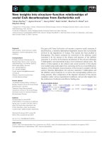
Conservation of CCM genes
The genes responsible for CCM are very well
conserved among different organisms (Fig. 1). These
genes are found not only in mammals, fish and other
vertebrates, but are also found in much more simple
and primitive organisms that lack a closed circulatory
system, such as Caenorhabditis elegans. The presence
of these genes in genetically tractable organisms has
allowed the development of numerous experimental
animal models, as discussed below.
Human phenotype
Although humans are generally not considered in the
category of animal models of disease, one can view the
field of human genetics as probing a vast natural
mutagenesis screen involving billions of individual
organisms. As in any mutagenesis screen, the impor-
tant information on genotype must be coupled with a
detailed characterization of phenotype. All other ani-
mal models are relevant to disease to the degree that
they help us further understand the human phenotype.
Recent investigations have further refined our under-
standing of this phenotype, and bear reviewing in this
manuscript.
In human CCM disease, the lesions exhibit a number
of characteristic features; these features will serve as
guideposts on the road to developing animal models of
CCM disease. Classically, a CCM consists of a cluster
of dilated blood vessels [12,13]. Each vessel in the clus-
ter is grossly dilated, earning the name of a cavern; each
vessel is lined only with a single layer of endothelium,
with the absence of normal vascular support cells, such
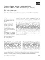
as smooth muscle cells. To be histologically classified as
a CCM, the lesion must contain multiple such vessels
adjacent to each other (Fig. 2). Grossly, this cluster
gives the lesion an appearance likened to a raspberry. In
addition, no brain parenchyma occurs in between the
vessels. Single dilated vessels, called capillary telangiec-
tasias, are not CCMs, although it has been hypothesized
that the disease progresses from a single capillary telan-
giectasia that blossoms into a multivessel CCM [13].
Functionally, the lesion vessels are subject to subclinical
bleeding, because hemosiderin, a breakdown product of
blood, is found in the brain tissues surrounding CCM
lesions [14]. Although CCMs have been clinically asso-
ciated as occurring with developmental venous malfor-
mations [15], it has been shown that these two types of
malformations are not linked genetically [16], and
familial cases of CCM are not generally associated
with venous malformations. Although these clinical
features define CCMs for physicians, little is known
about the cellular mechanisms that underlie and result
in such characteristics. These mechanisms are what
must be discovered, using either animal models or by
deeper study of human CCM patients.
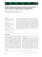
Fig. 1. Conservation of CCM proteins across species. Similarity
scores were generated for the three CCM proteins in comparison
with human protein sequences (KRIT1, accession number
AAH98442; CCM2, accession number AAH16832; PDCD10, acces-
sion number NP_665859). Protein sequences or predicted protein
sequences for a variety of vertebrate and nonvertebrate species
were included if similarity was detected by BLASTp algorithm
across a full-length protein sequence. Blank fields represent spe-
cies for which an orthologous gene has yet to be identified in avail-
able databases. All three proteins are well conserved across
species, and are found in nonvertebrate species. Conservation is
particularly strong for PDCD10, the smallest of the three proteins.
Note that Pdcd10 has been duplicated in the zebrafish genome; the
two proteins are denoted (a) and (b). C. elegans, Caenorhabd-
itis elegans.
A. C. Chan et al. CCM animal models
FEBS Journal 277 (2010) 1076–1083 ª 2010 The Authors Journal compilation ª 2010 FEBS 1077
One aspect of disease discovered in humans is that
CCMs are associated with an inflammatory response.
CCM lesions harbor a variety of immune cells [17],
and oligoclonal banding of IgG has been observed in
the CCM tissue [18]. What is still not known, however,
is whether this inflammatory response is a secondary
reaction to antigens exposed by the defective blood–
brain barrier of a CCM [19] or if inflammatory action
is part of the mechanism of pathogenesis leading to
formation of aberrant blood vessels. It is intriguing
that the mitogen-activated protein kinase kinase kinase
MEKK3 plays a key role in immune signaling [20],
and CCM2 protein has been shown to function as a
scaffold for MEKK3 in response to stress [6]. The
finding of immune involvement in CCM illustrates the
complexity of the disease; CCM is a vascular disease
localized mainly to neural tissues with an additional
immune component. The involvement of multiple cell
and tissue types raise the question of where the CCM
genes primarily function, and in which cell type their
loss leads to pathogenesis of disease.
Aside from the question of tissue specificity of CCM
gene function, another important question of disease
pathogenesis is that of a triggering event – what events
on a molecular, cellular or physiological level lead to
the formation of these isolated malformations? A clue
comes from studying patients with sporadic CCM and
those with familial CCM. People with an inherited
form of CCM have a larger number of lesions and
more frequent sequellae, such as seizure and hemor-
rhage. These features are reminiscent of the cancer ret-
inoblastoma, which led to the Knudson ‘two-hit’
hypothesis. Similarly, a two-hit hypothesis has been
proposed for the pathogenesis of CCM, in which an
inherited mutant allele is a silent, but predisposing hit,
and a second mutation acquired during life leads to a
disease phenotype. The data supporting this hypothesis
have been reviewed by Riant et al. [10] in an accompa-
nying minireview. In addition to genetic and epigenetic
events leading to CCM, these studies do not explore
physiologic stressors as potential disease triggers in the
heterozygous patient. For example, serum levels of the
angiogenic vascular endothelial growth factor have
been correlated with disease progression in case reports
[21,22].
Recent cell biology observations, supported by data
from mice, call to mind an important observational
study [19]. Using detailed ultrastructural examination
of surgically excised CCM specimens, the investigators
observed abnormal endothelial cell junctions from the
cavernous malformation. An important component of
the normal blood–brain barrier, tight junctions form
between endothelial cells and can be observed by elec-
tron microscopy. Although the cavernous malforma-
tion was found in the CNS where such tight junctions
are the rule, the investigators observed numerous
regions with impaired or deficient tight junctions
between adjacent endothelial cells. These areas of junc-
tional breakdown were associated with hemosiderin
pigment as functional evidence that junction break-
down was associated with pathologic vascular leak,
one of the defining features of CCMs.
Zebrafish
Hailed for its transparency and genetic tractability, a
significant body of work has been carried out in zebra-
fish to determine the functions of the CCM genes.
Initial results were described for santa (san, the zebrafish
orthologue of KRIT1) and valentine (vtn, the zebrafish
A
B
Fig. 2. Histology of CCM. Masson trichrome stain of surgically
excised cavernous malformation. (A) Low-magnification view of
CCM and surrounding brain. Hyalinized caverns of varying size are
observed, surrounded by a rim of collagen deposits (blue). The adja-
cent brain shows evidence of gliosis (red). (B) Higher magnification
view of boxed area. The caverns are lined by a single layer of endo-
thelium (arrowheads) without smooth muscle support. Rather than
smooth muscle cells or pericytes, a hyalinized rim of collagen sur-
rounds the caverns (asterisks). Brown hemosiderin deposits are
observed in the surrounding gliotic brain tissue (arrows).
CCM animal models A. C. Chan et al.
1078 FEBS Journal 277 (2010) 1076–1083 ª 2010 The Authors Journal compilation ª 2010 FEBS
orthologue of CCM2). Zebrafish with loss-of-function
mutations in san or vtn share a common phenotype
with fish lacking heart of glass (heg). Although muta-
tions in the human orthologue of heart of glass
(HEG1) have not been identified in patients with
CCM, this gene has been shown to be functionally and
genetically related to santa and valentine.
Heg is a single-pass transmembrane protein. Zebra-
fish with a nonsense heg mutation exhibit a dilated
heart phenotype. The myocardium proliferates to a
normal number of cells, but instead of building into
concentric layers to form the walls of the heart, the
myocardial cells form into a single layer, resulting in a
dilated, thin-walled heart whose structure is reminis-
cent of a CCM vessel. Heg has two soluble splice vari-
ants in addition to the transmembrane isoform, but it
is the transmembrane isoform that is essential in car-
diac patterning. Although the defect is one of myocar-
dial patterning, heg is expressed in the endocardial
cells, indicating that this cell layer signals to the myo-
cardium via Heg [23].
Interestingly, fish with nonsense mutations in san
and vtn were later shown to exhibit the same pheno-
type as the heg mutant fish – that of the dilated heart
covered by a single layer of myocardium. The similar-
ity of the phenotype in these nonsense alleles suggested
that these three proteins share a common developmen-
tal function. In addition, co-morpholino experiments
demonstrated synergy among the three genes, putting
them into a common genetic pathway [24]. Another
group refined the characterization of the santa and val-
entine phenotypes using different mutant alleles.
Focusing on the vasculature instead of the heart, they
found that these fish developed dilated, thin-walled
vessels that failed to form lumens. The dilated, thin-
walled, closed vessels, like the dilated, thin-walled
heart of these fish, are very reminiscent of human
CCM vessels and the closed vessels seen in CCM
knockout mice (see below). This dilation was attrib-
uted to abnormal endothelial cell spreading, a poten-
tial mechanistic insight into CCM pathogenesis. Of
note, these abnormal vessels were able to be rescued
by the transplantation of endothelial cells from wild-
type fish, again hinting that the endothelial cell is the
cell type that most needs the function of the CCM
proteins [25]. Later work also showed that loss of heg
or vtn via morpholino knockdown resulted in non-
patent vessels that patterned normally, similar to the
phenotypes seen in the Krit1 and Ccm2 knockout mice
(see below) [26]. Most recently, it has been shown that
a deletion mutation of pdcd10 (ccm3), which is dupli-
cated in the zebrafish genome, results in the same
developmental defects as mutations in santa and
valentine [27], making the zebrafish the first non-
human model organism to link all three CCM genes
phenotypically. Specifically, these defects are caused by
the loss of Ccm3 interaction with the kinases ser-
ine ⁄ threonine kinase 25 (STK25) and mammalian ster-
ile twenty-like 4 (MST4), giving a hint to the signaling
pathway in which Ccm3 belongs, as both STK25 and
MST4 belong to a family of kinases that are thought
to act upstream of the mitogen-activated protein kinas-
es (see the accompanying minireview [11] on the bio-
chemical interactions of the CCM proteins).
Furthering the pursuit of genetic interactions,
co-morpholino experiments were performed to examine
the interactions between the CCM genes and rap1b,a
Ras family small GTPase known to regulate cell
junctions [28] and notable as being closely related
to RAP1A, the binding partner bait originally used to
identify KRIT1 [29]. Knockdown of rap1b via mor-
pholino resulted in defective endothelial cell junctions
and intracerebral hemorrhage in the fish, reminiscent
of both the slow, unpredictable blood leak and the
frank hemorrhage associated with CCMs [30]. The
dose of rap1b morpholino was then titrated down so
that the hemorrhage phenotype was seen in only a
small percentage of fish. Combining this low dose of
rap1b morpholino with a similarly low dose of san
morpholino resulted in a synergistic increase in both
the intracerebral hemorrhage phenotype of rap1b and
the cardiac developmental phenotype of san.
The zebrafish experiments demonstrate the role of
the CCM genes in cardiac and vascular development;
the genetic tractability of the fish also provided a pow-
erful way to discover genetic interactions between the
CCM proteins and other proteins such as rap1b and
the previously unknown heg. A mystery remains as to
why HEG1 mutations are not found in humans with
CCMs. The synergistic effects of low-dose knockdown
of the CCM genes and their partners imply that a sim-
ilar mechanism may be responsible for pathogenesis in
humans; however, as previously stated, such polygenic
effects have yet to be identified in human tissue
samples.
Mouse
Mice have long been favored as a model organism for
laboratory studies and are the closest relative to
humans commonly used in genetic studies. Knockout
mice lacking Krit1 [31] and Ccm2 [26,32–34] have been
generated and described. Although an experimental
model of CCM lesions in the CNS was desired, neither
mice with heterozygous knockout of Krit1 nor Ccm2
develop CNS vascular lesions with any useful fre-
A. C. Chan et al. CCM animal models
FEBS Journal 277 (2010) 1076–1083 ª 2010 The Authors Journal compilation ª 2010 FEBS 1079
quency [26,31–34]. Although disappointing, this lack
of faithful disease modeling has generally been the case
for most mouse genotype equivalents of human disease
[35–38].
An important role for mouse models of genetic dis-
ease is to identify essential roles for protein function
in vivo, especially in development where the proof of
essential function is often embryonic lethality in com-
plete knockouts. Indeed, mice lacking either Krit1 or
Ccm2 die in mid-gestation with vascular defects at the
same developmental stage, and with a similar pheno-
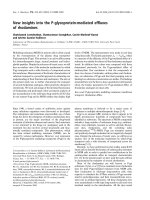
type [26,31,33,34]. The complete loss of Krit1 or Ccm2
results in vascular defects with a failure to connect the
developing heart to the developing aorta with a func-
tioning, patent first branchial arch artery. The associ-
ated rostral portions of the aorta are similarly
narrowed (Fig. 3). As a result, circulation is not estab-
lished as expected at E8.5 [33], and developmental
arrest and death ensue. Prior to developmental arrest,
cardiac and neural development proceeds normally.
Cavernous malformations are vascular lesions that
form predominantly in the CNS. The basis for this
anatomic predisposition is uncertain, but one possibil-
ity suggested by the abundant neuronal expression of
the CCM genes [32,39–42] is a mechanism by which
there is impaired signaling from neuronal cells to the
endothelium, with a primary defect in the neuronal
cell. Alternatively, the defect may lie primarily in endo-
thelial cells, and the CNS selectivity of the disease
could be a result of a unique sensitivity of the CNS
vasculature to CCM gene function. To address these
possibilities, mice with tissue-specific deletions of Ccm2
using the Cre–Lox inducible recombination system
have been generated and described. Two separate
floxed Ccm2 alleles were generated by different research
groups [33,34]. Using Cre recombinase driven by the
Tie2 promoter to direct recombination in endothelial
cells, both groups found an absolute requirement for
Ccm2 in the endothelium during development. The
neuronal expression of Ccm2 was not required for
development (as shown by deletion using the Nestin
promoter-driven Cre recombinase).
Whereas Krit1, Ccm2 and Pdcd10 have similar wide-
spread expression patterns in the mouse [32,39–42], the
expression of the mouse orthologue of heart of glass
(Heg1) is restricted to the endothelium and endocar-
dium. Unlike zebrafish, Heg1 knockout mice do not
phenocopy Krit1 or Ccm2 knockouts [26]. Rather,
Heg1 knockout mice die later in gestation or in early
postnatal stages with a variety of cardiac, vascular and
lymphatic defects. Although pulmonary hemorrhage,
cardiac rupture or chylous effusions may variably be
the mechanism of death, a common theme throughout
was disruption of the cell–cell junctions within the
endothelial or endocardial cells. Heg1 and Ccm2 were
also shown to genetically interact in the mouse as pre-
viously seen in fish [26]. Mice with both homozygous
knockout of Heg1 and heterozygous for Ccm2 were
found to have a much more severe phenotype than
either mutant in isolation. These dual knockouts phe-
nocopy mice with homozygous knockout of Ccm2 or
Krit1.
Multiple lines of investigation implicate a role for
impaired cell-to-cell communication and endothelial
cell junction integrity in states of CCM protein defi-
ciency. Endothelial cell tight junctions are required to
retain cells and macromolecules within the vasculature
and to prevent vascular leak. Although mice heterozy-
gous for Ccm2 do not frequently develop CCM lesions
like their human counterparts, these mice were shown
to have abnormal vascular leak in the dermis when
stressed with vascular endothelial growth factor [33].
Tight junctions are significantly regulated by the Rho
family of GTPases. Endothelial cell culture experi-
ments had implicated abnormally increased activity
of RhoA in vitro. A role for increased RhoA activity
in vivo was suggested by the ability of statins – known
inhibitors of Rho GTPases [43] – to rescue the
AB
Fig. 3. Narrowed arteries associated with
circulation failure in mice lacking Ccm2. The
connections of the heart to the aorta, and
the associated cranial portions of the dorsal
aorta are narrowed in mice lacking Ccm2.
The paired dorsal aortae in a wild-type
embryo at E9.0 are shown in (A) (arrows),
stained for the endothelial marker CD31.
Although endothelial cells are present in the
correct location in a Ccm2 gene trap mutant
littermate (arrows in B), little to no lumen is
formed to support circulation.
CCM animal models A. C. Chan et al.
1080 FEBS Journal 277 (2010) 1076–1083 ª 2010 The Authors Journal compilation ª 2010 FEBS
abnormal vascular leak of Ccm2 heterozygous knock-
out mice [33].
It is not clear what is responsible for the difference
in susceptibility between mice and humans for the cere-
bral vascular lesions. Although differences in lifespan
and brain mass may contribute to the lack of vascular
lesions in Krit1 or Ccm2 heterozygous knockout mice,
modifying factors are being sought which increase the
risk of CCM lesion formation. As discussed above,
observational studies in CCM patients suggest that a
‘two-hit hypothesis’ may underlie some lesions. Taking
advantage of the high rate of spontaneous mutations
in mice lacking the tumor suppressor p53, Krit1 het-
erozygous knockouts have been mated onto a p53
knockout background [44]. It was hoped that this
model would reproduce both the human genotype
(two genetic hits) and phenotype (cavernous malforma-
tions). Cerebral vascular lesions were observed in a
high proportion of animals on this background with
characteristics varying from capillary telangiectasias to
more complex cavernous malformations, but the
potential second hit mutation was not found. Unfortu-
nately, mice lacking p53 have a shortened lifespan
because of a high frequency of spontaneous tumors,
including occasional brain tumors [13,45], It is unfeasi-
ble to study the natural history of CCM disease in
these mice as they die from tumor burden shortly after
developing CCM lesions. Great caution must be taken
when interpreting the genetic contribution to vascular
lesions on this background with potential for cancer-
related vascular dysregulation and other physiologic
stressors that may contribute to CCM lesion develop-
ment. These results suggest, however, that a two-hit
model may produce malformations useful for study;
this second hit could come from recombination medi-
ated by an inducible promoter driving Cre recombin-
ase, thereby eliminating the confounding effects of the
p53 null background.
Discussion
Vascular malformations result in considerable morbid-
ity and mortality, especially with respect to lesions of
the central nervous system. The ability to prevent or
treat such lesions requires a greater understanding of
the underlying biology of lesion formation. In this
regard, cerebral cavernous malformation as a genetic
disorder offers unique opportunities to understand
the biology of vascular malformations. Initial insights
regarding the biochemistry of the CCM genes left a
considerable gap in understanding between protein
function and lesion biology. By exploring the function
of the CCM genes in animal models this gap is being
bridged. Animal models have demonstrated the cen-
tral importance of endothelial cell–cell interactions in
the pathogenesis of CCM vascular disease. Endothe-
lial cells need to be coordinated to organize into
proper sized lumens and to maintain vascular barrier
function. As a result of research into CCM, it
becomes apparent that vascular malformations
may result from the loss of genes crucial to vascular
stability.
In addition to providing the important in vivo con-
text for insights gained from biochemistry, animal
models can allow an acceleration of translational
research to ultimately impact the patients and families
with CCM. Recent work in mice shows the promise of
this approach, in that testable phenotypes can be iden-
tified and potential therapies can be evaluated in mice
genetically similar to CCM patients. Manipulations of
the current animal models to more closely mimic
human disease also appear promising. Ultimately, we
hope that a complete model of CCM lesion biology
can be developed to act as a vital link between bench
and bedside.
Acknowledgements
This work was funded by the US National Institutes of
Health (K.J.W. and D.Y.L.), including training grant
T32-GM007464 (A.C.C.), the American Heart Associa-
tion (K.J.W. and D.Y.L.), the H.A. and Edna Benning
Foundation, the Juvenile Diabetes Research Founda-
tion, the Burroughs Wellcome Fund and the Flight
Attendants Medical Research Institute (D.Y.L.).
References
1 Otten P, Pizzolato GP, Rilliet B & Berney J (1989) A
propos de 131 cas d’angiomes caverneux (cavernomes)
du S.N.C. repe
´
re
´
s par l’analyse re
´
trospective de 24 535
autopsies. Neuro-Chirurgie 35, 82–83, 128–131.
2 Robinson JR, Awad IA & Little JR (1991) Natural
history of the cavernous angioma. J neurosurg 75,
709–714.
3 Toldo I, Drigo P, Mammi I, Marini V & Carollo C
(2009) Vertebral and spinal cavernous angiomas asso-
ciated with familial cerebral cavernous malformation.
Surg neurol 71, 167–171.
4 Laberge-le Couteulx S, Jung HH, Labauge P, Houtte-
ville JP, Lescoat C, Cecillon M, Marechal E, Joutel A,
Bach JF & Tournier-Lasserve E (1999) Truncating
mutations in CCM1, encoding KRIT1, cause hereditary
cavernous angiomas. Nature genetics 23, 189–193.
5 Sahoo T, Johnson EW, Thomas JW, Kuehl PM, Jones
TL, Dokken CG, Touchman JW, Gallione CJ, Lee-Lin
A. C. Chan et al. CCM animal models
FEBS Journal 277 (2010) 1076–1083 ª 2010 The Authors Journal compilation ª 2010 FEBS 1081
SQ, Kosofsky B et al. (1999) Mutations in the gene
encoding KRIT1, a Krev-1 ⁄ rap1a binding protein,
cause cerebral cavernous malformations (CCM1). Hum
mol genet 8, 2325–2333.
6 Uhlik MT, Abell AN, Johnson NL, Sun W, Cuevas
BD, Lobel-Rice KE, Horne EA, Dell’Acqua ML &
Johnson GL (2003) Rac-MEKK3-MKK3 scaffolding
for p38 MAPK activation during hyperosmotic shock.
Nat Cell Biol 5, 1104–1110.
7 Liquori CL, Berg MJ, Siegel AM, Huang E,
Zawistowski JS, Stoffer T, Verlaan D, Balogun F,
Hughes L, Leedom TP et al. (2003) Mutations in
a gene encoding a novel protein containing a phos-
photyrosine-binding domain cause type 2 cerebral
cavernous malformations. Am J hum genet 73, 1459–
1464.
8 Denier C, Goutagny S, Labauge P, Krivosic V, Arnoult
M, Cousin A, Benabid AL, Comoy J, Frerebeau P,
Gilbert B et al. (2004) Mutations within the MGC4607
gene cause cerebral cavernous malformations. Am J
hum genet 74, 326–337.
9 Bergametti F, Denier C, Labauge P, Arnoult M, Boetto
S, Clanet M, Coubes P, Echenne B, Ibrahim R, Irthum
B et al. (2005) Mutations within the programmed cell
death 10 gene cause cerebral cavernous malformations.
Am J hum genet 76, 42–51.
10 Riant F, Bergametti F, Ayrignac X, Boulday G &
Tournier-Lasserve E (2010) Recent insights into cerebral
cavernous malformations: the molecular genetics of
CCM. FEBS J 277, 1070–1075.
11 Faurobert E & Albiges-Rizo C (2010) Recent insights
into cerebral cavernous malformations: a complex
jigsaw puzzle under construction. FEBS J 277, 1084–
1096.
12 Shenkar R, Venkatasubramanian PN, Zhao JC, Batjer
HH, Wyrwicz AM & Awad IA (2008) Advanced mag-
netic resonance imaging of cerebral cavernous malfor-
mations: part I. High-field imaging of excised human
lesions. Neurosurgery 63, 782–789; discussion 789.
13 Shenkar R, Venkatasubramanian PN, Wyrwicz AM,
Zhao JC, Shi C, Akers A, Marchuk DA & Awad IA
(2008) Advanced magnetic resonance imaging of cere-
bral cavernous malformations: part II. Imaging of
lesions in murine models. Neurosurgery 63, 790–797;
discussion 797-798.
14 Zhang J, Clatterbuck RE, Rigamonti D, Chang DD &
Dietz HC (2001) Interaction between krit1 and icap1-
alpha infers perturbation of integrin beta1-mediated
angiogenesis in the pathogenesis of cerebral cavernous
malformation. Hum Mol Genet 10, 2953–2960.
15 Abe T, Singer RJ, Marks MP, Norbash AM, Crowley
RS & Steinberg GK (1998) Coexistence of occult vascu-
lar malformations and developmental venous anomalies
in the central nervous system: MR evaluation. Ajnr 19,
51–57.
16 Guclu B, Ozturk AK, Pricola KL, Seker A, Ozek M &
Gunel M (2005) Cerebral venous malformations have
distinct genetic origin from cerebral cavernous malfor-
mations. Stroke 36, 2479–2480.
17 Shi C, Shenkar R, Du H, Duckworth E, Raja H, Batjer
HH & Awad IA (2009) Immune response in human cer-
ebral cavernous malformations. Stroke 40, 1659–1665.
18 Shi C, Shenkar R, Batjer HH, Check IJ & Awad IA
(2007) Oligoclonal immune response in cerebral caver-
nous malformations. Laboratory investigation. J Neuro-
surg 107, 1023–1026.
19 Clatterbuck RE, Eberhart CG, Crain BJ & Rigamonti
D (2001) Ultrastructural and immunocytochemical evi-
dence that an incompetent blood-brain barrier is related
to the pathophysiology of cavernous malformations.
J Neurol Neurosurg Psychiatry 71, 188–192.
20 Konno H, Yamamoto T, Yamazaki K, Gohda J,
Akiyama T, Semba K, Goto H, Kato A, Yujiri T, Imai
T et al. (2009) TRAF6 establishes innate immune
responses by activating NF-kappaB and IRF7 upon
sensing cytosolic viral RNA and DNA. PloS one 4,
e5674.
21 Jung KH, Chu K, Jeong SW, Park HK, Bae HJ &
Yoon BW (2003) Cerebral cavernous malformations
with dynamic and progressive course: correlation
study with vascular endothelial growth factor. Arch
neurol - Chicago 60, 1613–1618.
22 Abe T, Morishige M, Ooba H, Kamida T, Fujiki M,
Kobayashi H, Sakoda T & Kimba Y (2009) The asso-
ciation between high VEGF levels and multiple prob-
able punctuate cavernous malformations. Acta
neurochirurgica 151, 855–859.
23 Mably JD, Mohideen MA, Burns CG, Chen JN &
Fishman MC (2003) Heart of glass regulates the con-
centric growth of the heart in zebrafish. Curr Biol 13,
2138–2147.
24 Mably JD, Chuang LP, Serluca FC, Mohideen MA,
Chen JN & Fishman MC (2006) Santa and valentine
pattern concentric growth of cardiac myocardium in the
zebrafish. Development 133, 3139–3146.
25 Hogan BM, Bussmann J, Wolburg H & Schulte-Merker
S (2008) Ccm1 cell autonomously regulates endothelial
cellular morphogenesis and vascular tubulogenesis in
zebrafish. Hum Mol Genet 17, 2424–2432.
26 Kleaveland B, Zheng X, Liu JJ, Blum Y, Tung JJ, Zou
Z, Sweeney SM, Chen M, Guo L, Lu MM et al. (2009)
Regulation of cardiovascular development and integrity
by the heart of glass-cerebral cavernous malformation
protein pathway. Nature medicine 15, 169–176.
27 Voss K, Stahl S, Hogan BM, Reinders J, Schleider E,
Schulte-Merker S & Felbor U (2009) Functional
analyses of human and zebrafish 18-amino acid
in-frame deletion pave the way for domain mapping of
the cerebral cavernous malformation 3 protein. Hum
mutat 30, 1003–1011.
CCM animal models A. C. Chan et al.
1082 FEBS Journal 277 (2010) 1076–1083 ª 2010 The Authors Journal compilation ª 2010 FEBS
28 Cullere X, Shaw SK, Andersson L, Hirahashi J,
Luscinskas FW & Mayadas TN (2005) Regulation of
vascular endothelial barrier function by Epac, a cAMP-
activated exchange factor for Rap GTPase. Blood 105,
1950–1955.
29 Serebriiskii I, Estojak J, Sonoda G, Testa JR & Gole-
mis EA (1997) Association of Krev-1 ⁄ rap1a with Krit1,
a novel ankyrin repeat-containing protein encoded by a
gene mapping to 7q21-22. Oncogene 15, 1043–1049.
30 Gore AV, Lampugnani MG, Dye L, Dejana E & Wein-
stein BM (2008) Combinatorial interaction between
CCM pathway genes precipitates hemorrhagic stroke.
Disease models & mechanisms 1, 275–281.
31 Whitehead KJ, Plummer NW, Adams JA, Marchuk
DA & Li DY (2004) Ccm1 is required for arterial mor-
phogenesis: implications for the etiology of human
cavernous malformations. Development (Cambridge,
England) 131, 1437–1448.
32 Plummer NW, Squire TL, Srinivasan S, Huang E,
Zawistowski JS, Matsunami H, Hale LP & Marchuk
DA (2006) Neuronal expression of the Ccm2 gene in a
new mouse model of cerebral cavernous malformations.
Mamm Genome 17, 119–128.
33 Whitehead KJ, Chan AC, Navankasattusas S, Koh W,
London NR, Ling J, Mayo AH, Drakos SG, Jones CA,
Zhu W et al. (2009) The cerebral cavernous malforma-
tion signaling pathway promotes vascular integrity via
Rho GTPases. Nature medicine 15, 177–184.
34 Boulday G, Blecon A, Petit N, Chareyre F, Garcia LA,
Niwa-Kawakita M, Giovannini M & Tournier-Lasserve
E (2009) Tissue-specific conditional CCM2 knockout
mice establish the essential role of endothelial CCM2 in
angiogenesis: implications for human cerebral cavernous
malformations. Disease models & mechanisms 2, 168–177.
35 Ishibashi S, Brown MS, Goldstein JL, Gerard RD,
Hammer RE & Herz J (1993) Hypercholesterolemia in
low density lipoprotein receptor knockout mice and its
reversal by adenovirus-mediated gene delivery. J Clin
Invest 92, 883–893.
36 Pereira L, Andrikopoulos K, Tian J, Lee SY, Keene
DR, Ono R, Reinhardt DP, Sakai LY, Biery NJ,
Bunton T et al. (1997) Targetting of the gene encoding
fibrillin-1 recapitulates the vascular aspect of Marfan
syndrome. Nature genetics 17, 218–222.
37 Li DY, Sorensen LK, Brooke BS, Urness LD, Davis
EC, Taylor DG, Boak BB & Wendel DP (1999) Defec-
tive angiogenesis in mice lacking endoglin. Science 284,
1534–1537.
38 Urness LD, Sorensen LK & Li DY (2000) Arteriove-
nous malformations in mice lacking activin receptor-like
kinase-1. Nature genetics 26, 328–331.
39 Denier C, Gasc JM, Chapon F, Domenga V, Lescoat
C, Joutel A & Tournier-Lasserve E (2002) Krit1 ⁄ cer-
ebral cavernous malformation 1 mRNA is preferen-
tially expressed in neurons and epithelial cells in
embryo and adult. Mechanisms of development 117,
363–367.
40 Kehrer-Sawatzki H, Wilda M, Braun VM, Richter HP
& Hameister H (2002) Mutation and expression analysis
of the KRIT1 gene associated with cerebral cavernous
malformations (CCM1). Acta neuropathologica 104,
231–240.
41 Petit N, Blecon A, Denier C & Tournier-Lasserve E
(2006) Patterns of expression of the three cerebral
cavernous malformation (CCM) genes during embryo-
nic and postnatal brain development. Gene Expr Pat-
terns 6, 495–503.
42 Seker A, Pricola KL, Guclu B, Ozturk AK, Louvi A &
Gunel M (2006) CCM2 expression parallels that of
CCM1. Stroke 37, 518–523.
43 Park HJ, Kong D, Iruela-Arispe L, Begley U, Tang D
& Galper JB (2002) 3-hydroxy-3-methylglutaryl coen-
zyme A reductase inhibitors interfere with angiogenesis
by inhibiting the geranylgeranylation of RhoA. Circ res
91, 143–150.
44 Plummer NW, Gallione CJ, Srinivasan S, Zawistowski
JS, Louis DN & Marchuk DA (2004) Loss of p53
sensitizes mice with a mutation in Ccm1 (KRIT1) to
development of cerebral vascular malformations. The
Am J pathol 165, 1509–1518.
45 Jacks T, Remington L, Williams BO, Schmitt EM,
Halachmi S, Bronson RT & Weinberg RA (1994)
Tumor spectrum analysis in p53-mutant mice. Curr Biol
4, 1–7.
A. C. Chan et al. CCM animal models
FEBS Journal 277 (2010) 1076–1083 ª 2010 The Authors Journal compilation ª 2010 FEBS 1083