Báo cáo khoa học: Recent insights into cerebral cavernous malformations: a complex jigsaw puzzle under construction doc
Bạn đang xem bản rút gọn của tài liệu. Xem và tải ngay bản đầy đủ của tài liệu tại đây (298.84 KB, 13 trang )
MINIREVIEW
Recent insights into cerebral cavernous malformations:
a complex jigsaw puzzle under construction
Eva Faurobert and Corinne Albiges-Rizo
Centre de recherche, INSERM U823-CNRS ERL 3148, Universite
´
J. Fourier, Grenoble, France
Introduction
Cerebral cavernous malformations (CCM) are com-
mon vascular malformations with a prevalence of 1 in
every 200–250 individuals. Leakage of blood can be
detected by magnetic resonance imaging around each
lesion and individuals with these vascular lesions are
subject to an unpredictable risk of hemorrhage in mid-
life. Although lesions have been described in a variety
of vascular beds, clinical manifestations are most com-
mon in the central nervous system where the conse-
quences may be stroke, seizure or any kind of
neurological disorder, and can lead to death [1]. The
lesions consist of densely packed, grossly dilated, capil-
lary-like sinusoids lined by a single endothelial layer
embedded in a thick collagen matrix. Importantly,
these lesions lack the components of organized mature
vessels such as pericytes, astrocytic foot processes and
intact endothelial cell–cell junctions [2]. Both sporadic
and familial forms of CCM have been identified. The
genetics of the disease is developed in a minireview by
Riant et al. [3]. Briefly, three loci have been mapped
and the genes responsible for the disease, CCM1 to
CCM3, have been identified in these loci. Within the
Keywords
angiogenesis; blood brain barrier; cadherin;
CCM; cytoskeleton; endothelial cell; HEG;
hemorrhage; integrin; Krit1
Correspondence
E. Faurobert, Centre de recherche, INSERM
U823-CNRS ERL 3148, Universite
´
J. Fourier,
Site sante
´
La tronche, BP170 38042,
Grenoble, France
Fax: +33 476 54 94 25
Tel: +33 476 54 94 74
E-mail:
(Received 1 August 2009, revised 4 Novem-
ber 2009, accepted 25 November 2009)
doi:10.1111/j.1742-4658.2009.07537.x
Cerebral cavernous malformations (CCM) are common vascular malforma-
tions with an unpredictable risk of hemorrhage, the consequences of which
range from headache to stroke or death. Three genes, CCM1, CCM2 and
CCM3, have been linked to the disease. The encoded CCM proteins inter-
act with each other within a large protein complex. Within the past 2 years,
a plethora of new data has emerged on the signaling pathways in which
CCM proteins are involved. CCM proteins regulate diverse aspects of
endothelial cell morphogenesis and blood vessel stability such as cell–cell
junctions, cell shape and polarity, or cell adhesion to the extracellular
matrix. Although fascinating, a global picture is hard to depict because
little is known about how these pathways coordinate to orchestrate angio-
genesis. Here we present what is known about the structural domain
organization of CCM proteins, their association as a ternary complex and
their subcellular localization. Numerous CCM partners have been identified
using two-hybrid screens, genetic analyses or proteomic studies. We focus
on the best-characterized partners and review data on the signaling
pathways they regulate as a step towards a better understanding of the
etiology of CCM disease.
Abbreviations
CCM, cerebral cavernous malformation; FERM, band 4.1 ezrin radixin moesin; FN, fibronectin; HEG1, heart of glass 1; ICAP-1, integrin
cytoplasmic adaptor protein-1; Krit1, K-Rev interaction trapped 1; MAPK, mitogen-activated protein kinase; MEKK3, mitogen-activated protein
kinase kinase kinase 3; MKK, mitogen-activated protein kinase kinase; MST4, mammalian sterile twenty-like 4; OSM, osmosensing scaffold
for MEKK3; PTB, phosphotyrosine binding; STRIPAK, striatin interacting phosphatase and kinase; STK, serine ⁄ threonine kinase; vEGF,
vascular epidermal growth factor.
1084 FEBS Journal 277 (2010) 1084–1096 Journal compilation ª 2010 FEBS. No claim to original French government works
past two years, fascinating data have emerged on the
signaling pathways regulated by the products of these
three genes. However, a global picture is hard to depict
because not much is known about how these signaling
pathways coordinate. Many advances have been made
in describing the core complex formed by the associa-
tion of these three proteins and in the identification of
numerous CCM partners. In this minireview, we focus
on partners that open new avenues for CCM research
and discuss recent insights into their role in cytoskele-
tal remodeling, regulation of cell matrix adhesion and
cell–cell junction homeostasis.
Structural domain organization of
CCM1
⁄
Krit1, CCM2
⁄
OSM
⁄
MGC4607
and CCM3
⁄
PDCD10 proteins
CCM1 encodes a protein also named K-Rev interac-
tion trapped 1 (Krit1). Krit1 was first identified in
1997 as a partner of the small G-protein Krev-1 ⁄ Rap1
from a yeast two-hybrid screen [4]. Two years later,
the CCM1 locus was mapped to the gene encoding
Krit1 [5,6]. Krit1 is an 84 kDa scaffold protein with
no catalytic activity which contains several distinct
domains involved in protein–protein interaction
(Fig. 1). Remarkably, Krit1 possesses a C-terminal
band 4.1 ezrin radixin moesin (FERM) domain, a sig-
nature of membrane binding proteins like talin, ezrin,
radixin or moesin. FERM domains are composed of
three subdomains, F1–F3, arranged in cloverleaf
shape. The F3 subdomain has a phosphotyrosine bind-
ing (PTB) fold. PTB domains recognize a canonical
NPXY ⁄ F motif often found on the cytoplasmic tail of
transmembrane receptors. Recruitment of PTB or
FERM proteins to transmembrane receptors is a con-
served mechanism used by cells to build intracellular
signaling hubs. Remarkably, in addition to its FERM
domain, Krit1 possesses three N-terminal NPXY ⁄ F
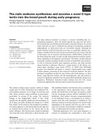
Fig. 1. Structural domains of CCM proteins. Krit1 ⁄ CCM1 bears a C-terminal FERM (band 4.1 erzin radixin moesin) domain and three N-terminal
NPXY ⁄ F motifs allowing either the folding of the protein on itself or its interaction with ICAP-1 and CCM2. ANK, ankyrin domain; MT, microtu-
bules; NLS, nuclear localization signal. The phosphotyrosine binding (PTB) domain of CCM2 ⁄ OSM interacts with a Krit1 NPXY ⁄ F motif. L198R
and F217A mutations prevent CCM2 interaction with Krit1. CCM3 has no homology with any known domain. Its N-terminal fragment (L33 to
K50) interacts with MST4, STK24 and STK25. Ser39 and Thr43 are the substrate of phosphorylation by STK25. HEG1 is a heavily glycosylated
(
) transmembrane protein carrying two extracellular EGF-like repeats and a C-terminal NPXY ⁄ F motif which interacts with Krit1. Its extracellu-
lar ligand is not known. ICAP-1 has a Ser ⁄ Thr riche N-terminus containing a NLS and sites of phosphorylation by calmodulin-dependent kinase II,
protein kinase A and protein kinase C. Reported interactions with b1 integrin, Krit1, Rho-associated kinase I-kinase and NM23.
E. Faurobert and C. Albiges-Rizo Emerging signaling pathways regulated by CCM proteins
FEBS Journal 277 (2010) 1084–1096 Journal compilation ª 2010 FEBS. No claim to original French government works 1085
motifs (Fig. 1). This peculiar structural organization
allows the N- and C-terminal halves of Krit1 to inter-
act with each other in a glutathione S-transferase pull-
down [7] or a yeast two-hybrid interaction assay [8],
suggesting that Krit1 may adopt a closed and open
conformation in vivo, resulting from either intramolec-
ular folding or dimerization. The first [7] or third [8]
NPXY ⁄ F motif may be involved in this interaction.
Systematic mutagenesis of each of the three motifs
should help to determine the contribution made by
each of them. Three ankyrin repeats are present
between the NPXY ⁄ F motifs and the FERM domain
(Fig. 1). Although ankyrin repeats are found in thou-
sands of proteins and support interaction with many
diverse proteins, no partner interacting with Krit1
ankyrin repeats has been found. Compared with Krit1,
CCM2 and CCM3 have a much simpler structural
domain organization. CCM2 encodes a scaffold pro-
tein of 51 kDa also containing a PTB domain [9,10],
but no other known domain (Fig. 1). It was identified
in a yeast two-hybrid screen using mitogen-activated
protein kinase kinase kinase 3 (MEKK3) as bait to
identify proteins involved in the cell response to hyper-
osmotic shock [11] and was named osmosensing scaf-
fold for MEKK3 (OSM). The last mutated gene
CCM3 or PDCD10 has been identified more recently
[12] and is upregulated in fibroblasts exposed to spe-
cific apoptosis inducers, such as staurosporine, cyclo-
heximide and tumor necrosis factor-a [13]. Apoptotic
or, by contrast, proliferative functions have been
attributed to CCM3 [14,15]. No homology with any
known domain is found on CCM3 but it has been sug-
gested that this small protein (25 kDa) folds as one
stable domain [16] (Fig. 1).
CCM complexes and their subcellular
localizations
Interactions within the CCM1, -2, -3 complex
Consistent with their involvement in the same pathology,
Krit1 ⁄ CCM1, CCM2 and CCM3 are able to interact.
Co-immunoprecipitations, glutathione S-transferase
pull-downs and mutagenesis have allowed us to identify
the interaction sites between the three proteins in this
complex.
Endogenous or overexpressed Krit1 and CCM2
interact with each other [17,18]. Mutations in the PTB
domain of CCM2 on conserved residues critical for the
NPXY ⁄ F motif binding (Fig. 1) are deleterious for the
Krit1–CCM2 interaction. One, L198R, a single mis-
sense mutation was found in a CCM patient [10], the
other, F217A, was engineered based on homology with
a known PTB domain [17]. The N-terminus of CCM2
also takes part in this interaction because a lack of
amino acid residues 11–68, an inframe deletion
observed in patients [19], prevents the interaction of
CCM2 with Krit1 [20]. Conversely, the binding
domain for CCM2 on Krit1 remains uncertain.
Because their interaction involves the CCM2 PTB
domain, it is likely that the counterpart on Krit1 is
one of its three NPXY ⁄ F motifs. Indeed, a yeast two-
hybrid assay using small fragments of CCM2 centered
on NPXY ⁄ F2 and -3 have identified these motifs as
CCM2 interacting sites [18]. However, single amino
acid substitution in each of these motifs has no effect
on the binding of Krit1 to CCM2 [17]. Additional
mutagenesis on residues immediately N- or C-terminal
of the NPXY ⁄ F might be required to significantly
reduce the affinity.
CCM2 interacts with CCM3 [16,20] but their respec-
tive interaction sites are not known. None of the three
CCM2 mutations cited above impairs its binding to
CCM3 [20] showing that CCM2 binding domains for
Krit1 and CCM3 are not redundant. Indeed, the three
overexpressed proteins form a complex. CCM2 is the
linker protein that brings together Krit1 and CCM3,
which otherwise have no affinity for each other
[16,20,21]. Remarkably, this ternary complex was
detected using proteomic approaches [21,22]. However,
CCM3 was also identified by proteomic analysis as a
component of another large complex named striatin-
interacting phosphatase and kinase (STRIPAK) which
assembles phosphatases and kinases arranged around a
protein phosphatase 2A core [22]. Interestingly, neither
Krit1 nor CCM2 was detected in the STRIPAK com-
plex, but small amounts of CCM3 could be pulled-
down along with Krit1 on CCM2 beads. This suggests
that CCM3 associates with (at least) two different
complexes; in substoichiometric amounts with the
Krit1–CCM2 complex and in large amounts with the
striatin-interacting phosphatase and kinase complex.
Shuttling of CCM proteins between the
membrane and nucleus
The in vitro data suggest that the three CCM proteins
associate in a ternary complex in vivo, but they are
also very likely engaged in several other complexes
having different localizations (Fig. 2). As such, Krit1
associates with the b1 integrin regulator integrin cyto-
plasmic adaptor protein-1 (ICAP-1; as discussed
below) and this complex can shuttle between the cyto-
sol and the nucleus. Both Krit1 and ICAP-1 have a
nuclear localization signal motif in their N-terminus
and both localize in a nuclear localization signal-
Emerging signaling pathways regulated by CCM proteins E. Faurobert and C. Albiges-Rizo
1086 FEBS Journal 277 (2010) 1084–1096 Journal compilation ª 2010 FEBS. No claim to original French government works
dependent manner to the nucleus of transfected cells
[8,17]. Interestingly, it has been shown that during cell
spreading, ICAP-1 shuttles from the plasma membrane
to the nucleus where it stimulates transcription and
cellular proliferation [23]. However, binding of CCM2
to Krit1 inhibits nuclear translocation of the Krit1–
ICAP-1 complex. Indeed, cotransfection of CCM2
with Krit1 and ICAP-1 induces the formation of a
ternary complex between the three proteins that
sequesters Krit1–ICAP-1 in the cytosol [8,17]. The
association of CCM2 with Krit1–ICAP-1 may there-
fore be a key event and the target of upstream signal-
ing pathways to control Krit1–ICAP-1 transcriptional
regulatory functions.
Transport along microtubules may be a way for
Krit1 and its partners to shuttle between the cytoplasm
and the nucleus. Interestingly, a- and b-tubulins have
been identified using proteomic analysis of proteins co-
immunoprecipitating with flagged CCM2 in stably
transfected macrophages [21]. The presence of tubulin
subunits in the pulled-down complex depended on
CCM2–Krit1 interaction because a functional PTB
domain was required on CCM2, suggesting that Krit1
is the direct partner of tubulin. In fact, Krit1 has been
shown to co-sediment with in vitro polymerized micro-
tubules [7], and to co-localize with microtubules in
bovine aortic endothelial cells [24]. Two binding sites
for microtubules have been mapped on Krit1: one
which contributes the most to the binding overlaps
with the nuclear localization signal sequence, the other
lies in its last 50 amino acids.
PTB and FERM domains have structural features
enabling their interaction with phosphoinositides in
membranes. As such, Krit1, CCM2 and CCM3 bind
to phosphoinositides [7,21]. Purified Krit1 binds to
liposomes only when supplemented with phosphoinosi-
tides [7]. Modeling of the Krit1 FERM domain using
known structures has highlighted a basic cleft between
the F1 and F3 subdomains which may interact with
the negative charges of phosphate groups. CCM2 and
CCM3 also bind directly to phospholipids, as shown
by overlay experiments on phosphatidylinositol phos-
phate arrays [21]. CCM2 most likely interacts via its
PTB domain. The CCM3 lipid-interacting domain is
not yet known. CCM2 binds preferentially to mono-
over biphosphorylated phosphatidylinositols, a result
also observed for Krit1 (our unpublished data).
Conversely, CCM3 has a higher affinity for bi- and
triphosphorylated phosphatidylinositols, an additional
argument suggesting that Krit1 together with CCM2
might localize to different membrane compartments
than CCM3.
Cell polarity
Adherens junction
formation and stability
Endothelial cell permeability
Myocardiac cells distribution
along endocardial-myocardial axis
Cell-cell junctions
Cell-matrix adhesion
Cell migration
ECM remodeling
Tubulogenesis
mural cell recruitment
-catenin
Krit1
p120
AF6
Rap1
Cadherin
HEG
Integrin
?
P
MST4
Lkb1
CCM3
?
?
?
ECM
Microtubules
Golgi
AB
Intracellular compartment Extracellular compartment
?
ICAP-1
ERM
Rac
Cdc42
MEKK3
MKK3
p38MAPK
RhoA degradation
Actin polymerization
Membrane ruffles
Actin stress fiber
Adherens junction formation and stabilization
Cell polarity
Cell polarity
Lumen Formation
Smurf1
RhoA
Krit1
CCM2
CCM2
CCM2
CCM2
MST4
Krit1
Rap1
RhoA
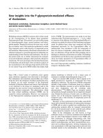
Fig. 2. Emerging signaling pathways and vascular processes controlled by the CCM proteins. (A) Cadherins, HEG1 and integrins are three
transmembrane receptors connected to CCM proteins or functions. All three receptors are known to have roles in different steps of vessel
morphogenesis. Possible cross-talk between their dependent signaling pathways through CCM proteins are represented by arrows. (B) CCM2
is a scaffold for small GTPases of the Rho family and for p38MAPK kinase. It is involved in actin cytoskeleton remodeling through scaffolding
of Rac, activation of the p38 MAPK kinase pathway and proteosomal degradation of RhoA. CCM2 may also be involved directly or indirectly in
Cdc42 activation. As a result, cell–cell junctions, cell polarity and lumen formation are likely to be dependent on CCM2 signaling.
E. Faurobert and C. Albiges-Rizo Emerging signaling pathways regulated by CCM proteins
FEBS Journal 277 (2010) 1084–1096 Journal compilation ª 2010 FEBS. No claim to original French government works 1087
New partners for the CCM proteins:
what they tell us on putative regulated
signaling pathways
CCM proteins are expressed in many different cell
types. Thus, a crucial and intriguing question about
the etiology of the cavernous malformations in blood
vessels is to ask what is unique to endothelial cells.
Indeed, depletion of CCM2 targeted to the endothe-
lium and not to the surrounding tissue results in
vascular defects in mouse embryos [25,26] (see also
Chan et al. [27]). One possibility is that specific sub-
sets of interactions occur in endothelial cells. Even
though many studies have not been conducted in
endothelial cells, they have been very helpful in iden-
tifying new partners for CCM proteins. As such,
proteomic studies performed in macrophages and
astrocytes have helped identify no fewer than 114
proteins interacting with CCM2 [21]. Here, we review
only the best-characterized partners which may give
clues to the function of CCM proteins in vascular
integrity.
An increasing amount of data indicates that CCM
proteins are connected to the plasma membrane and
regulate cell–cell adhesion, cell shape and polarity, and
most likely cell adhesion to the extracellular matrix
(Fig. 2). This makes sense with regard to the pheno-
type of CCM lesions in which endothelial cells are
joined loosely to each other, mural cells (i.e. pericytes
and astrocytes) are absent, and the basal lamina sur-
rounding the endothelium is abnormal [2]. Both cell–
cell adhesion and cell polarity require the assembly of
two specialized intercellular adhesion structures that
regulate vascular permeability. Adherens junctions ini-
tiate and maintain strong contacts between endothelial
cells and promote tight junction assembly. Tight junc-
tions are specialized for the passage of ions and solutes
through the paracellular route. They may also act as a
physical barrier along the cell surface allowing the
asymmetrical distribution of proteins and lipids
between apical and basolateral domains, a phenome-
non known as cell polarization. Cell adhesion to the
extracellular matrix requires integrins clustered in
highly dynamic adhesive structures which regulate
cytoskeleton rigidity, extracellular matrix remodeling
and probably cell–cell junctions.
Rap1, the master regulator of cell–cell and
cell–extracellular matrix adhesion
It has previously been established that the Ras family
small G protein Rap1 stimulates cell adhesion to the
extracellular matrix by activating integrins and cell–cell
adhesion by stimulating the formation and mainte-
nance of adherens junctions. It does so by activating a
large number of effectors most of which are involved
in regulating actin dynamics [28,29]. Rap1 was the first
reported Krit1 partner and was used as the bait to
clone Krit1 in a yeast two-hybrid screen [4]. This inter-
action was questioned until 2007 when two groups
used biochemical in vitro assays [7] and functional
studies [30] to confirm that Krit1 is a Rap1 effector.
However, Rap1 is not found in the CCM complex
defined by proteomic analysis, suggesting that Rap1–
Krit1 may form an independent complex. Interestingly,
Rap1a and -1b knockout mice show defective angio-
genesis, characterized by delayed perinatal retinal vas-
cularization, reduced microvessel sprouting from aortic
rings in response to angiogenic factors or reduced neo-
vascularization of ischemic hind limbs [31–33]. Reduc-
tion of the function of Rap1b using morpholinos in
zebrafish embryos disrupts endothelial junctions and
provokes intracranial hemorrhage. Importantly, a
minor reduction in Rap1b, in combination with a simi-
lar reduction in Krit1 results in a high incidence of
intracranial hemorrhage, whereas injection of each
morpholino independently has almost no effect [34].
This indicates that Rap1 and Krit1 act in a common
molecular pathway. Indeed, Glading et al. [30] showed
that small interfering RNA depletion of Krit1 blocks
the ability of Rap1 to stabilize endothelial cell–cell
junctions in culture cells [30].
CCM partners in cell–cell junctions
Proteins of adherens junctions
Endogenous Krit1 localizes to cell–cell junctions on a
bovine aortic endothelial cell confluent monolayer
and co-immunoprecipitates with the Rap1 effector
AF-6 ⁄ afadin, b-catenin and p120-catenin. This locali-
zation requires a Krit1 FERM domain and is depen-
dent upon activation of Rap1 [30]. It has consistently
been shown that in vitro Rap1 binding to the Krit1
FERM domain enhances the association of Krit1 with
liposomes, most likely by inducing a conformational
change in its basic pocket which gives Krit1 a better
affinity for phosphoinositides [7]. Depletion of Krit1
by small interfering RNA leads to disruption of b-cate-
nin localization to adherens junctions and increases the
permeability of the monolayer barrier [30], a pheno-
type reminiscent of that observed in human lesions.
Therefore, by localizing b-catenin to adherens junction,
Krit1 is likely to be involved in the formation and
maintenance of the endothelial barrier (Fig. 2A). How-
ever, it is not yet known whether the Krit1–b-catenin
interaction is direct.
Emerging signaling pathways regulated by CCM proteins E. Faurobert and C. Albiges-Rizo
1088 FEBS Journal 277 (2010) 1084–1096 Journal compilation ª 2010 FEBS. No claim to original French government works
The transmembrane glycosylated protein heart of
glass 1
Heart of glass 1 (HEG1) is a transmembrane protein of
unknown function bearing a large extracellular domain
with two epidermal growth factor-like domains, a
transmembrane segment and a short cytoplasmic tail
(100 amino acids) with a conserved C-terminal
NPXY ⁄ F motif (Fig. 1). Its extracellular domain is pre-
dicted to be highly glycosylated. It is expressed specifi-
cally in the endothelium and the endocardium. No
extracellular ligand is known. HEG1 is the mammalian
homolog of the zebrafish heart of glass. Zebrafish heart
of glass mutants show enlarged cardiac chambers
resulting from improper distribution of myocardiac
cells along the endocardial-to-myocardial axis [35].
Two other genes, santa and valentine, functioning in
the same molecular pathways, were identified and
found to be Krit1 and CCM2, respectively. They
display the same phenotype as heart of glass when
disrupted in zebrafish or when a combination of low-
dose morpholinos against the three proteins is injected
[36]. Recently, HEG1 and CCM2 were also shown to
interact genetically in the mouse [37]. Indeed,
Heg1
) ⁄ )
;Ccm2
lacZ ⁄ +
[37] like Ccm2
) ⁄ )
mice [25,26]
have severe cardiovascular defects and die early in
development owing to a failure of nascent endothelial
cells to form patent vessels. Both mice displayed short-
ened endothelial junctions compared with control litter-
mates [37]. More details can be found in the
accompanying minireview on animal models of CCM
disease [27]. In addition, the ternary complex between
HEG1, Krit1 and CCM2 has been demonstrated bio-
chemically [37] (Fig. 2A). A CCM2 mutant unable to
bind Krit1 is not recruited in the HEG1–Krit1 com-
plex, suggesting that Krit1 is the adaptor connecting
CCM2 to the transmembrane receptor. It is very likely
that the association of HEG1 with Krit1 requires
HEG1 NPXY ⁄ F motif and Krit1 FERM domain but
this remains to be tested.
As a hint toward its function, HEG1 is evolution-
ary related to mucin 13 [38]. Mucins are either
secreted or inserted as transmembrane glycoproteins
in polarized epithelia. Transmembrane mucin 1 can
associate with fibroblast growth factor receptor 3 [39]
and b-catenin to activate b-catenin-driven transcrip-
tion of Wnt target genes [40,41]. Interestingly, an
emerging idea concerning mucin function is that loss
of polarity through a breach in the cell layer could
enable growth factor receptors and mucins to associ-
ate and engage in signaling, which would activate
gene transcription designed to repair the breach and
re-establish cell polarity [42]. This signaling pathway
would make sense with regard to loss of the integrity
of the endothelial barrier and a putative dysfunction
of repair mechanisms in CCM lesions. Consistent
with this, Liebner et al. [43] have shown that Wnt ⁄ b-
catenin signaling is required for the endothelial cell
expression of proteins necessary for the development
of the blood–brain barrier [43]. Therefore, under the
control of HEG1, Krit1 and b-catenin may be
involved in the dual role of stabilizing cell–cell junc-
tions and regulating the expression of blood–brain
barrier-specific players.
Partners in cell-shape remodeling and polarity
Along with a role for Krit1 in cell–cell adhesion, a net-
work of data identifies the CCM complex as a scaffold
for the Rho family GTPases RhoA, Rac and Cdc42,
and for mitogen-activated protein kinase (MAPK) and
Ser ⁄ Thr kinases. These proteins regulate endothelial
cell shape and polarity. How RhoA, Rac and Cdc42
interplay to orchestrate cell–cell junction formation
and polarity is still under active investigation, and is
reviewed in Iden & Collard [44]. Nevertheless, emerging
data suggest that CCM proteins are involved in the
spatiotemporal tuning of these small GTPases and
consequently are able to remodel the actin cytoskeleton
(Fig. 2B).
CCM2 as a scaffold of actin cytoskeleton machinery
CCM2 ⁄ OSM was first identified by two-hybrid screen-
ing as a scaffold for the MEKK3 ⁄ mitogen-activated
protein kinase kinase (MKK)3 complex [11] which is
needed to restore cell volume and shape in response to
hyperosmotic shock. p38 MAPK is a downstream sub-
strate of MEKK3. MAPKs are ubiquitously expressed
and contribute to a wide variety of cell responses to
very diverse stimuli. MAPKs are the terminal kinases
in a three-kinase phospho-relay module, in which
MAPKs are phosphorylated and activated by MKKs,
which are themselves phosphorylated and activated
by mitogen-activated protein kinase kinase kinase like
MEKK3 [45].
p38 MAPK is a critical kinase for long-term cellu-
lar adaptation to prolonged hyperosmotic exposure.
It regulates gene transcription and actin remodeling.
This pathway is conserved from yeast to mammals
and in multiple tissues, suggesting its importance in
cellular physiology beyond that of hyperosmolarity
responses. Indeed, the p38 MAPK pathway has also
been shown to play an important role in angiogenesis.
Deletion of MEKK3 causes severe vascular defects
[46], and defective angiogenesis in Rap1b-deficient mice
E. Faurobert and C. Albiges-Rizo Emerging signaling pathways regulated by CCM proteins
FEBS Journal 277 (2010) 1084–1096 Journal compilation ª 2010 FEBS. No claim to original French government works 1089
is associated with an impaired p38 MAPK signaling
pathway [32]. Moreover, p38 MAPK is required for
the effect of vascular epidermal growth factor (vEGF)
on actin remodeling in human vein umbilical endothe-
lial cells [47]. The p38 MAPK signaling pathway
leads to the activation of heat shock protein 27, an
F-actin cap-binding protein which in turn activates
actin polymerization and stabilization. It is proposed
that CCM2 exists in a stable complex with MEKK3.
Upon hyperosmotic stress, CCM2 and MEKK3
are recruited to membrane ruffles through direct
interaction of CCM2 with Rac, where they co-localize
with F-actin [11]. Therefore, CCM2 may serve as a
scaffold for the actin polymerization machinery
(Fig. 2B). A link between CCM2, Rac and MEKK3
has been confirmed by proteomic analysis of the
CCM complex [21].
Control of RhoA degradation and actin stress fibers
formation by CCM2
More recently, the effects of the depletion of CCM2
on endothelial cell cytoskeletal architecture and signal-
ing have been studied [26]. CCM2 depletion by small
interfering RNA leads to an increased number of actin
stress fibers and enhanced permeability of the endothe-
lial layer, a phenotype also observed upon depletion
of Krit1 [30]. In addition to Rac1, CCM2 also co-
immunoprecipitates with RhoA. CCM2 depletion
leads to increased activated RhoA, whereas it has no
effect on Rac1 activation [26]. By contrast to hyperos-
motic shock, CCM2 depletion does not affect p38
MAPK signaling but rather another MAPK module,
i.e. the c-Jun N-terminal kinase, MKK4, MKK7 path-
way [26]. c-Jun N-terminal kinase activation is blocked
by the Rho-associated kinase inhibitor Y-27632 sug-
gesting that CCM2 loss activates the c-Jun N-terminal
kinase pathway through RhoA. Therefore, a physio-
logical function of CCM2 may be to limit RhoA acti-
vation. Crose et al. [48] recently gave a molecular
explanation for this inhibitory effect by identifying the
E3 ubiquitin ligase Smurf1 as a new CCM2 partner.
They showed by co-immunoprecipitation on overex-
pressed proteins that Smurf1 interacts with CCM2
through a PTB ⁄ NPXY interaction and that this inter-
action leads to loss of RhoA (Fig. 2B). Proteosomal
degradation is one of the modes used by cells to spa-
tially restrict small G-protein signaling. In particular,
localized degradation of RhoA has already been
involved in the control of cell polarity or migration
[49,50].
Importantly, HEG1, expressed only in endothelial
cells, may be a long sought after piece of the puzzle
which gives the CCM pathway its endothelial-specific
nature. Interaction of Krit1 with HEG1 and VE-cadh-
erin in the endothelial monolayer might create a physi-
cal link between these receptors to negatively control
RhoA-dependent stress fiber formation and promote a
Rac-dependent cell–cell junction.
Putative regulation of lumenogenesis by CCM2 via
Cdc42 activation
By contrast to Rac and RhoA, no interaction has been
observed between CCM2 and Cdc42. However, deple-
tion of CCM2 leads to less basal-activated Cdc42,
implying that CCM2 is somehow involved in activating
Cdc42 [26]. In addition to its role in actin filament
bundling during filopodia formation and cell migra-
tion, Cdc42 has a conserved role in regulating cell
polarity in many eukaryotic cells, mainly by interac-
tion with the polarity complex PAR (PAR6–PAR3–
aPKC). Cdc42 affects cell–cell junction formation and
the polarized trafficking of proteins to the apical and
basal domains [51].
Concomitant with a decrease in the level of activated
Cdc42 [26], knockdown of CCM2 in human vein
umbilical endothelial cells has been reported to
decrease lumen formation in 3D in vitro culture [26,37]
(Fig. 2B). This is consistent with the previously
described role of Cdc42 in lumenogenesis. During cap-
illary formation, endothelial cells assemble into chains,
polarize and generate apical membrane vesicles via
pinocytosis. The intracellular vesicles then coalesce into
an elongated vacuole-like structure spanning the length
of the cell, which fuses with the plasma membrane to
open to the exterior and establish luminal continuity
with the next cell in the chain [52]. Cdc42 and Rac1
are both required for lumenogenesis by involving
Pak2, Pak4 and the PAR complex [53]. Consistent
with this, in CCM2-depleted mice or zebrafish, endo-
thelial cells failed to organize in lumenized vessels.
However, endothelial vacuole-like structures form nor-
mally in the intersegmental vessels of zebrafish
embryos lacking CCM2, as visualized using green fluo-
rescent protein–Cdc42 to label these vacuoles [37]. By
contrast, CCM2-deficient human vein umbilical endo-
thelial cells showed a strong decrease in vacuoles and
lumen formation in a 3D in vitro culture [26]. Whereas
it is proposed in Kleaveland et al. [37] that steps
downstream of vacuole formation might be affected by
the loss of CCM2 and lead to the absence of a lumen,
the quantification of intracellular vacuoles in White-
head et al. [26] pinpoints a default at the level of vacu-
ole formation. Further experiments are needed to solve
the discrepancy between these results.
Emerging signaling pathways regulated by CCM proteins E. Faurobert and C. Albiges-Rizo
1090 FEBS Journal 277 (2010) 1084–1096 Journal compilation ª 2010 FEBS. No claim to original French government works
Putative control of cell polarization by CCM3 through
germinal center kinase III kinases
Using yeast two-hybrid screen and proteomic analysis,
serine ⁄ threonine kinase (STK)24, STK25 and mamma-
lian sterile twenty-like 4 (MST4) were identified as
partners of CCM3 [15,16,22]. These STKs belong to
the germinal center kinase III (GCKIII) subfamily,
and are related to the yeast protein kinase sterile 20
(Ste20). STK25 and MST4 bind at the N-terminus of
CCM3 between Leu33 and Lys50 [54], a region
removed by an inframe deletion of exon 5 in a family
of patients [12]. CCM3 is phosphorylated by STK25
at Ser39 and Thr43 [54], but the role of this phosphor-
ylation is not yet known. Both STK25 and MST4
localize to the Golgi apparatus in unpolarized cells
and regulate cell migration and polarity [55]. Interest-
ingly, MST1, a germinal center kinase kinase which
interacts with the Rap1 effector RAPL, translocates
from the Golgi on vesicles moving along microtubules
aimed at assembling specialized plasma membrane
domains such as leading edge during T-cell polariza-
tion [56].
The recent connection of MST4 with Lkb1 function
in cell polarity might help in understanding the role of
CCM3. Lkb1 is a tumor suppressor gene responsible
for Peutz–Jeghers syndrome, a cancer predisposition
disorder characterized by gastrointestinal polyps. Lkb1
regulates cell polarity in epithelial cells in a cell auton-
omous fashion. ten Klooster et al. [57] recently showed
that, upon Lkb1 activation, MST4 translocates from
the Golgi to the subapical domain of the epithelial cell
near the brush border where it phosphorylates ezrin, a
membrane–actin microfilaments linker necessary for
normal microvilli. Whereas Lkb1 seems to control
MST4 subcellular localization, CCM3 might regulate
MST4 kinase activity (Fig. 2A). Indeed, it has been
shown that CCM3 enhances MST4 activity in vitro
[15]. It would therefore be very interesting to place
CCM3 in the newly described Lkb1 pathway and to
check whether it also applies to endothelial polariza-
tion by regulating the function of ezrin radixin moesin
proteins. Interestingly, phosphorylated ezrin is local-
ized to the cell–cell junction in endothelial cells and
regulates junction formation and stability [58]. Impor-
tantly, conditional Lkb1 deletion targeted to endothe-
lial cells leads to embryonic death with loss of vascular
smooth muscle cells (vSMCs) around the vessels and
vascular disruption [59], a phenotype also observed in
CCM lesions. This phenotype is attributed to a loss of
transforming growth factor-b production in endothelial
cells and blocking of subsequent signaling to adjacent
differenciating vSMCs.
Partners in cell–extracellular matrix adhesion
The most recent articles strongly emphasize the role of
CCM proteins on the formation of cell–cell junctions.
However, we think that a control of the interaction of
endothelial cells with their surrounding environment
should not be ruled out. Indeed, ultrastructural analy-
ses of CCM lesions clearly demonstrated the absence
of perivascular ensheating cells or astrocytic foot pro-
cesses around the vessel, and the presence of a thicker
and multilayered collagenous matrix [2]. Moreover and
strikingly, no defects in cell junctions between endo-
thelial cells was observed in zebrafish CCM1 and
CCM2 mutants, but rather increased spreading of
endothelial cell around dilated vessels [60]. Finally, the
first chronologically identified CCM partner, ICAP-1
is involved in regulating cell adhesion to the extracellu-
lar matrix. ICAP-1 was identified as a Krit1 partner in
a yeast two-hybrid screen and their interaction con-
firmed by co-immunoprecipitation [61,62]. ICAP-1 is
present in the CCM complex identified by proteomic
analysis [21]. Like CCM2, ICAP-1 has a C-terminal
PTB domain linked to a short N-terminal moiety (60
amino acids) containing several consensus sites for
kinases (Fig. 1). The ICAP-1 PTB domain interacts
with the first NPXY ⁄ F motif of Krit1. Importantly,
a ternary complex can form between ICAP-1, Krit1
and CCM2 [17], suggesting that Krit1 may act as a
scaffold for ICAP-1- and CCM2-dependent signaling
pathways.
ICAP-1 inhibits b1 integrin activation and focal
adhesion assembly
Although its role in the CCM complex is not known,
ICAP-1 has been well characterized as inhibitor of
b1 integrin activation by talin. ICAP-1 binds specifi-
cally to the b1 integrin cytoplasmic tail [63]. Its overex-
pression in cells leads to disruption of b1 integrin focal
adhesions, subsequent decreased cell adhesion to fibro-
nectin and increased cell migration [64,65]. ICAP-1
competes in vitro with talin for binding to b1 integrin.
Consistent with this, live cell imaging performed in
Icap-1-deficient mouse embryonic fibroblasts confirmed
that ICAP-1 inhibits the b1 integrin high-affinity state
favored by talin, slows down the rate of focal adhesion
assembly and controls matrix sensing [66]. In addition,
ICAP-1 interacts with Rho-associated kinase and
recruits it to b1 integrin in the lamellipodia [67]. The
most evident phenotype of ICAP-1-deficient mice is
their smaller size and weight, their craniofacial abnor-
malities and a general skeletal defect because of a
reduced proliferation and differentiation defect in
E. Faurobert and C. Albiges-Rizo Emerging signaling pathways regulated by CCM proteins
FEBS Journal 277 (2010) 1084–1096 Journal compilation ª 2010 FEBS. No claim to original French government works 1091
osteoblast cells [68]. In addition, C57Bl6 ICAP-1-defi-
cient mice display a high rate of perinatal mortality
(D. Bouvard & R. Fa
¨
ssler, personal communication).
Whether ICAP-1-deficient mice suffer from vascular
defects is not yet known. Importantly, depletion of
Krit1 by small interfering RNA leads to the depletion
of ICAP-1 in HeLa or human vein umbilical endothe-
lial cells [69]. This reduced level of ICAP-1 is not
because of a downregulation of its mRNA [69], imply-
ing that ICAP-1 is stabilized upon its association with
Krit1. This observation suggests that ICAP-1 might
also be reduced in patients with a mutated CCM1
gene.
b1 Integrin regulates vascular morphogenesis: a target
for CCM proteins?
Ligand-activated integrins are essential to control
intracellular actin cytoskeleton organization [70] and
extracellular matrix remodeling [71]. Mouse models have
been very valuable in highlighting the role of b1 integrin
in blood vessel morphogenesis. Indeed, conditional
deletion of b1 integrin in endothelial cells induces
general vascular defects, including reduced branching
and sprouting and is embryonic lethal [72–74]. Interest-
ingly, blood vessels are frequently discontinuous [73],
cranial vessels are dilated [73,74] and sporadic large
cerebral hemagiomas can be seen [74]. Moreover, the
staining of fibronectin (FN), a ligand of a5b1 integrin, is
reduced and more diffused in mutant embryo basement
membranes around the vessels [73].
b1 integrin regulates several processes involved in
vascular morphogenesis such as extracellular matrix
remodeling and growth factor delivery, lumen for-
mation and the recruitment of mural cells [75–77].
Three-dimensional in vitro culture experiments and
chorioallantoic membrane assays in chicken embryos
have shown that FN fibrillogenesis is required for
endothelial cell tubulogenesis [78]. In vivo, FN fibrillo-
genesis is likely to be a a5b1 integrin-driven process
resulting in extracellular FN organization in fibrils
[71,79] which modulates environment rigidity. Remark-
ably, at identical substrate densities, plating endothelial
cells on rigid surfaces promotes cell–extracellular
matrix interactions and endothelial cell dispersion,
whereas plating endothelial cells on softer surfaces pro-
motes cell–cell interactions and network formation
[80]. In addition, FN fibrillogenesis organizes the depo-
sition of collagen [81]. This regulates cell contractility
and migration and might be crucial for proper tubulo-
genesis. Moreover, organized matrix can tether soluble
growth factors like vEGF or transforming growth
factor-b and generate gradients that elicit endothelial
chemotactic responses. It has been shown that matrix-
bound vEGF induces capillary sprouting with a small
lumen, whereas soluble vEGF induces capillary hyper-
plasia and lumen enlargement [82]. The major dilation
observed in CCM lesions in humans may be a conse-
quence of an incorrect growth factor gradient. Lume-
nogenesis per se is another process possibly involving
the b1 integrin family. It is proposed that integrins sig-
nal to Rac and Cdc42 to activate vacuolization [76,83].
Finally, b1 integrin promotes blood vessel maturation
by stimulating the adhesion of mural cells to endothe-
lial cells. For example, a4b1 integrin on endothelial
cells can interact with vascular cell adhesion molecule-
1, a transmembrane adhesion receptor present on
mural cells to mediate apposition of the two cell types
[84]. Conversely, b1 integrin in pericytes is necessary
for their correct spreading along the vessels [85,86].
The defect in coverage with mural cells in CCM lesions
might be a consequence of b1 integrin dysfunction
either in endothelial or mural cells.
Because ICAP-1 regulates b1 integrin function, CCM
proteins may regulate processes involving b1 integrin
(Fig. 2A). Interestingly, it has been reported using
yeast two-hybrid assays that Krit1 can compete with
b1 integrin for binding to ICAP-1 [62], suggesting
that Krit1 may regulate the ICAP-1 inhibitory effect
on b1 integrin. Conversely, b1 integrin and ICAP-1
may regulate Krit1 functions on cell–cell adhesion.
These intriguing hypotheses need further work to be
tested.
What about HEG1?
The numerous HEG1 glycosylated moieties might bind
to galactoside-binding lectins, named galectins, as muc-
ins do. Upon binding to galectin-3, epithelial cell
MUC1 clusters on the cell surface, possibly unraveling
adhesion sites, and this leads to epithelial cell to endo-
thelial cell binding [87]. Moreover, galectin-3 has been
reported to regulate a2b1 binding to collagen I and
collagen IV [88]. Consistent with this, early adhesion
of cells to the extracellular matrix involving receptors
other than integrins, for example proteoglycan or hyal-
uronan receptors, was reported to precede the forma-
tion of adhesive structures driven by integrins [89].
Therefore, HEG1, together with integrins, may partici-
pate in a temporally regulated adhesion process to
either extracellular matrix or mural cells.
Perspectives
The last two years have been extraordinarily rewarding
in that new avenues have opened for the comprehension
Emerging signaling pathways regulated by CCM proteins E. Faurobert and C. Albiges-Rizo
1092 FEBS Journal 277 (2010) 1084–1096 Journal compilation ª 2010 FEBS. No claim to original French government works
of CCM protein physiology. Although many hints
about various signaling pathways have been collected,
numerous gaps in the jigsaw puzzle persist, making it
difficult to catch sight of the whole. In future, effort will
be needed to describe the cross-talk between these dif-
ferent pathways. What stands out for now is that
HEG1 may ignite endothelial-specific pathways involv-
ing CCM proteins necessary for the morphogenesis of
blood vessels. Putative molecular links between HEG1
and adherens junctions, on the one hand, and integrins,
on the other hand, deserve to be thoroughly explored.
If molecular links between the two types of cell adhe-
sion are found to involve CCM partners, they may lift
the veil on the long known but poorly understood
cross-talk between integrins and cadherins.
Acknowledgements
We thank Olivier Destaing, Daniel Bouvard, Sophie
Be
´
raud Dufour, and Mireille Faurobert for helpful dis-
cussions and comments on the manuscript. This work
was supported by the CNRS, INSERM, the Re
´
gion
Rhoˆ ne-Alpes and the association pour la recherche
contre le cancer (ARC).
References
1 Marchuk DA, Srinivasan S, Squire TL & Zawistowski
JS (2003) Vascular morphogenesis: tales of two syn-
dromes. Hum Mol Genet 12 Spec No 1, R97–112.
2 Clatterbuck RE, Eberhart CG, Crain BJ & Rigamonti
D (2001) Ultrastructural and immunocytochemical evi-
dence that an incompetent blood-brain barrier is related
to the pathophysiology of cavernous malformations.
J Neurol Neurosurg Psychiatry 71, 188–192.
3 Riant F, Bergametti F, Ayrignac X, Boulday G &
Tournier-Lasserve E (2010) Recent insights into cerebral
cavernous malformations: the molecular genetics of
CCM. FEBS J 277, 1070–1075.
4 Serebriiskii I, Estojak J, Sonoda G, Testa JR &
Golemis EA (1997) Association of Krev-1 ⁄ rap1a with
Krit1, a novel ankyrin repeat-containing protein
encoded by a gene mapping to 7q21–22. Oncogene 15 ,
1043–1049.
5 Sahoo T, Johnson EW, Thomas JW, Kuehl PM, Jones
TL, Dokken CG, Touchman JW, Gallione CJ, Lee-Lin
SQ, Kosofsky B, Kurth JH, Louis DN, Mettler G,
Morrison L, Gil-Nagel A, Rich SS, Zabramski JM,
Boguski MS, Green ED & Marchuk DA (1999) Muta-
tions in the gene encoding KRIT1, a Krev-1 ⁄ rap1a
binding protein, cause cerebral cavernous malforma-
tions (CCM1). Hum Mol Genet 8, 2325–2333.
6 Laberge-le Couteulx S, Jung HH, Labauge P, Houtte-
ville JP, Lescoat C, Cecillon M, Marechal E, Joutel A,
Bach JF & Tournier-Lasserve E (1999) Truncating
mutations in CCM1, encoding KRIT1, cause hereditary
cavernous angiomas. Nat Genet 23, 189–193.
7 Beraud-Dufour S, Gautier R, Albiges-Rizo C, Chardin
P & Faurobert E (2007) Krit 1 interactions with micro-
tubules and membranes are regulated by Rap1 and
integrin cytoplasmic domain associated protein-1. FEBS
J 274, 5518–5532.
8 Francalanci F, Avolio M, De Luca E, Longo D,
Menchise V, Guazzi P, Sgro F, Marino M, Goitre L,
Balzac F, Trabalzini L & Retta SF (2009) Structural
and functional differences between KRIT1A and
KRIT1B isoforms: a framework for understanding
CCM pathogenesis. Exp Cell Res 315, 285–303.
9 Liquori CL, Berg MJ, Siegel AM, Huang E,
Zawistowski JS, Stoffer T, Verlaan D, Balogun F,
Hughes L, Leedom TP, Plummer NW, Cannella M,
Maglione V, Squitieri F, Johnson EW, Rouleau GA,
Ptacek L & Marchuk DA (2003) Mutations in a
gene encoding a novel protein containing a
phosphotyrosine-binding domain cause type 2
cerebral cavernous malformations. Am J Hum Genet
73, 1459–1464.
10 Denier C, Goutagny S, Labauge P, Krivosic V, Arnoult
M, Cousin A, Benabid AL, Comoy J, Frerebeau P, Gil-
bert B, Houtteville JP, Jan M, Lapierre F, Loiseau H,
Menei P, Mercier P, Moreau JJ, Nivelon-Chevallier A,
Parker F, Redondo AM, Scarabin JM, Tremoulet M,
Zerah M, Maciazek J & Tournier-Lasserve E (2004)
Mutations within the MGC4607 gene cause cerebral
cavernous malformations. Am J Hum Genet 74, 326–
337.
11 Uhlik MT, Abell AN, Johnson NL, Sun W, Cuevas
BD, Lobel-Rice KE, Horne EA, Dell’Acqua ML &
Johnson GL (2003) Rac-MEKK3-MKK3 scaffolding
for p38 MAPK activation during hyperosmotic shock.
Nat Cell Biol 5, 1104–1110.
12 Bergametti F, Denier C, Labauge P, Arnoult M, Boetto
S, Clanet M, Coubes P, Echenne B, Ibrahim R, Irthum
B, Jacquet G, Lonjon M, Moreau JJ, Neau JP, Parker
F, Tremoulet M & Tournier-Lasserve E (2005) Muta-
tions within the programmed cell death 10 gene cause
cerebral cavernous malformations. Am J Hum Genet 76,
42–51.
13 Busch CR, Heath DD & Hubberstey A (2004) Sensitive
genetic biomarkers for determining apoptosis in the
brown bullhead (Ameiurus nebulosus). Gene 329, 1–10.
14 Chen L, Tanriover G, Yano H, Friedlander R, Louvi A
& Gunel M (2009) Apoptotic Functions of
PDCD10 ⁄ CCM3, the Gene Mutated in Cerebral
Cavernous Malformation 3. Stroke 40, 1474–1481.
15 Ma X, Zhao H, Shan J, Long F, Chen Y, Zhang Y,
Han X & Ma D (2007) PDCD10 interacts with
Ste20-related kinase MST4 to promote cell growth and
E. Faurobert and C. Albiges-Rizo Emerging signaling pathways regulated by CCM proteins
FEBS Journal 277 (2010) 1084–1096 Journal compilation ª 2010 FEBS. No claim to original French government works 1093
transformation via modulation of the ERK pathway.
Mol Biol Cell 18, 1965–1978.
16 Voss K, Stahl S, Schleider E, Ullrich S, Nickel J, Muel-
ler TD & Felbor U (2007) CCM3 interacts with CCM2
indicating common pathogenesis for cerebral cavernous
malformations. Neurogenetics 8, 249–256.
17 Zawistowski JS, Stalheim L, Uhlik MT, Abell AN,
Ancrile BB, Johnson GL & Marchuk DA (2005) CCM1
and CCM2 protein interactions in cell signaling: impli-
cations for cerebral cavernous malformations pathogen-
esis. Hum Mol Genet 14, 2521–2531.
18 Zhang J, Rigamonti D, Dietz HC & Clatterbuck RE
(2007) Interaction between krit1 and malcavernin:
implications for the pathogenesis of cerebral cavernous
malformations. Neurosurgery 60, 353–359. discussion
359.
19 Liquori CL, Berg MJ, Squitieri F, Leedom TP, Ptacek
L, Johnson EW & Marchuk DA (2007) Deletions in
CCM2 are a common cause of cerebral cavernous mal-
formations. Am J Hum Genet 80, 69–75.
20 Stahl S, Gaetzner S, Voss K, Brackertz B, Schleider
E, Surucu O, Kunze E, Netzer C, Korenke C, Finckh
U, Habek M, Poljakovic Z, Elbracht M, Rudnik-
Schoneborn S, Bertalanffy H, Sure U & Felbor U
(2008) Novel CCM1, CCM2, and CCM3 mutations in
patients with cerebral cavernous malformations: in-
frame deletion in CCM2 prevents formation of a
CCM1 ⁄ CCM2 ⁄ CCM3 protein complex. Hum Mutat
29, 709–717.
21 Hilder TL, Malone MH, Bencharit S, Colicelli J,
Haystead TA, Johnson GL & Wu CC (2007) Proteomic
identification of the cerebral cavernous malformation
signaling complex. J Proteome Res 6, 4343–4355.
22 Goudreault M, D’Ambrosio LM, Kean MJ, Mullin
MJ, Larsen BG, Sanchez A, Chaudhry S, Chen GI,
Sicheri F, Nesvizhskii AI, Aebersold R, Raught B &
Gingras AC (2009) A PP2A phosphatase high density
interaction network identifies a novel striatin-interacting
phosphatase and kinase complex linked to the cerebral
cavernous malformation 3 (CCM3) protein. Mol Cell
Proteomics 8, 157–171.
23 Fournier HN, Dupe-Manet S, Bouvard D, Luton F,
Degani S, Block MR, Retta SF & Albiges-Rizo C
(2005) Nuclear translocation of integrin cytoplasmic
domain-associated protein 1 stimulates cellular prolif-
eration. Mol Biol Cell 16, 1859–1871.
24 Gunel M, Laurans MS, Shin D, DiLuna ML, Voorhees
J, Choate K, Nelson-Williams C & Lifton RP (2002)
KRIT1, a gene mutated in cerebral cavernous malfor-
mation, encodes a microtubule-associated protein. Proc
Natl Acad Sci U S A 99, 10677–10682.
25 Boulday G, Blecon A, Petit N, Chareyre F, Garcia LA,
Niwa-Kawakita M, Giovannini M & Tournier-Lasserve
E (2009) Tissue-specific conditional CCM2 knockout
mice establish the essential role of endothelial CCM2 in
angiogenesis: implications for human cerebral cavernous
malformations. Dis Model Mech 2, 168–177.
26 Whitehead KJ, Chan AC, Navankasattusas S, Koh W,
London NR, Ling J, Mayo AH, Drakos SG, Marchuk
DA, Davis GE & Li DY (2009) The cerebral cavernous
malformation signaling pathway promotes vascular
integrity via Rho GTPases. Nat Med 15, 177–184.
27 Chan AC, Li DY, Berg MJ & Whitehead KJ (2010)
Recent insights into cerebral cavernous malformations:
animal models of CCM and the human phenotype.
FEBS J 277, 1076–1083.
28 Bos JL (2005) Linking Rap to cell adhesion. Current
Opinion in Cell Biology 17, 123–128.
29 Ponsioen B, Gloerich M, Ritsma L, Rehmann H, Bos
JL & Jalink K (2009) Direct spatial control of Epac1 by
cAMP. Mol Cell Biol 29, 2521–2531.
30 Glading A, Han J, Stockton RA & Ginsberg MH
(2007) KRIT-1 ⁄ CCM1 is a Rap1 effector that regulates
endothelial cell cell junctions. J Cell Biol 179, 247–254.
31 Carmona G, Gottig S, Orlandi A, Scheele J, Bauerle T,
Jugold M, Kiessling F, Henschler R, Zeiher AM, Dim-
meler S & Chavakis E (2009) Role of the small GTPase
Rap1 for integrin activity regulation in endothelial cells
and angiogenesis. Blood 113, 488–497.
32 Chrzanowska-Wodnicka M, Kraus AE, Gale D, White
GC II & Vansluys J (2008) Defective angiogenesis,
endothelial migration, proliferation, and MAPK signal-
ing in Rap1b-deficient mice. Blood 111, 2647–2656.
33 Yan J, Li F, Ingram DA & Quilliam LA (2008) Rap1a
is a key regulator of fibroblast growth factor 2-induced
angiogenesis and together with Rap1b controls human
endothelial cell functions. Mol Cell Biol 28, 5803–5810.
34 Gore AV, Lampugnani MG, Dye L, Dejana E & Wein-
stein BM (2008) Combinatorial interaction between
CCM pathway genes precipitates hemorrhagic stroke.
Dis Model Mech 1, 275–281.
35 Mably JD, Mohideen MA, Burns CG, Chen JN &
Fishman MC (2003) Heart of glass regulates the con-
centric growth of the heart in zebrafish. Curr Biol 13,
2138–2147.
36 Mably JD, Chuang LP, Serluca FC, Mohideen MA,
Chen JN & Fishman MC (2006) Santa and valentine
pattern concentric growth of cardiac myocardium in the
zebrafish. Development 133, 3139–3146.
37 Kleaveland B, Zheng X, Liu JJ, Blum Y, Tung JJ, Zou
Z, Chen M, Guo L, Lu MM, Zhou D, Kitajewski J,
Affolter M, Ginsberg MH & Kahn ML (2009) Regula-
tion of cardiovascular development and integrity by the
heart of glass-cerebral cavernous malformation protein
pathway. Nat Med 15, 169–176.
38 Lang T, Hansson GC & Samuelsson T (2006) An inven-
tory of mucin genes in the chicken genome shows that
the mucin domain of Muc13 is encoded by multiple
exons and that ovomucin is part of a locus of related
gel-forming mucins. BMC Genomics 7, 197.
Emerging signaling pathways regulated by CCM proteins E. Faurobert and C. Albiges-Rizo
1094 FEBS Journal 277 (2010) 1084–1096 Journal compilation ª 2010 FEBS. No claim to original French government works
39 Ren J, Raina D, Chen W, Li G, Huang L & Kufe D
(2006) MUC1 Oncoprotein Functions in Activation of
Fibroblast Growth Factor Receptor Signaling. Mol
Cancer Res 4, 873–883.
40 Gopal U, Venkatraman J, Niranjali D & Halagowder
D (2007) Interaction of MUC1 with beta-catenin modu-
lates the Wnt target Gene cyclinD1 in H. pylori-induced
gastric cancer. Mol Carcinog 46, 807–817.
41 Huang L, Ren J, Chen D, Li Y, Kharbanda S & Kufe
D (2003) MUC1 cytoplasmic domain coactivates Wnt
target gene transcription and confers transformation.
Cancer Biol Ther 2, 702–706.
42 Singh PK & Hollingsworth MA (2006) Cell surface-
associated mucins in signal transduction. Trends Cell
Biol 16, 467–476.
43 Liebner S, Corada M, Bangsow T, Babbage J, Taddei
A, Czupalla CJ, Reis M, Felici A, Wolburg H, Fruttiger
M, Taketo MM, von Melchner H, Plate KH, Gerhardt
H & Dejana E (2008) Wnt ⁄ beta-catenin signaling con-
trols development of the blood-brain barrier. J Cell Biol
183, 409–417.
44 Iden S & Collard JG (2008) Crosstalk between small
GTPases and polarity proteins in cell polarization. Nat
Rev Mol Cell Biol 9, 846–859.
45 Cuevas BD, Abell AN & Johnson GL (2007) Role of
mitogen-activated protein kinase kinase kinases in sig-
nal integration. Oncogene 26 , 3159–3171.
46 Yang J, Boerm M, McCarty M, Bucana C, Fidler IJ,
Zhuang Y & Su B (2000) Mekk3 is essential for early
embryonic cardiovascular development. Nat Genet 24,
309–313.
47 Rousseau S, Houle F, Landry J & Huot J (1997) p38
MAP kinase activation by vascular endothelial
growth factor mediates actin reorganization and cell
migration in human endothelial cells. Oncogene 15,
2169–2177.
48 Crose LE, Hilder TL, Sciaky N & Johnson GL (2009)
Cerebral cavernous malformation 2 protein promotes
Smad ubiquitin regulatory factor 1-mediated RhoA
degradation in endothelial cells. J Biol Chem 284,
13301–13305.
49 Ozdamar B, Bose R, Barrios-Rodiles M, Wang HR,
Zhang Y & Wrana JL (2005) Regulation of the polarity
protein Par6 by TGFbeta receptors controls epithelial
cell plasticity. Science 307, 1603–1609.
50 Sahai E, Garcia-Medina R, Pouyssegur J & Vial E
(2007) Smurf1 regulates tumor cell plasticity and
motility through degradation of RhoA leading to
localized inhibition of contractility. J Cell Biol 176,
35–42.
51 Heasman SJ & Ridley AJ (2008) Mammalian Rho
GTPases: new insights into their functions from in vivo
studies. Nat Rev Mol Cell Biol 9, 690–701.
52 Kamei M, Saunders WB, Bayless KJ, Dye L, Davis GE
& Weinstein BM (2006) Endothelial tubes assemble
from intracellular vacuoles in vivo. Nature 442,
453–456.
53 Koh W, Mahan RD & Davis GE (2008) Cdc42- and
Rac1-mediated endothelial lumen formation requires
Pak2, Pak4 and Par3, and PKC-dependent signaling.
J Cell Sci 121, 989–1001.
54 Katrin V, Sonja S, Benjamin MH, Joerg R, Elisa S,
Stefan S-M & Ute F (2009) Functional analyses of
human and zebrafish 18-amino acid in-frame deletion
pave the way for domain mapping of the cerebral
cavernous malformation 3 protein. Hum Mutat 30,
1003–1011.
55 Preisinger C, Short B, De Corte V, Bruyneel E, Haas
A, Kopajtich R, Gettemans J & Barr FA (2004) YSK1
is activated by the Golgi matrix protein GM130 and
plays a role in cell migration through its substrate
14-3-3f. J Cell Biol 164, 1009–1020.
56 Katagiri K, Imamura M & Kinashi T (2006) Spatiotem-
poral regulation of the kinase Mst1 by binding protein
RAPL is critical for lymphocyte polarity and adhesion.
Nat Immunol 7, 919–928.
57 ten Klooster JP, Jansen M, Yuan J, Oorschot V,
Begthel H, Di Giacomo V, Colland F, de Koning J,
Maurice MM, Hornbeck P & Clevers H (2009) Mst4
and Ezrin induce brush borders downstream of the
Lkb1 ⁄ Strad ⁄ Mo25 polarization complex. Dev Cell 16,
551–562.
58 Pujuguet P, Del Maestro L, Gautreau A, Louvard D &
Arpin M (2003) Ezrin Regulates E-Cadherin-dependent
Adherens Junction Assembly through Rac1 Activation.
Mol Biol Cell 14, 2181–2191.
59 Londesborough A, Vaahtomeri K, Tiainen M, Katajisto
P, Ekman N, Vallenius T & Makela TP (2008) LKB1 in
endothelial cells is required for angiogenesis and
TGFbeta-mediated vascular smooth muscle cell recruit-
ment. Development 135, 2331–2338.
60 Hogan BM, Bussmann J, Wolburg H & Schulte-Merker
S (2008) Ccm1 cell autonomously regulates endothelial
cellular morphogenesis and vascular tubulogenesis in
zebrafish. Hum Mol Genet 17, 2424–2432.
61 Zhang J, Clatterbuck RE, Rigamonti D, Chang DD &
Dietz HC (2001) Interaction between krit1 and icap1al-
pha infers perturbation of integrin beta1-mediated
angiogenesis in the pathogenesis of cerebral cavernous
malformation. Hum Mol Genet 10, 2953–2960.
62 Zawistowski JS, Serebriiskii IG, Lee MF, Golemis EA
& Marchuk DA (2002) KRIT1 association with the
integrin-binding protein ICAP-1: a new direction in the
elucidation of cerebral cavernous malformations
(CCM1) pathogenesis. Hum Mol Genet 11, 389–396.
63 Chang DD, Wong C, Smith H & Liu J (1997) ICAP-1,
a novel beta1 integrin cytoplasmic domain-associated
protein, binds to a conserved and functionally impor-
tant NPXY sequence motif of beta1 integrin. J Cell Biol
138, 1149–1157.
E. Faurobert and C. Albiges-Rizo Emerging signaling pathways regulated by CCM proteins
FEBS Journal 277 (2010) 1084–1096 Journal compilation ª 2010 FEBS. No claim to original French government works 1095
64 Bouvard D, Vignoud L, Dupe-Manet S, Abed N,
Fournier HN, Vincent-Monegat C, Retta SF, Fassler R
& Block MR (2003) Disruption of focal adhesions by
integrin cytoplasmic domain-associated protein-1 alpha.
J Biol Chem 278, 6567–6574.
65 Zhang XA & Hemler ME (1999) Interaction of the
integrin beta1 cytoplasmic domain with ICAP-1 protein.
J Biol Chem 274, 11–19.
66 Millon-Fremillon A, Bouvard D, Grichine A, Manet-
Dupe S, Block MR & Albiges-Rizo C (2008) Cell adap-
tive response to extracellular matrix density is con-
trolled by ICAP-1-dependent beta1-integrin affinity.
J Cell Biol 180, 427–441.
67 Peter JMS, Bele
´
n A, Jacco van R, Yvonne MW, Dirk
G, Kees J & Ed R (2006) Integrin cytoplasmic domain-
associated protein-1 (ICAP-1) interacts with the
ROCK-I kinase at the plasma membrane. J Cell Physiol
208, 620–628.
68 Bouvard D, Aszodi A, Kostka G, Block MR,
Albiges-Rizo C & Fassler R (2007) Defective osteoblast
function in ICAP-1-deficient mice. Development 134,
2615–2625.
69 Zhang J, Basu S, Rigamonti D, Dietz HC & Clatter-
buck RE (2008) krit1 modulates beta1-integrin-mediated
endothelial cell proliferation. Neurosurgery 63, 571–578.
discussion 578.
70 Geiger B, Spatz JP & Bershadsky AD (2009) Environ-
mental sensing through focal adhesions. Nat Rev Mol
Cell Biol 10, 21–33.
71 Leiss M, Beckmann K, Giro
´
s A, Costell M & Fa
¨
ssler R
(2008) The role of integrin binding sites in fibronectin
matrix assembly in vivo. Current Opinion in Cell Biology
20, 502–507.
72 Tanjore H, Zeisberg EM, Gerami-Naini B & Kalluri R
(2008) Beta1 integrin expression on endothelial cells is
required for angiogenesis but not for vasculogenesis.
Dev Dyn 237, 75–82.
73 Carlson TR, Hu H, Braren R, Kim YH & Wang RA
(2008) Cell-autonomous requirement for {beta}1 integ-
rin in endothelial cell adhesion, migration and survival
during angiogenesis in mice. Development 135, 2193–
2202.
74 Lei L, Liu D, Huang Y, Jovin I, Shai S-Y, Kyriakides
T, Ross RS & Giordano FJ (2008) Endothelial Expres-
sion of b1 Integrin Is Required for Embryonic Vascular
Patterning and Postnatal Vascular Remodeling. Mol
Cell Biol 28, 794–802.
75 Davis GE & Senger DR (2005) Endothelial extracellular
matrix: biosynthesis, remodeling, and functions during
vascular morphogenesis and neovessel stabilization. Circ
Res 97, 1093–1107.
76 Iruela-Arispe ML & Davis GE (2009) Cellular and
Molecular Mechanisms of Vascular Lumen Formation.
Developmental Cell 16, 222–231.
77 Astrof S & Hynes RO (2009) Fibronectins in vascular
morphogenesis. Angiogenesis 12, 165–175.
78 Zhou X, Rowe RG, Hiraoka N, George JP, Wirtz D,
Mosher DF, Virtanen I, Chernousov MA & Weiss SJ
(2008) Fibronectin fibrillogenesis regulates three-dimen-
sional neovessel formation. Genes Dev 22, 1231–1243.
79 Mao Y & Schwarzbauer JE (2005) Fibronectin fibrillo-
genesis, a cell-mediated matrix assembly process. Matrix
Biology 24, 389–399.
80 Deroanne CF, Lapiere CM & Nusgens BV (2001) In
vitro tubulogenesis of endothelial cells by relaxation of
the coupling extracellular matrix-cytoskeleton. Cardio-
vasc Res 49, 647–658.
81 Sottile J, Shi F, Rublyevska I, Chiang HY, Lust J &
Chandler J (2007) Fibronectin-dependent collagen I
deposition modulates the cell response to fibronectin.
Am J Physiol Cell Physiol 293, C1934–1946.
82 Lee S, Jilani SM, Nikolova GV, Carpizo D &
Iruela-Arispe ML (2005) Processing of VEGF-A by
matrix metalloproteinases regulates bioavailability
and vascular patterning in tumors. J Cell Biol 169,
681–691.
83 Bayless KJ & Davis GE (2002) The Cdc42 and Rac1
GTPases are required for capillary lumen formation in
three-dimensional extracellular matrices. J Cell Sci 115,
1123–1136.
84 Garmy-Susini B, Jin H, Zhu Y, Sung RJ, Hwang R &
Varner J (2005) Integrin a4b1-VCAM-1-mediated
adhesion between endothelial and mural cells is required
for blood vessel maturation. J Clin Invest 115, 1542–
1551.
85 Abraham S, Kogata N, Fassler R & Adams RH (2008)
Integrin beta1 subunit controls mural cell adhesion,
spreading, and blood vessel wall stability. Circ Res 102 ,
562–570.
86 Grazioli A, Alves CS, Konstantopoulos K & Yang JT
(2006) Defective blood vessel development and pericy-
te ⁄ pvSMC distribution in alpha 4 integrin-deficient
mouse embryos. Dev Biol 293, 165–177.
87 Yu LG, Andrews N, Zhao Q, McKean D, Williams
JF, Connor LJ, Gerasimenko OV, Hilkens J,
Hirabayashi J, Kasai K & Rhodes JM (2007)
Galectin-3 interaction with Thomsen-Friedenreich
cdisaccharide on cancer-associated MUC1 causes
increased cancer cell endothelial adhesion. J Biol
Chem 282, 773–781.
88 Friedrichs J, Manninen A, Muller DJ & Helenius J
(2008) Galectin-3 regulates integrin a2b1-mediated
adhesion to collagen-I and -IV. J Biol Chem 283,
32264–32272.
89 Cohen M, Kam Z, Addadi L & Geiger B (2006)
Dynamic study of the transition from hyaluronan- to
integrin-mediated adhesion in chondrocytes. EMBO J
25, 302–311.
Emerging signaling pathways regulated by CCM proteins E. Faurobert and C. Albiges-Rizo
1096 FEBS Journal 277 (2010) 1084–1096 Journal compilation ª 2010 FEBS. No claim to original French government works