Evaluation of two rapid immunochromatographic assays for diagnosis of dengue among vietnamese febrile patients (2)
Bạn đang xem bản rút gọn của tài liệu. Xem và tải ngay bản đầy đủ của tài liệu tại đây (98.99 KB, 3 trang )
CLINICAL AND VACCINE IMMUNOLOGY, June 2007, p. 799–801
1556-6811/07/$08.00ϩ0 doi:10.1128/CVI.00483-06
Copyright © 2007, American Society for Microbiology. All Rights Reserved.
Vol. 14, No. 6
Evaluation of Two Rapid Immunochromatographic Assays for
Diagnosis of Dengue among Vietnamese Febrile Patientsᰔ
Tran Thi Thanh Nga,1,2 Khoa T. D. Thai,1 Hoang Lan Phuong,1,3 Phan Trong Giao,3 Le Quoc Hung,3
Tran Quang Binh,3 Vo Thi Chi Mai,4 Nguyen Van Nam,5 and Peter J. de Vries1*
Division of Infectious Diseases, Tropical Medicine and AIDS, Academic Medical Center F4-217, P.O. Box 22700, 1100 DE Amsterdam,
The Netherlands1; Department of Virology, Cho Ray Hospital, 201 B Nguyen Chi Thanh, District 5, Ho Chi Minh City, Vietnam2;
Department of Tropical Diseases, Cho Ray Hospital, 201 B Nguyen Chi Thanh, District 5, Ho Chi Minh City, Vietnam3;
Department of Microbiology, Cho Ray Hospital, 201 B Nguyen Chi Thanh, District 5, Ho Chi Minh City, Vietnam4; and
Binh Thuan Provincial Malaria Center, 133 A Hai Thuong Lan Ong, Phan Thiet City, Vietnam5
Received 27 December 2006/Returned for modification 5 February 2007/Accepted 12 April 2007
Results from two dengue rapid tests, the PanBio Duo cassette and the SD Bioline strip test, were compared
to those of enzyme-linked immunosorbent assays (Focus Diagnostics) from sera of 200 Vietnamese febrile
patients. The PanBio assay was superior, with sensitivity and specificity values for acute-phase serum samples
of 54% and 70% (immunoglobulin M) and 70% and 88% (immunoglobulin G), respectively.
Vietnam is a region of endemicity for dengue virus, with an
annual incidence of first infections exceeding 10% (9). Dengue
is often misdiagnosed or ignored, and routine notification of
dengue virus infections grossly underestimates the burden of
disease (6, 7). Routine serological diagnosis is usually based on
the detection of dengue-specific serum immunoglobulin M
(IgM) and IgG in paired sera using enzyme-linked immunosorbent assay (ELISA) (8, 11). ELISA is also the basis for an
abundance of rapid tests (1). The latest models make use of the
lateral flow principle, with the advantage that the test sample
can be applied directly on the test pad together with a buffer
solution (2). In this study we compared a newly developed
lateral flow test for dengue to another commercially available
product with a population of febrile patients from Vietnam, an
area where dengue is highly endemic.
The study site has been described elsewhere (5, 6). Patients
with acute fever without signs of severe systemic or organspecific disease were included. Two blood samples were collected by venous puncture on presentation (at time 0 [t0]) and
after 3 weeks (t3 weeks); sera were stored in a freezer at Ϫ70°C
until analysis. Paired serum samples were tested with a commercially available IgM capture and an IgG ELISA (Focus
Diagnostics Inc., Cypress, CA) (4). Details regarding both the
IgM capture ELISA and the IgG ELISA and the interpretation of the results have been described previously (4, 10). Two
rapid tests were evaluated: the PanBio Dengue Duo Cassette
(PanBio Ltd., Queensland, Australia) and the SD Bioline Dengue IgG/IgM strip test (Standard Diagnostics Inc., Kyonggi-do,
Korea). The rapid tests were read according to the manufacturers’ instructions by two independent investigators. Results
were read as positive and negative. Indeterminate results were
recorded as such. Acute primary dengue infection was defined
TABLE 1. Two rapid test results compared to ELISA of dengue in sera from Vietnamese febrile patientsa
Sample type (collection point) and test
and/or antibody
Convalescent-phase serum (t3 weeks)
PanBio Dengue Duo Cassette
IgM
IgG
SD Bioline Dengue IgG/IgM strip
IgM
IgG
Acute-phase serum (t0)
IgM
IgG
Sensitivity
Specificity
PPV
NPV
67.3 (57.8–75.6)
66.4 (58.4–73.6)
91.7 (84.4–95.7)
94.4 (84.9–98.1)
89.7 (81.1–94.7)
97.0 (91.6–99.0)
72.1 (63.6–79.3)
51.0 (41.4–60.6)
10.6 (6.0–18.0)
90.4 (84.6–94.2)
99.0 (94.3–99.8)
88.9 (77.8–94.8)
91.7 (64.6–98.5)
95.7 (90.8–98.0)
50.5 (43.5–57.6)
77.4 (65.6–86.0)
54.3 (42.7–65.4)
70.0 (62.0–77)
69.7 (61.0–77.1)
88.3 (77.8–94.2)
50.1 (39.6–61.7)
93.3 (86.9–96.7)
72.7 (64.0–79.9)
55.8 (45.8–65.4)
a
Serum samples from 200 patients were tested. Values are percentages. Values in parentheses are 95% CI. PPV, positive predictive value; NPV, negative predictive
value.
* Corresponding author. Mailing address: Division of Infectious Diseases, Tropical Medicine and AIDS, Academic Medical Center, F4-217,
Meibergdreef 9 1105 AZ Amsterdam, The Netherlands. Phone: 31 20
5664380. Fax: 31 20 6972286. E-mail:
ᰔ
Published ahead of print on 25 April 2007.
799
800
NOTES
CLIN. VACCINE IMMUNOL.
even after a second reading 15 minutes later. In the following
analysis, these were classified as negative.
With the PanBio test, 78 samples were positive for IgM and
100 for IgG. With the SD tests, 12 samples were positive for
IgM and 138 for IgG. Sensitivity and specificities, positive
predictive values, and negative predictive values using the Focus ELISA as the gold standard are presented in Table 1. The
results of the PanBio test were regarded as the best, and
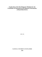
therefore, this assay was also applied to the t0 sample. Figure
1 shows the relationship between the PanBio cassette test and
the optical density (OD) values of the Focus ELISA. Especially
for IgM, there is no clear distinction between positive and
negative results.
The interpretation of results for the PanBio cassette test for
four diagnostic groups in comparison to that for the Focus ELISA
is shown in Table 2. The overall agreement was poor (kappa,
0.426; 2, 160; P Ͻ 0.001). The rapid test, applied to the t0
sample, had difficulties in discriminating acute from past disease
and acute primary from acute secondary dengue infection.
When the outcomes were combined to determine acute or
not acute dengue infection, the Focus ELISA identified 95
patients with acute dengue infection and 105 patients with no
acute dengue infection. Of these patients, the PanBio rapid
test, applied to the acute sample, identified 80 patients with
acute dengue and 120 without acute dengue (positive predictive value, 77.5%; negative predictive value, 72.5%; kappa,
0.485; P Ͻ 0.001).
The manufacturers have succeeded in making tests that are
easy to handle, but the sensitivity of both assays is poor. The
high cutoff levels that both manufacturers apply are necessary
to increase the specificity in areas where dengue is endemic,
but this is done at the expense of the sensitivities of both tests.
The poor sensitivities of both rapid tests, especially that of the
SD IgM test (ϳ11%), compares to results of a recent study of
a series of rapid tests that included the previous generation
PanBio test, a strip test, and the SD test (1).
The Focus ELISA is a rather sensitive assay, and the algorithm that was used for diagnostic classification in this study
was also sensitive, with small increases of antibody concentrations leading to a diagnosis of acute dengue infection. However, the lack of sensitivity of the rapid assays for detecting
antibodies in this study was at the basis of the diagnostic
misclassification.
Although the PanBio cassette test performs somewhat better than its paper strip predecessor and much better than the
FIG. 1. The Focus Dengue ELISA OD values for acute-phase serum samples tested with the PanBio Dengue Duo Cassette test.
as an IgM-positive and IgG-negative result; acute secondary
dengue infection was defined as an IgM-positive and IgGpositive or IgM-negative and IgG-positive result.
The overall agreement, sensitivities, specificities, and predictive
values and their 95% confidence intervals (95% CI) were determined using SPSS for Windows (version 12.2; SPSS, Inc., Chicago, IL). Agreement was assessed by calculating the kappa value.
Serum samples from 200 febrile patients were tested, of
whom 162 had dengue antibodies. Based on the Focus ELISA
results, 51 patients were classified as having acute primary
dengue infection, 44 as having acute secondary dengue infection, and 67 as having had past dengue infections. In 38 patients, no dengue antibodies were demonstrated.
The results of the rapid tests, applied to the t3-week sera,
were read as positive or negative and compared to the results
of the ELISA. The IgG test was always very clear. Five PanBio
IgM and three SD IgM test samples showed a very faint band,
TABLE 2. Diagnostic classification of dengue with the PanBio Dengue Duo Cassette test on acute-phase serum of Vietnamese febrile
patients, compared to ELISA on paired serum samplesa
No. of samples found positive by ELISA for paired sera
Diagnostic group
Acute primary
dengue
Acute secondary
dengue
No
dengue
Recent or
past dengue
Total
Acute primary dengue
Acute secondary dengue
No dengue
Recent or past dengue
10
15
21
5
12
25
3
4
0
0
38
0
2
16
9
40
24
56
71
49
Total
51
44
38
67
200
a
The diagnostic groups were assigned based on the results of the PanBio cassette test conducted at t0. The paired sera were collected at t0 and t3 weeks and tested
by the Focus Diagnostics ELISA.
VOL. 14, 2007
NOTES
SD strip test, the results of this study again confirm that rapid
tests that are based on antibody detection still have a limited
value for diagnosing dengue infection in its acute stage. Rapid
tests for the detection of dengue NS1 antigen are being developed and are a promising alternative for tests that are based on
antibody detection (3, 12).
In conclusion, the PanBio cassette and SD strip tests for
dengue infection are easy to use with clear results. They show
a high specificity with poor sensitivity, especially with respect to
the detection of IgM. The PanBio cassette test performed
better than the SD test but, applied to the acute-phase serum
sample, also did not discriminate clearly between infection at
the acute primary or secondary dengue or past dengue phase.
4.
5.
6.
7.
8.
9.
This study was carried out with the support of The Netherlands
Foundation for the Advancement of Tropical Research (WOTRO).
10.
REFERENCES
1. Blacksell, S. D., P. N. Newton, D. Bell, J. Kelley, M. P. Mammen, Jr., D. W.
Vaughn, V. Wuthiekanun, A. Sungkakum, A. Nisalak, and N. P. Day. 2006.
The comparative accuracy of 8 commercial rapid immunochromatographic
assays for the diagnosis of acute dengue virus infection. Clin. Infect. Dis.
42:1127–1134.
2. Charrel, R. N., and X. de Lamballerie. 2002. Low specificity of an immunochromatographic serological assay for diagnosis of dengue fever in travelers
returning with malaria. Clin. Diagn. Lab. Immunol. 9:1400.
3. Dussart, P., B. Labeau, G. Lagathu, P. Louis, M. R. Nunes, S. G. Rodrigues,
C. Storck-Herrmann, R. Cesaire, J. Morvan, M. Flamand, and L. Baril.
11.
12.
801
2006. Evaluation of an enzyme immunoassay for detection of dengue virus
NS1 antigen in human serum. Clin. Vaccine Immunol. 13:1185–1189.
Groen, J., P. Koraka, J. Velzing, C. Copra, and A. D. Osterhaus. 2000.
Evaluation of six immunoassays for detection of dengue virus-specific immunoglobulin M and G antibodies. Clin. Diagn. Lab. Immunol. 7:867–871.
Phuong, H. L., P. J. de Vries, N. Nagelkerke, P. T. Giao, L. Q. Hung, T. Q.
Binh, T. T. Nga, N. V. Nam, and P. A. Kager. 2006. Acute undifferentiated
fever in Binh Thuan province, Vietnam: imprecise clinical diagnosis and
irrational pharmacotherapy. Trop. Med. Int. Health 11:869–879.
Phuong, H. L., P. J. de Vries, T. T. Nga, P. T. Giao, L. Q. Hung, T. Q. Binh,
N. V. Nam, N. Nagelkerke, and P. A. Kager. 2006. Dengue as a cause of acute
undifferentiated fever in Vietnam. BMC Infect. Dis. 6:123.
Phuong, H. L., P. J. de Vries, K. T. Thai, T. T. Nga, L. Q. Hung, P. T. Giao,
T. Q. Binh, N. V. Nam, and P. A. Kager. Dengue virus infections in Vietnam:
the tip of the iceberg. Dengue Bull., in press.
Teles, F. R., D. M. Prazeres, and J. L. Lima-Filho. 2005. Trends in dengue
diagnosis. Rev. Med. Virol. 15:287–302.
Thai, K. T., T. Q. Binh, P. T. Giao, H. L. Phuong, L. Q. Hung, N. Van Nam,
T. T. Nga, J. Groen, N. Nagelkerke, and P. J. de Vries. 2005. Seroprevalence
of dengue antibodies, annual incidence and risk factors among children in
southern Vietnam. Trop. Med. Int. Health 10:379–386.
Tran, T., P. de Vries, L. Hoang, G. Phan, H. Le, B. Tran, C. Vo, N. Nguyen,
P. Kager, N. Nagelkerke, and J. Groen. 2006. Enzyme-linked immunoassay
for dengue virus IgM and IgG antibodies in serum and filter paper blood.
BMC Infect. Dis. 6:13.
World Health Organization. 1997. Dengue haemorrhagic fever: diagnosis,
treatment, prevention, and control, 2nd ed. World Health Organization,
Geneva, Switzerland.
Xu, H., B. Di, Y. X. Pan, L. W. Qiu, Y. D. Wang, W. Hao, L. J. He, K. Y. Yuen,
and X. Y. Che. 2006. Serotype 1-specific monoclonal antibody-based antigen
capture immunoassay for detection of circulating nonstructural protein NS1:
implications for early diagnosis and serotyping of dengue virus infections.
J. Clin. Microbiol. 44:2872–2878.