Báo cáo khoa học: Proteoglycans in health and disease: the multiple roles of syndecan shedding ppt
Bạn đang xem bản rút gọn của tài liệu. Xem và tải ngay bản đầy đủ của tài liệu tại đây (442.46 KB, 14 trang )
MINIREVIEW
Proteoglycans in health and disease: the multiple roles of
syndecan shedding
Tina Manon-Jensen
1
, Yoshifumi Itoh
2
and John R. Couchman
1
1 Deparment of Biomedical Sciences, University of Copenhagen, Denmark
2 Kennedy Institute of Rheumatology, Imperial College London, UK
Introduction
Syndecans are type 1 transmembrane heparan sulfate
proteoglycans (HSPGs) that have important roles dur-
ing development, wound healing and tumour progres-
sion by controlling cell proliferation, differentiation,
adhesion and migration. The heparan sulfate (HS)
chains substituted on the extracellular domains interact
with a wide range of ligands such as extracellular
matrix glycoproteins, collagens, cytokines, chemokines,
growth factors and enzymes, including metzincin pro-
teinases. The ectodomain of each syndecan is constitu-
tively shed in some cultured cells, but is accelerated in
response to wound healing, and some pathophysio-
logical events. Ectodomain shedding is an important
regulatory mechanism, because it can rapidly generate
soluble ectodomains that can function as paracrine or
autocrine effectors or competitors. Mammals have four
syndecan family members, syndecan-1 to -4 (Fig. 1),
whereas invertebrates and primitive chordates possess
only one syndecan, which is essential for neuronal
development and axon guidance [1,2]. All cells express
at least one member of the syndecan family [3], with
the exception of erythrocytes. Syndecan-4 can be found
in most tissues, but seems to be less abundant and is
frequently coexpressed with other syndecans. Syndec-
an-1 is highly expressed in epithelia, syndecan-2 in
endothelia and fibroblasts, whereas high expression of
Keywords
cell adhesion; cell migration;
glycosaminoglycan; growth factor; heparan
sulfate; metzincin; proteinase; proteoglycan;
receptors; signaling
Correspondence
J. R. Couchman, Department of Biomedical
Sciences, University of Copenhagen
Biocenter, Ole Maaløes Vej 5, 2200
Copenhagen N, Denmark
Fax: +45 353 25669
Tel: +45 353 25670
E-mail:
(Received 5 May 2010, revised 26 July
2010, accepted 28 July 2010)
doi:10.1111/j.1742-4658.2010.07798.x
Proteolytic processes in the extracellular matrix are a major influence on
cell adhesion, migration, survival, differentiation and proliferation. The
syndecan cell-surface proteoglycans are important mediators of cell spread-
ing on extracellular matrix and respond to growth factors and other bio-
logically active polypeptides. The ectodomain of each syndecan is
constitutively shed from many cultured cells, but is accelerated in response
to wound healing and diverse pathophysiological events. Ectodomain shed-
ding is an important regulatory mechanism, because it rapidly changes sur-
face receptor dynamics and generates soluble ectodomains that can
function as paracrine or autocrine effectors, or competitive inhibitors. It is
known that the family of syndecans can be shed by a variety of matrix pro-
teinase, including many metzincins. Shedding is particularly active in prolif-
erating and invasive cells, such as cancer cells, where cell-surface
components are continually released. Here, recent research into the shed-
ding of syndecans and its physiological relevance are assessed.
Abbreviations
ADAM, a disintegrin and metalloproteinase; GAG, glycosaminoglycan; GlcA, glucuronic acid; GalNAc, N-acetylgalactosamine; GlcNAc,
N-acetylglucosamine, HS, heparan sulfate; HSPG, heparan sulfate proteoglycan; MMP, matrix metalloproteinase; PKC, protein kinase C;
PMA, phorbol myristate acetate; TIMP, tissue inhibitor of metalloproteinases.
3876 FEBS Journal 277 (2010) 3876–3889 ª 2010 The Authors Journal compilation ª 2010 FEBS
syndecan-3 can mostly be found in neuronal tissues
and some musculoskeletal tissue. Here, our under-
standing of syndecan shedding and its function in
wound healing and tumour progression is reviewed.
Other reviews on syndecan structure and function have
been recently published [4–6].
Structural organization of syndecans
The syndecan core proteins range from 20 to 40 kDa
and have cytoplasmic domains that are highly con-
served across species, but have diversity in their ectod-
omains. All comprise an ectodomain, a single
transmembrane domain and a short cytoplasmic
domain (Fig. 1). The cytoplasmic domain consists of
membrane-proximal C1 and distal C2 conserved region
flanking a variable region (V) that is unique to each
syndecan, but highly conserved across species within
each individual syndecan gene. The C2 region inter-
acts with a number of PSD-95 ⁄ Discs-large ⁄ Zonula
occludens-domain-containing proteins such as syntenin,
Ga-interacting protein (GAIP)-interacting C-terminus ⁄
synectin and calc ium ⁄ calmodulin-associated serine kinase,
since the C2 region contains a class II PSD-95 ⁄
Discs-large ⁄ Zonula occludens protein-binding motif
FXF, where F represent a hydrophobic residue and X
any amino acid residue. Although information is
sparse, current evidence suggests that the C1 region
can interact with ezrin, at least for syndecan-2, which
provides a link to the actin cytoskeleton [7]. The cen-
tral V-region probably contains sites for syndecan-spe-
cific interaction partners, although this is only well
understood for syndecan-4 [4,8]. A ternary signalling
complex with phosphatidylinositol 4,5-bisphosphate
and protein kinase Ca has been described [9], whereas
others partners are the actin-associated protein a-acti-
nin as well as syndesmos, about whose function rather
little is known [10]. The transmembrane domain of all
syndecans contains a GXXXG motif that promotes
formation of SDS-resistant dimers [11,12]. The N-ter-
minal ectodomain has glycosaminoglycan (GAG) chain
substitution sites. These are predominantly HS cova-
lently linked to serine residues in a serine–glycine motif
surrounded by acidic residues. In addition to HS,
syndecan-1 and -3 can be substituted with chondroitin
or dermatan sulfate at sites closer to the transmem-
brane domain.
The synthesis of GAG chains in the Golgi apparatus
is a highly complex process, but both HS and chon-
droitin sulfate chains are linked to serine residues on
Syndecan-1
Syndecan-2
Syndecan-4Syndecan-3
Chondroitin sulphate
Heparan sulphate
33 kDa
23 kDa
43 kDa 22 kDa
C1
C2
V
Ser
Xyl
Gal
GlcA
GlcNAc
IdoA
GalNAc
Ser
6-O
6-O
2-0 2-0
2-0 6-0
NNN
6-0
NN
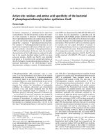
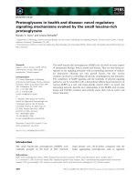
Fig. 1. Schematic of the four vertebrate syndecans. Syndecans-1 and -3 core proteins are larger than those of syndecan-2 and -4, and can
bear both heparan and chondroitin sulfate chains. The GAG chains are substituted on core protein serine residues and have a common stem
tetrasaccharide of xylose (xyl), two galactose units (gal) and a glucuronic acid residue (GlcA). The repeating disaccharide of HS is N-acetylglu-
cosamine and uronic acid, followed by several modifications in terms of sulfate and uronic acid epimerization to iduronic acid. The glucosa-
mine can be N-, 6-O or (rarely) 3-O sulfated, whereas the iduronic acid can be 2-O sulfated. In most cases, there are regions of low
sulfation, for example, adjacent to the core protein, with regions of intermediate or high sulfation. This yields a polysaccharide of immense
variability and complexity. Chondroitin sulfate contains N-acetylgalactosamine, which may be 6-O or 4-O sulfated. The cytoplasmic domains
have two highly conserved regions (C1 and C2) with an intervening syndecan-specific variable (V) region.
T. Manon-Jensen et al. Syndecan shedding at the cell surface
FEBS Journal 277 (2010) 3876–3889 ª 2010 The Authors Journal compilation ª 2010 FEBS 3877
the core protein through a tetrasaccharide linker con-
sisting of xylose–galactose–galactose–uronic acid resi-
dues, followed by the repeating disaccharide units. The
repeating unit of HS and chondroitin sulfate back-
bones are glucuronic acid (GlcA)–N-acetylglucosamine
(GlcNAc) or GlcA–N-acetylgalactosamine (GalNAc),
respectively. These chains range from 50 to 200 disac-
charides in length and undergo extensive modification
in which some uronic acid residues are epimerized and
a number of sulfation events occur (Fig. 1). In the case
of HS, chain modifications are not uniform but local-
ized along the chain. Subdomains of low sulfation are
interspersed among regions that are highly sulfated,
and small regions of intermediate sulfate lie at the
boundaries of these subdomains [13,14]. How the syn-
thesis of such complex polysaccharides is controlled
remains unknown.
Syndecan shedding
Syndecans undergo regulated proteolytic cleavage, usu-
ally near the plasma membrane, in a process known as
shedding. The release of syndecan extracellular
domains may not only downregulate signal transduc-
tion, but also convert the membrane-bound receptors
into soluble effectors ⁄ or antagonists. Soluble syndecan
ectodomain can compete with intact syndecans for
extracellular ligands in the pericellular environment
[15] (Fig. 2). The remaining portion of the membrane-
bound receptor loses its ability to bind ligands, and
can be further processed by the presenilin ⁄ c-secretase
complex. Like many other type I transmembrane
proteins [16], syndecan-3 has been shown to undergo
restricted intramembrane proteolysis by the membrane
presenilin ⁄ c-secretase complex within the hydrophobic
environment (mainly between Leu403 and Val404) of
the phospholipid bilayer of the membrane [17]. In
turn, there is decreased plasma membrane targeting of
the transcriptional cofactor calcium ⁄ calmodulin-associ-
ated serine kinase. Signaling is not restricted to the
syndecan proteoglycans but can be evoked by extracel-
lular proteoglycans binding to cell-surface receptors.
The leucine-rich proteoglycans are discussed in this
context by Iozzo & Schaefer [18] in this minireview
series.
Matrix metalloproteinases
Ectodomain shedding itself is a highly regulated pro-
cess that requires the direct action of enzymes gener-
ally referred to as sheddases. All mammalian syndecan
family members can be cleaved by extracellular prote-
ase [3]. The matrix metalloproteinases (MMPs) are
known sheddases of syndecans, and are endopeptidases
belonging to the family of metzincins (zinc endopeptid-
ases) which contain three major multigene families:
seralysins, astacins and a disintegrin and metallo-
proteinase (ADAM) ⁄ adamlysins. Substrate specificity
for MMPs is broad, therefore they function in many
physiological processes and are key to normal matrix
turnover, but also have essential roles in development
and reproduction, and in pathological tissue remodel-
ling during inflammatory disease, cancer invasion and
metastasis. Normally, MMPs cleave substrates before
HS
MMP9
Heparanase
ERK
Syndecan
CS
Soluble ectodomain
Intramembrane proteolysis by the
membrane presenilin/γ-secretase complex
Intracellular
Extracellular
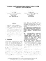
Fig. 2. Shedding of syndecans by metzincin
proteinases. Several metzincin enzymes can
cleave the syndecan core proteins, for
example MMP9, the site(s) being mem-
brane-proximal. Shedding is reported to be
enhanced if the HS chains are first cleaved
by heparanase. The shed syndecan may be
deposited in the pericellular matrix, whereas
the remnant core protein at the cell surface
may be further processed by intramembrane
cleavage by the presenilin ⁄ c-secretase com-
plex. There may also be signalling through
MAP kinases.
Syndecan shedding at the cell surface T. Manon-Jensen et al.
3878 FEBS Journal 277 (2010) 3876–3889 ª 2010 The Authors Journal compilation ª 2010 FEBS
a hydrophobic residue like Leu, Ile, Met, Phe or Tyr,
whereas cleavage before a charged residue is rarely
seen [19].
Twenty-three human MMPs have been identified
which can be divided into eight distinct structural
groups, five of which are secreted and three are mem-
brane-bound (MT-MMPs) (Fig. 3). The general form
of MMPs include an N-terminal signal sequence that
directs them to the endoplasmic reticulum, a propep-
tide (Pro) containing a cysteine switch motif
PRCGXPD (except for MMP23 which lacks the
cysteine switch motif) that maintains them as inactive
zymogens, and a catalytic domain with a z inc-binding site
(Zn, HEXXHXXGXXH) and a conserved methionine
(Met-turn) supporting the catalytic zinc. Interaction
between cysteine–zinc maintains proMMPs in an inac-
tive state by preventing a water molecule from binding
to the zinc atom. All MMPs, with the exception
of MMP-7, MMP-23 and MMP-26, also contain a
hemopexin-like domain that is connected to the
catalytic domain by a hinge region and mediates inter-
actions with tissue inhibitors of metalloproteinases,
cell-surface molecules and proteolytic substrates. The
first and last of the four repeats in the hemopexin-like
domains are linked by a disulfide bond (S–S) [19].
Two gelatinase MMPs (MMP-2 and MMP-9) con-
tain additional inserts that resemble collagen-binding
type II repeats of fibronectin. MMP-11 and MMP-28
contain a basic amino acid motif [KX(R ⁄ K)R] recog-
nized by furin-like serine proteinases between their
propeptide and catalytic domains that results in their
intracellular activation. This motif is also found in
MMP-21 with the vitronectin-like insert (Vn), MMP-
23 and the membrane-type MMPs (MT-MMPs) [19].
All soluble MMPs that do not harbour the basic motif
at the end of propeptide are secreted as zymogens and
activated extracellularly through proteolytic removal of
propeptide. Active MMPs, plasmin, cathepsin G and
neutrophil elastase have all been associated with
this function. MT-MMPs can be subdivided into
transmembrane (TM) forms and those that are
glycosylphosphatidylinositol anchored. The TM-type
MT-MMPs (MMP-14, MMP-15 and MMP-24) have a
single-span transmembrane domain and a very short
cytoplasmic domain. Alternately MMP-17 and MMP-
25 are glycosylphosphatidylinositol-anchored MMPs.
The type II membrane-linked MMP, MMP-23, has an
N-terminal signal anchor targeting it to the cell mem-
brane. Also, it is characterized by unique cysteine
array and immunoglobulin-like domains.
In healthy adults, activity of MMPs is difficult to
detect, except under conditions of tissue remodelling,
for example, in wound healing and menstrual endo-
metrium. Under physiological conditions, the activity
of MMPs is regulated by transcription, activation of
the precursor zymogen and by interactions with spe-
cific extracellular matrix components. In addition,
endogenous tissue inhibitors of metalloproteinases
provide a balance to prevent excessive degradation of
extracellular matrix. This physiological balance may
be disrupted in cancer. In many cancers, MMP
expression is upregulated and correlates with poor
prognosis [20,21]. Nevertheless, under some circum-
stances specific MMPs have a dual antitumour effect
[22].
Tissue inhibitor of metalloproteinases
The catalytic activity of MMPs can be inhibited by the
family of tissue inhibitor of metalloproteinases
(TIMP), of which there are four members (TIMP1-4).
TIMP-1, -2 and -4 are diffusible secreted proteins,
Type I transmembrane
GPI-anchored
Gelatin-binding
Minimal
Simple hemopexin-containing
Furin-activated secreted
MMP17 (MT4-MMP)
MMP25 (MT6-MMP)
MMP7
MMP26
MMP1 MMP12
MMP10
MMP8
MMP3
MMP28
MMP11
MMP9
MMP2
MMP27
MMP20
MMP19
MMP18
MMP13
MMP15 (MT2-MMP)
MMP14 (MT1-MMP)
Type II transmembrane
MMP23
MMP24 (MT5-MMP)
MMP16 (MT3-MMP)
Vitronectin-like
MMP21
pros cat
Hpx Hpx Hpx Hpx
FNII
Fu
V
TM Cyt
TM
IgCysR
GPI
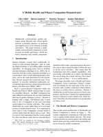
Fig. 3. Schematic of mammalian matrix me-
talloproteinases. The domain structures of
the various groups are shown, with
a list of some members. S, signal peptide;
Cat, catalytic domain; Pro, pro domain; TM,
transmembrane domain; Cyt, cytoplasmic
domain; Fu, furin cleavage site; Hpx,
hemopexin domain; Fn, fibronectin type II
repeats; V, vitronectin-like domain; CysR,
cysteine array; Ig, immunoglobin-like
domain, GPI, glycosylphosphatidylinositol
linker.
T. Manon-Jensen et al. Syndecan shedding at the cell surface
FEBS Journal 277 (2010) 3876–3889 ª 2010 The Authors Journal compilation ª 2010 FEBS 3879
whereas TIMP-3 is matrix associated because of its
heparin-binding characteristics which promote its asso-
ciation with matrix proteoglycans [23,24]. TIMP-3
binds to sulfated glycosaminoglycans such as heparin,
HS, chondroitin 4- and 6-sulfates, dermatan sulfate,
and sulfated compounds such as suramin and pento-
san, enabling interaction with GAG chains of synde-
cans [25]. Only TIMP-3 of the TIMP family has been
shown to effectively block shedding of syndecan-1 and -4
in mouse mammary epithelial cells [26].
Each TIMP can inhibit most MMPs, except TIMP-1
that, in particular, fails to inhibit several of the mem-
brane-type MMPs, MMP-14, -15, -16 and -24. The
inhibitory effect of TIMP-3 is different from the oth-
ers, as it also inhibits other metzincin subgroups, for
example the ADAM ⁄ adamlysins, including ADAM-17
(TACE) [27], ADAM-10 [28] and ADAM-12 [29], and
the ADAMs with thrombospondin motifs (ADAMTS)
including the aggrecanases ADAMTS4 and ADAM-
TS5 [30]. Kinetic studies have shown that TIMP-3 is
effective inhibitor of ADAM-17 (TACE) and aggre-
canases [27,30]. All mammalian TIMPs consist of two
distinct domains, N-terminal ( 125 amino acids) and
C-terminal ( 65 amino acids), where the N-terminal
domain usually is responsible for inhibition of protein-
ase activity. However, recently it has been shown that
the isolated N-terminal domains of TIMP-1 and
TIMP-3 are insufficient for ADAM10 inhibition,
whereas full-length TIMP-1 and TIMP-3 are [31]. The
C-terminal domain of TIMPs can stabilize proMMP
by binding to its hemopexin domain, leaving the N-ter-
minal fully capable of interacting with other MMPs.
Most cell types secrete proMMP-9 in complex with
TIMP-1, which complex can be found in the Golgi
apparatus [32]. TIMPs -2, -3 or -4 can bind proMMP2,
whereas TIMP-1 and -3 can interact with proMMP9.
TIMPs also facilitate activation of MMPs, by for
example, functioning as an adaptor between MT1-
MMP and Pro-MMP-2. MT1-MMP alone cannot bind
proMMP2, but the N-terminal region of TIMP-2 binds
the catalytic domain of MT1-MMP inhibiting its
activity, whereas its C-terminal domain binds to the
hemopexin-like domain of Pro-MMP-2 forming a
ternary complex. The complexed MT1-MMP cannot
cleave Pro-MMP-2, but requires a second MT1-MMP
molecule (without TIMP-2). Thus cleavage and activa-
tion of proMMP-2 require both active and inactive
MT1-MMP [33,34]. This process is facilitated by ho-
modimerization of two MT1-MMP molecules through
its hemopexin and transmembrane domains [35].
Syndecan sheddases
The glycosaminoglycan-bearing ectodomains of mam-
malian and Drosophila syndecans can be constitutively
shed from the cell surface as part of the normal turn-
over [3,26,36–39]. This constitutive shedding involves
metalloproteinases, but may be distinct from the metal-
loproteinase activity that mediates accelerated shedding
in response to wound healing, for example [26].
Evidence indicates the involvement of several MMPs
in syndecan cleavage in vitro and in vivo (Fig. 4).
Matrilysin (MMP-7) cleaves syndecan-1 [40], gelatinas-
es MMP-2 and MMP-9 can cleave syndecans-1, -2 and
Syndecan-1 Syndecan-4
CS
HS
ADAMT-S1 and -S4
Plasmin
Lys114-Arg115 and
Lys 129-Val130
Thrombin
Lys114-Arg115
MMP2
MMP9
MMP7
MT1-MMP
Near the 1st GAG chain
MMP2
MMP9
Intracellular
Extracellular
Human: Gly245-Leu246
Mouse: Ala243-Ser244
MT3-MMP
ADAM17
ADAM17
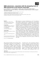
Fig. 4. Documented examples of metzincin
proteinases that shed syndecans-1 and -4.
Only in a few cases are the precise cleavage
sites known. Most sites are believed to be
membrane-proximal, although ADAMTS-1
and -4 may cleave syndecan-4 close to the
N-terminus. CS, chondroitin sulphate; HS,
heparan sulphate.
Syndecan shedding at the cell surface T. Manon-Jensen et al.
3880 FEBS Journal 277 (2010) 3876–3889 ª 2010 The Authors Journal compilation ª 2010 FEBS
-4 [41,42], whereas the membrane-associated metallo-
proteinases MT1-MMP and MT3-MMP are known to
cleave syndecan-1 [43]. However, current knowledge of
precise cleavage-specific sites on syndecan core proteins
is sparse. Human syndecan-4 is cleaved by the serine
proteases, plasmin and thrombin, at Lys114–
Arg115 ⁄ Lys192–Val130 and Lys114–Arg115, respec-
tively [44]. Despite high sequence homology between
human and mouse syndecan-1, they have distinct
MT1-MMP cleavage sites: human syndecan-1 is
cleaved at Gly245–Leu246, whereas cleavage of mouse
syndecan-1 occurs at Ala243–Ser244 [43,45].
The ADAM family of disintegrin and metallopro-
teinase membrane-anchored proteinases [46] also par-
ticipate in syndecan shedding. ADAM17 (TACE) has
recently been reported to shed syndecan-1 and syndec-
an-4 [47]. The cysteine-rich domain of human
ADAM12 was shown to associate with the ectodomain
of syndecan-4 and is regulated by HS; however, direct
ectodomain interactions with other members of the
ADAM family are not known [48,49].
The ADAMTS family (disintegrin and metallopro-
teinase with thrombospondin motifs) [50] also associ-
ates with syndecans. It has been reported that the p53
form ADAMTS4 binds HS and chondroitin sulfate
chains of syndecan-1 and aggrecan [51,52]. A recent
study also reported that syndecan-4 may regulate acti-
vation of ADAMTS-5 via engagement of HS chains
and regulation of MAPK-dependent synthesis of
MMP3 during cartilage damage in osteoarthritis [53].
Therefore, lack of syndecan-4 may be chondroprotec-
tive in some models of osteoarthritis. Both ADAMTS-1
and ADAMTS-4 have been demonstrated to cleave
syndecan-4 near the first GAG-attachment site, rather
than close to the membrane. This was shown to
decrease cell adhesion and promote cell migration [54].
Regulation of syndecan shedding
Syndecan shedding occurs through the direct action of
sheddases, although a variety of extracellular stimuli
including growth factors [55], chemokines [40,41,56],
bacterial virulence factors [57,58], trypsin [36], insulin
[59], heparanase [60] and cell stress [26] are known to
induce syndecan shedding. It is not yet clear how
extracellular stimuli influence sheddases to mediate
syndecan cleavage, but different agonists appear to
activate distinct intracellular signalling pathways to
activate shedding. Chemical inhibitor studies suggest
involvement of various signal transduction cascades,
such as protein kinase C (PKC), protein tyrosine
kinase, nuclear factor jB and mitogen-activated pro-
tein kinase pathways. For example, epidermal growth
factor- and thrombin receptor-mediated shedding cor-
relates with activation of the ERK ⁄ MAPK pathway,
and does not appear to involve PKC activation.
Inhibition of PKC activity prevents phorbol myristate
acetate (PMA)- and cellular stress-induced shedding of
syndecans, but does not affect thrombin or epidermal
growth factor receptor-activated shedding [26,55].
Interestingly, some pathogens usurp the host cell
shedding machinery to neutralize the host innate sys-
tem to promote their own pathogenesis by elevation of
syndecan shedding in response to bacterial virulence
factors [61–63]. For example, Staphylococcus aureus,
a common Gram-positive bacterium implicated in life-
threatening diseases like endocarditis and osteomyeli-
tis, enhances shedding of syndecan-1 through a-toxin
and b-toxin [58]. Beta-toxin, but not a-toxin, also
mediates shedding of syndecan-4. Alpha- and b-toxins
do not directly trigger syndecan-1 shedding, but acti-
vate protein tyrosine kinase-dependent intracellular sig-
nalling pathways that stimulate syndecan-1 shedding
[58]. Bacterial proteases can also enhance syndecan
shedding by mimicking the direct shedding effect of
syndecan sheddases [64]. For example, Streptococ-
cus pneumoniae sheds syndecan-1 directly through
ZmpC, a metalloproteinase virulence factor, where the
size of the shed soluble ectodomain is smaller than that
derived from a-orb-toxin mediated shedding [57].
Other pathogens may utilize HSPGs as attachment
receptors to facilitate either their entry into the host
cells or their survival in the host environment. For
example, the capsid ORF2 protein of hepatitis E virus
interacts mainly with 6-O-sulfate of syndecan-1 in
Huh-7 liver cells for productive infection [65].
Intracellular regulatory mechanisms play important
roles in agonist-induced shedding. Syndecans possess
highly conserved transmembrane and cytoplasmic
domains, the latter having three conserved tyrosine res-
idues and a variable number of serine ⁄ threonine resi-
dues that can serve as phosphorylation sites [66].
Phosphorylation of tyrosine residues has been sug-
gested to positively regulate syndecan-1 shedding
[26,55,67]. The phosphatase inhibitor pervanadate and
activation of intracellular kinases leads to tyrosine
phosphorylation and shedding of syndecan-1 [68].
Hayashida et al. [69] confirmed the pervanadate effect
on syndecan-1 shedding, but showed that S. aureus
b-toxin and PMA-mediated shedding was not accom-
panied by tyrosine phosphorylation. However, tyrosine
to phenylalanine mutation reduced the syndecan
shedding, suggesting mechanisms other than phosphor-
ylation, such as binding to other cytoplasmic compo-
nents is critical in agonist-mediated shedding. For
example, syndecan-1 cytoplasmic domain interacts with
T. Manon-Jensen et al. Syndecan shedding at the cell surface
FEBS Journal 277 (2010) 3876–3889 ª 2010 The Authors Journal compilation ª 2010 FEBS 3881
the inactive, GDP-bound form of Rab5, a small
GTPase that regulates intracellular trafficking and
triggers its conversion to an active GTP-bound state in
response to shedding promoters. A dominant negative
form of Rab5, unable to switch between active and
inactive states, significantly inhibited syndecan-1 shed-
ding, suggesting that trafficking is a key regulator of
syndecan-1 shedding [69].
Wound healing
Wound healing is a regulated process that can be
divided into three sequential, yet overlapping, phases;
inflammation, proliferation and remodelling [70]. Synd-
ecan-4 is upregulated in a range of inflammatory con-
ditions like ischaemic myocardial injury [71], and
dermal wound repair [72]. For example, atherosclerosis
is a chronic inflammatory disease marked by aberra-
tions in cell migration, proliferation and low-density
lipoprotein internalization [73]. Oxidized linoleic acid,
the major oxidized fatty acid in low-density lipopro-
tein, upregulates expression of syndecan-4, and as a
consequence, accelerated shedding of syndecans-4
involving the MEK pathway [74]. Increased levels of
syndecan-1 ectodomain are present in dermal wound
fluid, and in serum from patients with acute graft-ver-
sus-host disease [75].
A key inflammatory response is chemokine-mediated
recruitment of leukocytes into sites of inflammation
[76]. Many chemokines bind HS chains of syndecans
and evoke MMP-mediated shedding of syndecans with
potential loss from the site of injury [40,41,56]. MMP-
7 is upregulated in injured mucosal epithelium of the
lung, and promotes inflammation by shedding a synd-
ecan-1 ⁄ KC (CXCL8) complex that directs neutrophil
influx to the sites of injury [40]. Soluble syndecan-1
may maintain the proteolytic balance of acute wound
fluids, because it can bind the inflammation-related
neutrophil proteases cathepsin G and elastase, conse-
quently decreasing their affinity for their physiological
targets [37].
The function of syndecan-1 shedding in wound heal-
ing is not restricted to inflammation, but serves also to
promote re-epithelialization; however, this is not fully
clarified. Proliferating keratinocytes at the wound edge
and endothelial cells in the wound bed transiently
express syndecan-1 [77], whereas keratinocytes migrat-
ing into the wound lose their cell-surface syndecan-1
expression [37]. Syndecan-1 and syndecan-4 are shed
and may accumulate in dermal wound fluids [55].
Using a noncancerous simple epithelium cell line
(BEAS-2b) and organotypic cultures derived from pri-
mary epithelial cells, it has been demonstrated that
syndecan-1 is shed primarily by MMP-7 from epithe-
lial cells after injury [78], which enhances cell migra-
tion and facilitates wound closure. Therefore,
syndecan-1 shedding appears to be an important
response in wound healing. MMP-7 null mice demon-
strate a severely diminished re-epithelialization in
response to lung injury. Suppression of syndecan-1
expression in simple epithelial cells induces a promigra-
tory phenotype [79,80], consistent with decreased synd-
ecan levels in injured stratified epithelium (cornea and
skin) during repair [81,82]. Furthermore, knockdown
of syndecan-1 expression resulted in slowed cell migra-
tion in an A549 (a carcinoma-derived alveolar type II)
cell line [83]. Interestingly, soluble syndecan-1 ectodo-
main inhibited wound repair in mice overexpressing
syndecan-1, by exhibiting delay in wound closure,
re-epithelialization, granulation tissue formation and
remodelling [84]. Overall, the studies reveal that
MMP-7 cleavage of syndecan-1 is essential for effective
re-epithelialization; however, a balance is critical
because soluble syndecan-1 overexpression or complete
absence of syndecan-1 in the knockout lead to impair-
ment. The function of syndecan-1 may be tissue spe-
cific, because syndecan-1 null primary dermal
fibroblasts migrated faster than wild-type cells [85].
E-cadherin, a known mediator of cell–cell contact, is
also shed in vivo from injured lung epithelium by
MMP-7 [86], and has been shown to be coordinately
regulated with syndecan-1 [79]. It is not known if
shedding of E-cadherin and syndecan-1 happen contig-
uously, but could synergistically promote a migratory
epithelial phenotype.
It is well known that syndecans are functionally cou-
pled to integrins [4], which represent the major group
of cell-surface receptors for extracellular matrix macro-
molecules. There are 24 heterodimeric integrins in
mammals, each composed of an a and a b subunit
derived from combinations of 8 b and 18 a subunits.
Interaction between syndecan and integrins may be
direct [87] or indirect through an intermediate ‘recep-
tor’ [88]. This adhesion mechanism can be HS indepen-
dent, because the cell adhesion properties of syndecans
are not only limited to the HS chains, but can also be
mediated through the ectodomain core protein. The
evolutionarily conserved NXIP motif of syndecan-4
has been shown to promote b1-integrin-dependent cell
adhesion [89]. Syndecan-1 ectodomain regulates avb3
and avb5 integrin-mediated attachment and spreading
in human mammary carcinoma cells and B82L fibro-
blasts, respectively. The activity has been mapped to
residues 88–252 within the syndecan-1 ectodomain
[90,91]. This association can be blocked by synstatin, a
peptide inhibitor corresponding to the active site of the
Syndecan shedding at the cell surface T. Manon-Jensen et al.
3882 FEBS Journal 277 (2010) 3876–3889 ª 2010 The Authors Journal compilation ª 2010 FEBS
syndecan-1 core protein, and which can suppress
angiogenesis in vitro and in vivo, perhaps signifying
syndecan-1 as a critical mediator of tumour progres-
sion [87].
Another motif, the AVAAV (amino acids 222-226),
only present within the syndecan-1 ectodomain, has
been suggested to be an invasion regulatory domain,
because mutation within this region abolishes syndec-
an-1-mediated inhibition of cell invasion [92]. How-
ever, the mechanism remains unknown.
Integrins and syndecans together may influence the
outcome of cell adhesion and migration because their
different activation states and clustering on the cell
surface result in varying degrees of mechanical force
exerted on the extracellular matrix [5]. Syndecan-1
shedding by MMP-7 from repairing simple epithelial
(BEAS-2b) cells after injury [77] enhances cell migra-
tion and facilitates wound closure by causing the a2b1
integrin to assume a less-active conformation, compati-
ble with migration. It has previously been shown that
syndecan-1 facilitates integrin a2b1-mediated adhesion
to collagen [93].
Tumour progression
In addition to genetic and epigenetic changes, tumour
progression links a series of steps involving adhesion,
motility and growth, resulting in metastatic spread,
a major cause of death among cancer patients. These
steps are influenced by the activity of tumour-derived
MMPs. MMPs facilitate metastasis by degrading extra-
cellular matrix components, such as collagens, laminins
and proteoglycans, and they modulate cell adhesion,
enabling turnover of matrix contacts or adhesions.
Novel roles for proteoglycans in malignancy are also
discussed elsewhere in this volume [94].
As part of the regulation of MMPs, rate-limiting
effects, such as zymogen activation and the availability
of TIMPs are important. Another control element may
be contributed by HS chains of proteoglycans, which
interact with many extracellular protease, with exam-
ples from all four classes of proteases (aspartyl-, seryl-,
cysteyl-protease and metalloproteases). Heparan sulfate
also interacts with protease inhibitors, for example
TIMP-3 [95] and antithrombin III (ATIII). These
interactions may control extracellular matrix degrada-
tion, by either modifying enzymatic activity through
activation or inhibition, or providing a reservoir of
latent enzyme that is positioned for directed proteolytic
attack on extracellular matrix proteins. For example,
highly sulfated HS has been shown to inhibit the
proteolytic degradation of aggrecan, in part through
direct inhibition of aggrecanase activity [96]. Further-
more, HS chains of syndecans bind tumour-associated
MMPs, MMP-2, -7, -9 and -13 [97], in which MMP-2
catalytic activity is inhibited by its interaction with HS
chains of syndecan-2 [98], whereas MMP-1, -7 and -13
catalytic activity increases in the presence of heparin
[97]. MMP-7 has been shown to promote syndecan-1
shedding upon growth factor activation (FGF-2),
achieving its own release although still being attached
to HS chains [97]. Other attributes of HS chains
include the ability of TIMP-3 to interact with cell-sur-
face HS. This may lead to inhibition or internalization
of cell-surface MMPs or ADAMs, because it has been
discovered that TIMP-3 is internalized in HEK293 and
HTB94 chondrosarcoma cells [99], a process that is
mediated by cell-surface glycosaminoglycans [99,100].
Overall, HS chains of syndecans may support inva-
sion of tumour cells by protecting and anchoring
matrix-degrading proteases, while also harbouring sig-
nalling molecules that promote growth and directional
migration. However, the MMP-13 C-terminal domain
has been shown using yeast two-hybrid analysis to
associate with syndecan-4 without HS chains, suggest-
ing alternative MMP interaction sites than GAG
chains [101].
The role of syndecans in tumour progression may
vary with tumour stage and type, because syndecan-1
is reported to be downregulated in several types of
breast cancer [102], but upregulated in several
tumours, such as pancreatic cancer. Soluble syndecan-
1 ectodomain can be found in the serum of lung can-
cer patients [103] and Hodgkin’s lymphoma patients
[104], in the extracellular matrix of myeloma biopsies,
as well in the serum of myeloma patients [105,106], to
a much greater degree than in healthy individuals
[107].
A recent study has distinguished the roles between
membrane-bound and shed form of syndecan-1 in
breast cancer epithelial cells (MCF-7) in vitro. The
membrane-bound form of syndecan-1 increased prolif-
eration and inhibited invasiveness, whereas the soluble
form had the opposite effect, by promoting invasive-
ness and inhibiting proliferation [108].
Perhaps the best evidence for the importance of
shedding in cancer is shown for syndecan-1 in mye-
loma. Multiple myeloma is a malignant proliferation
of the bone marrow plasma cells increasing angiogene-
sis and development of osteolytic bone disease. Soluble
syndecan-1 promotes the growth of myeloma tumours
in vivo [109]. High levels of shed syndecan-1 in the sera
of myeloma patients are a marker of poor prognosis
[105,107,110].
Heparanase seems to play a distinct role in shedding
syndecans in myeloma. Mammalian heparanase
T. Manon-Jensen et al. Syndecan shedding at the cell surface
FEBS Journal 277 (2010) 3876–3889 ª 2010 The Authors Journal compilation ª 2010 FEBS 3883
(endo-b-d-glucuronidase) is known to modulate synde-
cans by cleaving the less-sulfated regions along the HS
chain releasing fragments of 10–20 sugar residues [111]
(Fig. 2). It may function in tumour progression by
promoting tumour growth, angiogenesis and metastasis
[112] by both enzymatic and nonenzymatic mecha-
nisms. A recently described nonenzymatic mechanism
of heparanase is its ability to facilitate cell adhesion
and spreading by clustering of syndecan-1 and syndec-
an-4 through interaction with their HS chains [113].
Knockdown of heparanase in myeloma cell lines
decreases soluble syndecan-1 [114]. In support, active
heparanase was shown to accelerate myeloma cell
growth and promote bone metastasis by increasing the
number and size of blood vessels within the tumour
[115,116]. Heparanase function in tumour progression
is discussed by Barash et al. [117] in this minireview
series.
Elevated active heparanase has been demonstrated
to enhance syndecan-1 shedding through ERK signal-
ling, which in turn upregulates expression of two pro-
teases, MMP-9 and urokinase-type plasminogen
activator [118]. Recently, it has been shown that hepa-
ranase-enhanced shedding of syndecan-1 by myeloma
cells promoted endothelial invasion and angiogenesis
[118]. Heparanase also increased urokinase-type plas-
minogen activator receptor expression levels [119], and
can even initiate syndecan-1 expression in the ARH-77
(human plasma cell leukemia) cell line that is normally
negative for syndecan-1 [60]. The expression of uroki-
nase-type plasminogen activator and its receptor may
also be a predictor of poor prognosis, just as with shed
syndecan-1 and heparanase [120].
The gelatinase MMP-9 sheds syndecan-1 directly
[41], and has been suggested as a useful prognostic
index of bone disease [121]. In addition, myeloma cell
invasiveness can be promoted by MMP-9 in vitro [122],
consistent with data suggesting that MMP-9 inhibition
has antimyeloma effects [123]. Urokinase, by contrast,
has a more indirect effect on syndecan shedding. Its
activity in generating plasmin from plasminogen has
been suggested to be a major activator of MMPs
in vivo, where it can process proMMP into active
MMP. In turn, these shed syndecans directly and⁄ or
activate other MMP sheddases. For example, plasmin
directly activates proMMP-1, proMMP-3, proMMP-9,
proMMP-10 and proMMP-13 in vitro [124].
Conclusions and perspectives
Syndecan shedding is subject to highly complex regula-
tion. In tissue culture, there may be constitutive shed-
ding, and in vivo enhanced shedding in cases of injury
and disease. Because syndecans are important co-
receptors for adhesion and growth factor receptors,
their loss from the cell surface may have multiple
effects. There is certainly a need for a deeper under-
standing of these processes, because they may relate to
diagnosis, prognosis or even treatment options for
some diseases. Better reagents for detecting syndecan
cleavage will be a valuable aid in these analyses, both
in vitro and in vivo. This may be difficult, not least
because so many different proteases can cleave the
syndecan core proteins. There is much to learn about
when and where these events take place.
References
1 Rawson JM, Dimitroff B, Johnson KG, Rawson JM,
Ge X, Van Vactor D & Selleck SB (2005) The heparan
sulfate proteoglycans Dally-like and Syndecan have
distinct functions in axon guidance and visual-system
assembly in Drosophila. Curr Biol 15, 833–838.
2 Rhiner C, Gysi S, Frohli E, Hengartner MO & Hajnal
A (2005) Syndecan regulates cell migration and axon
guidance in C. elegans . Development 132, 4621–4633.
3 Kim CW, Goldberger OA, Gallo RL & Bernfield M
(1994) Members of the syndecan family of heparan sul-
fate proteoglycans are expressed in distinct cell-, tissue-,
and development-specific patterns. Mol Biol Cell 5,
797–805.
4 Morgan MR, Humphries MJ & Bass MD (2007)
Synergistic control of cell adhesion by integrins and
syndecans. Nat Rev Mol Cell Biol 8 , 957–969.
5 Okina E, Manon-Jensen T, Whiteford JR & Couch-
man JR (2009) Syndecan proteoglycan contributions to
cytoskeletal organization and contractility. Scand
J Med Sci Sports 19, 479–489.
6 Xian X, Gopal S & Couchman JR (2010) Syndecans as
receptors and organizers of the extracellular matrix.
Cell Tissue Res 339, 31–46.
7 Grane
´
s F, Urena JM, Rocamora N & Vilaro
´
S (2000)
Ezrin links syndecan-2 to the cytoskeleton. J Cell Sci
113, 1267–1276.
8 Couchman JR (2003) Syndecans: proteoglycan regula-
tors of cell-surface microdomains? Nat Rev Mol Cell
Biol 4, 926–937.
9 Oh ES, Couchman JR & Woods A (1997) Serine phos-
phorylation of syndecan-2 proteoglycan cytoplasmic
domain. Arch Biochem Biophys 1, 67–74.
10 Baciu PC, Saoncella S, Lee SH, Denhez F, Leuthardt
D & Goetinck PF (2000) Syndesmos, a protein that
interacts with the cytoplasmic domain of syndecan-4,
mediates cell spreading and actin cytoskeletal organiza-
tion. J Cell Sci 113, 315–324.
11 Choi S, Lee E, Kwon S, Park H, Yi JY, Kim S,
Han IO, Yun Y & Oh ES (2005) Transmembrane
Syndecan shedding at the cell surface T. Manon-Jensen et al.
3884 FEBS Journal 277 (2010) 3876–3889 ª 2010 The Authors Journal compilation ª 2010 FEBS
domain-induced oligomerization is crucial for the func-
tions of syndecan-2 and syndecan-4. J Biol Chem 280,
42573–42579.
12 Dews IC & Mackenzie KR (2007) Transmembrane
domains of the syndecan family of growth factor corecep-
tors display a hierarchy of homotypic and heterotypic
interactions. Proc Natl Acad Sci USA 104, 20782–20787.
13 Bishop JR, Schuksz M & Esko JD (2007) Heparan sul-
phate proteoglycans fine-tune mammalian physiology.
Nature 446, 1030–1037.
14 Nadanaka S & Kitagawa H (2008) Heparan sulphate
biosynthesis and disease. J Biochem 144, 7–14.
15 Steinfeld R, Van Den Berghe H & David G (1996)
Stimulation of fibroblast growth factor receptor-1
occupancy and signaling by cell surface-associated
syndecans and glypican. J Cell Biol 133, 405–416.
16 Brown MS, Ye J, Rawson RB & Goldstein JL (2000)
Regulated intramembrane proteolysis: a control mecha-
nism conserved from bacteria to humans. Cell 100,
391–398.
17 Schulz JG, Annaert W, Vandekerckhove J, Zimmer-
mann P, De Strooper B & David G (2003) Syndecan 3
intramembrane proteolysis is presenilin ⁄ gamma-
secretase-dependent and modulates cytosolic signaling.
J Biol Chem 278, 48651–48657.
18 Iozzo RV & Schaefer L (2010) Proteoglycans in health
and disease: novel regulatory signaling mechanisms
evoked by the small leucine-rich proteoglycans. FEBS J
277, 3864–3875.
19 Visse R & Nagase H (2003) Matrix metalloproteinases
and tissue inhibitors of metalloproteinases: structure,
function, and biochemistry. Circ Res 92, 827–839.
20 Cho NH, Shim HS, Rha SY, Kang SH, Hong SH,
Choi YD, Hong SJ & Cho SH (2003) Increased expres-
sion of matrix metalloproteinase 9 correlates with poor
prognostic variables in renal cell carcinoma. Eur Urol
44, 560–566.
21 Curran S, Dundas SR, Buxton J, Leeman MF,
Ramsay R & Murray GI (2004) Matrix metalloprotein-
ase ⁄ tissue inhibitors of matrix metalloproteinase
phenotype identifies poor prognosis colorectal cancers.
Clin Cancer Res 10, 8229–8234.
22 Deryugina EI & Quigley JP (2005) Matrix metallopro-
teinases and tumor metastasis. Cancer Metastasis Rev
25, 9–34.
23 Leco KJ, Khokha R, Pavloff N, Hawkes SP &
Edwards DR (1994) Tissue inhibitor of metalloprotein-
ases-3 (TIMP-3) is an extracellular matrix-associated
protein with a distinctive pattern of expression in
mouse cells and tissues. J Biol Chem 269, 9352–9360.
24 Nagase H, Visse R & Murphy G (2006) Structure and
function of matrix metalloproteinases and TIMPs.
Cardiovasc Res 69, 562–573.
25 Yu WH, Yu S, Meng Q, Brew K & Woessner JF
(2000) TIMP-3 binds to sulfated glycosaminoglycans of
the extracellular matrix. J Biol Chem 275, 31226–
31232.
26 Fitzgerald ML, Wang Z, Park PW, Murphy G &
Bernfield M (2000) Shedding of syndecan-1 and -4
ectodomains is regulated by multiple signaling
pathways and mediated by a TIMP-3-sensitive metallo-
proteinase. J Cell Biol 148, 811–824.
27 Amour A, Slocombe PM, Webster A, Butler M,
Knight CG, Smith BJ, Stephens PE, Shelley C, Hutton
M, Kna
¨
uper V et al. (1998) TNF-alpha converting
enzyme (TACE) is inhibited by TIMP-3. FEBS Lett
435, 39–44.
28 Amour A, Knight CG, Webster A, Slocombe PM,
Stephens PE, Kna
¨
uper V, Docherty AJ & Murphy
G (2000) The in vitro activity of ADAM-10 is inhib-
ited by TIMP-1 and TIMP-3. FEBS Lett 473, 275–
279.
29 Loechel F, Fox JW, Murphy G, Albrechtsen R &
Wewer UM (2000) ADAM 12-S cleaves IGFBP-3 and
IGFBP-5 and is inhibited by TIMP-3. Biochem Biophys
Res Commun 278, 511–515.
30 Kashiwagi M, Tortorella M, Nagase H & Brew K
(2001) TIMP-3 is a potent inhibitor of aggrecanase 1
(ADAMTS-4) and aggrecanase 2 (ADAMTS-5). J Biol
Chem 276, 12501–12504.
31 Rapti M, Atkinson SJ, Lee MH, Trim A, Moss M &
Murphy G (2008) The isolated N-terminal domains of
TIMP-1 and TIMP-3 are insufficient for ADAM10
inhibition. Biochem J 411, 433–439.
32 Roderfeld M, Graf J, Giese B, Salguero-Palacios R,
Tschuschner A, Mu
¨
ller-Newen G & Roeb E (2007)
Latent MMP-9 is bound to TIMP-1 before secretion.
Biol Chem 388, 1227–1234.
33 Sato H, Takino T, Okada Y, Cao J, Shinagawa A,
Yamamoto E & Seiki M (1994) A matrix metallopro-
teinase expressed on the surface of invasive tumour
cells. Nature 370, 61–65.
34 Strongin AY, Collier I, Bannikov G, Marmer BL,
Grant GA & Goldberg GI (1995) Mechanism of cell
surface activation of 72-kDa type IV collagenase.
Isolation of the activated form of the membrane
metalloprotease J Biol Chem 270, 5331–5338.
35 Itoh Y, Ito N, Nagase H & Seiki M (2008) The second
dimer interface of MT1-MMP, the transmembrane
domain, is essential for ProMMP-2 activation on the
cell surface. J Biol Chem 283, 13053–13062.
36 Jalkanen M, Rapraeger A, Saunders S & Bernfield MJ
(1987) Cell surface proteoglycan of mouse mammary
epithelial cells is shed by cleavage of its matrix-binding
ectodomain from its membrane-associated domain.
Cell Biol 105, 3087–3096.
37 Kainulainen V, Wang H, Schick C & Bernfield M
(1998) Syndecans, heparan sulfate proteoglycans,
maintain the proteolytic balance of acute wound fluids.
J Biol Chem 273, 11563–11569.
T. Manon-Jensen et al. Syndecan shedding at the cell surface
FEBS Journal 277 (2010) 3876–3889 ª 2010 The Authors Journal compilation ª 2010 FEBS 3885
38 Spring J, Paine-Saunders SE, Hynes RO & Bernfield
M (1994) Drosophila syndecan: conservation of a cell-
surface heparan sulfate proteoglycan. Proc Natl Acad
Sci USA 91, 3334–3338.
39 Yanagishita M & Hascall VC (1992) Cell surface hepa-
ran sulfate proteoglycans. J Biol Chem 267, 9451–9454.
40 Li Q, Park PW, Wilson CL & Parks WC (2002)
Matrilysin shedding of syndecan-1 regulates chemokine
mobilization and transepithelial efflux of neutrophils in
acute lung injury. Cell 111, 635–646.
41 Brule S, Charnaux N, Sutton A, Ledoux D, Chaigneau
T, Saffar L & Gattegno L (2006) The shedding of
syndecan-4 and syndecan-1 from HeLa cells and
human primary macrophages is accelerated by
SDF-1 ⁄ CXCL12 and mediated by the matrix
metalloproteinase-9. Glycobiology 16, 488–501.
42 Fears CY, Gladson CL & Woods A (2006) Syndecan-2
is expressed in the microvasculature of gliomas and
regulates angiogenic processes in microvascular endo-
thelial cells. J Biol Chem 281, 14533–14536.
43 Endo K, Takino T, Miyamori H, Kinsen H, Yoshizaki
T, Furukawa M & Sato H (2003) Cleavage of syndec-
an-1 by membrane type matrix metalloproteinase-1
stimulates cell migration. J Biol Chem 278, 40764–
40770.
44 Schmidt A, Echtermeyer F, Alozie A, Brands K &
Buddecke E (2005) Plasmin- and thrombin-accelerated
shedding of syndecan-4 ectodomain generates cleavage
sites at Lys(114)–Arg(115) and Lys(129)–Val(130)
bonds. J Biol Chem 280, 34441–34446.
45 Wang Z, Go
¨
tte M, Bernfield M & Reizes O (2005) Con-
stitutive and accelerated shedding of murine syndecan-1
is mediated by cleavage of its core protein at a specific
juxtamembrane site. Biochemistry 44, 12355–12361.
46 Edwards DR, Handsley MM & Pennington CJ (2008)
The ADAM metalloproteinases. Mol Aspects Med 29,
258–289.
47 Pruessmeyer J, Martin C, Hess FM, Schwarz N,
Schmidt S, Kogel T, Hoettecke N, Schmidt B, Sechi A,
Uhlig S et al. (2010) A disintegrin and metalloprotein-
ase 17 (ADAM17) mediates inflammation-induced
shedding of syndecan-1 and -4 by lung epithelial cells.
J Biol Chem 285, 555–564.
48 Iba K, Albrechtsen R, Gilpin B, Fro
¨
hlich C, Loechel
F, Zolkiewska A, Ishiguro K, Kojima T, Liu W,
Langford JK et al. (2000) The cysteine-rich domain of
human ADAM 12 supports cell adhesion through
syndecans and triggers signaling events that lead to
beta1 integrin-dependent cell spreading. J Cell Biol
149, 1143–1156.
49 Sørensen HP, Vive
`
s RR, Manetopoulos C, Albrechtsen
R, Lydolph MC, Jacobsen J, Couchman JR & Wewer
UM (2008) Heparan sulfate regulates ADAM12
through a molecular switch mechanism. J Biol Chem
283, 31920–31932.
50 Porter S, Clark IM, Kevorkian L & Edwards DR
(2005) The ADAMTS metalloproteinases. Biochem J
386, 15–27.
51 Gao G, Plaas A, Thompson VP, Jin S, Zuo F & Sandy
JD (2004) ADAMTS4 (aggrecanase-1) activation on
the cell surface involves C-terminal cleavage by glyco-
sylphosphatidyl inositol-anchored membrane type 4-
matrix metalloproteinase and binding of the activated
proteinase to chondroitin sulfate and heparan sulfate
on syndecan-1. J Biol Chem 279, 10042–10051.
52 Tortorella MD, Burn TC, Pratta MA, Abbaszade I,
Hollis JM, Liu R, Rosenfeld SA, Copeland RA,
Decicco CP, Wynn R et al. (1999) Purification and
cloning of aggrecanase-1: a member of the ADAMTS
family of proteins. Science
284, 1664–1666.
53 Echtermeyer F, Bertrand J, Dreier R, Meinecke I,
Neugebauer K, Fuerst M, Lee YJ, Song YW, Herzog
C, Theilmeier G et al. (2009) Syndecan-4 regulates
ADAMTS-5 activation and cartilage breakdown in
osteoarthritis. Nat Med 15, 1072–1076.
54 Rodrı
´
guez-Manzaneque JC, Carpizo D, Plaza-Calonge
Mdel C, Torres-Collado AX, Thai SN, Simons M,
Horowitz A & Iruela-Arispe ML (2008) Cleavage of
syndecan-4 by ADAMTS1 provokes defects in
adhesion. Int J Biochem Cell Biol 41, 800–810.
55 Subramanian SV, Fitzgerald ML & Bernfield M (1997)
Regulated shedding of syndecan-1 and -4 ectodomains
by thrombin and growth factor receptor activation.
J Biol Chem 272, 14713–14720.
56 Charnaux N, Brule S, Chaigneau T, Saffar L, Sutton
A, Hamon M, Prost C, Lievre N, Vita C & Gattegno
L (2005) RANTES (CCL5) induces a CCR5-dependent
accelerated shedding of syndecan-1 (CD138) and synd-
ecan-4 from HeLa cells and forms complexes with the
shed ectodomains of these proteoglycans as well as
with those of CD44. Glycobiology 15, 119–130.
57 Chen Y, Hayashida A, Bennett AE, Hollingshead SK
& Park PW (2007) Streptococcus pneumoniae sheds
syndecan-1 ectodomains through ZmpC, a metal-
loproteinase virulence factor. J Biol Chem 282, 159–
167.
58 Park PW, Foster TJ, Nishi E, Duncan SJ, Klagsbrun
M & Chen Y (2004) Activation of syndecan-1 ectodo-
main shedding by Staphylococcus aureus alpha-toxin
and beta-toxin. J Biol Chem 279, 251–258.
59 Wang JB, Guan J, Shen J, Zhou L, Zhang YJ, Si YF,
Yang L, Jian XH & Sheng Y (2009) Insulin increases
shedding of syndecan-1 in the serum of patients with
type 2 diabetes mellitus. Diabetes Res Clin Pract 86,
83–88.
60 Yang Y, Macleod V, Miao HQ, Theus A, Zhan F,
Shaughnessy JD Jr, Sawyer J, Li JP, Zcharia E, Vlo-
davsky I et al. (2007) Heparanase enhances syndecan-1
shedding: a novel mechanism for stimulation of tumor
growth and metastasis. J Biol Chem 282 , 13326–13333.
Syndecan shedding at the cell surface T. Manon-Jensen et al.
3886 FEBS Journal 277 (2010) 3876–3889 ª 2010 The Authors Journal compilation ª 2010 FEBS
61 Andrian E, Grenier D & Rouabhia M (2005)
Porphyromonas gingivalis lipopolysaccharide induces
shedding of syndecan-1 expressed by gingival epithelial
cells. J Cell Physiol 204, 178–183.
62 Park PW, Pier GB, Hinkes MT & Bernfield M (2001)
Exploitation of syndecan-1 shedding by Pseudomonas
aeruginosa enhances virulence. Nature 411, 98–102.
63 Popova TG, Millis B, Bradburne C, Nazarenko S,
Bailey C, Chandhoke V & Popov SG (2006) Accelera-
tion of epithelial cell syndecan-1 shedding by anthrax
hemolytic virulence factors. BMC Microbiol 6,8.
64 Chung MC, Popova TG, Millis BA, Mukherjee DV,
Zhou W, Liotta LA, Petricoin EF, Chandhoke V,
Bailey C & Popov SG (2006) Secreted neutral metallo-
proteases of Bacillus anthracis as candidate pathogenic
factors. J Biol Chem 281, 31408–31418.
65 Kalia M, Chandra V, Rahman SA, Sehgal D & Jameel
S (2009) Heparan sulfate proteoglycans are required
for cellular binding of the hepatitis E virus ORF2 cap-
sid protein and for viral infection. J Virol 83, 12714–
12724.
66 Horowitz A & Simons M (1998) Regulation of syndec-
an-4 phosphorylation in vivo. J Biol Chem 273, 10914–
10918.
67 Ott VL & Rapraeger AC (1998) Tyrosine phosphoryla-
tion of syndecan-1 and -4 cytoplasmic domains in
adherent B82 fibroblasts. J Biol Chem 273, 35291–
35298.
68 Reiland J, Ott VL, Lebakken CS, Yeaman C, McCarthy
J & Rapraeger AC (1996) Pervanadate activation of
intracellular kinases leads to tyrosine phosphorylation
and shedding of syndecan-1. Biochem J 319, 39–47.
69 Hayashida K, Stahl PD & Park PW (2008) Syndecan-1
ectodomain shedding is regulated by the small GTPase
Rab5. J Biol Chem 283, 35435–35444.
70 Martin P & Leibovich SJ (2005) Inflammatory cells
during wound repair: the good, the bad and the ugly.
Trends Cell Biol 15, 599–607.
71 Cizmeci-Smith G, Langan E, Youkey J, Showalter LJ
& Carey DJ (1997) Syndecan-4 is a primary-response
gene induced by basic fibroblast growth factor and
arterial injury in vascular smooth muscle cells.
Arterioscler Thromb Vasc Biol 17, 172–180.
72 Gallo R, Kim C, Kokenyesi R, Adzick NS & Bernfield
M (1996) Syndecans-1 and -4 are induced during
wound repair of neonatal but not fetal skin. J Invest
Dermatol 107, 676–683.
73 Lusis AJ (2000) Atherosclerosis. Nature 407, 233–241.
74 Houston M, Julien MA, Parthasarathy S & Chaikof
EL (2005) Oxidized linoleic acid regulates expression
and shedding of syndecan-4. Am J Physiol Cell Physiol
288, 458–466.
75 Seidel C, Ringde
´
n O & Remberger M (2003) Increased
levels of syndecan-1 in serum during acute graft-
versus-host disease. Transplantation 76, 423–426.
76 Zlotnik A & Yoshie O (2000) Chemokines: a new
classification system and their role in immunity.
Immunity 12, 121–127.
77 Elenius K, Vainio S, Laato M, Salmivirta M, Thesleff
I & Jalkanen MJ (1991) Induced expression of syndec-
an in healing wounds. Cell Biol 114, 585–595.
78 Chen P, Abacherli LE, Nadler ST, Wang Y, Li Q &
Parks WC (2009) MMP7 shedding of syndecan-1 facili-
tates re-epithelialization by affecting alpha(2)beta(1)
integrin activation. PLoS ONE 10, e6565.
79 Leppa
¨
S, Vleminckx K, Van Roy F & Jalkanen M
(1996) Syndecan-1 expression in mammary epithelial
tumor cells is E-cadherin-dependent. J Cell Sci 109,
1393–1403.
80 Kato M, Saunders S, Nguyen H & Bernfield M (1995)
Loss of cell surface syndecan-1 causes epithelia to
transform into anchorage-independent mesenchyme-
like cells. Mol Biol Cell 6, 559–576.
81 Grushkin-Lerner LS & Trinkaus-Randall V (1991)
Localization of integrin and syndecan in vivo in a
corneal epithelial abrasion and keratectomy. Curr Eye
Res 10, 75–85.
82 Oksala O, Salo T, Tammi R, Ha
¨
kkinen L, Jalkanen
M, Inki P & Larjava H (1995) Expression of proteo-
glycans and hyaluronan during wound healing.
J Histochem Cytochem 43, 125–135.
83 Kliment CR, Englert JM, Gochuico BR, Yu G,
Kaminski N, Rosas I & Oury TD (2009) Oxidative
stress alters syndecan-1 distribution in lungs with
pulmonary fibrosis. J Biol Chem 284, 3537–3545.
84 Elenius V, Go
¨
tte M, Reizes O, Elenius K & Bernfield
M (2004) Inhibition by the soluble syndecan-1 ectodo-
mains delays wound repair in mice overexpressing
syndecan-1. J Biol Chem 279, 41928–41935.
85 Jurjus RA, Liu Y, Pal-Ghosh S, Tadvalkar G & Stepp
MA (2008) Primary dermal fibroblasts derived from
sdc-1 deficient mice migrate faster and have altered
alphav integrin function. Wound Repair Regen 16 ,
649–660.
86 McGuire JK, Li Q & Parks WC (2003) Matrilysin
(matrix metalloproteinase-7) mediates E-cadherin
ectodomain shedding in injured lung epithelium. Am
J Pathol 162, 1831–1843.
87 Beauvais DM, Ell BJ, McWhorter AR & Rapraeger
AC (2009) Syndecan-1 regulates alphavbeta3 and
alphavbeta5 integrin activation during angiogenesis
and is blocked by synstatin, a novel peptide inhibitor.
J Exp Med 206, 691–705.
88 Whiteford JR, Behrends V, Kirby H, Kusche-Gullberg
M, Muramatsu T & Couchman JR (2007) Syndecans
promote integrin-mediated adhesion of mesenchymal
cells in two distinct pathways. Exp Cell Res 313, 3902–
3913.
89 Whiteford JR & Couchman JR (2006) A conserved
NXIP motif is required for cell adhesion properties of
T. Manon-Jensen et al. Syndecan shedding at the cell surface
FEBS Journal 277 (2010) 3876–3889 ª 2010 The Authors Journal compilation ª 2010 FEBS 3887
the syndecan-4 ectodomain. J Biol Chem 281, 32156–
32163.
90 Beauvais DM, Burbach BJ & Rapraeger AC (2004)
The syndecan-1 ectodomain regulates alphavbeta3 inte-
grin activity in human mammary carcinoma cells.
J Cell Biol 167, 171–181.
91 McQuade KJ, Beauvais DM, Burbach BJ & Rapraeger
AC (2006) Syndecan-1 regulates alphavbeta5 integrin
activity in B82L fibroblasts. J Cell Sci 119, 2445–2456.
92 Langford JK, Yang Y, Kieber-Emmons T & Sander-
son RD (2005) Identification of an invasion regulatory
domain within the core protein of syndecan-1. J Biol
Chem 280, 3467–3473.
93 Vuoriluoto K, Jokinen J, Kallio K, Salmivirta M,
Heino J & Ivaska J (2008) Syndecan-1 supports
integrin alpha2beta1-mediated adhesion to collagen.
Exp Cell Res 314, 3369–3381.
94 Theocharis AD, Skandalis SS, Tzanakakis GN & Ka-
ramanos NK (2010) Proteoglycans in health and dis-
ease: novel roles for proteoglycan in malignancy and
their pharmacological targeting. FEBS J 277, 3904–
3923.
95 Yu WH & Woessner JF (2000) Heparan sulfate proteo-
glycans as extracellular docking molecules for matrily-
sin (matrix metalloproteinase 7). J Biol Chem 275,
4183–4191.
96 Munteanu SE, Ilic MZ & Handley CJ (2002) Highly
sulfated glycosaminoglycans inhibit aggrecanase degra-
dation of aggrecan by bovine articular cartilage explant
cultures. Matrix Biol 21, 429–440.
97 Yu WH & Woessner JF (2001) Heparin-enhanced
zymographic detection of matrilysin and collagenases.
Anal Biochem 293, 38–42.
98 Munesue S, Yoshitomi Y, Kusano Y, Koyama Y,
Nishiyama A, Nakanishi H, Miyazaki K, Ishimaru T,
Miyaura S, Okayama M et al. (2007) A novel function
of syndecan-2, suppression of matrix metalloproteinase-
2 activation, which causes suppression of metastasis.
J Biol Chem 282, 28164–28174.
99 Troeberg L, Fushimi K, Scilabra SD, Nakamura H,
Dive V, Thøgersen IB, Enghild JJ & Nagase H (2009)
The C-terminal domains of ADAMTS-4 and ADAM-
TS-5 promote association with N-TIMP-3. Matrix Biol
28, 463–469.
100 Safaiyan F, Kolset SO, Prydz K, Gottfridsson E,
Lindahl U & Salmivirta M (1999) Selective effects of
sodium chlorate treatment on the sulfation of heparan
sulfate. J Biol Chem 274, 36267–36273.
101 Zhang L, Yang M, Yang D, Cavey G, Davidson P &
Gibson G (2010) Molecular interactions of MMP-13
C-terminal domain with chondrocyte proteins. Connect
Tissue Res 51, 230–239.
102 Beauvais DM & Rapraeger AC (2004) Syndecans in
tumor cell adhesion and signaling. Reprod Biol
Endocrinol 2,3.
103 Joensuu H, Anttonen A, Eriksson M, Ma
¨
kitaro R,
Alfthan H, Kinnula V & Leppa
¨
S (2002) Soluble
syndecan-1 and serum basic fibroblast growth factor
are new prognostic factors in lung cancer. Cancer Res
62, 5210–5217.
104 Vassilakopoulos TP, Kyrtsonis MC, Papadogiannis A,
Nadali G, Angelopoulou MK, Tzenou T, Dimopoulou
MN, Siakantaris MP, Kontopidou FN, Kalpadakis C
et al. (2005) Serum levels of soluble syndecan-1 in
Hodgkin’s lymphoma. Anticancer Res 25 , 4743–4746.
105 Bayer-Garner IB, Sanderson RD, Dhodapkar MV,
Owens RB & Wilson CS (2001) Syndecan-1 (CD138)
immunoreactivity in bone marrow biopsies of multiple
myeloma: shed syndecan-1 accumulates in fibrotic
regions. Modern Pathol 14, 1052–1058.
106 Lovell R, Dunn JA, Begum G, Barth NJ, Plant T,
Moss PA, Drayson MT & Pratt G (2005) Soluble
syndecan-1 level at diagnosis is an independent prog-
nostic factor in multiple myeloma and the extent of fall
from diagnosis to plateau predicts for overall survival.
Br J Haematol 130, 542–548.
107 Seidel C, Sundan A, Hjorth M, Turesson I, Dahl IM,
Abildgaard N, Waage A & Borset M (2000) Serum
syndecan-1: a new independent prognostic marker in
multiple myeloma. Blood 95, 388–392.
108 Nikolova V, Koo CY, Ibrahim SA, Wang Z, Spill-
mann D, Dreier R, Kelsch R, Fischgra
¨
be J, Smollich
M, Rossi LH et al. (2009) Differential roles for mem-
brane-bound and soluble syndecan-1 (CD138) in breast
cancer progression. Carcinogenesis 30, 397–407.
109 Yang Y, Yaccoby S, Liu W, Langford JK, Pumphrey
CY, Theus A, Epstein J & Sanderson RD (2002) Solu-
ble syndecan-1 promotes growth of myeloma tumors
in vivo. Blood 100, 610–617.
110 Dhodapkar MV, Kelly T, Theus A, Athota AB, Barlo-
gie B & Sanderson RD (1997) Elevated levels of shed
syndecan-1 correlate with tumour mass and decreased
matrix metalloproteinase-9 activity in the serum of
patients with multiple myeloma. Br J Haematol 99,
368–371.
111 Kato M, Wang H, Kainulainen V, Fitzgerald ML,
Ledbetter S, Ornitz DM & Bernfield M (1998)
Physiological degradation converts the soluble
syndecan-1 ectodomain from an inhibitor to a potent
activator of FGF-2. Nat Med 4, 691–697.
112 Ilan N, Elkin M & Vlodavsky I (2006) Regulation,
function and clinical significance of heparanase in
cancer metastasis and angiogenesis. Int J Biochem Cell
Biol 38, 2018–2039.
113 Levy-Adam F, Feld S, Suss-Toby E, Vlodavsky I &
Ilan N (2008) Heparanase facilitates cell adhesion and
spreading by clustering of cell surface heparan sulfate
proteoglycans. PLoS ONE 3, e2319.
114 Zcharia E, Jia J, Zhang X, Baraz L, Lindahl U, Peretz
T, Vlodavsky I & Li JP (2009) Newly generated
Syndecan shedding at the cell surface T. Manon-Jensen et al.
3888 FEBS Journal 277 (2010) 3876–3889 ª 2010 The Authors Journal compilation ª 2010 FEBS
heparanase knock-out mice unravel co-regulation of
heparanase and matrix metalloproteinases. PLoS ONE
4, e5181.
115 Yang Y, Macleod V, Bendre M, Huang Y, Theus AM,
Miao HQ, Kussie P, Yaccoby S, Epstein J, Suva LJ et al.
(2005) Heparanase promotes the spontaneous metastasis
of myeloma cells to bone. Blood 105, 1303–1309.
116 Kelly T, Miao HQ, Yang Y, Navarro E, Kussie P,
Huang Y, MacLeod V, Casciano J, Joseph L, Zhan F
et al. (2003) High heparanase activity in multiple
myeloma is associated with elevated microvessel
density. Cancer Res 63, 8749–8756.
117 Barash U, Cohen-Kapaln V, Dowek J, Sanderson RD,
Ilan N & Vlodavsky I (2010) Proteoglycans in health
and disease: new concepts arising for heparanase func-
tion in tumour progression and metastasis. FEBS J
277, 3890–3903.
118 Purushothaman A, Chen L, Yang Y & Sanderson RD
(2008) Heparanase stimulation of protease expression
implicates it as a master regulator of the aggressive
tumor phenotype in myeloma. J Biol Chem 283,
32628–32636.
119 Purushothaman A, Uyama T, Kobayashi F, Yamada
S, Sugahara K, Rapraeger AC & Sanderson RD (2010)
Heparanase-enhanced shedding of syndecan-1 by
myeloma cells promotes endothelial invasion and
angiogenesis. Blood 115, 2449–2457.
120 Hjertner O, Qvigstad G, Hjorth-Hansen H, Seidel C,
Woodliff J, Epstein J, Waage A, Sundan A & Bo
¨
rset
M (2000) Expression of urokinase plasminogen activa-
tor and the urokinase plasminogen activator receptor
in myeloma cells. Br J Haematol 109, 815–822.
121 Sfiridaki A, Miyakis S, Tsirakis G, Alegakis A, Passam
AM, Kandidaki E, Margioris AN & Alexandrakis MG
(2005) Systemic levels of interleukin-6 and matrix
metalloproteinase-9 in patients with multiple myeloma
may be useful as prognostic indexes of bone disease.
Clin Chem Lab Med 43, 934–938.
122 Van de Broek I, Asosingh K, Allegaert V, Leleu X,
Facon T, Vanderkerken K, Van Camp B & Van Riet I
(2004) Bone marrow endothelial cells increase the
invasiveness of human multiple myeloma cells through
upregulation of MMP-9: evidence for a role of hepato-
cyte growth factor. Leukemia 18, 976–982.
123 Van Valckenborgh E, Croucher PI, De Raeve H,
Carron C, De Leenheer E, Blacher S, Devy L, Noe
¨
lA,
De Bruyne E, Asosingh K et al. (2004) Multifunctional
role of matrix metalloproteinases in multiple myeloma:
a study in the 5T2MM mouse model. Am J Pathol
165, 869–878.
124 Nagase H (1997) Activation mechanisms of matrix
metalloproteinases. Biol Chem 378, 151–160.
T. Manon-Jensen et al. Syndecan shedding at the cell surface
FEBS Journal 277 (2010) 3876–3889 ª 2010 The Authors Journal compilation ª 2010 FEBS 3889