Báo cáo khoa học: The structural basis for catalytic function of GMD and RMD, two closely related enzymes from the GDP-D-rhamnose biosynthesis pathway pdf
Bạn đang xem bản rút gọn của tài liệu. Xem và tải ngay bản đầy đủ của tài liệu tại đây (1.18 MB, 15 trang )
The structural basis for catalytic function of GMD and
RMD, two closely related enzymes from the
GDP-
D-rhamnose biosynthesis pathway
Jerry D. King
1,
*, Karen K. H. Poon
1,
*
,
, Nicole A. Webb
2,
*, Erin M. Anderson
1
, David J. McNally
3,
à,
Jean-Robert Brisson
3
, Paul Messner
4
, R. M. Garavito
2
and Joseph S. Lam
1
1 Department of Molecular and Cellular Biology, University of Guelph, Canada
2 Department of Biochemistry and Molecular Biology, Michigan State University, East Lansing, MI, USA
3 Institute for Biological Sciences, National Research Council, Ottawa, Canada
4 Zentrum fu
¨
r NanoBiotechnologie, Universita
¨
tfu
¨
r Bodenkultur Wien, Austria
Keywords
Aneurinibacillus thermoaerophilus;
D-rhamnose; GMD/RMD; Pseudomonas
aeruginosa; real-time NMR
Correspondence
J. S. Lam, Department of Molecular and
Cellular Biology, University of Guelph,
Ontario N1G 2W1, Canada
Fax: +1 519 837 1802
Tel: +1 519 824 4120 extension 53823
E-mail:
Present address
Department of Physiology and Biophysics,
University of Calgary, Canada
àDepartment of Chemistry, University of
Toronto, Canada
Database
Protein structure model data are available in
the Protein Data Bank database under the
accession number 2PK3
*These authors contributed equally to this
work
(Received 26 November 2008, revised 28
February 2009, accepted 4 March 2009)
doi:10.1111/j.1742-4658.2009.06993.x
The rare 6-deoxysugar d-rhamnose is a component of bacterial cell sur-
face glycans, including the d-rhamnose homopolymer produced by Pseu-
domonas aeruginosa, called A-band O polysaccharide. GDP-d-rhamnose
synthesis from GDP-d-mannose is catalyzed by two enzymes. The first is
a GDP-d-mannose-4,6-dehydratase (GMD). The second enzyme, RMD,
reduces the GMD product (GDP-6-deoxy-d-lyxo-hexos-4-ulose) to GDP-
d-rhamnose. Genes encoding GMD and RMD are present in P. aerugin-
osa, and genetic evidence indicates they act in A-band O-polysaccharide
biosynthesis. Details of their enzyme functions have not, however, been
previously elucidated. We aimed to characterize these enzymes biochemi-
cally, and to determine the structure of RMD to better understand what
determines substrate specificity and catalytic activity in these enzymes. We
used capillary electrophoresis and NMR analysis of reaction products to
precisely define P. aeruginosa GMD and RMD functions. P. aeruginosa
GMD is bifunctional, and can catalyze both GDP-d-mannose
4,6-dehydration and the subsequent reduction reaction to produce GDP-
d-rhamnose. RMD catalyzes the stereospecific reduction of GDP-6-deoxy-
d-lyxo-hexos-4-ulose, as predicted. Reconstitution of GDP-d-rhamnose
biosynthesis in vitro revealed that the P. aeruginosa pathway may be regu-
lated by feedback inhibition in the cell. We determined the structure of
RMD from Aneurinibacillus thermoaerophilus at 1.8 A
˚
resolution. The
structure of A. thermoaerophilus RMD is remarkably similar to that of
P. aeruginosa GMD, which explains why P. aeruginosa GMD is also able
to catalyze the RMD reaction. Comparison of the active sites and amino
acid sequences suggests that a conserved amino acid side chain (Arg185 in
P. aeruginosa GMD) may be crucial for orienting substrate and cofactor
in GMD enzymes.
Abbreviations
APPR, adenine-phosphoribose-pyrophosphate-ribose; CE, capillary electrophoresis;
D-Man, a-D-mannose; D-Rha, a-D-rhamnose; GMD,
GDP-
D-mannose-4,6-dehydratase; HMBC, heteronuclear multiple bond correlation; HSQC, heteronuclear single-quantum correlation; LPS,
lipopolysaccharide; PBCV, Paramecium bursaria chlorella virus; RMD, GDP-6-deoxy-
D-lyxo-hexos-4-ulose-4-reductase (GDP-D-rhamnose
forming); SDR, short-chain dehydrogenase/reductase.
2686 FEBS Journal 276 (2009) 2686–2700 ª 2009 The Authors Journal compilation ª 2009 FEBS
The rare sugar d-rhamnose (6-deoxy-d-mannose;
d-Rha) has been unambiguously identified only in
bacteria, including pathogens of animals [1,2] and
plants [3], where it is a component of cell surface
polysaccharides. It is also believed to be present in
the major viral capsid glycoprotein of Paramecium
bursaria chlorella virus-1 (PBCV-1) [4].
The precursor for d-Rha in glycan biosynthesis
is GDP-d-Rha. The biosynthetic pathway for GDP-
d-Rha has been elucidated [5] in one bacterial species,
the Gram-positive thermophile Aneurinibacillus thermo-
aerophilus L420-91
T
, in which d-Rha is a component
of the S-layer protein glycan. Two enzymes, GDP-d-
mannose-4,6-dehydratase (GMD; EC 4.2.1.47) and
GDP-6-deoxy-d-lyxo-hexos-4-ulose-4-reductase (GDP-
d-rhamnose forming) (RMD; EC 1.1.1.281), catalyze
the conversion of GDP-d-mannose (GDP-d-Man) to
GDP-d-Rha (Fig. 1). GMD (note that this enzyme is
distinct from GDP-d-Man dehydrogenase, which has
also been called GMD in Pseudomonas aeruginosa),
catalyzes the dehydration of GDP-d-Man to produce
GDP-6-deoxy-d-lyxo-hexos-4-ulose. RMD then
reduces the 4-keto moiety to produce GDP-d-Rha [5].
Both proteins are members of the sugar nucleotide-
modifying subfamily of the short-chain dehydrogenase/
reductase (SDR) family. Members of this large family
typically share low sequence identity and can catalyze
a wide range of different reactions [6], almost all of
which involve oxidoreductase chemistry mediated by a
dinucleotide cofactor. GMD is widespread in nature,
and catalyzes the first step in the biosynthesis of the
6-deoxy sugars l-fucose [7], 6-deoxy-d-talose [8,9], and
d-perosamine [10], as well as d-Rha [5]. For this
reason, GMDs from a variety of organisms have been
studied [11–15]. Only one RMD, from A. thermoaero-
philus, has been purified and characterized in vitro [5].
Bioinformatic analysis indicates that the closest paral-
og of RMD is GMD. The similarity of these proteins
is also suggested by the fact that a number of GMDs
are bifunctional, being able to catalyze the same
stereospecific reduction as RMD, in addition to their
4,6-dehydratase function [5,16,17].
P. aeruginosa is a Gram-negative, opportunistic
pathogen that accounts for approximately one in 10 of
hospital-acquired infections [18]. It also establishes
chronic lung infections in cystic fibrosis patients, in
whom it is a major cause of morbidity and mortality.
This bacterium produces a cell surface polymer known
as A-band O polysaccharide, which consists of a linear
d-Rha homopolymer attached to lipopolysaccharide
[19]. The function of A-band lipopolysaccharide (LPS)
in infection has not been defined, but this molecule is
produced by the majority of P. aeruginosa isolates,
and is maintained on the cell surface in chronic infec-
tions. A-band O polysaccharide is apparently immuno-
logically invisible to the host in the initial stages of
infection, but becomes a major antigen over time as
other LPS forms are selectively lost. The appearance
of antibodies against A band in host serum correlates
with extended duration of disease and reduced lung
function [20].
An eight-gene cluster encodes functions for synthesis
and export of A-band O polysaccharide [21,22], and
contains genes for the expression of GMD and RMD
homologs, gmd (originally called gca) and rmd, respec-
tively. Genetic evidence supports the annotation of
these genes, but their functions have not been con-
firmed biochemically. The gmd gene was identified in a
1 kb region on plasmid pFV36 that could restore
A-band synthesis in the A-band-deficient P. aeruginosa
strain, rd7513. This region encodes a protein of
approximately 37 kDa, and conferred the ability to
Escherichia coli lysates to synthesize
14
C-labeled GDP-
Rha from labeled GDP-Man [23]. Mutation of rmd in
P. aeruginosa abrogated A-band O polysaccharide
production [24], and coexpression in Saccharomyces
cerevisiae of rmd from P. aeruginosa and gmd from
Helicobacter pylori enabled the yeast cell lysates to
convert GDP-Man to GDP-Rha [25].
A specific question about the activity of RMD arises
from early work on 6-deoxyhexose biosynthesis in
Pseudomonas. A soil isolate known as ‘strain GS’ pro-
duces a capsular polysaccharide containing d-Rha and
6-deoxy-d-talose, two residues that differ only in the
stereochemistry at C4. A cellular fraction was able to
nonstereospecifically reduce the ketone in GDP-6-
deoxy-d-lyxo-hexos-4-ulose, thereby producing both
A
O
O-GDP
OH
HO
HO
OH
B
O
O-GDP
OH
HO
O
C
O
O-GDP
OH
HO
HO
OH
D
O
O-GDP
OH
HO
HO
GMD
H
2
O
RMD
NADPH
NADP
+
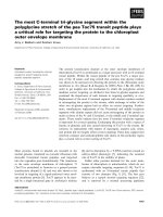
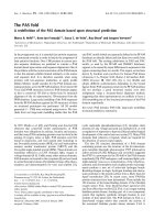
Fig. 1. The biosynthetic pathway leading to the production of GDP-
a-
D-Rha in P. aeruginosa. GMD catalyzes the 4,6-dehydration of
GDP-
D-Man (A), resulting in the production of GDP-6-deoxy-lyxo-
hexos-4-ulose (B), which exists in equilibrium with its gem-diol form
(C). RMD catalyzes the stereoselective reduction of compound B at
C4, resulting in the production of GDP-
D-Rha (D). Although GMD
can catalyze this final reduction reaction, our data indicate that
GMD does so much less rapidly than RMD.
J. D. King et al. GMD and RMD in bacterial GDP-
D-Rha synthesis
FEBS Journal 276 (2009) 2686–2700 ª 2009 The Authors Journal compilation ª 2009 FEBS 2687
epimers: GDP-d-Rha and GDP-6-deoxy-d-talose [26].
That study did not establish whether this activity was
due to an RMD homolog or to more than one
enzyme, but biochemical characterization of P. aeru-
ginosa RMD will show whether or not this enzyme is a
stereospecific reductase.
Crystal structures have been determined for the
GMDs from P. aeruginosa [27], E. coli [28], Arabidop-
sis thaliana [29], and PBCV-1 [30]. Up to now, no
RMD structure has been reported.
Here, we report the biochemical characterization of
purified His6-tag fusions of GMD and RMD from
P. aeruginosa, and the structural characterization of
RMD from A. thermoaerophilus.
Results
Purification and stability of His6-GMD and
His6-RMD
Pa
We purified N-terminally His6-tagged fusions of
P. aeruginosa GMD and RMD (His6-GMD and His6-
RMD
Pa
, respectively) in two chromatography steps to
greater than 95% purity (as judged by Coomassie-
stained SDS/PAGE; not shown). In 25% glycerol,
both enzymes retained activity after storage for more
than 1 year at )80 °C. We made qualitative observa-
tions that His6-GMD lost activity slowly in the course
of enzyme–substrate incubations, particularly at 37 °C,
but 4,6-dehydratase activity was still detectable after
incubation for 16 h at 25 °C (not shown). Addition of
BSA (10 mgÆmL
)1
) and glycerol [10% (v/v)] improved
the stability of His6-GMD during incubations at
37 °C, but these additives were not routinely included
in assays, as the proteins were stable during the time-
scales of the experiments, and both additives prevented
accurate measurement of reaction products by capil-
lary electrophoresis (CE) or NMR.
CE analysis of His6-GMD functions
CE is a technique that is able to resolve closely related
sugar nucleotides, and was our method of choice for
initial in vitro characterization of these enzymes. Anal-
ysis of the products that were formed after incubation
of His6-GMD with GDP-d-Man indicated that this
enzyme catalyzes quantitative 4,6-dehydration of this
substrate to GDP-6-deoxy-d-lyxo-hexos-4-ulose
(Fig. 2A,B). The chemical structures of sugar nucleo-
tide compounds in these reactions were unambiguously
identified by NMR spectroscopy (see below). The pro-
posed mechanism for GMD requires oxidoreductase
chemistry mediated by a dinucleotide cofactor [31].
Addition of exogenous NAD(P) was not required for
catalytic activity of the P. aeruginosa enzyme, indicat-
ing that the cofactor had copurified with the protein.
Some GMDs are bifunctional, being able to catalyze
the subsequent reduction of their initial 4-ketosugar
nucleotide products to produce 6-deoxysugar nucleo-
tides [5,16,17]. Addition of NADPH to the incubation
of the P. aeruginosa enzyme resulted in the gradual,
His6-GMD-dependent reduction of GDP-6-deoxy-d-
lyxo-hexos-4-ulose to GDP-d-Rha (Fig. 2C,D). There-
fore, like its homologs from Klebsiella pneumonia,
A. thermoaerophilus and PBCV-1, His6-GMD from
P. aeruginosa is a bifunctional 4,6-dehydratase, and a
stereospecific 4-reductase.
CE analysis of His6-RMD
Pa
incubations and
His6-RMD
Pa
–His6-GMD coincubation
RMDs use the product of the GMD-catalyzed 4,6-
dehydration reaction as substrate, and employ an
NAD(P)H cofactor as an electron donor. We incu-
bated His6-GMD with GDP-d-Man for 1 h, which
was enough time for complete conversion of the GDP-
d-Man, and then removed the enzyme by filtration.
His6-RMD
Pa
and NADPH were then added to the
reaction mixture. The GDP-6-deoxy-d-lyxo-hexos-4-
ulose substrate was generated in situ because it is
unstable, and its purification is therefore impractical
[5,17]. CE analysis of the reaction products (Fig. 3)
Retention time (min)
Absorbance at 254 nm
10 12 14 16
GDP-Man GDP-Rha
NADP
+
GDP-Rha
GDP
-6-deoxy-lyxo-hexos-4-ulose
GDP
-6-deoxy-lyxo-hexos-4-ulose
GDP-Man
C
B
A
D
NADPH
NADPHNADP
+
NADP
+
NADPHGDP-Rha
E
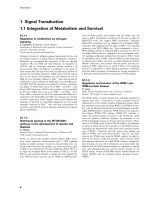
Fig. 2. CE analysis of dehydratase and reductase activities exhib-
ited by His6-GMD. Sugar nucleotide peaks were identified by
NMR; other peaks were identified by comparison with standards.
His6-GMD catalyzes the production of GDP-6-deoxy-
D-lyxo-hexos-4-
ulose from GDP-
D-Man. When reduced cofactor (NADPH) is added,
His6-GMD can catalyze the reduction of this intermediate to GDP-
D-Rha, but the reaction is incomplete after 1 h. Traces: (A) stan-
dard, GDP-
D-Man; (B) product of incubation of His6-GMD with
GDP-
D-Man; (C) products of incubation of His6-GMD with GDP-6-
deoxy-
D-lyxo-hexos-4-ulose (generated in situ) and NADPH for 1 h;
(D) as in (C), but incubated for 2 h; (E) reaction in (D) spiked with
GDP-
D-Man. Spiking demonstrates that the final product is not the
same as the starting material.
GMD and RMD in bacterial GDP-
D-Rha synthesis J. D. King et al.
2688 FEBS Journal 276 (2009) 2686–2700 ª 2009 The Authors Journal compilation ª 2009 FEBS
showed that, in the presence of excess NADPH, His6-
RMD
Pa
catalyzed the conversion of GDP-6-deoxy-d-
lyxo-hexos-4-ulose to GDP-d-Rha. When His6-GMD
and His6-RMD
Pa
were coincubated with GDP-d-Man
and NADPH, however, no reaction was observed by
CE (not shown).
Identification of reaction products by NMR
spectroscopy
To precisely define the functions of His6-GMD and
His6-RMD
Pa
in vitro, we identified the products of
these enzyme–substrate incubations by NMR spectros-
copy. As it is not possible to purify the labile product
of the GDP-d-Man 4,6-dehydration, we performed the
enzyme incubation in an NMR spectrometer, and
monitored the reaction directly in the tube (Figs 4
and 5). This technique was previously used for identifi-
cation of labile 4-keto UDP-sugars [32]. Monitoring of
the anomeric region of the 1D-
1
H-spectrum over the
course of incubation with His6-GMD, without
NADPH, showed progressive depletion of signals from
GDP-d-Man (compound A) and the growth of peaks
corresponding to the anomeric resonances of the
4-keto (compound B) and gem-diol (compound C)
forms of GDP-6-deoxy-d-lyxo-hexos-4-ulose (Fig. 4B).
On the basis of integration of the anomeric signals, the
4-keto and gem-diol forms of this compound coexist in
equilibrium at an approximately 5 : 2 ratio. Full
assignment of the NMR spectra of compounds B and
C and measurement of coupling constants (Table 1)
was achieved after removal of enzyme by filtration at
the 16-h time point, using selective 1D-TOCSY and
NOESY NMR experiments.
One-dimensional TOCSY of compound A H1
revealed a single J-coupled signal corresponding to H2
(Fig. 5B). The small J
1,2
coupling observed for
Retention time (min)
Absorbance at 254 nm
GDP-Rha
NADP
+
NADPH
GDP
-6-deoxy-lyxo-hexos-4-ulose
GDP-Man
C
B
A
10 12 14 16
Fig. 3. CE analysis of His6-RMD
Pa
reactions. His6-RMD converts
the product of the His6-GMD-catalyzed reaction, GDP-6-deoxy-
D-
lyxo-hexos-4-ulose, to GDP-
D-Rha, in the presence of NADPH.
Traces: (A) standard, GDP-
D-Man; (B) the product of His6-GMD
incubation with GDP-
D-Man after removal of His6-GMD by filtration;
(C) the His6-GMD product shown in (B), after subsequent incuba-
tion with His6-RMD
Pa
and NADPH.
A
B
C
1
H (p.p.m.)
Fig. 4. NMR spectroscopy of the active His6-GMD reaction directly
in aqueous reaction buffer. The reaction buffer was: 5 m
M GDP-a-
D-mannose, 90 l g of His6-GMD, 25 mM NaPO
4
,50mM NaCl
(pH 7.2), and 90% H
2
O/10% D
2
O. (A)
1
H-NMR spectrum of the
His6-GMD reaction buffer at the beginning of the reaction, showing
the anomeric region. (B)
1
H-NMR spectrum of the His6-GMD
reaction buffer after 16 h. (C)
1
H-NMR spectrum of the His6-GMD
reaction buffer after 16 h following the addition of NADPH. A,
GDP-
D-Man; B, GDP-6-deoxy-D-lyxo-hexos-4-ulose; C, gem-diol form
of compound B; D, GDP-
D-Rha; *unknown impurities.
A
B
C
D
Fig. 5. NMR spectroscopy of GDP-6-deoxy-a-D-lyxo-hexos-4-ulose
(B). These spectra were measured directly in aqueous reaction buf-
fer (5 m
M GDP-D-Man, 90 lg of His6-GMD, 25 mM NaPO
4
,50mM
NaCl, pH 7.2, 90% H
2
O/10% D
2
O). (A)
1
H-NMR spectrum. (B) 1D-
TOCSY of compound B H1 (80 ms). (C) 1D-TOCSY of compound B
H2 (80 ms). (D)
13
C-HSQC spectrum (128 transients, 128 incre-
ments,
1
J
C,H
= 140 Hz, 12 h). For selective 1D experiments,
excited resonances are underlined. A, GDP-
D-Man; C, gem-diol
form of compound B; R, ribose.
J. D. King et al. GMD and RMD in bacterial GDP-
D-Rha synthesis
FEBS Journal 276 (2009) 2686–2700 ª 2009 The Authors Journal compilation ª 2009 FEBS 2689
compound B is consistent with a manno-configured
sugar ring [33]. Owing to this small J
1,2
coupling, a
1D-TOCSY experiment on compound B H2 was
needed to assign H3 (Fig. 5C). Proton assignments for
compound C were also made on the basis of the
results of 1D-TOCSY experiments on H1 and H2 (not
shown). Selective 1D-NOESY experiments revealed
NOEs between H3 and H5 for compounds B and C,
which indicated that these protons were in close spatial
proximity, and thus occupied the trans position on the
sugar ring (data not shown). These NOEs, along with
the small J
1,2
-confirmed compounds B and C, had the
manno configuration. Carbon assignments were made
on the basis of the results of a
13
C heteronuclear single
quantum correlation (HSQC) experiment (Fig. 5D).
Whereas
13
C–
1
H correlations were readily observed for
compounds A and B, signals for compound C were
only visible at higher intensity. Three-bond
13
C–
1
H
correlations observed using a heteronuclear multiple
bond correlation (HMBC) experiment were used to
assign C4 of compounds B and C. It is of importance
that a signal corresponding to C4 of compound B was
observed at d
C
= 208.8 p.p.m. and was indicative of a
carbonyl group, whereas that of compound C at
d
C
= 94.0 p.p.m. was consistent with a diol form [34].
Together, these spectroscopy results for the ‘real-time’
enzyme–substrate reaction in the NMR tube contain-
ing the His6-GMD–substrate reaction mixture pro-
vided unambiguous identification of the structure of
compound B as GDP-6-deoxy-a-d-lyxo-hexos-4-ulose
and that of compound C as the gem-diol form of com-
pound B.
The product of the His6-RMD-catalyzed reaction
(compound D) was purified by anion exchange
chromatography before being analyzed by NMR. This
sample contained NADP
+
as a minor contaminant,
but this did not prevent identification of the reaction
product. Proton chemical shifts and J
H,H
coupling
constants determined using 1D-TOCSY experiments
agreed well with those previously reported for
GDP-d-Rha [5] (Fig. 6A–C, Table 1). Results from
a
31
P-HMQC experiment showed a
1
H–
31
P correlation
for the anomeric signal of compound D at d
P
= )13.2
p.p.m., and another at d
P
= )10.8 p.p.m., correspond-
ing to H5/5¢ of ribose (Fig. 6D). Carbon chemical
shifts and connectivities determined using
13
C-HSQC
(Fig. 6E) and HMBC were nearly identical to
those reported for GDP-d-Rha [5]. On the basis
of these NMR findings, compound D was concluded
to be GDP-a-d-Rha. These results for His6-RMD
Pa
therefore confirm that this enzyme is a GDP-6-
deoxy-a-d-lyxo-hexos-4-ulose-4-reductase (GDP-d-Rha-
forming).
Time courses of His6-GMD and His6-RMD
Pa
reactions determined by in-NMR-tube enzyme
incubation
The NMR spectroscopic measurement of substrate and
product concentrations during enzyme incubations in
Table 1. NMR data for sugar nucleotide metabolites in the GDP-D-Rha pathway of P. aeruginosa. Resonances were referenced to an internal
acetone standard at d
H
= 2.225 p.p.m. and d
C
= 31.07 p.p.m.
Compound
1
H and
13
C chemical shifts [d (p.p.m.)], and proton coupling constants (J
H,H
(Hz)]
H1
C1
J
1,2
H2
C2
J
2,3
H3
C3
J
3,4
H4
C4
J
4,5
H5
C5
J
5,6
H6/6¢
C6
GDP-a-
D-mannose (A) d
H
5.51 4.05 3.92 3.68 3.84 3.75/3.85
d
C
97.1 70.9 69.6 67.2 74.1 61.6
3
J
H,H
1.8 3.4 10.3 10.0
3
J
H,P
7.9
GDP-a-
D-6-deoxy-D-lyxo-hexos-4-ulose (B) d
H
5.58 4.46 4.84 4.68 1.22
d
C
96.3 76.0 73.1 208.8 71.9 13.4
3
J
H,H
2.2 3.8 6.5 6.5
3
J
H,P
7.6
gem-Diol form of
GDP-a-
D-6-deoxy-D-lyxo-hexos-4-ulose (C)
d
H
5.45 4.01 3.93 4.06 1.20
d
C
97.4 71.5 69.6 94.0 71.0 12.3
3
J
H,H
1.8 3.5 6.5 6.5
3
J
H,P
7.6
GDP-a-
D-rhamnose (D) d
H
5.43 4.03 3.86 3.42 3.89 1.25
d
C
97.2 71.2 70.4 72.8 70.4 17.6
3
J
H,H
1.2 3.5 9.7 9.8 6.1
3
J
H,P
7.6
GMD and RMD in bacterial GDP-
D-Rha synthesis J. D. King et al.
2690 FEBS Journal 276 (2009) 2686–2700 ª 2009 The Authors Journal compilation ª 2009 FEBS
NMR tubes enables sensitive observation, in real time,
of the course of enzyme-catalyzed reactions, and is
particularly suitable when these reactions have labile
starting materials or products [32,35]. To assess the rel-
ative reaction rates for the enzyme-catalyzed conver-
sions described above, we performed His6-GMD
incubations in an NMR tube. The progress of the
reaction was monitored by acquiring a proton spec-
trum (
1
H) every 2.8 min. Time course graphs were
created using vnmrj software (Varian, Palo Alto, CA,
USA) by plotting the integrals for the anomeric signals
for compounds A, B, C and D versus time. Lines of
best fit (solid lines) were generated through the data
points using vnmrj software. In the reaction contain-
ing His6-GMD and NADPH, build-up of com-
pounds B and C in the reaction tube was observed,
and these were slowly converted into compound D
(Fig. 7B), indicating that the reductase activity of
His6-GMD is much slower than its 4,6-dehydratase
activity in these conditions. The 4,6-dehydration reac-
tion creating compounds B and C proceeded at very
similar rates in the presence and absence of NADPH
(Fig. 7A,B).
We also used this technique to corroborate our
observation, by CE analysis, that His6-GMD–His6-
RMD–NADPH coincubation inhibits the 4,6-dehydra-
tion reaction. We observed the same phenomenon
(Fig. 7C). This more sensitive technique revealed that
a small amount of compound D was produced but the
majority of the GDP-d-Man starting material
remained unchanged. Neither of the intermediate
E
D
C
B
A
Fig. 6. NMR spectroscopy of the purified product from the
His6-RMD
Pa
-catalyzed reaction, GDP-a-D-rhamnose (D). (A)
1
H-NMR
spectrum. (B) 1D-TOCSY of compound D H1 (80 ms). (C) 1D-TOCSY
of compound D H6 (80 ms). (D)
31
P-HMQC spectrum (128
transients, 32 increments,
1
J
H,P
= 8 Hz, 4 h). (E)
13
C-HSQC spec-
trum (128 transients, 32 increments,
1
J
C,H
= 150 Hz, 15 h). For
selective 1D experiments, excited resonances are underlined. R
represents ribose.
A
B
C
Fig. 7. NMR spectroscopy of active enzyme–substrate incubations
directly in aqueous reaction buffer. The time course of reactions
was monitored by
1
H-NMR over a 4 h period. The changing relative
concentrations of each sugar nucleotide are shown here in plots of
their anomeric signal integrals versus time. GDP-
D-Man was incu-
bated with the enzyme(s), with or without NADPH. Coincubation of
His6-RMD
Pa
with His6-GMD and NADPH inhibits the 4,6-dehydra-
tase activity of His6-GMD. A, GDP-
D-Man; B, GDP-6-deoxy-D-lyxo-
hexos-4-ulose, C, gem-diol form of compound B; D, GDP-
D-Rha.
J. D. King et al. GMD and RMD in bacterial GDP-
D-Rha synthesis
FEBS Journal 276 (2009) 2686–2700 ª 2009 The Authors Journal compilation ª 2009 FEBS 2691
compounds, B or C, was detected, indicating that the
ketone was converted to compound D faster than it
was produced, probably by His6-RMD. The activity
of the His6-RMD
Pa
protein preparation used in this
His6-GMD–His6-RMD
Pa
–NADPH experiment was
confirmed by incubation with the product of the
His6-GMD incubation; all of the 25 mm compound
B plus compound C present was converted to com-
pound D by His6-RMD
Pa
within 4 min (data not
shown).
Kinetic analysis of the His6-GMD GDP-
D-Man
4,6-dehydratase activity
To compare the P. aeruginosa GMD with GMDs from
other organisms, we determined its kinetic parameters.
His6-GMD exhibits non-Michaelis–Menten kinetics
producing typical curves corresponding to the sub-
strate inhibition model with the following kinetic
parameters: K
m
= 14.02 ± 6.05 lm; V
max
= 3.64 ±
1.37 lmolÆmin
)1
Æmg
)1
; k
cat
= 8.82 s
)1
; K
i
= 2.859 ±
1.31 lm; k
cat
/K
m
= 6.3 · 10
5
m
)1
Æs
)1
.
Structural characterization of His6-RMD from
A. thermoaerophilus
To gain further understanding of the second step in
the GDP-d-Rha pathway, we set out to structurally
characterize RMD. Attempts to obtain high-quality
crystals of His6-RMD
Pa
were unsuccessful, but we
were able to determine the crystal structure of His6-
RMD
At
to 1.8 A
˚
resolution, in complex with the prod-
uct analog GDP-d-Man and a partially disordered
NADP(H) cofactor. The nicotinamide ring was not
resolved in the electron density (Fig. 8), so this mole-
cule was modeled into the structure as adenine-phos-
phoribose-pyrophosphate-ribose (APPR). The protein
has the typical architecture of the sugar nucleotide-
modifying SDR family, folding into two domains: a
Rossmann fold domain, which binds cofactor, and a
mixed a/b domain, which binds substrate and confers
substrate nucleotide specificity (Fig. 9A). The catalytic
triad is located at the interface between these
two domains. The structure also exhibits the typical
dimer interface for this protein family, consisting of a
four-helix bundle, where each monomer provides two
helices (Fig. 9B).
Comparison of the RMD structure with the struc-
ture of P. aeruginosa GMD [27] indicates that the
dinucleotide cofactor-binding site is more open to
solvent in RMD (Fig. 10). The active form of
P. aeruginosa GMD is a tetramer, and has a structural
feature called the ‘RR loop’ (comprising Arg35–
Arg43). The RR loop stretches from each molecule
into the adjacent monomer, and undergoes interactions
with the neighboring protein and cofactor across the
tetramer interface. In sequence alignments with GMD,
both the P. aeruginosa and A. thermoaerophilus RMD
sequences have gaps in the region of the RR loop, and
in the His6-RMD
At
structure, the b2–b3 loop is nota-
bly shorter than in GMD. The truncation of this struc-
tural feature probably explains why the RMD
structure does not exhibit a tetramer-forming interac-
tion like GMD, and why the RMD cofactor-binding
site is less occluded than in GMD.
A curious feature of RMD, which was suggested by
sequence alignments and confirmed by the RMD struc-
ture, is that the active site of this enzyme is very simi-
lar to that of GMD. The two protein structures
superimpose quite well, with an rmsd of 1.2 A
˚
over
281 equivalent Ca atoms. In addition, not only is the
SDR catalytic triad present in the RMD structure
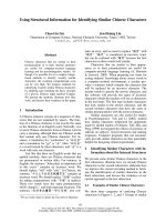
Fig. 8. The RMD active site. Stereoview of the RMD active site showing the refined 2F
o
)F
c
electron density map around GDP-D-Man and
APPR, contoured at 1.0r. Ser114, Tyr140 and Lys144 form the catalytic triad. No clear electron density is seen for the nicotinamide ring,
and even the nicotinamide ribose shows some indications of disorder; in fact, the average B-factor for the nicotinamide ribose is significantly
higher than for either the adenine or guanine riboses. An asterisk is placed in the expected position of the disordered nicotinamide moiety.
His170 is positioned in part of this ‘open’ space, a change of about 3.4 A
˚
as compared with the position of the equivalent residue, His180,
in P. aeruginosa GMD.
GMD and RMD in bacterial GDP-
D-Rha synthesis J. D. King et al.
2692 FEBS Journal 276 (2009) 2686–2700 ª 2009 The Authors Journal compilation ª 2009 FEBS
(Ser105, Tyr131, and Lys135), but so is the conserved
4,6-dehydratase active site glutamate (Glu128 in
P. aeruginosa GMD, and Glu116 in A. thermoaerophi-
lus RMD). This glutamate is proposed to be the active
site base that abstracts the C5 proton in the dehydra-
tion reaction [36]. Sequence alignments suggest, how-
ever, that this glutamate is not conserved in RMD
from P. aeruginosa (Asp107 occupies this position).
Comparison of other amino acid side chains lining
the active sites of A. thermoaerophilus RMD and
P. aeruginosa GMD shows that all residues are con-
served, with the exception of RMD Gln175 (Arg185
in GMD). This arginine is conserved in all characteri-
zed GMD sequences, and in the Ar. thaliana MUR1
structure this side chain is close enough to suggest
hydrogen-bonding interactions with a cofactor phos-
phate, the nicotinamide carboxyamide, and the
rhamnosyl O2 hydroxyl of the substrate analog
(Fig. 11). The degree of conservation for Gln175
among RMD enzymes is unclear, because so few
bona fide RMDs have been identified and character-
ized. In a blast search (using the blastp algorithm
[37]) of the P. aeruginosa RMD sequence, however, 89
of the top 100 hits had glutamine in this position; 10
of the others had arginine in its place, and the final
variant had glutamate.
Discussion
We present the biochemical characterization of His6-
GMD and His6-RMD from P. aeruginosa. Despite
being the focus of some research interest in the past,
A
B
Fig. 9. Structure of RMD from A. thermoaerophilus. (A) Stereoview of the RMD monomer. The cofactor-binding domain and the substrate-
binding domain are shown in aqua and light sand, respectively; the APPR portion of the cofactor (dark gray) and the ligand analog GDP-
D-
Man (light gray) are represented as space-filling models. Termini and secondary structural elements are labeled. (B) View of the RMD
homodimeric structure; an asterisk highlights the four-helix bundle, the typical SDR enzyme dimerization mode.
A
B
Fig. 10. The RMD cofactor-binding site is readily accessible to sol-
vent. Surface representations of (A) A. thermoaerophilus RMD and
(B) P. aeruginosa GMD, looking into the cofactor-binding site. Cor-
responding monomers from RMD and GMD are colored the same.
An additional monomer of the GMD tetramer (gray) significantly
reduces the accessibility of cofactor to bulk solvent.
J. D. King et al. GMD and RMD in bacterial GDP-
D-Rha synthesis
FEBS Journal 276 (2009) 2686–2700 ª 2009 The Authors Journal compilation ª 2009 FEBS 2693
including the publication of the P. aeruginosa GMD
crystal structure [27], the enzymatic functions of these
proteins have not previously been characterized using
in vitro assays with purified proteins. Their functions
had only been inferred from genetic experiments
[23,24] and functional assays using cell lysates as
enzyme source [23,25]. We have reconstituted the path-
way in vitro using both P. aeruginosa proteins, and
used NMR to unambiguously identify the reaction
products (Fig. 1). We have also defined conditions for
purification, long-term storage, and the performance of
enzyme–substrate incubations, so that these enzymes
can be used as synthetic tools to prepare GDP-d-Rha,
or its 4-keto precursor. The stability of P. aeruginosa
His6-GMD and His6-RMD
Pa
makes them suitable for
this application, and the kinetic parameters for
P. aeruginosa GMD are comparable with those of
GMD enzymes from other organisms [12,16,38–40].
Bifunctionality of P. aeruginosa GMD
We observed that P. aeruginosa GMD, like the
enzymes from K. pneumoniae, A. thermoaerophilus, and
PBCV-1 [5,16,17], is able to catalyze the reductase
reaction leading to GDP-d-Rha. This is consistent with
previous observations: when P. aeruginosa GMD was
expressed from plasmid pFV39 (which contains the
full-length gmd gene and a nonfunctional fragment of
rmd that lacks the first 97 rmd codons), it was able to
catalyze the conversion of GDP-d-Man to GDP-d-
Rha [23], although this assay was conducted with
E. coli cell lysates, and the reaction product was only
identified at that time by paper chromatography. The
ability of GMD to catalyze the reduction reaction indi-
cates that exchange of cofactor with solution must be
possible for this enzyme. In the current understanding
of the mechanism, the 4,6-dehydratase reaction cata-
lyzed by these enzymes involves an initial oxidation of
the sugar nucleotide, followed by subsequent reduc-
tion, and the cofactor, presumed to be bound as
NAD(P)
+
in the resting state, is therefore regenerated
in the catalytic cycle [31]. Conversely, reduction of
GDP-6-deoxy-d-lyxo-hexos-4-ulose to GDP-d-Rha
requires the formation of an initial enzyme–NAD(P)H
complex, whereupon the cofactor is oxidized to
NAD(P)
+
during the reaction. Therefore, the reduced
cofactor must be replaced from the solution before the
next reaction cycle. Recent evidence suggests that sev-
eral different GMDs contain a proportion or majority
of cofactor bound in the reduced state, and that this is
important for protein stability in solution [38]. Facile
exchange of cofactor with bulk solution is sometimes
reflected by binding of cofactor in a solvent-exposed
groove (e.g. RmlD [41]). In contrast, the P. aeruginosa
GMD structure [27] shows that the cofactor’s access to
solvent is blocked in large part by interactions with the
RR loop. Given that the dimer–dimer interface in
P. aeruginosa GMD is apparently stabilized by interac-
tions between cofactor and the neighboring monomer,
it is conceivable that oxidation of the cofactor may
alter the conformation of the RR loop or destabilize
the dimer–dimer interface in a manner that allows
cofactor exchange. In support of this hypothesis, the
oligomerization state of PBCV-1 GMD is responsive
to the oxidation state of bound NADP. In this viral
enzyme, addition of NADPH, but not NADP
+
,
induces dimerization of the apoenzyme, and oxidation
of the bound NADPH results in dimer dissociation
[38].
Unlike the case of PBCV-1, where the GMD has a
higher specific activity as a reductase than as a 4,6-de-
hydratase, the bifunctionality of P. aeruginosa GMD is
unlikely to be metabolically significant in vivo, at least
in terms of biosynthesis, because P. aeruginosa
expresses a dedicated reductase, RMD, to perform this
synthetic step. It is still possible, however, that the
GMD-catalyzed reductase reaction is functionally
important, either in regulation of enzyme activity or as
a mechanism to change the oxidation state of bound
cofactor. There are properties of GMDs, e.g. stimula-
tion of catalytic activity by addition of micromolar
NADPH [39] and the exclusive presence of NADPH in
GMD crystals, which are unexplained by the current
mechanism [38].
Fig. 11. The potential hydrogen-bonding interactions of a con-
served GMD arginine. The active sites of A. thermoaerophilus
RMD and Ar. thaliana MUR1 are shown in equivalent orientations
for comparison. MUR1 Arg220 is conserved in all GMDs, and dur-
ing catalysis may coordinate with a cofactor phosphate, the sub-
strate hexose, and the nicotinamide carboxyamide. The distances
between these groups in the MUR1 crystal structure are indicated.
In the RMD structure, the position of the MUR1 Arg220 is occu-
pied by a glutamine, and this amino acid side chain is too short to
mediate the same protein–ligand interactions. This may account for
the disordering of the nicotinamide ring in the RMD crystal.
GMD and RMD in bacterial GDP-
D-Rha synthesis J. D. King et al.
2694 FEBS Journal 276 (2009) 2686–2700 ª 2009 The Authors Journal compilation ª 2009 FEBS
Feedback inhibition
Our observation of strong inhibition of GDP-d-Man
consumption by His6-GMD when incubated with
NADPH and His6-RMD indicates that a feedback
mechanism inhibits the 4,6-dehydratase activity of
His6-GMD in these conditions. Such feedback inhibi-
tion, by which a sugar nucleotide controls the rate of
its own synthesis, is not unusual, and is well docu-
mented for GMDs: there are multiple examples of
organisms that incorporate l-fucose into oligosaccha-
rides or polysaccharides, where GMD is inhibited by
GDP-l-fucose [12,13,15,28,42,43]. Presumably, this
feedback inhibition has evolved to prevent build-up of
excess GDP- l-fucose and/or excessive consumption of
the starting material. In the case of P. aeruginosa
GMD, tight control of GDP-d-Man consumption may
be important, because this sugar nucleotide is also an
intermediate in the biosynthetic pathway for another
virulence factor, alginate [44].
In preliminary experiments to elucidate the inhibi-
tory mechanism, we have observed that strong inhibi-
tion of the His6-GMD reaction only occurs in the
presence of His6-RMD, raising the possibility that the
mechanism of inhibition involves a GMD–RMD pro-
tein–protein interaction. The reaction was also strongly
inhibited when His6-GMD was incubated with His6-
RMD and NADP
+
, which rules out the possibility
that GMD is inhibited simply by the exchange of
bound NADP
+
with NADPH preventing the first oxi-
dative step of the 4,6-dehydratase reaction (data not
shown). At the present time, the inhibitory mechanism
remains unclear, but this will be an interesting subject
for further study.
RMD structure
The similarity of the A. thermoaerophilus RMD struc-
ture to GMD structures is, in some respects, unsurpris-
ing. Where a bifunctional GMD enzyme is able to
catalyze the same reaction as RMD, a close resem-
blance between the two active sites makes sense, at least
as far as substrate binding and the SDR Ser/Thr-Tyr-
Lys catalytic triad are concerned. What is more intrigu-
ing is that all of the amino acid side chains that have
been proposed to function in acid–base catalysis of the
4,6-dehydratase reaction of GMD [27] are conserved in
A. thermoaerophilus RMD. The conservation of these
residues has been noted previously [45]; the RMD struc-
ture confirms that their orientation in space is also con-
served. Why, then, is this RMD protein unable to
catalyze the GDP-d-Man 4,6-dehydration reaction?
Previously, the absence of such potential catalytic side
chains has been used to rule out possible functions for
SDRs [46]. The structure of RMD that we report here
emphasizes that the inverse argument does not apply:
the presence of such residues does not necessarily mean
that the catalytic competence is likewise conserved. The
disordered nature of the NADP nicotinamide ring in
the RMD crystal indicated, however, an important dif-
ference between the two active sites. We propose that
Arg185 in P. aeruginosa GMD is important for aligning
NADP and GDP-d-Man in the active site for the dehy-
dratase reaction. This role is suggested by the close rela-
tive positions of the corresponding side chain, Arg220,
the NADPH cofactor and the substrate analog hexose
in the MUR1 structure (Fig. 11). Some of the distances
between these groups are rather long for classic hydro-
gen bonding, but relative motion of ligand molecules is
expected during the catalytic cycle: In the GMD reac-
tion, the nicotinamide must extract hydride from the
substrate C4¢, and later donate it back at C6¢.In
RMD
At
, the amino acid occupying the position of the
conserved GMD arginine is Gln175, and the side chain
of this residue is too short to undergo these interac-
tions. This may be the reason why a productive ternary
RMD–NADP
+
–GDP-d-Man complex cannot assem-
ble in the configuration necessary for this reaction. The
bioinformatics analysis suggested that Arg185 is abso-
lutely conserved among GMDs from diverse organisms,
and that Gln175 is well conserved among close RMD
homologs. The 10 RMD homolog sequences examined
that had arginine in this position may, in fact, represent
GMDs. We are currently working to test experimentally
whether exchange of arginine and glutamine at this
position can interconvert the catalytic functions of these
GMD and RMD enzymes. Subject to this experimental
verification, this current report may have helped to
identify a diagnostic amino acid for distinction of
GMD/RMD enzyme functions from sequence alone.
As has been previously discussed [46], such indicators
are important to make full use of the vast amount of
sequence information available in the genome
databases, and to provide useful indicators for the
accurate annotation of this important class of enzymes.
Conclusions
We have verified, biochemically, the functions of
GMD and RMD from P. aeruginosa, and showed that
GMD from this organism is a bifunctional 4,6-dehy-
dratase and a stereospecific 4-reductase. Reconstitution
of the P. aeruginosa GDP-d-Rha pathway in vitro
revealed a feedback mechanism inhibiting the first step
that may be important for the regulation of GDP-d-
Man consumption. Finally, structural analysis of
J. D. King et al. GMD and RMD in bacterial GDP-D-Rha synthesis
FEBS Journal 276 (2009) 2686–2700 ª 2009 The Authors Journal compilation ª 2009 FEBS 2695
RMD from A. thermoaerophilus identified an amino
acid, Arg185, in P. aeruginosa GMD that may be criti-
cal for correct orientation of GDP-d-Man and
NADP
+
cofactor for the 4,6-dehydration reaction.
The corresponding residue in RMD
At
is Gln175. The
amino acid in this position may be a key indicator of
specificity among these closely related GMD/RMD
enzymes.
Experimental procedures
Materials
Unless stated otherwise, all materials were purchased from
Sigma-Aldrich (Oakville, Canada).
DNA methods
The rmd genes from P. aeruginosa and A. thermoaerophilus
were amplified from chromosomal DNA templates, using the
following primer pairs: P. aeruginosa,5¢-AGGCCCGCTTC
C
GGATCCACTCAGCGTCTG-3¢ and 5¢-AAAAAAGTCG
ACTTCTTCTCGTACCCGTGACTC-3¢; and A. thermo-
aerophilus,5¢-TT
GGATCCATGAGAGCCCTAATCACTG
GA-3¢ and 5¢-TA
GGTACCTTATGCTTGACGGTAACT
TTGT-3¢. Restriction enzyme sites included on the primers
are underlined. The amplified P. aeruginosa gene was ligated
into pQE-30 (Qiagen, Missisauga, Canada), using BamHI
and SalI restriction sites. The A. thermoaerophilus gene was
cloned into the BamHI–KpnI-digested pQE-80 (Qiagen).
Both expression vectors encode N-terminal His6-tag fusions
of their respective RMD proteins.
Protein expression and purification
His6-GMD was expressed from the pQE-30-gmd vector,
which has been described previously [27]. His6-tagged RMD
proteins (His6-RMD
Pa
and His6-RMD
At
) were expressed
from the vectors described above, and E. coli M15(pREP4)
or E. coli BL21(DE3) was used as the host strain. Cells from
an overnight culture were used to inoculate 1 L of LB
broth. When the attenuance at 600 nm (D
600 nm
) reached
0.5–0.6, protein expression was induced by addition of iso-
propyl-thio-b-d-galactoside to 0.25 mm, and the cultures
were shaken for a further 16 h at room temperature. Cells
were harvested by centrifugation (10 000 g, 10 min), and
then suspended in 50 mm Hepes, 300 mm NaCl, and 5 mm
imidazole (pH 8.0). Cell lysis was performed with
ultrasonication. After centrifugation (10 000 g, 20 min), the
supernatant was passed through a 4 mL column of Ni
2+
–
nitrilotriacetic acid resin (Qiagen). The column was
thoroughly washed (50 mm Hepes, 300 mm NaCl, 20 mm
imidazole, pH 7.5), and then eluted by increasing the
imidazole concentration to 200 mm in the same buffer.
Proteins were then purified by anion exchange chromatogra-
phy using a 1 mL HiTrap-Q column (GE Healthcare, Baie
d’Urfe
´
, Canada), pre-equilibrated with 20 mm Tris/HCl
(pH 8.5), and eluted with a linear, 40 mL gradient from 0 to
1 m NaCl. For NMR analysis of enzyme incubations, pro-
teins were buffer-exchanged using a PD-10 column (GE
Healthcare), into 25 mm sodium phosphate and 50 mm
NaCl (pH 7.5). For long-term storage, 25% glycerol was
added to protein aliquots, which were then frozen ()80 °C).
Enzyme–substrate incubations
The enzymes used for in vitro assays were the N-terminal
His6-tagged fusions of the P. aeruginosa proteins. Unless
otherwise stated, enzyme–substrate incubations were per-
formed in 40 mm Tris/HCl (pH 7.5) and 10 mm MgCl
2
with 1.0 mm GDP-d-Man. In reactions that were to be ana-
lyzed by CE, NADP
+
and/or NADPH was added at 0.01
or 0.1 mm to assist in alignment of CE traces. In reductase
reactions requiring cofactor as a reagent, NADPH was
added in molar excess with respect to the sugar nucleotide
substrate. Reactions were started by the addition of
enzyme, and were incubated at 37 °C. To determine enzyme
catalytic activities with GDP-6-deoxy-d-lyxo-hexos-4-ulose,
a GMD reaction was performed for 1 h, the enzyme was
removed by filtration, and then enzyme and cofactor for
the second reaction were added. To determine the optimal
pH for enzyme-catalyzed reactions, the extent of substrate
conversion was determined after 5 min in the standard
reaction mixture, but with Mes (pH 5, 5.5, 6, and 6.5) or
Bis/Tris propane (pH 7, 7.5, 8, 8.5, 9, 9.5, and 10) in place
of Tris/HCl (pH 7.5).
CE
CE analyses were performed using a P/ACE MDQ Glyco-
protein System (Beckman Coulter, Fullerton, CA, USA),
using a bare silica 75 lm · 57 cm capillary and a running
buffer consisting of 25 mm sodium tetraborate (pH 9.5).
Compound elution was monitored by measuring UV absor-
bance at 254 nm, with the UV detector positioned at
50 cm. The capillary was preconditioned before each run
by washing with 0.2 m sodium hydroxide, water, and run-
ning buffer, each for 2 min. Samples were introduced by
pressure injection for 8 s (for reaction composition analysis)
or 24 s (for kinetic analysis), and separation was performed
at 22 kV. Peak integration was performed using 32 karat
software (Beckman).
Determination of kinetic parameters for GMD
Reactions were performed in triplicate, and contained
0.25 lg of protein and 0.5–40 lm GDP-d-Man in a total
volume of 1 mL. Samples were incubated at 37 °C for
GMD and RMD in bacterial GDP- D-Rha synthesis J. D. King et al.
2696 FEBS Journal 276 (2009) 2686–2700 ª 2009 The Authors Journal compilation ª 2009 FEBS
5 min, the reaction was stopped by flash freezing in a dry
ice/ethanol bath, and this was followed by transfer to a
boiling water bath for 10 min to denature the enzymes.
Samples were refrozen, lyophilized and finally suspended in
25 lL of water prior to analysis by CE. Because of the
instability of the ketone product in the enzyme-inactivation
procedure, reaction product quantities were normalized by
comparison with no-enzyme controls. Data were fitted to
the substrate inhibition equation v = V
max
/(1 + K
m
/
[S] + [S]/K
i
), and the kinetic parameters were determined
by nonlinear regression using the sigmaplot enzyme
kinetics module. In the course of these experiments, we
found that MgCl
2
has a slight inhibitory effect on GMD
activity (data not shown), and so all kinetic parameters
were measured in the absence of MgCl
2
.
Purification of the product of the GMD/RMD
sequential reaction (GDP-
D-Rha)
In a large-scale reaction, 30 lmol of GDP-d-Man was com-
pletely converted to GDP-d-Rha in sequential reactions cata-
lyzed by His6-GMD and then His6-RMD (1.5 mg of each
enzyme) with a molar excess of NADPH. Protein was
removed from the completed reaction by ultrafiltration
through a Centriplus YM-3 cartridge (Millipore, Billerica,
MA, USA). The reaction product was then purified as previ-
ously reported [47]. Briefly, the filtrate was subjected to anion
exchange chromatography using an Econo-Pac High Q
anion exchange column (Bio-Rad, Hercules, CA, USA) with
a linear gradient of 0–500 mm triethylammonium bicarbon-
ate (pH 8.0). Fractions were monitored by CE, and those
containing the sugar nucleotide were pooled. Bicarbonate
was removed (as CO
2
gas) by addition of H
+
-charged
AG 50W-X4 resin (Bio-Rad) until pH 4.5 was achieved. The
resin was removed by filtration, and the solution was neutral-
ized by the addition of triethylamine. Finally, water and
triethylamine were removed by lyophilization.
NMR spectroscopy
NMR experiments were performed at 500 MHz (
1
H) in 10%
D
2
O (90% H
2
O) or 99% D
2
O with a Varian Z-gradient
3 mm triple resonance (
1
H,
13
C,
31
P) probe (Varian). Stan-
dard homonuclear-correlated and heteronuclear-correlated
2D pulse sequences from Varian, such as COSY, TOCSY,
HSQC, HMBC and
31
P-HMQC, were used for general
assignments. NMR experiments were typically performed at
25 °C with suppression of the deuterated H
2
O resonance.
The methyl resonance of acetone was used as an internal
reference at d
H
= 2.225 p.p.m. and d
C
= 31.07 p.p.m.
Selective 1D-TOCSY experiments with a Z-filter, as well as
1D-NOESY experiments, were used for complete residue
assignment and for the determination of J
H,H
coupling con-
stants [48,49]. In initial experiments (data shown in Fig. 4) to
observe the products of GMD-catalyzed, ‘in-NMR-tube’
reactions, the enzyme was first placed in a 3 mm NMR tube
suspended in 200 lL of its reaction buffer (25 mm NaPO
4
,
50 mm NaCl, pH 7.2, 90% H
2
O/10% D
2
O). The reaction
was started by the addition of 5 mm GDP-d-Man, ± 5 mm
NADPH, to the reaction buffer, and the proton spectrum
was taken at the start of the reaction and again 16 h later. To
follow His6-GMD-catalyzed and His6-RMD-catalyzed reac-
tions over time (for the data shown in Fig. 7), 25 mm GDP-
d-Man was incubated with 7.5 lgÆmL
)1
each enzyme,
±25mm NADPH, in 25 mm NaPO
4
,50mm NaCl
(pH 7.2), and 90% H
2
O/10% D
2
O. A proton spectrum was
acquired every 2.8 min over a 4 h period.
Crystallography
Purified His6-RMD
At
(i.e. the tagged A. thermoaerophilus
protein) was concentrated to 10 mgÆmL
)1
, and crystals were
grown by the sitting drop vapor diffusion method in 35%
pentaerythritol propoxylate (5/4 PO/OH; Hampton
Research, Aliso Viejo, CA, USA), 100 mm Tris (pH 8.5),
and 200 mm NaCl. As the natural substrate is unstable and
the natural product is not available commercially, the prod-
uct analog GDP-d-Man was added to the crystallization
conditions, along with NADP or NADPH. The presence of
GDP-d-Man was found to be absolutely necessary for crys-
tal growth. The best crystals grew in a combination of
5mm GDP-d-Man and NADPH. Prior to data collection,
crystals were flash-frozen in liquid nitrogen directly from
the crystallization drop.
X-ray diffraction data were collected at a wavelength of
1.0 A
˚
on a MAR CCD detector at the Advanced Photon
Source Beamline 5-ID (DND) (Argonne National Labora-
tory). Crystals were held at 100 K in a cryostream during
data collection. Data were processed using xds software
[50], and statistics are shown in Table 2.
The structure of RMD was determined by molecular
replacement with mrbump (R. M. Keegan & M. D. Winn,
unpublished data) in conjunction with the ccp4 suite [51],
using a search model based on the known structure of
GMD from P. aeruginosa (29% sequence identity; Protein
Data Bank code: 1RPN [27]). arp/warp [52] was used for
an initial round of automatic model building and refine-
ment of the protein portion. The initial 2F
o
)F
c
electron
density maps from arp/warp revealed clear density for a
GDP-sugar and the APPR portion of the cofactor. Further
refinement was carried out using the TLS option in ref-
mac5 [53], alternated with manual model building in coot
[54] using the 2F
o
)F
c
and F
o
)F
c
maps. Restraint libraries
were constructed for APPR and GDP-d-Man using
sketcher [51]. Water molecules were added using coot,
and checked for accuracy by hand. The final model
(R
factor
= 16.5%, R
free
= 19.8%) consists of an RMD dimer,
350 water molecules, and two molecules each of APPR and
GDP-d-Man. Although the density is a little weak in one
loop area (residues 33–36), there is clear density for
J. D. King et al. GMD and RMD in bacterial GDP-D-Rha synthesis
FEBS Journal 276 (2009) 2686–2700 ª 2009 The Authors Journal compilation ª 2009 FEBS 2697
residues 1–309 of 309 residues in each monomer. The model
conforms ideally to the geometry defined by procheck [55].
The refinement statistics are presented in Table 2, and the
final coordinates have been deposited in the Protein Data
Bank under the accession number 2PK3.
Acknowledgements
We are grateful to E. F. Mulrooney for technical assis-
tance. This work was supported by an operating grant
to J. S. Lam from the Canadian Cystic Fibrosis Foun-
dation (CCFF) and the Canadian Institutes of Health
Research (grant no. MOP14687), a grant (no. P18013-
B10) to P. Messner from the Austrian Science Fund,
and a grant from the NIH (GM65501) to R. M. Garav-
ito. K. K. H. Poon was a recipient of a postdoctoral
fellowship from CCFF. J. S. Lam holds a Canada
Research Chair in Cystic Fibrosis and Microbial Glyco-
biology funded jointly by the Canadian Foundation of
Innovation and the Ontario Innovation Trust.
References
1 Kocharova NA, Knirel YA, Widmalm G, Jansson PE
& Moran AP (2000) Structure of an atypical O-antigen
polysaccharide of Helicobacter pylori containing a novel
monosaccharide 3-C-methyl-d-mannose. Biochemistry
39, 4755–4760.
2 Senchenkova SN, Shashkov AS, Knirel YA, McGov-
ern JJ & Moran AP (1996) The O-specific polysaccha-
ride chain of Campylobacter fetus serotype B
lipopolysaccharide is a d-rhamnan terminated with
3-O-methyl-d-rhamnose (d-acofriose). Eur J Biochem
239, 434–438.
3 Molicaro A, Silip A, Lanzetta R, Newman MA, Dow
JM & Parrilli M (2003) Structural elucidation of the
O-chain of the lipopolysaccharide from Xanthomonas
campestris strain 8004. Carbohydr Res 338, 277–281.
4 Wang IN, Li Y, Que Q, Bhattacharya M, Lane LC,
Chaney WG & Van Etten JL (1993) Evidence for virus-
encoded glycosylation specificity. Proc Natl Acad Sci
USA 90, 3840–3844.
5 Kneidinger B, Graninger M, Adam G, Puchberger M,
Kosma P, Zayni S & Messner P (2001) Identification of
two GDP-6-deoxy-d-lyxo-4-hexulose reductases synthe-
sizing GDP-d-rhamnose in Aneurinibacillus thermoaero-
philus L420-91T. J Biol Chem 276, 5577–5583.
6 Persson B, Kallberg Y, Oppermann U & Jornvall H
(2003) Coenzyme-based functional assignments of short-
chain dehydrogenases/reductases (SDRs). Chem Biol
Interact 143-144, 271–278.
7 Chang S, Duerr B & Serif G (1988) An epimerase-
reductase in l-fucose synthesis. J Biol Chem 263, 1693–
1697.
8 Maki M, Jarvinen N, Rabina J, Maaheimo H, Mattila
P & Renkonen R (2003) Cloning and functional expres-
sion of a novel GDP-6-deoxy-d-talose synthetase from
Actinobacillus actinomycetemcomitans. Glycobiology 13,
295–303.
9 Markovitz A (1964) Biosynthesis of guanosine
diphosphate d-rhamnose and guanosine diphosphate d-
talomethylose from guanosine diphosphate alpha-
d-mannose. J Biol Chem 239, 2091–2098.
10 Albermann C & Piepersberg W (2001) Expression and
identification of the RfbE protein from Vibrio
cholerae O1 and its use for the enzymatic synthesis of
GDP-d-perosamine.
Glycobiology 11, 655–661.
11 Bonin CP, Potter I, Vanzin GF & Reiter WD (1997)
The MUR1 gene of Arabidopsis thaliana encodes an
isoform of GDP-d-mannose-4,6-dehydratase, catalyzing
the first step in the de novo synthesis of GDP-l-fucose.
Proc Natl Acad Sci USA 94, 2085–2090.
12 Bisso A, Sturla L, Zanardi D, De Flora A & Tonetti M
(1999) Structural and enzymatic characterization of
human recombinant GDP-d-mannose-4,6-dehydratase.
FEBS Lett 456, 370–374.
13 Sturla L, Bisso A, Zanardi D, Benatti U, De Flora A &
Tonetti M (1997) Expression, purification and charac-
terization of GDP-d-mannose 4,6-dehydratase from
Escherichia coli. FEBS Lett 412, 126–130.
Table 2. Data collection and refinement statistics. R-fac-
tor = R|F
o
)F
c
|/RF
o
, where F
o
and F
c
are observed and calculated
structure factors, respectively. R-free is the cross-validation R-factor
computed for the test set of 3207 reflections (5% of the total
unique reflections).
Data collection statistics
Space group P1
Unit cell parameters (A
˚
, °) a = 46.88, b = 55.74,
c = 79.24, a = 72.54,
b = 82.95, c = 75.61
Resolution range (A
˚
) 30.0–1.8
No. of observed reflections 215 748
No. of unique reflections 64 129
Completeness (%) 96.5 (95.2)
a
B-factor from Wilson plot 20.35
R
merge
(%) 7.6 (43.0)
a
Average I/r (I) 13.4 (3.7)
a
Refinement statistics
No. of residues 618/618
No. of water molecules 350
No. of heteromolecules 4
R
factor
16.5
R
free
19.8
rmsd bond lengths (A
˚
) 0.014
rmsd bond angles (°) 1.47
Average B-factor (A
˚
2
) 25.6
Ramachandran
Most favored (%) 93.8
Allowed (%) 6.2
a
Indicates statistic from highest-resolution shell.
GMD and RMD in bacterial GDP-
D-Rha synthesis J. D. King et al.
2698 FEBS Journal 276 (2009) 2686–2700 ª 2009 The Authors Journal compilation ª 2009 FEBS
14 Broschat KO, Chang S & Serif G (1985) Purification
and characterization of GDP-d-mannose 4,6-dehydra-
tase from porcine thyroid. Eur J Biochem 153, 397–401.
15 Rhomberg S, Fuchsluger C, Rendic D, Paschinger K,
Jantsch V, Kosma P & Wilson IB (2006) Reconstitution
in vitro of the GDP-fucose biosynthetic pathways of
Caenorhabditis elegans and Drosophila melanogaster.
FEBS J 273, 2244–2256.
16 Tonetti M, Zanardi D, Gurnon JR, Fruscione F, Armi-
rotti A, Damonte G, Sturla L, De Flora A & Van Etten
JL (2003) Paramecium bursaria chlorella virus 1 encodes
two enzymes involved in the biosynthesis of GDP-l-
fucose and GDP-d-rhamnose. J Biol Chem 278, 21559–
21565.
17 Yamamoto K, Katayama I, Onoda Y, Inami M, Kuma-
gai H & Tochikura T (1993) Evidence that the enzyme
catalyzing the conversion of guanosine diphosphate
d-mannose to a 4-keto sugar nucleotide intermediate
requires nicotinamide adenine dinucleotide phosphate.
Arch Biochem Biophys 300, 694–698.
18 Emori TG & Gaynes RP (1993) An overview of noso-
comial infections, including the role of the microbiology
laboratory. Clin Microbiol Rev 6, 428–442.
19 Rivera M & McGroarty EJ (1989) Analysis of a com-
mon-antigen lipopolysaccharide from Pseudomonas
aeruginosa. J Bacteriol 171, 2244–2248.
20 Lam MY, McGroarty EJ, Kropinski AM, MacDonald
LA, Pedersen SS, Høiby N & Lam JS (1989) Occur-
rence of a common lipopolysaccharide antigen in stan-
dard and clinical strains of Pseudomonas aeruginosa.
J Clin Microbiol 27, 962–967.
21 Rocchetta HL, Burrows LL, Pacan JC & Lam JS
(1998) Three rhamnosyltransferases responsible for
assembly of the A-band d-rhamnan polysaccharide in
Pseudomonas aeruginosa: a fourth transferase, WbpL, is
required for the initiation of both A-band and B-band
lipopolysaccharide synthesis. Mol Microbiol 28, 1103–
1119.
22 Rocchetta HL & Lam JS (1997) Identification and func-
tional characterization of an ABC transport system
involved in polysaccharide export of A-band lipopoly-
saccharide in Pseudomonas aeruginosa. J Bacteriol 179,
4713–4724.
23 Lightfoot J & Lam JS (1993) Chromosomal mapping,
expression and synthesis of lipopolysaccharide in Pseu-
domonas aeruginosa: a role for guanosine diphospho
(GDP)-D-mannose. Mol Microbiol 8, 771–782.
24 Rocchetta HL, Pacan JC & Lam JS (1998) Synthesis of
the A-band polysaccharide sugar d-rhamnose requires
Rmd and WbpW: identification of multiple AlgA
homologues, WbpW and ORF488, in Pseudomonas
aeruginosa. Mol Microbiol
29, 1419–1434.
25 Maki M, Jarvinen N, Rabina J, Roos C, Maaheimo H
& Renkonen R (2002) Functional expression of Pseudo-
monas aeruginosa GDP-4-keto-6-deoxy-d-mannose
reductase which synthesizes GDP-rhamnose. Eur J Bio-
chem 269, 593–601.
26 Markovitz A (1962) Isolation of d-talomethylose
(6-deoxy-d-talose) and d-rhamnose (6-deoxy-d-man-
nose) from capsular polysaccharide of a Gram-negative
bacterium. J Biol Chem 237, 1767–1771.
27 Webb NA, Mulichak AM, Lam JS, Rocchetta HL &
Garavito RM (2004) Crystal structure of a tetrameric
GDP-d-mannose 4,6-dehydratase from a bacterial
GDP-d-rhamnose biosynthetic pathway. Protein Sci 13,
529–539.
28 Somoza JR, Menon S, Schmidt H, Joseph-McCarthy
D, Dessen A, Stahl ML, Somers WS & Sullivan FX
(2000) Structural and kinetic analysis of Escherichia coli
GDP-mannose 4,6 dehydratase provides insights into
the enzyme’s catalytic mechanism and regulation by
GDP-fucose. Structure 8, 123–135.
29 Mulichak AM, Bonin CP, Reiter WD & Garavito RM
(2002) Structure of the MUR1 GDP-mannose 4,6-dehy-
dratase from Arabidopsis thaliana: implications for
ligand binding and specificity. Biochemistry 41, 15578–
15589.
30 Rosano C, Zuccotti S, Sturla L, Fruscione F, Tonetti
M & Bolognesi M (2006) Quaternary assembly and
crystal structure of GDP-d-mannose 4,6-dehydratase
from Paramecium bursaria chlorella virus. Biochem
Biophys Res Commun 339, 191–195.
31 Oths PJ, Mayer RM & Floss HG (1990) Stereochemis-
try and mechanism of the GDP-mannose dehydratase
reaction. Carbohydr Res 198, 91–100.
32 McNally DJ, Schoenhofen IC, Mulrooney EF, Whit-
field DM, Vinogradov E, Lam JS, Logan SM & Brisson
JR (2006) Identification of labile UDP-ketosugars in
Helicobacter pylori, Campylobacter jejuni and Pseudomo-
nas aeruginosa: key metabolites used to make glycan
virulence factors. Chembiochem 7, 1865–1868.
33 Kanipes MI, Ribeiro AA, Lin S, Cotter RJ & Raetz
CR (2003) A mannosyl transferase required for lipo-
polysaccharide inner core assembly in Rhizobium legu-
minosarum. Purification, substrate specificity, and
expression in Salmonella waaC mutants. J Biol Chem
278, 16356–16364.
34 Schoenhofen IC, McNally DJ, Vinogradov E, Whitfield
D, Young NM, Dick S, Wakarchuk WW, Brisson JR &
Logan SM (2006) Functional characterization of dehy-
dratase/aminotransferase pairs from Helicobacter and
Campylobacter: enzymes distinguishing the pseudaminic
acid and bacillosamine biosynthetic pathways. J Biol
Chem 281, 723–732.
35 Pfoestl A, Hofinger A, Kosma P & Messner P (2003)
Biosynthesis of dTDP-3-acetamido-3,6-dideoxy-alpha-d-
galactose in Aneurinibacillus thermoaerophilus L420-91T.
J Biol Chem 278, 26410–26417.
36 Gross JW, Hegeman AD, Gerratana B & Frey PA
(2001) Dehydration is catalyzed by glutamate-136 and
J. D. King et al. GMD and RMD in bacterial GDP-D-Rha synthesis
FEBS Journal 276 (2009) 2686–2700 ª 2009 The Authors Journal compilation ª 2009 FEBS 2699
aspartic acid-135 active site residues in Escherichia coli
dTDP-glucose 4,6-dehydratase. Biochemistry 40, 12497–
12504.
37 Altschul SF, Madden TL, Schaffer AA, Zhang J, Zhang
Z, Miller W & Lipman DJ (1997) Gapped BLAST and
PSI-BLAST: a new generation of protein database
search programs. Nucleic Acids Res 25, 3389–3402.
38 Fruscione F, Sturla L, Duncan G, Van Etten JL, Val-
buzzi P, De Flora A, Di Zanni E & Tonetti M (2008)
Differential role of NADP
+
and NADPH in the activ-
ity and structure of GDP-d-mannose 4,6-dehydratase
from two chlorella viruses. J Biol Chem 283, 184–193.
39 Sullivan FX, Kumar R, Kriz R, Stahl M, Xu GY,
Rouse J, Chang XJ, Boodhoo A, Potvin B & Cumming
DA (1998) Molecular cloning of human GDP-mannose
4,6-dehydratase and reconstitution of GDP-fucose bio-
synthesis in vitro. J Biol Chem 273, 8193–8202.
40 Wu B, Zhang Y & Wang PG (2001) Identification and
characterization of GDP-d-mannose 4,6-dehydratase
and GDP-l-fucose synthetase in a GDP-l-fucose bio-
synthetic gene cluster from Helicobacter pylori. Biochem
Biophys Res Commun 285 , 364–371.
41 Blankenfeldt W, Kerr ID, Giraud MF, McMiken HJ,
Leonard G, Whitfield C, Messner P, Graninger M &
Naismith JH (2002) Variation on a theme of SDR.
dTDP-6-deoxy-l-lyxo-4-hexulose reductase (RmlD)
shows a new Mg
2+
-dependent dimerization mode.
Structure 10, 773–786.
42 Albermann C, Distler J & Piepersberg W (2000) Pre-
parative synthesis of GDP-beta-l-fucose by recombi-
nant enzymes from enterobacterial sources.
Glycobiology 10, 875–881.
43 Kornfeld RH & Ginsburg V (1966) Control of synthesis
of guanosine 5¢-diphosphate d-mannose and guanosine
5¢-diphosphate l-fucose in bacteria. Biochim Biophys
Acta 117, 79–87.
44 Deretic V, Gill JF & Chakrabarty AM (1987) Gene
algD coding for GDP-mannose dehydrogenase is trans-
criptionally activated in mucoid Pseudomonas aerugin-
osa. J Bacteriol 169, 351–358.
45 Maki M & Renkonen R (2004) Biosynthesis of 6-deoxy-
hexose glycans in bacteria. Glycobiology 14, 1R–15R.
46 King JD, Harmer NJ, Preston A, Palmer CM, Rejzek
M, Field RA, Blundell TL & Maskell DJ (2007) Pre-
dicting protein function from structure – the roles of
short-chain dehydrogenase/reductase enzymes in Borde-
tella O-antigen biosynthesis. J Mol Biol 374, 749–763.
47 Mulrooney EF, Poon KK, McNally DJ, Brisson JR &
Lam JS (2005) Biosynthesis of UDP-N-acetyl-l-fucos-
amine, a precursor to the biosynthesis of lipopolysac-
charide in Pseudomonas aeruginosa serotype O11. J Biol
Chem 280, 19535–19542.
48 Brisson JR, Sue SC, Wu WG, McManus G, Nghia PT
& Uhrin D (2002) NMR Spectroscopy of Glycoconju-
gates. Wiley-VCH, Weinhem.
49 Uhrin D & Brisson JR (2000) Structure Determination
of Microbial Polysaccharide by High Resolution NMR
Spectroscopy. Horizon Scientific Press, Wymondham,
UK.
50 Kabsch WJ (1993) Automatic processing of rotation
diffraction data from crystals of initially unknown sym-
metry and cell constants. J Appl Crystallogr 26, 795–
800.
51 Collaborative Computational Project N (1994) The
CCP4 Suite: programs for protein crystallography. Acta
Crystallogr D 50, 760–763.
52 Perrakis A, Harkiolaki M, Wilson KS & Lamzin VS
(2001) ARP/wARP and molecular replacement. Acta
Crystallogr D 57, 1445–1450.
53 Winn M, Isupov M & Murshudov GN (2001) Use of
TLS parameters to model anisotropic displacements in
macromolecular refinement. Acta Crystallogr D 57,
122–133.
54 Emsley P & Cowtan K (2004) Coot: model-building
tools for molecular graphics. Acta Crystallogr D
60,
2126–2132.
55 Laskowski RA, MacArthur MW, Moss DS & Thornton
JM (1993) PROCHECK: a program to check the ste-
reochemical quality of protein structures. J Appl Crys-
tallogr 26 , 283–291.
GMD and RMD in bacterial GDP- D-Rha synthesis J. D. King et al.
2700 FEBS Journal 276 (2009) 2686–2700 ª 2009 The Authors Journal compilation ª 2009 FEBS